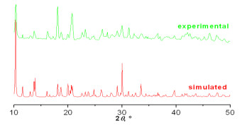
Figure 1.
PXRD pattern of compound 1

Most of the lanthanide elements (not including La and Lu) show fine photoluminescence performances and, recently, lanthanide materials with photoluminescence performances have drawn more and more attention from chemical, physical and material researchers[1-4]. Nowadays, a large number of researchers are devoting themselves to the design, synthesis, structures, physical and chemical characterization of novel lanthanide materials to investigate the potential and broad applications of lanthanide materials in cell imaging, magnetic materials, catalysts, electrochemical displays, light-emitting diodes, sensors, luminescent probes, and so forth[5-9]. Relative to the broad investigation on the photoluminescence performances of lanthanide materials, there are only few examples of investigation on the semiconductor performances of lanthanide materials and, therefore, more exploration is still necessary[10].
Transition metal materials usually possess attractive properties that enable them to have potential applications in the fields of materials, chemistry, physics, biology, etc. Therefore, transition metal materials have gained more and more attention since many years ago[11-23]. In recent years, a large number of transition metal materials have been documented[24-37]. Amongst the transition metal materials, group 12 (IIB) ones are fascinate due to the following reasons: the vital role of zinc played in biological systems, the broad applications of IIB materials, photoelectric and photo-luminescence behavior, as well as different coordination motifs offered by the d10 geometries of IIB cations[38-40]. Mercury is an interesting element and has also gained much attention over the years because of various coordination modes, semiconductor properties, photoluminescence properties, optoelectronic behaviors and so on.
To my knowledge, isonicotinic acid is an important organic molecule. It can be applied as a useful building ligand because it possesses two carboxyl oxygen atoms at one end and one nitrogen atom at the other end. These oxygen and nitrogen atoms enable isonicotinic acid to bind to several metal centers and yield an extended structure with different coordination motifs. It is well-known that oxygen atoms are favorable to bind to lanthanide ions, while nitrogen atoms tend to coordinate to transition metal ions. Therefore, it is deemed that isonicotinic acid can simultaneously coordinate to both lanthanide and transition metal ions. Over the years, the investigation of materials with attractive photoluminescence and semiconductor performances, especially lanthanide mercury materials, have become one of my research topics with a view to getting new findings in crystal structures, photoluminescence and semiconductor performances. In the present paper, the hydrothermal preparation, X-ray structure, photophysical performances and thermogravimetry of a novel holmium-mercury material, {[Ho(IA)(HIA)2(H2O)2]2(Hg3Br8)}n (nHgBr2)·2nNO3 (1, HIA is isonicotinc acid), are reported. It is characterized by a 2-D layer-like structure. It is noteworthy that this compound exhibits upconversion green emission.
In the present work, all chemicals and/or reagents for the preparation of complex 1 were of AR grade purity, commercially bought and used directly without purification. The infrared spectrum was carried out using a PE Spectrum-One FT-IR spectrophotometer with a KBr pellet. The solid-state photoluminescence spectra were carried out with a F97XP photoluminescence spectrometer. A solid-state UV-visible diffuse reflectance spectrum was carried out using a TU1901 UV-visible spectrometer. A powder X-ray diffraction (PXRD) pattern was measured on a AL-Y3000 powder diffractometer with Cu-Kα (λ = 1.54056 Å) at a step size of 0.02°. The simulated powder pattern was obtained by using single-crystal X-ray diffraction data and processed by the free Mercury v1.4 program provided by the Cambridge Crystallographic Data Centre. A thermogravimetry (TG) curve was measured on a NETZSCH TG 209F3 analyzer under nitrogen atmosphere.
A mixture of 2.5 mmol HgBr2 (900 mg), 1 mmol Ho(NO3)3·6H2O (459 mg), 3 mmol isonicotinic acid (369 mg) and distilled water (10 mL) was put into a stainless-steel Teflon-lined reactor (23 mL) and heated at 160 ℃ for 15 days, then powered off. Colorless prism-like crystals can be obtained once the reactor was cooled down to room temperature and, the crystal was filtered and cleaned with water. The yield of the crystal obtained was 47% based on Ho(NO3)3·6H2O. IR peaks (cm−1): 3442(vs), 3137(w), 3079(w), 2888(w), 2430(w), 1710(w), 1621(vs), 1500(w), 1411(vs), 1385(vs), 1302(w), 1232(w), 1054(w), 1010(w), 851(w), 768(m), 679(m), 551(w) and 412(m). The purity of the product was confirmed by PXRD, as shown in Fig. 1.
Using a carefully selected crystal, the single-crystal X-ray diffraction data set of complex 1 was obtained from a SuperNova CCD X-ray diffractometer equipped with a graphite-monochromated MoKα radiation (λ = 0.71073 Å). The data reduction and empirical absorption were performed with CrystalClear software. By using direct methods, the crystal structure of 1 was resolved with SHELXS software[41] and refined on F2 with a full-matrix least-square. All of the non-H atoms were found based on the difference Fourier spots and anisotropically refined, while all of the H atoms were theoretically identified, attached to the parent atoms and isotropically refined. Reflections collected are 17376; the final R = 0.0874 for 381 parameters and 3310 observed reflections with I > 2σ(I) and wR = 0.2233 (w = 1/[σ2(Fo2) + (0.1212P)2 + 30.1388P], where P = (Fo2 + 2Fc2)/3); S = 1.078, (Δρ)max = 3.874, (Δρ)min = –3.128 e/Å3 and (Δ/σ)max = 0.000. The selected bond distances and bond angles are present in Table 1, while hydrogen bonds are given in Table 2.
 DownLoad:
CSV
DownLoad:
CSV
| Bond | Dist. | Bond | Dist. | |
| Hg(1)–N(2) | 2.16(2) | Hg(3)–Br(5)#1 | 2.424(2) | |
| Hg(1)–N(2)#1 | 2.16(2) | Ho(1)–O(1) | 2.325(15) | |
| Hg(1)–Br(4) | 2.788(3) | Ho(1)–O(2)#2 | 2.331(14) | |
| Hg(1)–Br(4)#1 | 2.788(3) | Ho(1)–O(3) | 2.308(19) | |
| Hg(2)–Br(1) | 2.493(4) | Ho(1)–O(4)#2 | 2.230(17) | |
| Hg(2)–Br(2) | 2.516(4) | Ho(1)–O(5) | 2.381(14) | |
| Hg(2)–Br(3) | 2.663(3) | Ho(1)–O(6)#3 | 2.314(16) | |
| Hg(2)–Br(4) | 2.978(3) | Ho(1)–O(1W) | 2.474(16) | |
| Hg(3)–Br(5) | 2.424(2) | Ho(1)–O(2W) | 2.470(16) | |
| Angle | (°) | Angle | (°) | |
| N(2)#1–Hg(1)–N(2) | 165.4(11) | Br(5)–Hg(3)–Br(5)#1 | 171.99(16) | |
| N(2)#1–Hg(1)–Br(4) | 95.1(6) | Hg(1)–Br(4)–Hg(2) | 87.30(9) | |
| N(2)–Hg(1)–Br(4) | 94.5(5) | O(4)–Ho(1)–O(3)#2 | 96.3(7) | |
| N(2)#1–Hg(1)–Br(4)#1 | 94.5(5) | O(4)–Ho(1)–O(6)#3 | 146.0(6) | |
| N(2)–Hg(1)–Br(4)#1 | 95.1(6) | O(3)#2–Ho(1)–O(6)#3 | 81.1(6) | |
| Br(4)–Hg(1)–Br(4)#1 | 97.89(13) | O(4)–Ho(1)–O(1)#3 | 139.1(6) | |
| Br(1)–Hg(2)–Br(2) | 131.49(14) | O(1)#3–Ho(1)–O(2W) | 69.8(5) | |
| Br(1)–Hg(2)–Br(3) | 111.12(13) | O(4)–Ho(1)–O(2W) | 70.0(5) | |
| Br(2)–Hg(2)–Br(3) | 109.69(12) | O(4)–Ho(1)–O(2) | 81.5(6) | |
| Br(1)–Hg(2)–Br(4) | 99.73(12) | O(3)#2–Ho(1)–O(2) | 144.4(6) | |
| Br(2)–Hg(2)–Br(4) | 99.06(13) | O(5)–Ho(1)–O(2W) | 69.4(5) | |
| Br(3)–Hg(2)–Br(4) | 98.50(9) | O(1)#3–Ho(1)–O(2) | 125.1(6) | |
| Symmetry codes: #1: –x, y, –z + 1/2; #2: –x + 1, –y, –z + 1; #3: –x + 1, –y + 1, –z + 1 | ||||
DownLoad:
CSV
| D–H⋯A | D–H (Å) | H⋯A (Å) | D⋯A (Å) | D–H⋯A (º) |
| N(1)–H(1B)⋯O(7)#1 | 0.86 | 2.32 | 3.07(4) | 175 |
| N(3)–H(3A)⋯O(8)#2 | 0.86 | 2.17 | 2.92(4) | 146 |
| Symmetry codes: #1: –x, 2 – y, 1 – z; #2: x, 2 – y, –1/2 + z | ||||
The infrared spectrum shows that the peaks of complex 1 mainly reside in the frequency span of 400~1720 cm–1. A very strong absorption peak at 3442 cm–1 can be ascribed to the νO–H stretching vibration of coordination water molecules. The weak absorption peak residing at 3079 cm–1 should come from the νC–H stretching vibration of pyridyl ring of the isonicotinic acid molecule. The very strong absorption peaks at 1621 and 1411 cm–1 can be attributed to the νC–O stretching vibration of coordinating carboxylic moieties, which suggests the coordination of all carboxylic moieties to metal ions. The absorption peak at 768 cm–1 should be due to the νC–H bending vibration of pyridyl ring.
Complex 1 was prepared from a reaction of HgBr2, Ho(NO3)3·6H2O, isonicotinic acid and distilled water via a hydrothermal reaction. Single-crystal X-ray diffraction analysis results show that 1 crystallizes in space group P2/c of monoclinic system with the Z value being two. The asymmetric unit of 1 consists of three crystallographically independent Hg2+ ions (Two of them are 0.5 occupancies), one Ho3+ ion, five bromide ions, one NO3– anion, three isonicotinic acid molecules and two coordination water molecules, as depicted in Fig. 2. Three Hg2+ ions locate in different coordination spheres. The Hg(1) ion has a four-coordinated tetrahedral conformation and is bound by two μ2-bridging bromide ions and two nitrogen atoms of two isonicotinato ligands. The Hg(2) ion also shows a four-coordinated tetrahedral conformation and is surrounded by three terminal bromide ions and one μ2-bridging bromide ion. Differently, the Hg(3) ion is only coordinated by two terminal bromide ions. The bond distance of Hg–N is 2.16(2) Å, while that of Hg–Br falls in the range of 2.424(2)~2.978(3) Å averaged by 2.634(4) Å. The bond distances for both Hg–N and Hg–Br are normal and comparable with those in the literature[42, 43]. The N–Hg–N bond angle is 165.4(11)°. The N–Hg–Br bond angle falls in the range of 94.5(5)°~95.1(6)°, while that of Br–Hg–Br resides in the span of 97.89(13)°~171.99(16)°. The Hg(1)–Br(4)–Hg(2) bond angle is 87.30(9)°, close to a right-angle.
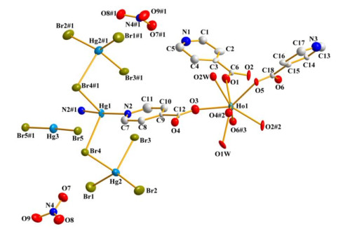
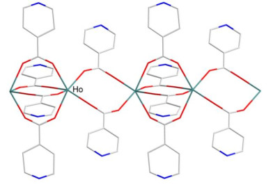
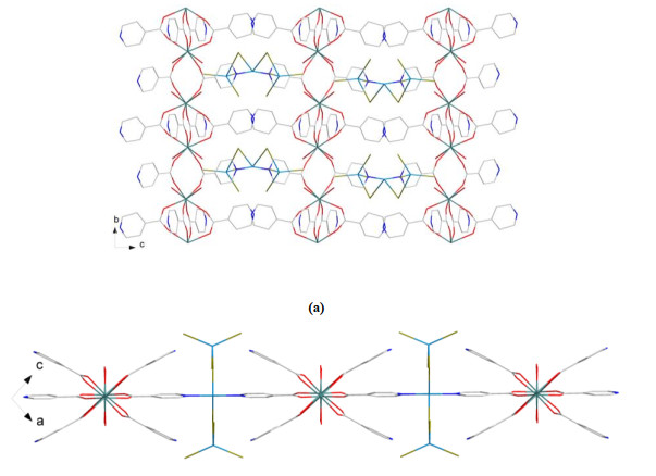
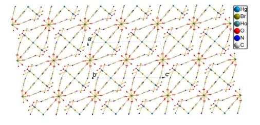
The Ho3+ ion displays a distorted square anti-prism conformation and is coordinated by six oxygen atoms from six isonicotinato ligands and two oxygen atoms of two coordination water molecules. The Ho(Ⅲ)–OIA bond distances in 1 fall in the span of 2.230(17)~2.381(14) Å with an average value of 2.314(19) Å, which is obviously shorter than that of Ho(Ⅲ)–Owater (2.474(16) and 2.470(16) Å). This suggests that the Ho(Ⅲ) ions have obviously stronger affinity to the isonicotinato ligands than to the water. All Ho(Ⅲ)–O bond distances reside in the normal range and are comparable with that reported[44]. The O–Ho–O bond angles locate in the range of 69.4(5)°~146.0(6)°. The neighboring Ho(Ⅲ) ions connect together via four or two isonicotinato ligands to yield a one-dimensional (1D) -Ho–(IA)4–Ho–(IA)2–Ho–(IA)4–Ho–(IA)2–Ho- chain running along the b direction, as presented in Fig. 3. The neighboring Ho(Ⅲ)⋯Ho(Ⅲ) distances are 4.3434(1) and 5.1335(2) Å in the chain. The 1D chains are interlinked by Hg(1) ions to give a two-dimensional (2D) layer parallel to the bc plane, as shown in Fig. 4. In complex 1, all of the NO3– anions are isolated. As presented in Table 2, there are only two hydrogen bonding interactions existing in 1, namely, N(1)–H(1B)⋯O(7) (–x, 2 – y, 1 – z) and N(3)–H(3A)⋯O(8) (x, 2 – y, –1/2 + z). Both of them just link the NO3– anions to the isonicotinato ligands, so they can not lead to a higher dimensional structure. With regard to complex 1, the Van der Waals force, hydrogen bonding interactions and electrostatic interactions between the [Ho(IA)(HIA)2(H2O)2]2+ ions, (Hg3Br8)2– ions, HgBr2 moieties and NO3– anions solidify the crystal packing structure (Fig. 5).
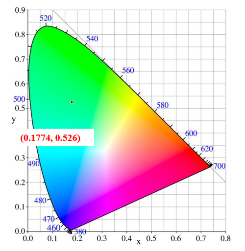
It is well-known that holmium and mercury compounds can usually display photoluminescence. Therefore, the photo-luminescence performance of the title compound was tested by using solid samples at room temperature. As given in Fig. 6, the photoluminescence adsorption of this compound locates in the range of 600~650 nm and the maximum peak is at 616 nm. When excited by the 616 nm wavelength, the compound shows two photoluminescence emission peaks that locate at 468 nm (blue region) and 545 nm (green region) and, the latter one is much stronger. These two emission peaks shall be attributed to the 5I8 → 5G6 and 5S2 → 5I8 characteristic emission of the 4f electron intrashell transition of the Ho(Ⅲ) ions[45, 46]. With regard to complex 1, it has a CIE chromaticity coordinate of (0.1774, 0.526) in the green region (Fig. 7). Therefore, 1 may be a potential green light emitting material. It should be pointed out that the excitation wavelength is 616 nm in the red region, but the emission wavelengths are in the blue and green regions. So, the photoluminescence of complex 1 is upconversion green emission.

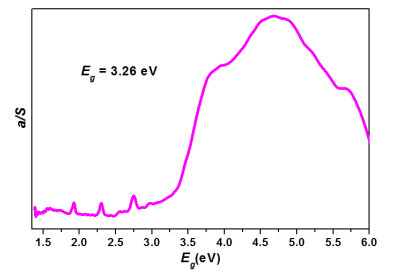
Generally speaking, mercury materials can display semiconductor behaviors. For the sake of further investigating its photophysical performances, the solid state UV-visible diffuse reflectance spectrum of complex 1 was carried out at room temperature by using a powder sample. The solid state UV-visible diffuse reflectance spectrum data were converted with the Kubelka-Munk formula α/S = (1 – R)2/2R that is commonly applied for such research. With regard to this formula, α indicates the absorption coefficient, S is the scattering coefficient, while R stands for the reflection rate. With the use of linear epitaxy from the maximum absorption edge of the α/S versus energy curve, its semiconductive band gap value could be ascertained. As a result, by using this method, the semiconductive band gap of complex 1 is found to be 3.26 eV, as depicted in Fig. 8. From the diagram, several small bands smaller than 3.0 eV observed can be ascribed to the Ho(Ⅲ) ions. Based on the band gap value of 1, this complex may be a wide band gap semiconductive material. The maximum absorption edge of the diagram is not steep, so it shall undergo an indirect transition in this complex[47].
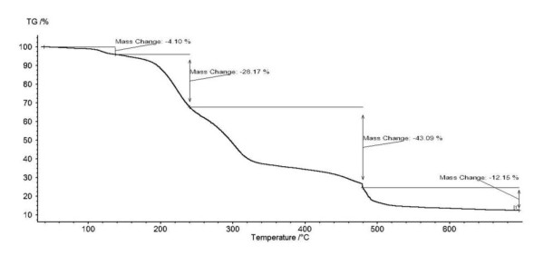
The TG measurement of complex 1 was carried out under nitrogen atmosphere. As depicted in Fig. 9, complex 1 undergoes a four-step decomposition process with the weight loss being 4.10%, 28.17%, 43.09% and 12.15%, respectively. The total weight loss is 88.48%. On the first step (until 137 ℃) the weight loss is 4.10% due to the removal of all NO3– anions (calcd. 4.19%). In the second step from 137 to 240 ℃, the weight loss is 28.17% assigned to the departure of all isonicotinato ligands and water molecules (calcd. 28.21%). In the third step (from 240 to 480 ℃), the mass change is 43.09% probably due to the loss of Hg3Br8 moiety (calcd. 43.33%). The last step is from 480 to 700 ℃ with the weight loss of 12.15% caused by the removal of HgBr2 moiety (calcd. 12.57%).
A novel holmium-mercury compound was synthesized and characterized. This compound displays a two-dimensional layer-like structure. Using solid-state samples, it shows upconversion green photoluminescence emission. The emission peaks should be ascribed to the 5I8 → 5G6 and 5S2 → 5I8 characteristic emission of the 4f electrons of Ho3+ ions. This compound has a CIE chromaticity coordinate (0.1774, 0.526).
Wen, G. X.; Han, M. L.; Wu, X. Q.; Wu, Y. P.; Dong, W. W.; Zhao, J.; Li, D. S.; Ma, L. F. A multi-responsive luminescent sensor based on a super-stable sandwich-type terbium(Ⅲ)-organic framework. Dalton Trans. 2016, 45, 15492–15499. doi: 10.1039/C6DT03057B
Liu, S. J.; Cao, C.; Xie, C. C.; Zheng, T. F.; Tong, X. L.; Liao, J. S.; Chen, J. L.; Wen, H. R.; Chang, Z.; Bu, X. H. Tricarboxylate-based GdⅢ coordination polymers exhibiting large magnetocaloric effects. Dalton Trans. 2016, 45, 9209–9215. doi: 10.1039/C6DT01349J
Qiu, L. Y.; Yu, C. F.; Wang, X. L.; Xie, Y. B.; Kirillov, A. M.; Huang, W.; Li, J. P.; Gao, P.; Wu, T.; Gu, X. W.; Nie, Q.; Wu, D. Y. Tuning the solid-state white light emission of postsynthetic lanthanide-encapsulated double-layer MOFs for three-color luminescent thermometry applications. Inorg. Chem. 2019, 58, 4524–4533. doi: 10.1021/acs.inorgchem.9b00084
Zhou, Z.; Gu, J. P.; Qiao, X. G.; Wu, H. X.; Fu, H. R.; Wang, L.; Li, H. Y.; Ma, L. F. Double protected lanthanide fluorescence core@shell colloidal hybrid for the selective and sensitive detection of ClO-. Sensor Actuat. B-Chem. 2019, 282, 437–442. doi: 10.1016/j.snb.2018.11.103
Wei, J. H.; Yi, J. W.; Han, M. L.; Li, B.; Liu, S.; Wu, Y. P.; Ma, L. F.; Li, D. S. A water-stable terbium(Ⅲ)-organic framework as a chemosensor for inorganic ions, nitro-containing compounds and antibiotics in aqueous solutions. Chem. Asian J. 2019, 14, 3694–3701. doi: 10.1002/asia.201900706
Samannan, B.; Selvam, J.; Thavasikani, J. Synthesis, characterization and anticancer activity of transition metal substituted polyoxometalate-β-cyclodextrin composites. Asian J. Chem. 2020, 32, 297–302. doi: 10.14233/ajchem.2020.22321
Xiong, X. H.; Tao, Y.; Yu, Z. W.; Yang, L. X.; Sun, L. J.; Fan, Y. L.; Luo, F. Selective extraction of thorium from uranium and rare earth elements using sulfonated covalent organic framework and its membrane derivate. Chem. Engin. J. 2020, 384, 123240–7. doi: 10.1016/j.cej.2019.123240
Yao, X.; An, G. H.; Li, Y. X.; Yan, P. F.; Li, W. Z.; Li, G. M. Effect of nuclearity and symmetry on the single-molecule magnets behavior of seven-coordinated β-diketonate Dy(Ⅲ) complexes. J. Solid State Chem. 2019, 274, 295–302. doi: 10.1016/j.jssc.2019.03.044
Liu, S. J.; Cao, C.; Yao, S. L.; Zheng, T. F.; Wang, Z. X.; Liu, C.; Liao, J. S.; Chen, J. L.; Li, Y. W.; Wen, H. R. Temperature- and vapor-induced reversible single-crystal-to-single-crystal transformations of three 2D/3D Gd(Ⅲ)-organic frameworks exhibiting significant magnetocaloric effects. Dalton Trans. 2017, 46, 64–70. doi: 10.1039/C6DT03589B
Ahmed, N.; Nisar, J.; Kouser, R.; Nabi, A. G.; Mukhtar, S.; Saeed, Y.; Nasim, M. H. Study of electronic, magnetic and optical properties of KMS2 (M = Nd, Ho, Er and Lu): first principle calculations. Mater. Res. Express 2017, 4, 065903–8. doi: 10.1088/2053-1591/aa75fc
Wu, H. Q.; Yan, C. S.; Luo, F.; Krishna, R. Beyond crystal engineering: significant enhancement of C2H2/CO2 separation by constructing composite material. Inorg. Chem. 2018, 57, 3679–3682. doi: 10.1021/acs.inorgchem.8b00341
Knoefel, N. D.; Schoo, C.; Seifert, T. P.; Roesky, P. W. A dimolybdenum paddlewheel as a building block for heteromultimetallic structures. Dalton Trans. 2020, 49, 1513–1521. doi: 10.1039/C9DT04167B
Li, J. Q.; Gong, L. L.; Feng, X. F.; Zhang, L.; Wu, H. Q.; Yan, C. S.; Xiong, Y. Y.; Gao, H. Y.; Luo, F. Direct extraction of U(VI) from alkaline solution and seawater via anion exchange by metal-organic framework. Chem. Eng. J. 2017, 316, 154–159. doi: 10.1016/j.cej.2017.01.046
Cai, H.; Li, N.; Zhang, N.; Yang, Z.; Cao, J.; Lin, Y.; Min, N.; Wang, J. Metal-directed supramolecular architectures based on the bifunctional ligand 2, 5-bis(1H-1, 2, 4-triazol-1-yl)terephthalic acid. Acta Crystallogr. C 2020, 76, 118–124. doi: 10.1107/S2053229620000248
Gong, L. L.; Feng, X. F.; Luo, F.; Yi, X. F.; Zheng, A. M. Removal and safe reuse of highly toxic allyl alcohol using a highly selective photo-sensitive metal-organic framework. Green Chem. 2016, 18, 2047–2055. doi: 10.1039/C5GC02182K
Zhao, Y.; Zhai, Z. M.; Liu, X. Y.; Yang, X. G.; Ma, L. F.; Wang, L. Y. Two cobalt(II) coordination polymers based on 5-i-butoxyisophthalate and dipyridyl: syntheses, structures and efficient oxygen evolution reaction. J. Solid State Chem. 2019, 278, 120913–6. doi: 10.1016/j.jssc.2019.120913
Anuja, K.; Reddy, K. H; Srinivasulu, K.; Dhanalakshmi, D. Synthesis, structural characterization and DNA binding studies on transition metal complexes with 2-formylpyridine benzoylhydrazone. Asian J. Chem. 2020, 32, 322–328. doi: 10.14233/ajchem.2020.22389
Yin, W. H.; Xiong, Y. Y.; Wu, H. Q.; Tao, Y.; Yang, L. X.; Li, J. Q.; Tong, X. L.; Luo, F. Functionalizing a metal-organic framework by a photoassisted multicomponent postsynthetic modification approach showing highly effective Hg(II) removal. Inorg. Chem. 2018, 57, 8722–8725. doi: 10.1021/acs.inorgchem.8b01457
Martinez, B.; Livache, C.; Goubet, N.; Jagtap, A.; Cruguel, H.; Ouerghi, A.; Lacaze, E.; Silly, M. G.; Lhuillier, E. Probing charge carrier dynamics to unveil the role of surface ligands in HgTe narrow band gap nanocrystals. J. Phys. Chem. C 2018, 122, 859–865.
Fan, C. B.; Gong, L. L.; Huang, L.; Luo, F.; Krishna, R.; Yi, X. F.; Zheng, A. M.; Zhang, L.; Pu, S. Z.; Feng, X. F.; Luo, M. B.; Guo, G. C. Significant enhancement of C2H2/C2H4 separation by a photochromic diarylethene unit: a temperature- and light-responsive separation switch. Angew. Chem. Int. Ed. 2017, 56, 7900–7906. doi: 10.1002/anie.201702484
Du, X.; Su, H.; Zhang, X. Metal-organic framework-derived M (M = Fe, Ni, Zn and Mo) doped Co9S8 nanoarrays as efficient electrocatalyst for water splitting: the combination of theoretical calculation and experiment. J. Catal. 2020, 383, 103–116. doi: 10.1016/j.jcat.2020.01.015
Cheng, Y. J.; Wang, R.; Wang, S.; Xi, X. J.; Ma, L. F.; Zang, S. Q. Encapsulating [Mo3S13]2– clusters in cationic covalent organic frameworks: enhancing stability and recyclability by converting a homogeneous photocatalyst to a heterogeneous photocatalyst. Chem. Commun. 2018, 54, 13563–13566. doi: 10.1039/C8CC07784C
Wu, Y. P.; Tian, J. W.; Liu, S.; Li, B.; Zhao, J.; Ma, L. F.; Li, D. S.; Lan, Y. Q.; Bu, X. Bi-microporous metal-organic-frameworks with cubane [M4(OH)4] (M = Ni, Co) clusters and pore space partition for electrocatalytic methanol oxidation reaction. Angew. Chem. Int. Ed. 2019, 58, 12185–12189. doi: 10.1002/anie.201907136
Wu, X. X.; Fu, H. R.; Han, M. L.; Zhou, Z.; Ma, L. F. Tetraphenylethylene immobilized metal-organic frameworks: highly sensitive fluorescent sensor for the detection of Cr2O72– and nitroaromatic explosives. Cryst. Growth Des. 2017, 17, 6041–6048. doi: 10.1021/acs.cgd.7b01155
Liu, S. J.; Cao, C.; Yang, F.; Yu, M. H.; Yao, S. L.; Zheng, T. F.; He, W. W.; Zhao, H. X.; Hu, T. L.; Bu, X. H. High proton conduction in two CoII and MnII anionic metal-organic frameworks derived from 1, 3, 5-benzenetricarboxylic acid. Cryst. Growth Des. 2016, 16, 6776–6780. doi: 10.1021/acs.cgd.6b00776
Yang, X. G.; Zhai, Z. M.; Lu, X. M.; Zhao, Y.; Chang, X. H.; Ma, L. F. Room temperature phosphorescence of Mn(II) and Zn(II) coordination polymers for photoelectron response applications. Dalton Trans. 2019, 48, 10785–10789. doi: 10.1039/C9DT02178G
Zhao, Y.; Deng, D. S.; Ma, L. F.; Ji, B. M.; Wang, L. Y. A new copper-based metal-organic framework as a promising heterogeneous catalyst for chemo- and regio-selective enamination of β-ketoesters. Chem. Commun. 2013, 49, 10299–10301. doi: 10.1039/c3cc45310c
Yang, X. G.; Ma, L. F.; Yan, D. P. Facile synthesis of 1D organic-inorganic perovskite micro-belts with high water stability for sensing and photonic applications. Chem. Sci. 2019, 10, 4567–4572. doi: 10.1039/C9SC00162J
Yao, S. L.; Liu, S. J.; Tian, X. M.; Zheng, T. F.; Cao, C.; Niu, C. Y.; Chen, Y. Q.; Chen, J. L.; Huang, H.; Wen, H. R. A Zn(II)-based metal-organic framework with a rare tcj topology as a turn-on fluorescent sensor for acetylacetone. Inorg. Chem. 2019, 58, 3578–3581. doi: 10.1021/acs.inorgchem.8b03316
Zhao, Y.; Wang, L.; Fan, N. N.; Han, M. L.; Yang, G. P.; Ma, L. F. Porous Zn(II)-based metal-organic frameworks decorated with carboxylate groups exhibiting high gas adsorption and separation of organic dyes. Cryst. Growth Des. 2018, 18, 7114–7121. doi: 10.1021/acs.cgd.8b01290
Luo, M. B.; Xiong, Y. Y.; Wu, H. Q.; Feng, X. F.; Li, J. Q.; Luo, F. The MOF+ technique: a significant synergic effect enables high performance chromate removal. Angew. Chem. Int. Ed. 2017, 56, 16376–16379. doi: 10.1002/anie.201709197
Fu, H. R.; Wang, N.; Qin, J. H.; Han, M. L.; Ma, L. F.; Wang, F. Spatial confinement of a cationic MOF: a SC-SC approach for high capacity Cr(VI)-oxyanion capture in aqueous solution. Chem. Commun. 2018, 54, 11645–11648. doi: 10.1039/C8CC05990J
Wang, H.; Meng, W.; Wu, J.; Ding, J.; Hou, H.; Fan, Y. Crystalline central-metal transformation in metal-organic frameworks. Coor. Chem. Rev. 2016, 307, 130–146. doi: 10.1016/j.ccr.2015.05.009
Cryer, M. E.; Fiedler, H.; Halpert, J. E. Photo-electrosensitive memristor using oxygen doping in HgTe nanocrystal films. ACS Appl. Mater. Inter. 2018, 10, 18927–18934. doi: 10.1021/acsami.8b05429
Fu, H. R.; Zhao, Y.; Zhou, Z.; Yang, X. G.; Ma, L. F. Neutral ligand TIPA-based two 2D metal-organic frameworks: ultrahigh selectivity of C2H2/CH4 and efficient sensing and sorption of Cr(VI). Dalton Trans. 2018, 47, 3725–3732. doi: 10.1039/C8DT00206A
Yao, S. L.; Zheng, T. F.; Tian, X. M.; Liu, S. J.; Cao, C.; Zhu, Z. H.; Chen, Y. Q.; Chen, J. L.; Wen, H. R. Dicarboxylate-induced structural diversity of luminescent ZnII/CdII coordination polymers derived from V-shaped bis-benzimidazole. CrystEngComm. 2018, 20, 5822–5832. doi: 10.1039/C8CE01261J
Zhai, Z. M.; Yang, X. G.; Yang, Z. T.; Lu, X. M.; Ma, L. F. Trinuclear Ni(II) oriented highly dense packing and π-conjugation degree of metal-organic framework for efficient water oxidation. CrystEngComm. 2019, 21, 5862–5866. doi: 10.1039/C9CE00944B
Zhao, Y.; Yang, X. G.; Lu, X. M.; Yang, C. D.; Fan, N. N.; Yang, Z. T.; Wang, L. Y.; Ma, L. F. {Zn6} Cluster based metal-organic framework with enhanced room-temperature phosphorescence and optoelectronic performances. Inorg. Chem. 2019, 58, 6215–6221. doi: 10.1021/acs.inorgchem.9b00450
Qin, J. H.; Huang, Y. D.; Zhao, Y.; Yang, X. G.; Li, F. F.; Wang, C.; Ma, L. F. Highly dense packing of chromophoric linkers achievable in a pyrene-based metal-organic framework for photoelectric response. Inorg. Chem. 2019, 58, 15013–15016. doi: 10.1021/acs.inorgchem.9b02203
Zhou, Z.; Han, M. L.; Fu, H. R.; Ma, L. F.; Luo, F.; Li, D. S. Engineering design toward exploring the functional group substitution in 1D channels of Zn-organic frameworks upon nitro explosives and antibiotics detection. Dalton Trans. 2018, 47, 5359–5365. doi: 10.1039/C8DT00594J
Sheldrick, G. M. Crystal structure refinement with SHELXL. Acta Crystallogr. Sec. C-Struct. Chem. 2015, 71, 3–8. doi: 10.1107/S2053229614024218
Lin, W. S.; Kuang, H. M.; Luo, H.; Chen, W. T. Upconversion photoluminescence and energy transfer mechanism of a novel terbium-mercury compound. Chin. J. Struct. Chem. 2019, 38, 1012–1020.
Lin, W. S.; Chen, W. T. Magnetic, photoluminescent and semiconductive properties of a novel 4f-5d bromide compound (La6Hg5Br26)[4(HgBr2)](2Br). Chin. J. Struct. Chem. 2020, 1, 154–163.
Kuang, H. M.; Zhang, Z. X.; Lin, L. Z.; Chen, H. L.; Chen, W. T. Preparation, structure, photoluminescence and energy transfer mechanism of a novel holmium complex. Chin. J. Struct. Chem. 2019, 38, 337–344.
Rajagopalan, K.; Jagannathan, T. Up/down conversion luminescence properties of (Na0.5Gd0.5)MoO4: Ln3+ (Ln = Eu, Tb, Dy, Yb/Er, Yb/Tm, and Yb/Ho) microstructures: synthesis, morphology, structural and magnetic investigation. New J. Chem. 2014, 38, 3480–3491. doi: 10.1039/C4NJ00165F
Tishchenko, M. A.; Gerasimenko, G. I.; Poluektov, N. S. Spectrophotometric study of the complexing of neodymium, holmium, and erbium ions with diantipyrylmethane and some of its homologs in aqueous-ethanol solutions. Doklady Akademii Nauk SSSR 1975, 222, 1107–1110.
Huang, F. Q.; Mitchell, K.; Ibers, J. A. New layered materials: syntheses, structures, and optical and magnetic properties of CsGdZnSe3, CsZrCuSe3, CsUCuSe3, and BaGdCuSe3. Inorg. Chem. 2001, 40, 5123–5126. doi: 10.1021/ic0104353

Figure 2 An ORTEP view of compound 1 with hydrogen atoms being omitted for clarity. Displacement ellipsoids are drawn at the 20% probability level. Symmetry codes: #1: –x, y, –z + 1/2; #2: –x + 1, –y, –z + 1; #3: –x + 1, –y + 1, –z + 1

Figure 5 A packing diagram of compound 1 with dashed lines representing the hydrogen bonding interactions (Å, °): N(1)–H(1B)⋯O(7) (–x, 2 – y, 1 – z); N(3)–H(3A)⋯O(8) (x, 2 – y, –1/2 + z)

Figure 6 Upconversion photoluminescence spectra of compound 1 measured using solid-state samples at room temperature with solid and dashed lines representing the emission and excitation spectra, respectively
Table 1. Selected Bond Lengths (Å) and Bond Angles (°)
| Bond | Dist. | Bond | Dist. | |
| Hg(1)–N(2) | 2.16(2) | Hg(3)–Br(5)#1 | 2.424(2) | |
| Hg(1)–N(2)#1 | 2.16(2) | Ho(1)–O(1) | 2.325(15) | |
| Hg(1)–Br(4) | 2.788(3) | Ho(1)–O(2)#2 | 2.331(14) | |
| Hg(1)–Br(4)#1 | 2.788(3) | Ho(1)–O(3) | 2.308(19) | |
| Hg(2)–Br(1) | 2.493(4) | Ho(1)–O(4)#2 | 2.230(17) | |
| Hg(2)–Br(2) | 2.516(4) | Ho(1)–O(5) | 2.381(14) | |
| Hg(2)–Br(3) | 2.663(3) | Ho(1)–O(6)#3 | 2.314(16) | |
| Hg(2)–Br(4) | 2.978(3) | Ho(1)–O(1W) | 2.474(16) | |
| Hg(3)–Br(5) | 2.424(2) | Ho(1)–O(2W) | 2.470(16) | |
| Angle | (°) | Angle | (°) | |
| N(2)#1–Hg(1)–N(2) | 165.4(11) | Br(5)–Hg(3)–Br(5)#1 | 171.99(16) | |
| N(2)#1–Hg(1)–Br(4) | 95.1(6) | Hg(1)–Br(4)–Hg(2) | 87.30(9) | |
| N(2)–Hg(1)–Br(4) | 94.5(5) | O(4)–Ho(1)–O(3)#2 | 96.3(7) | |
| N(2)#1–Hg(1)–Br(4)#1 | 94.5(5) | O(4)–Ho(1)–O(6)#3 | 146.0(6) | |
| N(2)–Hg(1)–Br(4)#1 | 95.1(6) | O(3)#2–Ho(1)–O(6)#3 | 81.1(6) | |
| Br(4)–Hg(1)–Br(4)#1 | 97.89(13) | O(4)–Ho(1)–O(1)#3 | 139.1(6) | |
| Br(1)–Hg(2)–Br(2) | 131.49(14) | O(1)#3–Ho(1)–O(2W) | 69.8(5) | |
| Br(1)–Hg(2)–Br(3) | 111.12(13) | O(4)–Ho(1)–O(2W) | 70.0(5) | |
| Br(2)–Hg(2)–Br(3) | 109.69(12) | O(4)–Ho(1)–O(2) | 81.5(6) | |
| Br(1)–Hg(2)–Br(4) | 99.73(12) | O(3)#2–Ho(1)–O(2) | 144.4(6) | |
| Br(2)–Hg(2)–Br(4) | 99.06(13) | O(5)–Ho(1)–O(2W) | 69.4(5) | |
| Br(3)–Hg(2)–Br(4) | 98.50(9) | O(1)#3–Ho(1)–O(2) | 125.1(6) | |
| Symmetry codes: #1: –x, y, –z + 1/2; #2: –x + 1, –y, –z + 1; #3: –x + 1, –y + 1, –z + 1 | ||||
 下载: 导出CSV
Table 2. Hydrogen Bonding Interactions
| D–H⋯A | D–H (Å) | H⋯A (Å) | D⋯A (Å) | D–H⋯A (º) |
| N(1)–H(1B)⋯O(7)#1 | 0.86 | 2.32 | 3.07(4) | 175 |
| N(3)–H(3A)⋯O(8)#2 | 0.86 | 2.17 | 2.92(4) | 146 |
| Symmetry codes: #1: –x, 2 – y, 1 – z; #2: x, 2 – y, –1/2 + z | ||||
下载: 导出CSV

 扫一扫看文章

扫一扫关注我们


 下载:
下载: