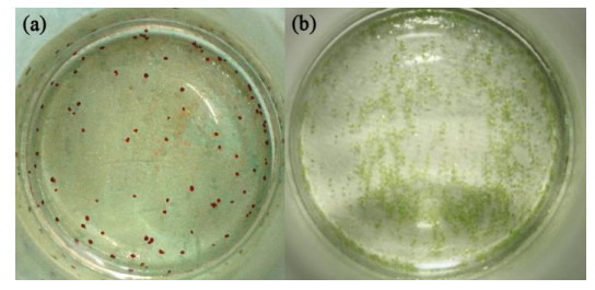
Figure 1.
Crystal photos of the complexes. (a) Red crystal of 1; (b) Green crystal of 2
Syntheses, Crystal Structures, Non-covalent Interactions and Properties of a Nickel(II) Complex Monomer and Its Dimer
Jin-Feng CHU , Shu-Ying WANG , Ming-Yu ZHANG , Qi-Xin XU , You-Qing WANG

Supramolecular complexes have attracted much attention in recent years due to their interesting structures and potential applications in fluorescence, electrochemistry, magnetism, catalysis, gas storage and so on[1-8]. So far, many interesting supramolecular complexes have been designed and prepared by molecular design and crystal engineering methods, but it is still very difficult to obtain the target complexes with expected structures and properties. In the preparation of complexes, many factors may affect the structures of complexes, such as the coordination geometry of metal center, the structure of organic ligand, metal ligand ratio, pH value, solvent and even temperature[9-13]. In addition, non-covalent interactions such as hydrogen bonds, π-π stacking interactions and anion-π interactions play important roles in the formation of various supramolecular structures[14-25].
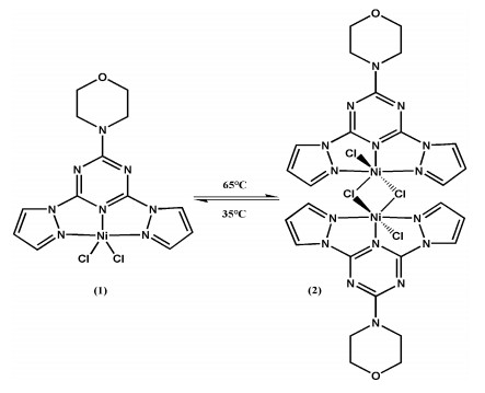
In our previous studies, we reported the diversity of reactions of a polydentate ligand 2-morpholine-4-yl-4, 6- di-pyrazol-1-yl-1, 3, 5-triazine (L) with different metal ions such as Co(II), Mn(II), Cr(III) and Fe(III)[22]. Recently, when we used ligand L to react with NiCl2·2H2O, an interesting coordination polymerization isomerism phenomenon appeared. A pair of nickel(II) supramolecular complexes monomer and dimer, (1) and (2), were synthesized under different temperature conditions, and their syntheses, crystal structures, non-covalent interactions and properties were discussed.
The solvents used in the reactions were dried according to standard procedures. All chemicals were obtained from commercial sources with reagent grade and used without further purification. The polydentate ligand L was prepared according to the literature[26]. C, H and N analyses were carried out using Vario elemental analysis III instrument. The FT-IR spectra were recorded on a Bruker Vector 22 infrared spectrometer using the KBr pellet method. Thermogravimetric (TG) analysis was carried out on a locally produced HCT-2 thermal analysis system. The sample was loaded in a sealed aluminum crucible (5~10 mg) and heated from room temperature to 800 ℃ at a heating rate of 10 ℃/min under nitrogen atmosphere in order to avoid oxidization. UV/Vis/NIR diffuse-reflectance spectra were obtained with a Cary 500 UV/Vis/NIR spectrophoto- meter. BaSO4 powder was used as reference (100% reflectance). The fluorescence analysis was applied to characterize 1, 2 and L in the aqueous solution (1 × 10⁻⁵ mol·L–1) at room temperature.
A solution of L (100 mg, 0.34 mmol) in acetonitrile (4 mL) was added dropwise to a solution of NiCl2·2H2O (98 mg, 0.34 mmol) in acetonitrile (15 mL) and the resulting solution was left undisturbed. Red block crystals (Fig. 1(a)) suitable for X-ray crystallography were deposited from the solution after several days by slow evaporation of the solvent at 35 ℃. Yield: 29% (based on Ni). Anal. Calcd. (%) for C13H14Cl2N8NiO: C, 36.49; H, 3.30; N, 26.19. Found (%): C, 36.36; H, 3.28; N, 26.15. IR (KBr, cm–1): 3125 (w), 1650 (s), 1591 (m), 1550 (w), 1513 (s), 1479 (s), 1445 (m), 1396 (s), 1295 (w), 1267 (m), 1178 (w), 1116 (w), 1069 (w), 1025 (m), 953 (w), 936 (w), 908 (w), 812 (m), 794 (m), 780 (m), 593 (w).
The preparation method of the reaction mixture of com- plex 2 was similar to that of 1. Green prism crystals (Fig. 1(b)) suitable for X-ray crystallography deposited from the solution after several days by slow evaporation of the solvent at 65 ℃. Yield: 27% (based on Ni). Anal. Calcd. (%) for C26H28Cl4N16Ni2O2: C, 36.49; H, 3.30; N, 26.19. Found (%): C, 36.28; H, 3.01; N, 26.22. IR (KBr, cm–1): 3085 (w), 1639 (s), 1593 (m), 1548 (w), 1512 (s), 1488 (s), 1471 (s), 1405 (s), 1304 (w), 1294 (w), 1266 (m), 1185 (w), 1109 (w), 1076 (w), 1022 (w), 957 (m), 938 (w), 906 (w), 812 (m), 795 (m), 780 (m), 595 (w).
Suitable single crystals of complexes 1 and 2 were mounted onto the end of a thin glass fiber using Fomblin oil. X-ray diffraction intensity data were measured on a Xcalibur, Eos, Gemini diffractometer using MoKα radiation (λ = 0.71073 Å). Using OLEX2[27], the structures were solved by direct methods with SHELXS program[28] and refined by full-matrix least-squares techniques on F2 with SHELXL[29]. H atoms on C atoms were positioned geometrically and refined as riding atoms. Crystal data, data collection and structure refinement details are summarized in Table 1. In the crystal structure of 1, the positions of N(5), O(1), C(6) and C(7) atoms of the morpholine ring were disordered between two sites. The occupancies were refined with the sum of the occupancies of disordered sites constrained to 1, which led to an occupancy ratio of 0.618(5): 0.382(5). Graphical pictures were prepared by Diamond software.
 DownLoad:
CSV
DownLoad:
CSV
| Complex | 1 | 2 |
| Chemical formula | C13H14Cl2N8NiO | C26H28Cl4N16Ni2O2 |
| Formula weight | 427.91 | 855.86 |
| Temperature (K) | 123.0 | 97.2 |
| Crystal system | Orthorhombic | Monoclinic |
| Space group | Cmca | P21/c |
| a (Å) | 12.6569(18) | 14.600(3) |
| b (Å) | 10.4622(7) | 13.11(2) |
| c (Å) | 25.66(2) | 9.183(3) |
| α (°) | 90.00 | 90.00 |
| β (°) | 90.00 | 105.29(3) |
| γ (°) | 90.00 | 90.00 |
| V (Å3) | 3398(3) | 1695(3) |
| Z | 8 | 2 |
| μ (mm–1) | 1.477 | 1.480 |
| Crystal size (mm3) | 0.25 × 0.20 × 0.20 | 0.10 × 0.10 × 0.03 |
| Index ranges | –15≤h≤15, –7≤k≤12, –31≤l≤30 | –18≤h≤14, –7≤k≤16, –9≤l≤11 |
| 2θ range for data collection (°) | 6.36 to 52 | 6.22 to 52 |
| F(000) | 1743 | 872 |
| ρcalc (g·cm-3) | 1.673 | 1.677 |
| Reflections collected | 4897 | 7129 |
| Independent reflections (Rint) | 1745 (0.0252) | 3326 (0.0676) |
| Data/restraints/parameters | 1745/122/155 | 3326/0/226 |
| Goodness-of-fit on F2 | 1.054 | 0.999 |
| R/wR (I > 2σ(I)) | 0.0282/0.0642 | 0.0613/0.0694 |
| R/wR (all data) | 0.0379/0.0680 | 0.1097/0.0806 |
| Largest diff. peak/hole (e·Å−3) | 0.289/–0.412 | 0.487/–0.553 |
Crystal structure analysis shows that complex 1 belongs to orthorhombic space group Cmca. The nickel(II) center is coordinated by three nitrogen atoms from triazine and pyrazolyl rings of L and two chloride anions with a NiN3Cl2 donor set. The coordination geometry around the metal center can be best described as a distorted square pyramid, where the three nitrogen atoms N(1), N(3) and N(1)i (atom at –x+1, y, z) of L and the Cl(2) atom occupy the basal posi- tions, and the Cl(1) atom located at the apical one (Fig. 2(a)). The three aromatic rings of the L ligand are nearly coplanar. The dihedral angles between the triazine and pyrazolyl planes are both 7.325°. The selected bond lengths and bond angles for complex 1 are listed in Table 2.
DownLoad:
CSV
| Bond | Dist. | Bond | Dist. | Bond | Dist. | ||
| Ni(1)–Cl(1) | 2.2902(14) | Ni(1)–Cl(2) | 2.2527(15) | Ni(1)–N(1) | 2.1601(17) | ||
| Ni(1)–N(3) | 1.981(2) | Ni(1)–N(1)i | 2.1601(17) | ||||
| Angle | (°) | Angle | (°) | Angle | (°) | ||
| Cl(1)–Ni(1)–Cl(2) | 109.12(5) | Cl(1)–Ni(1)–N(1) | 97.46(5) | Cl(1)–Ni(1)–N(3) | 96.44(8) | ||
| Cl(1)–Ni(1)–N1(i) | 97.46(5) | Cl(2)–Ni(1)–N(1) | 100.59(5) | Cl(2)–Ni(1)–N(1)i | 100.59(5) | ||
| Cl(2)–Ni(1)–N(3) | 154.44(7) | N(3)–Ni(1)–N(1) | 75.37(5) | N(3)–Ni(1)–N(1)i | 75.37(5) | ||
| N(1)–Ni(1)–N(1)i | 148.37(9) | ||||||
| Symmetry transformation: i: −x+1, y, z | |||||||
As shown in Fig. 2(b), the anion-π interactions on both sides of the triazine ring are observed between Cl(2)i (atom at x, –y + 1, –z), O(1)ii (atom at –x + 1, y + 1/2, –z + 1/2) and the triazine ring. The distance between Cl(2)i and the centroid of the triazine is 3.369 Å, while that between O(1)ii and the centroid of the triazine is 2.815 Å. The angles of the Cl(2)i···centroid and O(1)ii···centroid axes to the triazine ring plane are 73.22 and 77.69°, respectively. Though no classical hydrogen bonding interactions are observed between the molecules in 1, the C(3)–H(3)···Cl(1)i (atom at x + 1/2, y − 1/2, z) hydrogen bond interaction is present (Fig. 2(c) and Table 3). The molecules of 1 are interlinked through non-covalent interactions involving hydrogen bonding inter- actions, chloride-π interactions and oxygen-π interactions, which forms a three-dimensional network in the crystalline solid (Fig. 2(d)).
 DownLoad:
CSV
DownLoad:
CSV
| Complex | D–H···A | d(D–H) | d(H···A) | d(D···A) | ∠DHA |
| 1 | C(3)–H(3)···Cl(1)i | 0.95 | 2.67 | 3.437(1) | 138 |
| 2 | C(6)–H(6)···O(1)i | 0.93 | 2.52 | 3.361(5) | 151 |
| C(12)–H(12A)···O(1)ii | 0.97 | 2.80 | 3.730(6) | 162 | |
| C(13)–H(13A)···N(6)iii | 0.97 | 2.61 | 3.424(6) | 142 | |
| C(2)–H(2)···Cl(2)iv | 0.93 | 2.96 | 3.581(5) | 125 | |
| C(3)–H(3)···Cl(1)iii | 0.93 | 2.56 | 3.481(5) | 170 | |
| C(5)–H(5)···Cl(1)v | 0.93 | 2.66 | 3.578(7) | 171 | |
| C(11)–H(11A)···Cl(1)vi | 0.97 | 2.71 | 3.653(5) | 163 | |
| C(13)–H(13B)···Cl(1)iii | 0.97 | 2.82 | 3.654(6) | 144 | |
| Symmetry codes for 1: (i) x + 1/2, y − 1/2, z; for 2: (i) −x, y − 1/2, −z + 1/2; (ii) x, −y + 3/2, z + 1/2; (iii) x, −y + 3/2, z − 1/2; (iv) −x + 1, y + 1/2, −z + 5/2; (v) x, −y + 1/2, z − 1/2; (vi) −x, −y + 1, −z + 1 | |||||
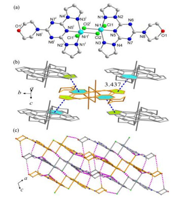
Complex 2 crystallizes in the monoclinic space group P21/c, with the asymmetric unit containing one nickel(II) cation, one L ligand, one monodentate chloride ion and one bridging chloride ion. All the atoms in the asymmetric unit are in general positions. As shown in Fig. 3(a), an inversion center is located at the core of the complex formed by two nickel(II) ions (Ni(1) and Ni(1)i (atom at –x + 1, –y + 1, –z + 2) and two bridging chloride anions (Cl(2) and Cl(2)i (atom at –x+1, –y+1, –z+2). Each nickel(II) ion also has one L ligand and one monodentate chloride anion completing the coordination environment with a NiN3Cl3 donor set. The nickel(II) center has a distorted octahedral geometry and is coordinated by three N atoms from one L ligand and by three Cl atoms. The selected bond lengths and bond angles for complex 2 are listed in Table 4. The dihedral angles between the triazine and two pyrazolyl planes of the L ligand are 6.493° and 11.011°, respectively, and the difference may be due to the π-π stacking interaction between a pyrazolyl ring from one L ligand and a triazine ring from another L ligand (Fig. 3(b)). The centroid distance between aromatic rings is 3.437 Å and the dihedral angle is 2.356°.
 DownLoad:
CSV
DownLoad:
CSV
| Bond | Dist. | Bond | Dist. | Bond | Dist. | ||
| Ni(1)–Cl(1) | 2.4040(14) | Ni(1)–Cl(2) | 2.3290(14) | Ni(1)–Cl(2)i | 2.5027(14) | ||
| Ni(1)–N(1) | 2.149(5) | Ni(1)–N(3) | 2.163(5) | Ni(1)–N(5) | 1.985(4) | ||
| Angle | (°) | Angle | (°) | Angle | (°) | ||
| N(1)–Ni(1)–Cl(1) | 92.08(10) | N(1)–Ni(1)–Cl(2) | 102.58(10) | N(1)–Ni(1)–Cl(2)i | 87.63(10) | ||
| N(1)–Ni(1)–N(3) | 150.95(14) | N(1)–Ni(1)–N(5) | 76.06(15) | N(3)–Ni(1)–Cl(1) | 89.37(11) | ||
| N(3)–Ni(1)–Cl(2) | 106.13(12) | N(3)–Ni(1)–Cl(2)i | 90.17(10) | N(3)–Ni(1)–N(5) | 74.94(16) | ||
| N(5)–Ni(1)–Cl(1) | 89.48(11) | N(5)–Ni(1)–Cl(2) | 174.64(11) | N(5)–Ni(1)–Cl(2)i | 89.02(11) | ||
| Cl(1)–Ni(1)–Cl(2) | 95.77(5) | Cl(1)–Ni(1)–Cl(2)i | 178.50(5) | Cl(2)–Ni(1)–Cl(2)i | 85.73(5) | ||
| Symmetry code: i: –x + 1, –y + 1, –z + 2 | |||||||
As in complex 1, no classical hydrogen bonding interac- tions are present in complex 2. Nevertheless, many C–H···Cl, C–H···O and C–H···N weak hydrogen bonds are observed in the three-dimensional network of 2 (Fig. 3(c)) and the relevant parameters are listed in Table 3. Compared with complex 1, more weak hydrogen bonds further stabilize the structure of 2.
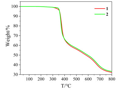
TG analysis was carried out to investigate the thermal stabilities of complexes 1 and 2. As shown in Fig. 4, complex 1 did not show any weight loss below 305 ℃ in accord with that no solvent molecules exist in the lattice of 1. It started to decompose from 305 ℃ and the total weight loss below 800 ℃ was observed to be 67.81%, which may correspond to the removal of 2-morpholine-4-yl-4, 6-di- pyrazol-1-yl-1, 3, 5-triazine ligand (calcd. 69.71%). The final product may be NiCl2 (calcd. 30.29%). The TG curve of complex 2 was very similar to that of 1, indicating that they have the same compositions and similar thermal stabilities.
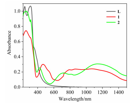
We used UV/Vis/NIR spectroscopy in diffuse reflectance mode to get the solid absorption spectra (Fig. 5) for complexes 1, 2 and the free ligand L. Complexes 1 and 2 showed absorption bands in 220~390 nm similar to that of ligand L, which were assigned as π → π* and n → π* transitions of the ligand. From 390 to 1500 nm, the absorbance of complexes 1 and 2 was different. Complex 1 exhibited a d-d band with the maximum at 468 nm and a broad d-d band ranging from 651 to 1481 nm, which was consistent with its color of red and a square-pyramidal coordination of nickel(II) complexes[30]. Complex 2 showed three d-d bands with the maximum at 423, 726 and 1140 nm, assignable to the transitions to 3A2g → 3T2g(F), 3A2g → 3T1g(P) and 3A2g → 3T1g(F), respectively. The presence and positions of these three bands suggested the green color of 2 and an octahedral environment for the nickel(II) ion[30, 31].
The fluorescence emission spectra of 1, 2 and L at room temperature are shown in Fig. 6. Complexes 1 and 2 exhibited a strong emission band at 429 nm upon excitation at 257 nm. The emissions of complexes 1 and 2 were similar to that of the free ligand L and can be attributed to intra-ligand π→π* transitions of L, not to LMCT (ligand-to-metal charge transfer) or MLCT (metal-to-ligand charge transfer). The difference of fluorescence intensity between 1 and 2 can be attributed to the fact that complex 2 is the dimer of 1.

We have obtained a nickel(II) complex monomer at 35 ℃ and its dimer at 65 ℃. As shown in Scheme 1, we speculate that there is a conversion reaction between the two complexes, and the transition from complexes 1 to 2 is an endothermic process[32]. The color difference of complexes 1 and 2 can be attributed to their structural differences[33-36].
In summary, two new nickel(II) supramolecular com- plexes monomer and dimer were successfully prepared using polydentate ligand L to react with NiCl2·2H2O under dif- ferent temperature conditions and characterized by thermal gravimetric analyses and spectroscopic methods. Complexes 1 and 2 showed different structures. Complex 1 is mononuclear, but complex 2 is binuclear. Non-covalent interactions such as π-π stacking interactions, anion-π interactions and weak hydrogen bonds were observed in the three-dimensional supramolecular structures of 1 and 2. This work not only realizes the controllable synthesis of nickel complex monomer and dimer under different temperature, but also provides a new pathway for the construction of chlorine bridge complex.
Wang, A. N.; Fan, R. Q.; Dong, Y. W.; Chen, W.; Song, Y. W.; Wang, P.; Hao, S.; Liu, Z. G.; Yang, Y. L. (E)-4-Methyl-N-((quinolin-2- yl)ethylidene)aniline as ligand for IIB supramolecular complexes: synthesis, structure, aggregation-induced emission enhancement and application in PMMA-doped hybrid material. Dalton Trans. 2017, 46, 71–85. doi: 10.1039/C6DT03853K
Strelnik, I. D.; Musina, E. I.; Karasik, A. A.; Sizov, V. V.; Grachova, E. V.; Gurzhiy, V. V.; Melnikov, A. S.; Kolesnikov, I. E. Binuclear gold(I) phosphine alkynyl complexes templated on a flexible cyclic phosphine ligand: synthesis and some features of solid-state luminescence. Inorg. Chem. 2020, 59, 244–253. doi: 10.1021/acs.inorgchem.9b02091
Yang, L. G.; Wang, X.; Zhang, Y. H.; Li, Z. M.; Song, H. X.; Niu, Y. S.; Tao, Z.; Liu, Q. Y.; Xiao, X. Structure and electrochemical studies on the complexation of hexacyclohexanocucurbit[6]uril with lead ion. Polyhedron 2018, 142, 58–62. doi: 10.1016/j.poly.2017.12.035
Zhang, Y.; Ali, B.; Wu, J. F.; Guo, M.; Yu, Y.; Liu, Z. L.; Tang, J. K. Construction of metallosupramolecular coordination complexes: from lanthanide helicates to octahedral cages showing single-molecule magnet behavior. Inorg. Chem. 2019, 58, 3167–3174. doi: 10.1021/acs.inorgchem.8b03249
Nakada, A.; Koike, K.; Nakashima, T.; Morimoto, T.; Ishitani, O. Photocatalytic CO2 reduction to formic acid using a Ru(II)-Re(I) supramolecular complex in an aqueous solution. Inorg. Chem. 2015, 54, 1800–1807. doi: 10.1021/ic502707t
Wenz, K. M.; Leonhardt-Lutterbeck, G.; Breit, B. Inducing axial chirality in a supramolecular catalyst. Angew. Chem. Int. Ed. 2018, 57, 5100–5104. doi: 10.1002/anie.201801048
Chen, L. D.; Zhou, L. X.; Zheng, Y. Q.; Zhu, H. L. Two new Ag(I) supramolecular complexes based on melamine: synthesis, structures and photocatalytic activity under visible light irradiation. Polyhedron 2017, 126, 150–158. doi: 10.1016/j.poly.2017.01.031
Freire, C.; Nunes, M.; Pereira, C.; Fernandes, D. M.; Peixoto, A. F.; Rocha, M. Metallo(salen) complexes as versatile building blocks for the fabrication of molecular materials and devices with tuned properties. Coord. Chem. Rev. 2019, 394, 104–134. doi: 10.1016/j.ccr.2019.05.014
Hu, M. L.; Morsali, A.; Aboutorabi, L. Lead(II) carboxylate supramolecular compounds: coordination modes, structures and nano-structure aspects. Coord. Chem. Rev. 2011, 255, 2821–2859. doi: 10.1016/j.ccr.2011.05.019
Walczak, A.; Stefankiewicz, A. R. pH-Induced linkage isomerism of Pd(II) complexes: a pathway to air- and water-stable suzuki-miyaura-reaction catalysts. Inorg. Chem. 2018, 57, 471–477. doi: 10.1021/acs.inorgchem.7b02711
Hema, M. K.; Karthik, C. S.; Pampa, K. J.; Manukumar, H. M.; Mallu, P.; Warad, I.; Lokanath, N. K. Solvent induced 4, 4, 4-trifluoro-1-(2-naphthyl)-1, 3-butanedione Cu(II) complexes: synthesis, structure, DFT calculation and biocidal activity. Polyhedron 2019, 168, 127–137. doi: 10.1016/j.poly.2019.04.028
Jiang, P.; Peng, F.; Chen, Y. M. Temperature-induced single-crystal-to-single-crystal transformation of a binuclear Mn(II) complex into a 1D chain polymer. RSC Adv. 2016, 6, 89192–89197. doi: 10.1039/C6RA14364D
Takjoo, R.; Mashmoul Moghadam, S. M.; Amiri Rudbari, H.; Bruno, G. Synthesis and X-ray crystal structures of some isothiosemicarbazone complexes. Transit. Met. Chem. 2019, 44, 525–534. doi: 10.1007/s11243-019-00310-w
Takai, A.; Chkounda, M.; Eggenspiller, A.; Gros, C. P.; Lachkar, M.; Barbe, J. M.; Fukuzumi, S. Efficient photoinduced electron transfer in a porphyrin tripod-fullerene supramolecular complex via π-π interactions in nonpolar media. J. Am. Chem. Soc. 2010, 132, 4477–4489. doi: 10.1021/ja100192x
Parra-Munoz, N.; Hidalgo, P. I.; Ripoll, G.; Belmar, J.; Pasan, J.; Jimenez, C. A. Influence of counterions on the supramolecular frameworks of isoquinoline-based silver(I) complexes. CrystEngComm. 2020, 22, 95–104. doi: 10.1039/C9CE01411J
Mohana, M.; Muthiah, P. T.; McMillen, C. D. Supramolecular architectures in two 1: 1 cocrystals of 5-fluorouracil with 5-bromothiophene-2-carboxylic acid and thiophene-2-carboxylic acid. Acta Cryst. 2017, C73, 481–485.
Nowroozi, A.; Ebrahimi, A.; Rezvani, R. O. Mutual effects of the cation-π, anion-π and intramolecular hydrogen bond in the various complexes of 1, 3, 5-triamino-2, 4, 6-trinitrobenzene with some cations (Li+, Na+, K+, Mg2+, Ca2+) and anions (F–, Cl–, Br–). Struct. Chem. 2018, 29, 129–137. doi: 10.1007/s11224-017-1010-3
Xiao, G. B.; Fang, Z. H.; Yao, X. Q. A new cadmium(II) coordination polymer extended through hydrogen bonds and π-π stacking interactions: synthesis and photoluminescence property. Chin. J. Struct. Chem. 2018, 37, 1987–1993.
Tom, L.; Aiswarya, N.; Sreejith, S. S.; Kurup, M. R. P. Self-organized three dimensional architectures based on non-covalent interactions in square planar Cu(II) thiosemicarbazone: solvent mediated crystallization and EPR based correlation study. Inorg. Chim. Acta 2018, 473, 223–235. doi: 10.1016/j.ica.2018.01.005
Wan, Q. Y.; Xiao, X. S.; To, W. P.; Lu, W.; Chen, Y.; Low, K. H.; Che, C. M. Counteranion- and solvent-mediated chirality transfer in the supramolecular polymerization of luminescent platinum(II) complexes. Angew. Chem. Int. Ed. 2018, 57, 17189–17193. doi: 10.1002/anie.201811943
Zhou, Q. K.; Wang, L.; Liu, D. Construction of a three-dimensional supramolecular framework based on an anionic cadmium(II) coordination network and protonated dipyridine organic cations. Acta Cryst. 2018, C74, 889–893.
Engeldinger, E.; Armspach, D.; Matt, D.; Jones, P. G.; Welter, R. A cyclodextrin diphosphane as a first and second coordination sphere cavitand: evidence for weak C–H···Cl–M hydrogen bonds within metal-capped cavities. Angew. Chem. Int. Ed. 2002, 41, 2593–2596. doi: 10.1002/1521-3773(20020715)41:14<2593::AID-ANIE2593>3.0.CO;2-M
Frontera, A.; Gamez, P.; Mascal, M.; Mooibroek, T. J.; Reedijk, J. Putting anion-π interactions into perspective. Angew. Chem. Int. Ed. 2011, 50, 9564–9583. doi: 10.1002/anie.201100208
Bettencourt-Dias, A.; Beeler, R. M.; Zimmerman, J. R. Anion-π and H-bonding interactions supporting encapsulation of [Ln(NO3)6/5]3–/2– (Ln = Nd, Er) with a triazine-based ligand. J. Am. Chem. Soc. 2019, 141, 15102–15110. doi: 10.1021/jacs.9b05566
Schottel, B. L.; Chifotides, H. T.; Dunbar, K. R. Anion-π interactions. Chem. Soc. Rev. 2008, 37, 68–83. doi: 10.1039/B614208G
Chen, W.; Chu, J. F.; Wang, Y. Q. Synthesis, characterization and preliminary reactivity behaviors with transitional metals of a new polydentate N-donor ligand. J. Mol. Struct. 2014, 1068, 237–244. doi: 10.1016/j.molstruc.2014.04.031
Dolomanov, O. V.; Bourhis, L. J.; Gildea, R. J.; Howard, J. A. K.; Puschmann, H. OLEX2: a complete structure solution, refinement and analysis program. J. Appl. Cryst. 2009, 42, 339–341. doi: 10.1107/S0021889808042726
Sheldrick, G. M. A short history of SHELX. Acta Cryst. 2008, A64, 112–122.
Sheldrick, G. M. SHELXL-97, Program for the Refinement of Crystal Structures. University of Göttingen. Göttingen, Germany 1997.
Yun, J. X.; Hu, Z. L.; Li, Y. M.; Jin, J.; Chen, C.; Yan, X.; Liu, Y. H.; Ding, Y.; Chi, Y. X.; Niu, S. Y. Synthesis, structure and photoelectric property of a series of Ni(Ⅱ) complexes constructed through aromatic carboxylic ligands. Chem. J. Chin. Univ. 2018, 39, 2161–2169.
Rodríguez, A.; García-Vázquez, J. A.; Romero, J.; Sousa-Pedrares, A.; Sousa, A.; Castro, J. Electrochemical synthesis and characterization of cobalt(II), cobalt(III), and nickel(II) complexes with pyrimidine-2-thionato ligands. Z. Anorg. Allg. Chem. 2007, 633, 763–770. doi: 10.1002/zaac.200600387
Vallarino, L. M.; Goedken, V. L.; Quagliano, J. V. Donor properties of positively charged ligands. Metal complexes of the N-chloromethyl-dabconium ligand. Inorg. Chem. 1973, 12, 102–107. doi: 10.1021/ic50119a026
Ainscough, E. W.; Brodie, A. M.; Depree, C. V.; Otter, C. A. Divalent cobalt, nickel and zinc halide complexes with multimodal ligands based on the cyclotriphosphazene platform: a structural study. Polyhedron 2006, 25, 2341–2352 doi: 10.1016/j.poly.2006.02.001
Beattie, J. W.; White, D. S.; Bheemaraju, A.; Martin, P. D.; Groysman, S. Recyclable chemosensor for oxalate based on bimetallic complexes of a dinucleating bis(iminopyridine) ligand. Dalton Trans. 2014, 43, 7979–7986. doi: 10.1039/C4DT00577E
Hamaguchi, T.; Ando, I. Synthesis and characterisation of a new six-coordinated thermochromic Ni complex. Inorg. Chim. Acta 2015, 427, 144–149. doi: 10.1016/j.ica.2014.12.013
Pitchaimani, J.; Karthikeyan, S.; Lakshminarasimhan, N.; Anthony, S. P.; Moon, D.; Madhu, V. Reversible thermochromism of nickel(II) complexes and single-crystal-to-single-crystal transformation. ACS Omega 2019, 4, 13756–13761. doi: 10.1021/acsomega.9b01263

Figure 1 Crystal photos of the complexes. (a) Red crystal of 1; (b) Green crystal of 2

Figure 2 Structure of complex 1. (a) ORTEP drawing, hydrogen atoms and solvent molecules have been omitted for clarity (symmetry code: (i) –x + 1, y, z); (b) Chloride-π interactions (red dashed lines) and the oxygen-π interaction (blue dashed line, symmetry codes: (i) x, –y + 1, –z; (ii) –x + 1, y + 1/2, –z + 1/2); (c) C(3)–H(3)···Cl(1) weak hydrogen bonding interaction (pink dashed lines) (Symmetry code: (i) x + 1/2, y − 1/2, z); (d) Molecular packing with non-covalent interactions

Figure 3 Structure of complex 2. (a) ORTEP drawing, hydrogen atoms and solvent molecules have been omitted for clarity (Symmetry code: (i) –x + 1, –y + 1, –z + 2); (b) π-π stacking interactions (blue dashed lines); (c) Molecular packing with weak hydrogen bonding interactions (pink dashed lines)

Figure 5 UV/Vis/NIR solid absorption spectra in diffuse reflectance mode of 1, 2 and L
Table 1. Crystal Data and Structure Refinement Parameters for Complexes 1 and 2
| Complex | 1 | 2 |
| Chemical formula | C13H14Cl2N8NiO | C26H28Cl4N16Ni2O2 |
| Formula weight | 427.91 | 855.86 |
| Temperature (K) | 123.0 | 97.2 |
| Crystal system | Orthorhombic | Monoclinic |
| Space group | Cmca | P21/c |
| a (Å) | 12.6569(18) | 14.600(3) |
| b (Å) | 10.4622(7) | 13.11(2) |
| c (Å) | 25.66(2) | 9.183(3) |
| α (°) | 90.00 | 90.00 |
| β (°) | 90.00 | 105.29(3) |
| γ (°) | 90.00 | 90.00 |
| V (Å3) | 3398(3) | 1695(3) |
| Z | 8 | 2 |
| μ (mm–1) | 1.477 | 1.480 |
| Crystal size (mm3) | 0.25 × 0.20 × 0.20 | 0.10 × 0.10 × 0.03 |
| Index ranges | –15≤h≤15, –7≤k≤12, –31≤l≤30 | –18≤h≤14, –7≤k≤16, –9≤l≤11 |
| 2θ range for data collection (°) | 6.36 to 52 | 6.22 to 52 |
| F(000) | 1743 | 872 |
| ρcalc (g·cm-3) | 1.673 | 1.677 |
| Reflections collected | 4897 | 7129 |
| Independent reflections (Rint) | 1745 (0.0252) | 3326 (0.0676) |
| Data/restraints/parameters | 1745/122/155 | 3326/0/226 |
| Goodness-of-fit on F2 | 1.054 | 0.999 |
| R/wR (I > 2σ(I)) | 0.0282/0.0642 | 0.0613/0.0694 |
| R/wR (all data) | 0.0379/0.0680 | 0.1097/0.0806 |
| Largest diff. peak/hole (e·Å−3) | 0.289/–0.412 | 0.487/–0.553 |
 下载: 导出CSV
Table 2. Selected Bond Lengths (Å) and Bond Angles (°) for 1
| Bond | Dist. | Bond | Dist. | Bond | Dist. | ||
| Ni(1)–Cl(1) | 2.2902(14) | Ni(1)–Cl(2) | 2.2527(15) | Ni(1)–N(1) | 2.1601(17) | ||
| Ni(1)–N(3) | 1.981(2) | Ni(1)–N(1)i | 2.1601(17) | ||||
| Angle | (°) | Angle | (°) | Angle | (°) | ||
| Cl(1)–Ni(1)–Cl(2) | 109.12(5) | Cl(1)–Ni(1)–N(1) | 97.46(5) | Cl(1)–Ni(1)–N(3) | 96.44(8) | ||
| Cl(1)–Ni(1)–N1(i) | 97.46(5) | Cl(2)–Ni(1)–N(1) | 100.59(5) | Cl(2)–Ni(1)–N(1)i | 100.59(5) | ||
| Cl(2)–Ni(1)–N(3) | 154.44(7) | N(3)–Ni(1)–N(1) | 75.37(5) | N(3)–Ni(1)–N(1)i | 75.37(5) | ||
| N(1)–Ni(1)–N(1)i | 148.37(9) | ||||||
| Symmetry transformation: i: −x+1, y, z | |||||||
下载: 导出CSV
Table 3. Hydrogen Bond Lengths (Å) and Bond Angles (°) for Complexes 1 and 2
| Complex | D–H···A | d(D–H) | d(H···A) | d(D···A) | ∠DHA |
| 1 | C(3)–H(3)···Cl(1)i | 0.95 | 2.67 | 3.437(1) | 138 |
| 2 | C(6)–H(6)···O(1)i | 0.93 | 2.52 | 3.361(5) | 151 |
| C(12)–H(12A)···O(1)ii | 0.97 | 2.80 | 3.730(6) | 162 | |
| C(13)–H(13A)···N(6)iii | 0.97 | 2.61 | 3.424(6) | 142 | |
| C(2)–H(2)···Cl(2)iv | 0.93 | 2.96 | 3.581(5) | 125 | |
| C(3)–H(3)···Cl(1)iii | 0.93 | 2.56 | 3.481(5) | 170 | |
| C(5)–H(5)···Cl(1)v | 0.93 | 2.66 | 3.578(7) | 171 | |
| C(11)–H(11A)···Cl(1)vi | 0.97 | 2.71 | 3.653(5) | 163 | |
| C(13)–H(13B)···Cl(1)iii | 0.97 | 2.82 | 3.654(6) | 144 | |
| Symmetry codes for 1: (i) x + 1/2, y − 1/2, z; for 2: (i) −x, y − 1/2, −z + 1/2; (ii) x, −y + 3/2, z + 1/2; (iii) x, −y + 3/2, z − 1/2; (iv) −x + 1, y + 1/2, −z + 5/2; (v) x, −y + 1/2, z − 1/2; (vi) −x, −y + 1, −z + 1 | |||||
下载: 导出CSV
Table 4. Selected Bond Lengths (Å) and Bond Angles (°) for 2
| Bond | Dist. | Bond | Dist. | Bond | Dist. | ||
| Ni(1)–Cl(1) | 2.4040(14) | Ni(1)–Cl(2) | 2.3290(14) | Ni(1)–Cl(2)i | 2.5027(14) | ||
| Ni(1)–N(1) | 2.149(5) | Ni(1)–N(3) | 2.163(5) | Ni(1)–N(5) | 1.985(4) | ||
| Angle | (°) | Angle | (°) | Angle | (°) | ||
| N(1)–Ni(1)–Cl(1) | 92.08(10) | N(1)–Ni(1)–Cl(2) | 102.58(10) | N(1)–Ni(1)–Cl(2)i | 87.63(10) | ||
| N(1)–Ni(1)–N(3) | 150.95(14) | N(1)–Ni(1)–N(5) | 76.06(15) | N(3)–Ni(1)–Cl(1) | 89.37(11) | ||
| N(3)–Ni(1)–Cl(2) | 106.13(12) | N(3)–Ni(1)–Cl(2)i | 90.17(10) | N(3)–Ni(1)–N(5) | 74.94(16) | ||
| N(5)–Ni(1)–Cl(1) | 89.48(11) | N(5)–Ni(1)–Cl(2) | 174.64(11) | N(5)–Ni(1)–Cl(2)i | 89.02(11) | ||
| Cl(1)–Ni(1)–Cl(2) | 95.77(5) | Cl(1)–Ni(1)–Cl(2)i | 178.50(5) | Cl(2)–Ni(1)–Cl(2)i | 85.73(5) | ||
| Symmetry code: i: –x + 1, –y + 1, –z + 2 | |||||||
下载: 导出CSV

 扫一扫看文章

扫一扫关注我们


 下载:
下载: