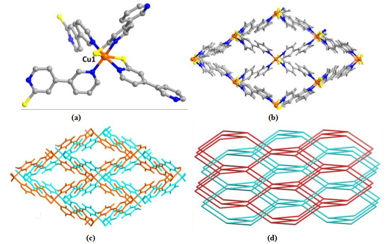
Figure 1.
(a) View of the coordination surrounding for the Cu(II) ion. (b) View for the single network of 1. (c) View for the 2-fold interpenetrated network of 1. (d) View of the 2-fold dia net for 1

New drugs for the treatment of cancer with better rates of cure and less severe side effects are needed. In this field, coordination chemistry has a great potential to offer a wide variety of compounds with different geometry, redox reactivity and a diversity of mechanisms related to DNA binding, some of which are unique to metals[1-3]. The usefulness of coordination metal complexes in cancer chemotherapy has been demonstrated by Cisplatin and other platinum coordination compounds which are amongst the most successfully used anticancer drugs[4]. As a result of an intense and continued research on coordination complexes with antitumor activity, compounds of different metals other than Pt are entering clinical studies[5].
Coordination polymers (CPs) with novel structures have recently drawn the attention of chemists as they possess intriguing topologies and potential applications in the fields of catalysis, sensing, nonlinear optics, gas adsorption, ion exchange, photoactive materials and so on[6]. Usually, the structure of the coordination polymer depends on many factors such as the coordination trend of metal ions, rigidity or flexibility of the organic ligands, substituents, solvent systems, reaction temperature, pH values, and so forth. A subtle change in the aforementioned factors may cause a drastic change in the crystallization of CPs[7]. On the other hand, recent studies have revealed that the Cu(II) and Co(II)-based coordination polymers could be used as potential anticancer reagents with promising results[8, 9]. In this study, two new Cu(II) and Co(II)-based coordination polymers with the chemical formulae of {Cu(L)2]·DMF}n (1) and {[Co(L)2]·2DMF}n (2) have been successfully constructed from a pyridine-substituted N-heterocyclic thioamide ligand, namely [3, 4΄-bipyridine]-2΄-thiol (HL). Complex 1 is composed of two identical coordination networks with dia topology interpenetrating into each other, and shows 1-D microporous channels along the a axis. Complex 2 is assembled by 2-D square-grid layers stacked in an eclipsed fashion to give 1-D large square channels along the b axis. The cell viability detected by CCK-8 kit indicated that compound 1 showed excellent anti-cancer activity on H1650 cancer cells. The inhibitory effect of compound 1 on cancer cells owns to the induction of apoptosis, mediated by PI3K/AKT pathway activation, which is confirmed by apoptosis assay and western blot.
All reagents and solvents were commercially available and used as received without further purification. Elemental analyses for C, H and N were performed on a CHN-O-Rapid analyzer or an Elementar Vario MICRO analyzer. IR spectra were recorded with a Thermo Scientific Nicolet 5700 FT-IR spectrophotometer with KBr pellets from 400 to 4000 cm-1.
For complex 1, Cu(NO3)2·6H2O (12 mg, 0.05 mmol) and L (9.45 mg, 0.05 mmol) in H2O (3.0 mL)-DMF(3.0 mL) mixture was added to a Teflon-lined autoclave (23 mL) and heated at 120 ℃ for 72 h. Blue block-shaped crystals were filtered, washed with DMF and dried under vacuum (Yield: 36% based on L). Anal. Calcd. for C23H19CuN5OS2 (%): C, 54.26; H, 3.76; N, 13.76. Found (%): C, 53.92; H, 3.68; N, 13.34.
For complex 2, Co(NO3)2·6H2O (29 mg, 0.1 mmol) and L (9.45 mg, 0.05 mmol) in H2O (1.0 mL)-DMF(4.0 mL) mixture was added to a Teflon-lined autoclave (23 mL). After the addition of HCl (aq, 20 μL, 2M), the mixture was heated at 120 ℃ for 72 h. Green block-shaped crystals were filtered, washed with DMF and dried under vacuum (Yield: 49% based on L). Anal. Calcd. for C26H28CoN6O2S2 (%): C, 53.88; H, 4.87; N, 14.50. Found (%): C, 53.38; H, 4.68; N, 14.34.
A green block-shaped single crystal of complex 1 (0.24mm × 0.21mm × 0.22mm) and a blue block-shaped single crystal of complex 2 (0.23mm × 0.20mm × 0.20mm) were placed on an APEX II CCD area detector equipped with a graphitemonochromatic MoKα radiation (λ = 0.71073 Å) at 293 (2) K. A total of 13233 and 10764 reflections were collected for complexes 1 and 2, of which 2369 (Rint = 0.0567) and 3553 (Rint = 0.0434) were independent in the φ-ω ranges of 2.480~27.865º and 2.508~29.302º, and 2066 and 2994 observed reflections with I > 2σ(I) were employed for structure refinements for complexes 1 and 2, respectively.
The empirical absorption corrections by SADABS were carried out. The structures were solved by direct methods with SHELXS-97 program and refined with SHELXL-97 by full-matrix least-squares techniques. The non-hydrogen atoms were refined anisotropically, and the hydrogen ones were determined with theoretical calculations. For complex 1, the final R = 0.0571, wR = 0.1586 (w = 1/[σ2(Fo2) + (0.1000P)2 + 0.0000P], where P = (Fo2 + 2Fc2)/3), (Δ/σ)max = 0.000, S = 1.080, (Δρ)max = 0.644 and (Δρ)min = –0.841 e/Å3. For complex 2, the final R = 0.0464, wR = 0.1251 (w = 1/[σ2(Fo2) + (0.1000P)2 + 0.0000P], where P = (Fo2 + 2Fc2)/3), (Δ/σ)max = 0.000, S = 1.051, (Δρ)max = 0.602 and (Δρ)min = –0.400 e/Å3. Complex 1 crystallizes in orthorhombic, space group Fddd with a = 13.6258(12), b = 18.3469(14), c = 32.846(3) Å, V = 8211.1(12) Å3, Z = 4, F(000) = 3536, Dc = 1.411 mg/m3 and μ = 1.278 mm-1. Complex 2 crystallizes in orthorhombic, space group P21212 with a = 12.942(3), b = 16.2436(14), c = 6.3569(11) Å, V = 1336.4(4) Å3, Z = 4, F(000) = 442, Dc = 1.082 Mg/m3 and μ = 0.809 mm-1.
The H1650 cancer cells proliferation was examined with CCK-8 assay under the treatment of compounds 1 and 2 according to the protocols[10]. In brief, the cells were plated into 96-well plates with the destiny of 1000 cell/well, and incubated in a 37 ℃, 5% CO2 humidified atmosphere overnight. Then, the indicated dose of compound 1 or 2 (1, 2, 4, 8, 10, 20, 40, 80 μg/mL) was added into cells for 24 h incubation. After that, the H1650 cancer cells were exposed to CCK-8 reagents in 100 μL medium for another 2 h. The absorbance was measured and recorded for cell proliferation calculation according to the formula: cell proliferation rate = (OD (experimental group)/OD (control group)) × 100%. Each group was repeated at least three times.
The H1650 cancer cells apoptosis and cell cycle distribution were assessed by Annexin V-FITC/PI apoptosis kit (BD Biosciences, San Jose, CA, USA) in flow cytometer[11]. Before exposed to indicated concentrations of compounds 1 and 2, the cells were plated onto 6-well plates (2 × 10⁵ cells per well) and incubated at 37 ℃ in 5% CO2 humidified atmosphere overnight. The dimethyl sulfoxide (0.1% DMSO) and apoptosis inducer kit (Beyotime, Shanghai, China) were used as the negative and positive controls. After compounds treatment, the cells were harvested and labeled with 5 μL Annexin V-FITC and 5 μL propidium iodide (PI) for cell apoptosis detection, and the PI/RNase staining buffer (BD, San Diego, USA) was recommended for cell cycle progression. The samples were detected by flow cytometry (BD, NJ, USA), and the results were analyzed using flow cytometry (FACSCalibur, BD Biosciences, USA). All experiments were repeated three times.
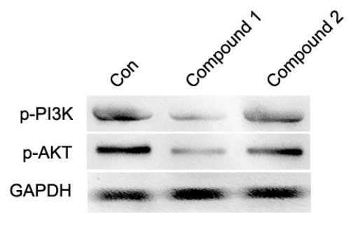
The total cell protein was extracted from H1650 cancer cells after being treated with indicated doses of compounds 1 and 2 using total Protein Extraction Kit (Invent Biotechnologies, USA) followed by the manufacturer's protocol[12]. The total protein concentrations were determined by BCA Protein Assay Kit (Beyotime Biotechnology) and diluted by 5× loading buffer. Then, equal amount of protein samples was separated by 12% sodium dodecyl sulfate polyacrylamide gel, and transferred onto 0.22 mm polyvinylidene fluoride (PVDF) membrane (Millipore, Bedford, MA, USA). The membranes were blocked with 5% BSA and incubated with appropriate dilution of primary antibody at 4 ℃ overnight. GAPDH was used as internal reference. After being washed with PBS, the membranes were incubated with appropriate secondary horseradish peroxidase (HRP)-conjugated secondary antibodies (Jackson Immuno Research, West Grove, PA) for 1 h at room temperature. Images were captured using Millipore Immobilon ECL illuminating solution (Millipore Corporation, USA), and the Quantity One software was used to quantify the density of bands.
Single-crystal X-ray diffraction analysis reveals that complex 1 crystallizes in orthorhombic space group Fddd and reveals a 3D framework structure. The basic repeating unit of 1 contains one Cu2+ ion locating on the 4-fold axis along with one L ligand. As shown in Fig. 1a, each Cu(II) atom in 1 adopts a distorted octahedral coordination geometry defined by a N4S2 donor set from four anionic L ligands (Cu–N pyridine 2.076(2) Å; Cu–N pyrimidine 2.110(3) Å; Cu–S 2.482(9) Å), in which two are bound in a N, S-chelation fashion to metal atom with the pyrimidinethione end, and the other two in cis-positions are with the monodentate pyridine end. As a whole, each L ligand connects two Cu(II) atoms in a μ2-1, κS κNpyrimidine: 2, κNpyridine bridging mode that combines one monodentate pyridine and one N, S-chelated pyrimidinethione. In the L ligand, the pyridine ring is slightly tilted to the pyrimidine ring with a small dihedral angle of 11.42°. Single network of 1 shows diamondoid cavity that is large enough to permit another independent network to interpenetrate, leading to a two-fold interpenetrating 3-D network (Fig. 1b and Fig. 1c). Albeit 2-fold interpenetration, complex 1 still possesses 1-D microporous channels running along the b-axis, and the solvent-accessible volume was calculated by PLATON to be about 16.8% of the total crystal volume. To the best of our knowledge, complex 1 is the first two-fold interpenetrating 3-D porous network built from N-heterocyclic thioamide ligand. From the topological point of view, each Cu atom is linked to its four neighbors through four L ligands which could be viewed as a four-connected node, and the L ligand could be considered as a linker node, so the whole framework of 1 could be judged as a 3-D dia network with the point symbol of {6^6}, which belongs to the class IIa type interpenetrating pattern (Fig. 1d).
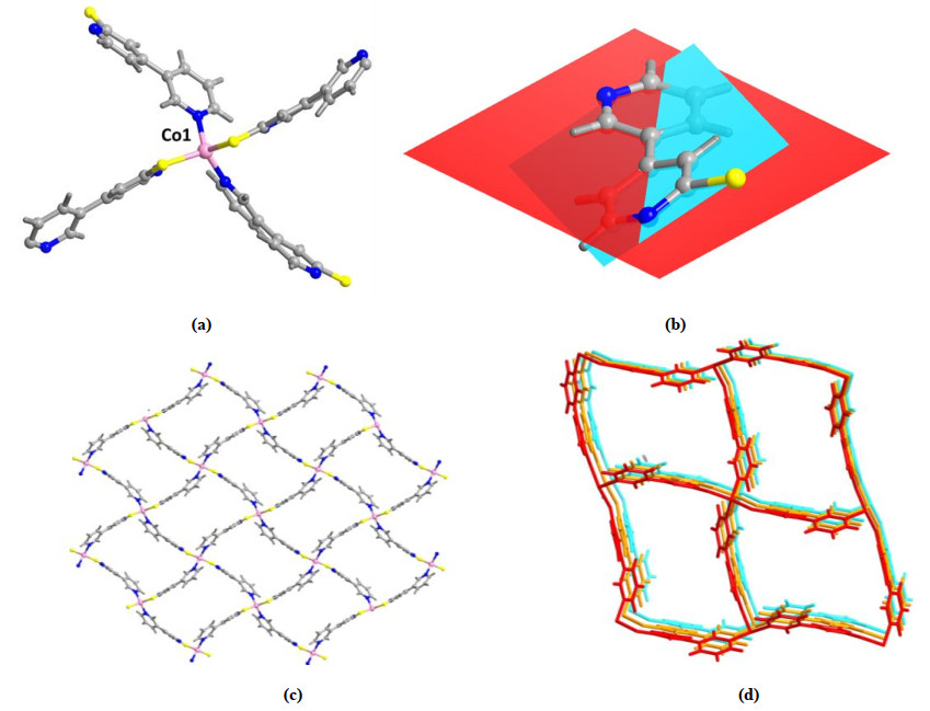
Complex 2 is produced by the reaction of Co(NO3)2·6H2O with L ligand in the mixed solvent of DMF and H2O. The single-crystal X-ray diffraction analysis shows that complex 2 crystallizes in orthorhombic space group P21212 and reveals a 2D layered network. The basic repeating unit of 1 is composed of one Co(II) ion locating on the 2-fold axis, one L ligand and one DMF molecule as revealed via the elemental analyses. Different from 1, each Co(II) atom in 2 adopts a tetrahedral coordination geometry completed by a N2S2 donor set from four L ligands, and each L ligand connects two Co(II) atoms in a μ2-1, κS: 2, κNpyridine bridging manner that integrates one N-monodentate pyridine and one S-monodentate pyrimidinethione (Fig. 2a). The Co–N bond distance is 2.017(3) Å and the Co–S bond length is 2.262(4) Å. Meanwhile, a larger torsion between pyridine and pyrimidine rings in L ligand is observed with a dihedral angle of 26.63° (Fig. 2b). Although each Co atom is also linked to its four neighbors through four L ligands, only a 2-D square-grid layer with 4-connected sql topology is formed in the ab plane, and the size of square grid is about 10.4 × 10.4 Å2 measured by the Co···Co distance (Fig. 2c). Particularly, 2-D square-grid layers are stacked in an eclipsed fashion along the b axis, giving rise to a 3-D porous coordination network with 1-D square channels (Fig. 2d).
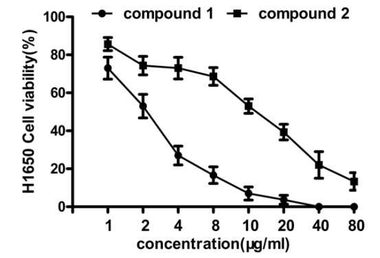
The CCK-8 assays were carried out for H1650 cell viability assay after being treated with compounds 1 and 2. We found that compared with 2 at the same concentration, 1 could reduce the viability of cancer cell strongly, as shown in Fig. 3. This indicated that compound 1 may has a better in vitro anti-cancer activity than 2.
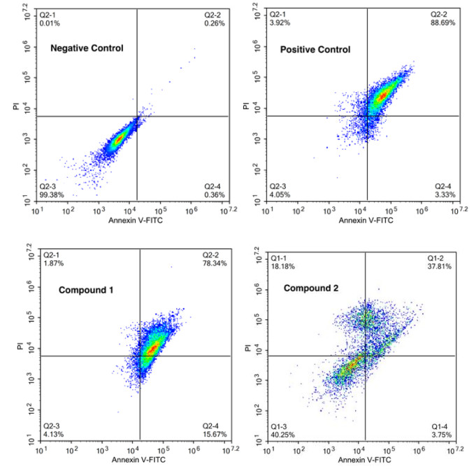
As the main manner of cancer cell death, the apoptosis of H1650 was measured by Annexin V-FITC/PI experiment. The cells were treated with the same concentration of compounds 1 and 2 for 24 h, followed by apoptosis detection. As shown in Fig. 4, compound 1 could significantly increase the percentage of apoptotic cells up to 78.34%, which is much higher than the apoptotic percentage induced by compound 2. This result also convinced that compound 1 has a significantly higher anti-cancer activity than 2 in H1650 cells.
The PI3K/AKT pathway is reported to participate in the cancer cell proliferation and apoptosis progression, so the protein levels of this signaling pathway were measured by western blot. As shown in Fig. 5, the results suggested that compound 1 could reduce the PI3K/AKT protein level remarkably, and compound 2 could only down-regulate the protein level slightly. This figure supplementary proved the stronger anti-cancer activity of compound 1 than that of 2.
In summary, by using a pyridine-substituted N-heterocyclic thioamide ligand, two new Cu(II) and Co(II)-based coordination polymers with interesting structural diversity have been successfully prepared under solvothermal conditions. The single-crystal X-ray study reveals that complex 1 is composed of two identical coordination networks with dia topology interpenetrating into each other, and shows 1-D microporous channels along the a axis. Complex 2 is assembled by 2-D square-grid layers stacked in an eclipsed fashion to give 1-D large square channels along the b axis. In addition, we firstly evaluated the anti-viability activity of compounds 1 and 2 on H1650 cancer cells, and found 1 showed better activity than 2. The following apoptosis and western blot assay also convinced the conclusion that compound 1 could induce the cancer cell apoptosis via inhibiting the PI3K/AKT pathway.
Novio, F.; Simmchen, J.; Vázquez-Mera, N.; Amorín-Ferré, L.; Ruiz-Molina, D. Coordination polymer nanoparticles in medicine. Coord. Chem. Rev. 2013, 257, 2839–2847. doi: 10.1016/j.ccr.2013.04.022
Lian, H. Y.; Hu, M.; Liu, C. H.; Yamauchi, Y.; Wu, K. C. W. Highly biocompatible, hollow coordination polymer nanoparticles as cisplatin carriers for efficient intracellular drug delivery. Chem. Commun. 2012, 48, 5151–5153. doi: 10.1039/c2cc31708g
Li, F.; Li, T.; Cao, W.; Wang, L.; Xu, H. Near-infrared light stimuli-responsive synergistic therapy nanoplatforms based on the coordination of tellurium-containing block polymer and cisplatin for cancer treatment. Biomaterials 2017, 133, 208–218. doi: 10.1016/j.biomaterials.2017.04.032
Abu-Surrah, A.; Kettunen, M. Platinum group antitumor chemistry: design and development of new anticancer drugs complementary to cisplatin. Curr. Med. Chem. 2006, 13, 1337–1357. doi: 10.2174/092986706776872970
Muhammad, N.; Guo, Z. Metal-based anticancer chemotherapeutic agents. Curr. Opin. Chem. Biol. 2014, 19, 144–153. doi: 10.1016/j.cbpa.2014.02.003
Jin, L. N.; Liu, Q.; Sun, W. Y. An introduction to synthesis and application of nanoscale metal-carboxylate coordination polymers. CrystEngComm. 2014, 16, 3816–3828. doi: 10.1039/c3ce41962b
Zhang, J. P.; Huang, X. C.; Chen, X. M. Supramolecular isomerism in coordination polymers. Chem. Soc. Rev. 2009, 38, 2385–2396. doi: 10.1039/b900317g
Abbasi, Z.; Salehi, M.; Khaleghian, A.; Kubicki, M. Co(III), V(IV) and Cu(II) complexes of bidentate N, O-donor Schiff base ligands: characterization, anticancer activities and metal oxide nanoparticles preparation via solid state thermal decomposition. Appl. Organomet. Chem. 2018, 32, e4542. doi: 10.1002/aoc.4542
Gao, E.; Xing, J.; Qu, Y.; Qiu, X.; Zhu, M. Synthesis, characterization, DNA binding, cytotoxicity and molecular docking properties of Cu(II) and Mn(II) complexes with 1, 4-bis (pyrazol-1-yl) terephthalic acid. Appl. Organomet. Chem. 2018, 32, e4469. doi: 10.1002/aoc.4469
Wang, T.; Chen, Y.; Nie, H.; Huang, Y.; Zhao, Y.; Yang, J. IL-27 inhibits non-small-cell lung cancer cell metastasis by miR-935 in vitro. Onco. Targets. Ther. 2019, 12, 1447–1454. doi: 10.2147/OTT.S173207
Wang, H.; Lou, C.; Ma, N. miR-140-5p alleviates the aggressive progression of Wilms' tumor through directly targeting TGFBR1 gene. Cancer Manag. Res. 2019, 11, 1641–1651. doi: 10.2147/CMAR.S177508
Sun, B.; Dong, C.; Lei, H.; Gong, Y.; Li, M.; Zhang, Y.; Zhang, H.; Sun, L. Propranolol inhibits proliferation and invasion of hemangioma-derived endothelial cells by suppressing the DLL4/Notch1/Akt pathway. Chem. Biol. Interact. 2018, 294, 28–33. doi: 10.1016/j.cbi.2018.08.018

Figure 1 (a) View of the coordination surrounding for the Cu(II) ion. (b) View for the single network of 1. (c) View for the 2-fold interpenetrated network of 1. (d) View of the 2-fold dia net for 1

Figure 2 (a) View of the coordination surrounding for the Co(II) ion. (b) View of the molecular conformation for the L ligand. (c) View of the 2D layered network of 1. (d) View of the 3D packing diagram for complex 2

Figure 3 Viability of H1650 cancer cells. The H1650 cancer cells were treated with serious doses of compounds 1 and 2 for 24 h. The cell viability rate was determined by CCK-8 assay. Data were expressed as mean ± SD from three repeats. *p < 0.05 was regarded as significant difference

Figure 4 H1650 cancer cell apoptosis induced by compounds 1 and 2. After treated with compound 1 or 2 at the same indicated concentration for 24 h, the cell apoptosis was analyzed with annexin V‑FITC apoptosis detection kit in flow cytometry. The experiment was performed in triplicate.

 扫一扫看文章

扫一扫关注我们
 DownLoad:
 下载:
下载:

 下载:
下载: