Table 1.
Hydrogen Bond Lengths (Å) and Bond Angles (°) for C-H2IA
Citation:
Wen-Xu ZHENG, Tao LI, Zheng XIANG, Chang-Cang HUANG. Cytosinium Isophthalate: Crystal Structure Redetermination and Room Temperature Phosphorescence[J]. Chinese Journal of Structural Chemistry,
2020, 39(9): 1707-1713.
doi:
10.14102/j.cnki.0254-5861.2011-2647

Cytosinium Isophthalate: Crystal Structure Redetermination and Room Temperature Phosphorescence
English
Cytosinium Isophthalate: Crystal Structure Redetermination and Room Temperature Phosphorescence
Abstract:
The title compound cytosinium isophthalate (C-H2IA) self-assembly of cytosine (C) and isophthalic acid (H2IA) in aqueous media has been synthesized and the crystal structure with a reasonable protonation state is redetermined. Single-crystal X-ray diffraction analysis reveals that each asymmetric unit contains one protoned cytosine molecule and one deprotoned isophthalic acid. The proton transferred from carboxylic acid to the pyrimidine ring is disordered across an inversion center with occupancy of 0.5 and the proton located to one of the carboxylate group lies on an inversion center shared by two crystallographically equivalent oxygen atoms. In addition, the cytosine molecules are connected by complementary hydrogen bonds to form a one-dimensional tape structure. The neighboring isophthalic acids are connected via hydrogen bonds between carboxyl groups to form a one-dimensional lattice like tape. Furthermore, the adjacent organic base tapes and organic acid tapes are stacked one with another through π-π stacking interactions to form a three-dimensional supramolecular structure. Interestingly, C-H2IA displays a green phosphorescence in solid state at room temperature with the lifetime of 0.7 s determined by time resolved studies, indicating that supramolecular C-H2IA is a potential pure organic phosphorescent luminogens.
-
1. INTRODUCTION
Room temperature phosphorescence (RTP) has attracted considerable attention in the fields of organic light-emitting diodes, biological imaging and sensing devices due to their long-lived emission, large stokes shifts and high quantum yields[1]. Heretofore, most of the room temperature phosphorescence compounds contain noble metals, such as Ir(Ⅲ), Ru(Ⅲ) and Pt(Ⅱ)[2]. Considering the intrinsic problems of noble metals including high cost and human toxicity, it is vital to develop highly efficient metal-free pure organic compounds with RTP. However, because of the inefficient intersystem crossing caused by intramolecular motions and collisions with quenching species of oxygen and moisture, achieving organic room temperature phosphorescence is still of great challenge[3].
Supramolecular organic frameworks[4], a kind of crystalline solids constructed via non-covalent interactions (e.g. hydrogen bonds, halogen bonds, van der Waals contacts and π-π stacking) to form one-, two-, or three-dimensional infinite networks, have attracted immense attention not only because of their intriguing architectures but also of their exploitable properties for potential applications in the fields of absorption[5], luminescence[6], sensing[7], catalysis[8], molecular recognition[9], and so on. According to literatures, in a self-assembly system the intermolecular interactions such as hydrogen bonds can provide a rigid molecular environment for suppressing non-radiative transition to enhance the phosphorescence[10]. With this in mind, a native nuclo-bases cytosine with rigid plane organic molecular posing both acceptor and donor functionalities was selected to construct hydrogen bonding net with complementary carboxylic acid[11], and it is a supramolecular framework with isophthalic acid (C-H2IA) synthesized in water media. The crystal structure redetermination and room temperature phosphorescence are reported here.
2. EXPERIMENTAL
2.1 Materials and methods
Cytosine (C) and isophthalic acid (H2IA) were purchased from Energy Chemical without further purification. The IR spectrum was recorded in the range of 4000~400 cm−1 on a Nicolet IS50 FTIR spectrometer with ATR accessory. A thermogravimetric analysis (TGA) experiment was performed using a Mettler Toledo TG/DSC3+ instrument (heating rate of 10 K·min-1, argon stream) from room temperature to 1000 ℃. Powder X-ray diffraction measurements were performed with a Rigaku Ultima Ⅳ at room temperature. The luminescence and phosphorescence spectra for the solid samples were recorded at room temperature on an Edinburgh Instrument FLS980 luminescence spectrophotometer.
2.2 Synthesis and crystallization
The supramolcular framework of C-H2IA was synthesized from the mixture of cytosine (C) and isophthalic acid (H2IA) in an aqueous solution at 120 ℃. A 25 mL oven-dried two-necked round bottom flask was charged with cytosine (0.0555 g, 0.5 mmol), isophthalic acid (0.0831 g, 0.5 mmol) and deionized water (15 mL). After being heated in an oil bath at 120 ℃ for 30 min, the turbid solution got clear. Then it was cooled to room temperature naturally, obtaining colorless acicular crystals (yield: 72.6%). IR (cm-1): 3000 (m, br), 1695 (vs), 1658(vs), 1446 (w), 1370(w), 1253 (m), 1202 (m), 1150 (m), 1073 (w), 973 (w), 930 (w), 792 (w), 735 (m), 873 (w), 645 (w), 568 (w).
2.3 X-ray crystallography
Data of compound C-H2IA were collected on a Rigaku Saturn 724 CCD diffractometer by a graphite-monochromatized MoKα radiation (λ = 0.71073 Å) by using an ω (2.62≤θ≤29.43º) scan mode at 293(2) K. A total of 10529 reflections with 2716 independent ones (Rint = 0.0391) were collected, in which 2208 were observed (I > 2σ(I)). The structure was solved by direct methods and refined with full-matrix least-squares methods on F2 by using the Olex2 program package. The final R = 0.0408 and wR = 0.1061 with 185 parameters; S = 1.122, (Δ/σ)max = 0.000, (Δρ)max = 0.261 and (Δρ)min = –0.264 e/Å3. All non-hydrogen atoms were refined anisotropic. C-bound H atoms were positioned geometrically, with C–H = 0.93 Å and Uiso(H) = 1.2Ueq(C). Other hydrogen atoms were located in difference Fourier maps, with Uiso(H) = 1.5Ueq(N) and Uiso(H) = 1.5Ueq(O), respectively. Distances and angles of hydrogen bonds for C-H2IA are given in Table 1.
Table 1

 DownLoad:
CSV
DownLoad:
CSV
D–H⋅⋅⋅A d(D–H) d(H⋅⋅⋅A) d(D⋅⋅⋅A) < (DHA) N(4)–H(4)⋅⋅⋅N(4)#1 0.88 1.96(3) 2.829(2) 174 N(6)–H(6A)⋅⋅⋅O(2)#1 0.85 2.03 2.8670(15) 171 O(9)–H(9)⋅⋅⋅O(9)#2 1.23 1.23 2.4612(18) 180 N(1)–H(1)⋅⋅⋅O(2)#3 0.93 1.91 2.8304(15) 173 O(11)–H(11)⋅⋅⋅O(10)#4 0.92 1.80 2.6179(14) 147 Symmetry transformations used to generate the equivalent atoms:
#1: –x+1, –y+2, –z+1; #2: –x, –y+1, –z+1; #3: –x+2, –y+2, –z+1; #4: x+1, y, z3. RESULTS AND DISCUSSION
3.1 Structural description
According to literatures, the nucleobase cytosine molecule can be protonated under acidic conditions to form three types of motifs, cytosinium ion (CH+)[12], cytosine-cytosinium ion dimer (C·CH+) which was first observed by Eichhorn et al. with acetyl cytosine[13] and cytosinium ion-cytosinium ion dimer (CH+·CH+), the only one example reported by Pedireddi et al. previously[14]. However, it is difficult to form a cytosinium ion-cytosinium ion dimer due to the electrostatic repulsion between two face-to-face protonated cytosine molecules. The crystal structure of C-H2IA was reported by Pedireddi et al. previously (Cambridge Structural Database with a refcode TAZXAU)[14] containing a cytosinium ion-cytosinium ion dimer. In this previous structure determination, one hydroxyl proton of carboxylic acid transferred to the nitrogen atom of pyrimidine ring, and the short inter H···H contact 1.2227 Å for the adjacent pyrimidine rings and the short inter O···O contact 2.4585 Å for the adjacent carboxyl groups imply the unambiguous protonation state of the compound considering electrostatic repulsion. As a result, it is important to redetermine the crystal structure of cytosinium isophthalate, not only to reveal the actual position of proton but also help to better understand the biological processes for the fact that many cytosine derivatives are active pharmaceutical ingredients.
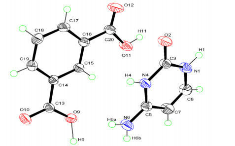
The crystal structure of C-H2IA was determined by single-crystal X-ray diffraction (SC-XRD). As depicted in Fig. 1, single-crystal X-ray diffraction analysis revealed that the supramolecular framework of C-H2IA crystallizes in triclinic space group P
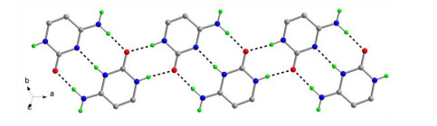
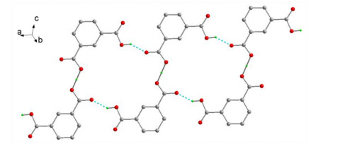
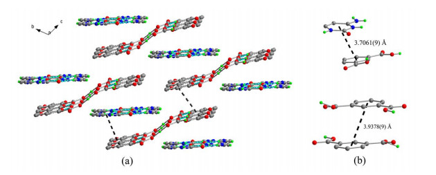
$ \overline 1 $ with a = 8.5825(4), b = 8.9568(5), c = 9.0642(5) Å, α = 100.843(5), β = 105.240(5), γ = 113.056(5)°, containing one protoned cytosine molecule and one deprotoned isophthalic acid in the asymmetric unit, revealing proton transfer from the carboxylate group to the pyrimidine ring occurs. In contrast to the previous structure determination, the hydrogen atom labeled H(9) which located to one of the carboxylate group oxygen atoms O(9) lies on an inversion center shared by two crystallo-graphically equivalent oxygen atoms O(9). Simultaneously, the hydrogen atom labeled H(4) bound to the pyrimidine N(4) atom disorder across a crystallographic inversion center with the occupancy of 0.5 rather than 1.0. As shown in Fig. 2, the cytosine dimeric unit is formed via complementary triple hydrogen bonds utilizing its dual nature of acceptor and donor properties, involving heterocyclic N–H, carbonyl, and the amino groups, thus resembling a Watson-Crick like pattern, by forming N–H···O and N–H···N hydrogen bonds with the N···O and N···N distances of 2.8670(15) and 2.829(2) Å, respectively. In the dimers, the half occupancy hydrogen atom residing on N(2) atom is disordered and shared by two cytosine molecules. The adjacent triplet hydrogen-bonded dimers are connected via N–H···O hydrogen bonds performed by pyridyl NH and oxygen atom with the N···O distances of 2.8304(15) Å, forming a one-dimensional infinite tape structure of [HC2]nn+ along the [1 0 0] direction. As shown in Fig. 3, the neighboring isophthalic acids are connected via O–H–O symmetric hydrogen bonds to form a one-dimensional lattice like infinite tape of [H(HIA)2]nn- with O···O distances of 2.4612(18) Å. In the symmetric hydrogen bond, the H(9) atom is equally shared between two oxygen atoms, and the donor and acceptor can not be distinguished. The short distances between two crystallographically equivalent oxygen atom O(9) indicate a strong symmetric hydrogen bond, which is not found in the previous report[14] due to the incorrect proton determination. The organic base tapes and organic acid tapes are stacked one with another through π-π stacking interactions involving benzene and pyrimidine rings. π-π interaction can be observed between two neighboring parallel benzene rings with the centroid-centroid distance of 3.9378(9) Å stacking between the pyrimidine and benzene rings involved with the centroid-to-centroid (Cg−Cg) distance of 3.7061(9) Å and dihedral angle of 9.38(7)° (Fig. 4).Figure 1
 Figure 1. A perspective view with atomic numbering scheme. Displacement ellipsoids are drawn at the 50% probability level
Figure 2
 Figure 2. A view of infinite tape of [HC2]nn+ connected via hydrogen bonds (dashed line). Hydrogen atoms on carbons and one moiety of disorder hydrogen atoms are omitted for clarity
Figure 3
 Figure 3. A view of infinite tape of [H(HIA)2]nn- connected via hydrogen bonds (dashed line). The hydrogen atoms on benzene ring are omitted for clarity
Figure 4
 Figure 4. (a) Three-dimensional supramolecular net formed by π-π stacking of tapes; (b) Illustration of the molecular packing in C-H2IA
3.2 Powder X-ray diffraction (PXRD)
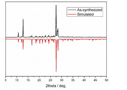
To verify the sample purity after crystallization process, X-ray powder diffractogram of C-H2IA was performed at room temperature. As shown in Fig. 5, experimental X-ray powder diffractograms are almost in agreement with the simulated pattern calculated from signal-crystal diffraction data using the Mercury software[15], stating the purity of the compound. The intensity difference between both diffractograms could be attributed to the preferred orientation effects.
Figure 5
 Figure 5. Powder X-ray diffraction (PXRD) patterns of C-H2IA
3.3 Thermogravimetric analysis (TGA)
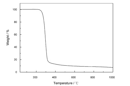
In order to estimate the thermal stability of C-H2IA, thermogravimetric analysis (TGA) was performed under argon gas atmosphere. In the TGA curve of C-H2IA (Fig. 6), there was no obvious weight loss above temperature 200 ℃, showing a considerable thermal stability of C-H2IA. The rapid massive weight loss from 250 to 350 ℃ can be attributed to the decomposition of organic compound. The remainder after treating under 1000 ℃ can be attributed to the carbon formed at high temperature.
Figure 6
 Figure 6. Thermogravimetric analysis (TGA) curves of C-H2IA
3.4 Photoluminescence analysis
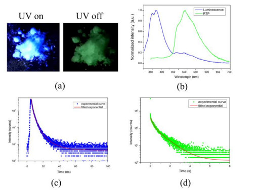
The photoluminescence properties of C-H2IA in crystalline state were first explored under atmospheric conditions. Exposed to the radiation of a UV-lamp, the crystals showed a blue light emission. Interestingly, after UV-light cessation, the solid sample displayed nearly 10 seconds lasting green glow which can be easily observed by naked eyes. As shown in Fig. 7b, the steady solid-state photoluminescence spectrum showed a blue emission band with peaks at 350 and 380 nm and a green emission band with peaks at 480 and 500 nm observed from room temperature phosphorescence spectrum (Fig. 7b green line). Further researches on time-resolved photoluminescence decay of C-H2IA were carried out. As shown in Fig. 7c, the blue emission at 375 nm decay plot was fitted by a tri-exponential curve with lifetime of 10 ns, which can be assigned to the fluorescence properties. As shown in Fig. 7d, the ultra long-lived green emission at 500 nm decay plot was fitted by a bi-exponential curve with lifetime of 0.7 s, which can be assigned to the room temperature phosphorescence properties.
Figure 7
 Figure 7. (a) Photographs of C-H2IA crystals with a hand-held 300 nm UV lamp irradiation on and off; (b) Normalized photoluminescence (PL, blue line) and room temperature phosphorescence (green line) spectra of C-H2IA under 300 nm excitation; (c) Time-resolved fluorescence decay (blue dot) and fitting curves (red line) of the emission bands at 375 nm; (d) Time-resolved phosphorescence decay (green dot) and fitting curves (red line) of the emission bands at 500 nm
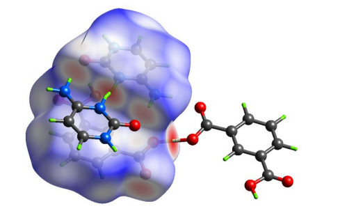
Suppressing the motion of organic molecules to increase the possibility of radiative transition and decrease the non-radiative transitions plays a key role to the room temperature phosphorescence in this report. Thus, Hirshfeld surface calculation was performed with CrystalExplorer program[16] to analyze the non-covalent interactions in the crystal lattice. Fig. 8 shows the Hirshfeld surface of C-H2IA mapped over dnorm used a red-white-blue color scheme. In the Hirshfeld surface, the dark red color highlights shorter contacts with the negative dnorm values which are shorter than the sum of van der Waals radii, whereas the dark blue color represents the contacts longer than the van der Waals radii with the positive dnorm values and white represents the contact around the van der Waals radii. The hydrogen bonding interactions, such as N–H···O and N–H···N interactions in the cytosine-cytosinium ion dimer, N–H···O interactions between cytosine-cytosinium ion dimers and O–H···O interactions between isophthalic acid molecules, can be observed in the Hirshfeld surface plot as the bright red shaded areas. The Hirshfeld surface plot is in coincidence with the result of single-crystal analysis.
Figure 8
 Figure 8. Hirschfeld surfaces for C-H2IA mapped with dnorm
4. CONCLUSION
In summary, a supramolecular framework cytosinium isophthalate was synthesized by self-assembly of cytosine and isophthalic acid in aqueous media and the crystal structure was redetermined revealing the partial proton transfer from carboxylate group to the pyrimidine ring. In the supramolecular framework C-H2IA, the hydrogen bonds take over to form the one-dimensional infinite cytosine and isophthalic acid tape structures. On this basis, the adjacent arranged organic base and acid tapes are further extended into a three-dimensional network through π-π interactions. Interesting, the framework C-H2IA shows a green phosphorescence emission visible to the naked eyes under ambient conditions. The room temperature solid-state photoluminescence analysis reveals C-H2IA possesses the ultra-long emission lifetime up to 0.7 s and is thus a potential organic luminescent material with ultra-long phosphorescence feature.
-
-
[1]
Yang, Z.; Mao, Z.; Zhang, X. Intermolecular electronic coupling of organic units for efficient persistent room-temperature phosphorescence. Angew. Chem., Int. Ed. Engl. 2016, 55, 2181–2185. doi: 10.1002/anie.201509224
-
[2]
(a) Kuei, C. Y.; Liu, S. H.; Chou, P. T. Room temperature blue phosphorescence: a combined experimental and theoretical study on the bis-tridentate Ir(Ⅲ) metal complexes. Dalton Trans. 2016, 45, 15364–15373; (b) Hirata, S. Recent advances in materials with room-temperature phosphorescence: photophysics for triplet exciton stabilization. Adv. Optical Mater. 2017, 5, 1700116–1700165.
-
[3]
(a) Gong, Y.; Chen, G.; Peng, Q. Achieving persistent room temperature phosphorescence and remarkable mechanochromism from pure organic luminogens. Adv. Mater. 2015, 27, 6195–6201; (b) Xiao, L.; Wu, Y.; Chen, J. Highly efficient room-temperature phosphorescence from halogen-bonding-assisted doped organic crystals. J. Phys. Chem. A 2017, 121, 8652–8658.
-
[4]
(a) Ge, C. H.; Zhang, X. Y.; Yu, F. Hydrogen-bonded assembly of Ni(Ⅱ) and Cu(Ⅱ) complexes generate 3D supramolecular frameworks. J. Chem. Crystallogr. 2008, 38, 501–505; (b) Nirmalram, J. S.; Tamilselvi, D.; Muthiah, P. T. Hydrogen bonded supramolecular assembly in N6-benzyladeninium nitrate and N6-benzyladeninium 3-hydroxy picolinate: a synthetic cytokinin. J. Chem. Crystallogr. 2011, 41, 864–867.
-
[5]
Chen, J. The complexation between 'texas sized' molecular box and linear n-aliphate dianion: en route to supramolecular organic frameworks (SOFs) for selectively CO2 absorption. Tetrahedron 2016, 72, 431–435. doi: 10.1016/j.tet.2015.11.062
-
[6]
Ni, X. L.; Chen, S.; Yang, Y. Facile cucurbit[8]uril-based supramolecular approach to fabricate tunable luminescent materials in aqueous solution. J. Am. Chem. Soc. 2016, 138, 6177–6183. doi: 10.1021/jacs.6b01223
-
[7]
Han, Z. B.; Xiao, Z. Z.; Hao, M. Functional hydrogen-bonded supramolecular framework for K+ ion sensing. Cryst. Growth Des. 2015, 15, 531–533. doi: 10.1021/cg501259g
-
[8]
Lin, X. M.; Niu, J. L.; Wen, P. X. A polyhedral metal-organic framework based on supramolecular building blocks: catalysis and luminescent sensing of solvent molecules. Cryst. Growth Des. 2016, 16, 4705–4710. doi: 10.1021/acs.cgd.6b00779
-
[9]
Zhang, Y.; Zhan, T. G.; Zhou, T. Y. Fluorescence enhancement through the formation of a single-layer two-dimensional supramolecular organic framework and its application in highly selective recognition of picric acid. Chem. Comm. 2016, 52, 7588–7591. doi: 10.1039/C6CC03631G
-
[10]
Bian, L.; Shi, H.; Wang, X. Simultaneously enhancing efficiency and lifetime of ultralong organic phosphorescence materials by molecular self-assembly. J. Am. Chem. Soc. 2018, 140, 10734–10739. doi: 10.1021/jacs.8b03867
-
[11]
Ebenezer, S.; Muthiah, P. T. Design of co-crystals/salts of aminopyrimidines and carboxylic acids through recurrently occurring xynthons. Cryst. Growth Des. 2012, 12, 3766–3785. doi: 10.1021/cg3005954
-
[12]
Ayed, M.; Ayed, B.; Haddad, A. Synthesis and structural characterization of new inorganic-organic hybrid: arsenomolybdate compound with cytosinium cations. Bull. Mater. Sci. 2015, 38, 13–21. doi: 10.1007/s12034-014-0805-8
-
[13]
Marsh, R. E.; Bierstedt, R.; Eichhorn, E. L. The crystal structure of cytosine-5-acetic acid. Acta Crystallogr. 1962, 15, 310–316. doi: 10.1107/S0365110X62000791
-
[14]
Perumalla, S. R.; Suresh, E.; Pedireddi, V. R. Nucleobases in molecular recognition: molecular adducts of adenine and cytosine with COOH functional groups. Angew. Chem., Int. Ed. Engl. 2005, 44, 7752–7757. doi: 10.1002/anie.200502434
-
[15]
Macrae, C. F.; Bruno, I. J.; Chisholm, J. A. Mercury CSD 2.0 - new features for the visualization and investigation of crystal structures. Journal of Applied Crystallogr. 2008, 41, 466–470. doi: 10.1107/S0021889807067908
-
[16]
(a) Turner, M. J.; McKinnon, M. J. J.; Wolff, S. K.; Grimwood, D. J.; Spackman, P. R.; Jayatilaka, D.; Spackman, M. A. CrystalExplorer17 (2017). University of Western Australia. http://hirshfeldsurface.net 2017; (b) Mackenzie, C. F.; Spackman, P. R.; Jayatilaka, D.; Spackman, M. A. CrystalExplorer model energies and energy frameworks: extension to metal coordination compounds, organic salts, solvates and open-shell systems. IUCrJ 2017, 4, 575–587; (c) Bakavoli, M.; Rahimizadeh, M.; Feizyzadeh, B. 3,6-di(p-chlorophenyl)-2,7-dihydro-1,4,5-thiadiazepine: crystal structure and decoding intermolecular interactions with hirshfeld surface analysis. J. Chem. Crystallogr. 2010, 40, 746–752.
-
[1]
-
Figure 1 A perspective view with atomic numbering scheme. Displacement ellipsoids are drawn at the 50% probability level
Figure 2 A view of infinite tape of [HC2]nn+ connected via hydrogen bonds (dashed line). Hydrogen atoms on carbons and one moiety of disorder hydrogen atoms are omitted for clarity
Figure 3 A view of infinite tape of [H(HIA)2]nn- connected via hydrogen bonds (dashed line). The hydrogen atoms on benzene ring are omitted for clarity
Figure 4 (a) Three-dimensional supramolecular net formed by π-π stacking of tapes; (b) Illustration of the molecular packing in C-H2IA
Figure 7 (a) Photographs of C-H2IA crystals with a hand-held 300 nm UV lamp irradiation on and off; (b) Normalized photoluminescence (PL, blue line) and room temperature phosphorescence (green line) spectra of C-H2IA under 300 nm excitation; (c) Time-resolved fluorescence decay (blue dot) and fitting curves (red line) of the emission bands at 375 nm; (d) Time-resolved phosphorescence decay (green dot) and fitting curves (red line) of the emission bands at 500 nm
Table 1. Hydrogen Bond Lengths (Å) and Bond Angles (°) for C-H2IA
D–H⋅⋅⋅A d(D–H) d(H⋅⋅⋅A) d(D⋅⋅⋅A) < (DHA) N(4)–H(4)⋅⋅⋅N(4)#1 0.88 1.96(3) 2.829(2) 174 N(6)–H(6A)⋅⋅⋅O(2)#1 0.85 2.03 2.8670(15) 171 O(9)–H(9)⋅⋅⋅O(9)#2 1.23 1.23 2.4612(18) 180 N(1)–H(1)⋅⋅⋅O(2)#3 0.93 1.91 2.8304(15) 173 O(11)–H(11)⋅⋅⋅O(10)#4 0.92 1.80 2.6179(14) 147 Symmetry transformations used to generate the equivalent atoms:
#1: –x+1, –y+2, –z+1; #2: –x, –y+1, –z+1; #3: –x+2, –y+2, –z+1; #4: x+1, y, z 下载: 导出CSV
-


 扫一扫看文章
计量
- PDF下载量: 5
- 文章访问数: 747
- HTML全文浏览量: 7
 下载:
下载:
