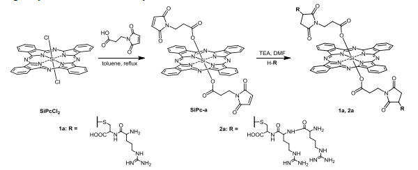
Scheme 1a.
Synthesis of 1a and 2a
Synthesis, Spectroscopic Properties, and Photodynamic Anticancer Activities of Novel Arginine-modified Silicon(IV) Phthalocyanines
Jia-Wen YING , Qun SUN , Li-Fang YANG , Mei-Rong KE , Jian-Dong HUANG

As a non-invasive therapeutic modality for cancers, photodynamic therapy (PDT) has been approved for treating various cancers since 1993. It utilizes the photochemical reactions of three components, including light, photosensitizers, and oxygen, to generate cytotoxic reactive oxygen species (ROS), particularly singlet oxygen, leading to irreversible damage to cancer cells and tissues[1, 2]. PDT has received incessant attention for cancer therapy due to several unique advantages, including non-invasion, high spatiotemporal selectivity, no drug resistance, and antitumor immune response against metastases[3-6].
Owning to some desirable features, such as strong absorption in photo-therapeutic window (600~800 nm) and easily structural modification, phthalocyanines have been widely used for PDT[7, 8]. However, with the 18π electron conjugated macrocyclic skeleton, they generally show poor hydrophilicity and tend to aggregate in aqueous solutions, resulting in self-quenching of photosensitizing activities, which is unfavorable for PDT. Thus, various strategies have been developed to address this issue. One of the most facile and effective approaches is modification with biocompatible hydrophilic molecules, such as peptides[9-11], saccharides[12, 13], nucleosides[14, 15], protein[16, 17] and amino acids[18]. Amino acids, which are essentially biological small molecules to human body, have been usually used to modify various drugs including photosensitizers to improve their biocompatibility and hydrophilicity[19]. Recently, we have reported a series of silicon(IV) phthalocyanines (SiPcs) axially substituted with L-tyrosine moieties, which could enhance the hydrophilicity of phthalocyanines and relieve aggregation in aqueous solution, leading to extremely high photodynamic activities against cancer cells[20].
With a highly basic guanidine group, arginine can be easily protonated in a wide range of pH and turn into a positively charged cation, which is liable to bind to the negatively charged plasma membrane by electrostatic interaction, and could facilitate cellular uptake[21, 22]. Thus, the arginine-rich peptides have widely been used as cell penetrating peptides (CPP) to deliver various biomolecules or drugs into cells[23-25]. Moreover, it has been reported that the arginine-rich CPP is greatly more efficient than the lysine-rich analogues even with the same net charges, probably resulting from the formation of stable bidentate hydrogen bonds between the guanidine group and polarizable oxo-anions such as sulfates or phosphates[26-28].
Inspired by the results, we are interested that whether less arginine units such as one or two arginine(s) could also bind plasma membrane and improve the cellular uptake. Thus in this work, we carefully design a series of novel SiPcs (1a, 2a, 1b, and 2b) modified with arginine and arginine-containing oligopeptides (Arg-Arg, Cys-Arg, Cys-Arg-Arg) through different connection ways to demonstrate the effect on photodynamic behavior. The photophysical and photochemical properties, aggregation behavior, plasma membrane localization, in vitro and in vivo photodynamic activities of these SiPcs have been investigated and the difference has been discussed. Thus, this work would provide a new reference for the development of phthalocyanine-based photosensitizers.
All the reactions were performed under an atmosphere of nitrogen. Toluene was dried by a Deminex EX-SPS-800 solvent purification system. Maleic anhydride, alpha-alanine and N-(tert-butoxycarbonyl)-L-arginine hydrochloride (N-Boc-L-Arg) were purchased from Adamas Reagent Co., Ltd., (Switzerland). N-(tert-butoxycarbonyl)-arginine-arginine (N-Boc-Arg-Arg) was obtained from GL Biochem Ltd., (Shanghai, China). Cysteinearginine (Cys-Arg) and cysteine-arginine-arginine (Cys-Arg-Arg) were purchased from DGpeptides Co., Ltd., (Hangzhou, China). 1-Hydroxybenzotriazole (HOBt) and N, N-diisopropylethylamine (DIEA) were purchased from Acros organics (Belgium).1-Ethyl-3-(3-dimethylaminopropyl)carbodiimide hydrochloride (EDCI) was bought from TCI (Shanghai, China). SiPc-b and 3-(maleimido)propanoic acid were synthesized according to previous methods[29, 30]. Chromatographic purifications were performed on silica gel columns (300~400 mesh, Qingdao Haiyang Chemical Co., Ltd., China) with the indicated eluents. Size exclusion chromatography was performed on Bio-Rad Bio-Beads S-X1 beads with the indicated eluents.
1H NMR spectra were recorded on a Bruker AVANCE III 400 spectrometer in CDCl3 or DMSO-d6. High resolution mass spectra (HRMS) were recorded on a Thermo Scientific Exactive Plus LC/MS spectrometer. The electronic absorption spectra and fluorescence spectra were measured on a Shimadzu UV-2450 UV-vis spectrophotometer and an Edinburgh FS5 spectrofluorometer, respectively. The fluorescence quantum yields (ФF) and singlet oxygen quantum yields (Ф△) in DMF were measured by the methods described in previous work[31-33].
The analytical HPLC experiments were performed on a Kromasir KR100-10 C18 column (5 μm, 4.6mm × 250mm) by using a Shimadzu LC-10AT controller with a SPD-M10A diode array detector. The conditions were set as follows: 1a, 2a, and 1b: 50~90% B for 30 min (solvent A: 100% H2O with 1% trifluoroacetic acid; solvent B: 100% methanol (MeOH)). 2b: 20~70% B for 40 min (solvent A: a mixed solvent containing an aqueous solution of sodium dodecyl sulfate (10 mM) and phosphoric acid (26 mM), acetonitrile and methanol (250/250/30, v/v/v); solvent B: 100% N, N-dimethylformamide (DMF)). The column temperature was set at 30 ℃ at a flow-rate of 1.0 mL/min.
A mixture of silicon(IV) phthalocyanine dichloride (SiPcCl2) (100.0 mg, 0.164 mmol) and 3-maleimidopropionic acid (110.8 mg, 0.654 mmol) in anhydrous toluene (50 mL) was refluxed for 24 h. The solvent was evaporated in vacuum distillation and the obtained solid was purified by silica column chromatography using dichloromethane (DCM)/MeOH (40/1, v/v) as the eluents to give blue solid SiPc-a (45.0 mg, 31%). 1H NMR (400 MHz, CDCl3, ppm): δ 9.65~9.75 (m, 8H, Pc-Hα), 8.34~8.44 (m, 8H, Pc-Hβ), 6.00 (s, 4H, CH), 1.59 (t, J = 8.0 Hz, 4H, CH2), –0.41 (t, J = 8.0 Hz, 4H, CH2). H RMS (ESI) m/z: calcd. for C46H28N10O8Si: [M]+ 876.1861, found 876.1898.
A mixture of SiPc-a (20.0 mg, 0.023 mmol), Cys-Arg (25.3 mg, 0.091 mmol), and trimethylamine (TEA) (5 μL) in DMF (5 mL) was stirred at 30 ℃ for 24 h. The solvent was evaporated in vacuum distillation and the residue was dissolved in DMF (2 mL) to be purified by size-exclusion column chromatography using DMF as the eluent. The obtained crude was further dissolved in DMF (4 mL), precipitated with a large amount of DCM, filtered, and then washed with DCM and ethyl acetate (EA), respectively, to give 1a as a blue-green solid (21.0 mg, 64%). 1H NMR (400 MHz, DMSO-d6, ppm) δ 9.71~9.81 (m, 8H, Pc–H), 8.55~8.65 (m, 8H, Pc–H), 8.20~8.29 (m, 2H, NH), 7.04~7.30 (m, 8H, NH), 4.19 (br., 4H, NH2), 3.07~3.10 (m, 2H, CH), 2.65~2.77 (m, 4H, CH), 2.29~2.47 (m, 8H, CH, CH2), 1.40~1.81 (m, 12H, CH2), 1.23 (t, J = 8.0, 4H, CH2), –0.48 (t, J = 8.0, 4H, CH2). H RMS (ESI) m/z: calcd. for C64H66N20O14S2Si: [M+2H]2+ 716.2212, found 716.2180. The purity was found to be 97.3% by HPLC analysis.
According to the above procedure, SiPc-a (20.0 mg, 0.023 mmol) was treated with Cys-Arg-Arg (29.6 mg, 0.068 mmol) and TEA (5 ul) in DMF (5 mL) to give 2a as a blue-green solid (23.0 mg, 58%). 1H NMR (400 MHz, DMSO-d6, ppm) δ 9.71~9.78 (m, 8H, Pc-H), 8.54~8.63 (m, 8H, Pc–H), 8.13~8.24 (m, 4H, NH), 7.61~7.74 (m, 4H, NH), 6.98~7.35 (m, 12H, NH), 4.27 (br., 4H, NH2), 4.06~4.11 (m, 4H, CH), 3.10~3.14 (m, 2H, CH), 3.00~3.06 (m, 8H, CH2), 2.37~2.47 (m, 6H, CH, CH2), 1.37~1.81 (m, 20H, CH2), 1.22 (t, J = 7.6, 4H, CH2), –0.50 (t, J = 7.6, 4H, CH2). H RMS (ESI) m/z: calcd. for C76H90N28O16S2Si: [M+2H]2+ 872.3223, found 872.3192. The purity was found to be 96.3% by HPLC analysis.
The SiPc-b (50.0 mg, 0.062 mmol), N-Boc-L-Arg (50.63 mg, 0.185 mmol), EDCI (35.4 mg, 0.185 mmol), HOBt (24.9 mg, 0.185 mmol) and DIEA (40 ul) in DMF (10 mL) were stirred at 30 ℃ for 24 h. The solvent was evaporated in vacuum distillation and the residue was dissolved in DMF (2 mL) to be purified by size-exclusion column chromatography using DMF as the eluent to give 1b as a blue-green solid (20.0 mg, 24%). 1H NMR (400 MHz, DMSO-d6, ppm) δ 9.61~9.82 (m, 8H, Pc-H), 8.50~8.63 (m, 8H, Pc-H), 7.51 (br., 2H, NH), 7.27 (br., 2H, NH), 6.85~7.20 (m, 6H, NH, NH2), 6.53 (d, J = 6.4 Hz, 2H, NH), 5.42 (d, J = 7.2 Hz, 4H, Ar–H), 3.55 (br., 2H, CH), 2.40 (br., 4H, CH2), 2.24 (d, J = 7.2 Hz, 4H, Ar–H), 1.75 (br., 4H, CH2), 1.29~1.41 (m, 4H, CH2), 1.18 (s, 18H, CH3). H RMS (ESI) m/z: calcd. for C70H76N20O8Si: [M+2H]2+ 663.3009, found 663.2996. The purity was found to be 96.6% by HPLC analysis.
According to the above procedure, SiPc-b (20.0 mg, 0.025 mmol) was treated with N-Boc-Arg-Arg (31.8 mg, 0.074 mmol), EDCI (14.2 mg, 0.074 mmol), HOBt (9.9 mg, 0.074 mmol), and DIEA (16 ul) in DMF (10 mL) to give 2b as a blue-green solid (13.0 mg, 32%). HRMS (ESI) m/z: calcd. for C82H102N26O10Si: [M+2H]2+ 819.4021, found 819.4019. The purity was found to be 95.5% by HPLC analysis.
The HepG2 human hepatocarcinoma cells and Hela human cervical cancer cells were maintained in RPMI 1640 cell culture medium (Hyclone™), and supplemented with 10% fetal bovine serum, penicillin (50 units/mL), and streptomycin (50 mg/mL) at 37 ℃ in a humidified 5% CO2 incubator.
The solution of 1a, 2a, 1b, and 2b (1 mM) was prepared in dimethyl sulfoxide (DMSO) and stored at 4 ℃ in the dark. The stock solution was diluted to 80 μM with Phosphate Buffered Saline (PBS) containing 1% Cremophor EL™. The solutions were sterilized by filtration through 0.22 μm syringe filters, and then diluted with the cellular culture medium to required concentrations.
HepG2 and Hela cells (about 1 × 10⁴ cells per well) were maintained in 96-well plates overnight at 37 ℃ in a humidified 5% CO2 atmosphere. The cells were then respectively incubated with 1a, 2a, 1b, and 2b (100 μL) at various concentrations in the dark for 2 h. After removing the samples, the cells were rinsed with PBS and re-fed with 100 μL of the culture medium before illumination at ambient temperature. The fluence rate (λ > 610 nm) was 15 mW/cm2. An illumination of 30 min led to a total fluence of 27 J/cm2. After illumination, the cells were then incubated in a humidified 5% CO2 incubator overnight.
Cell viability was determined by the colorimetric MTT assay and carried on by our previous methods[34].
About 6 × 10⁴ HepG2 or Hela cells suspension in RPMI 1640 cell culture medium (0.4 mL) were seeded on a confocal dish and incubated overnight at 37 ℃ with 5% CO2. After removing the medium, the cells were incubated with 1a, 2a, 1b and 2b (4 μM, 0.4 mL), respectively in the dark for different incubation time (0.5, 1 and 2 h), then the cells were rinsed with PBS twice and imaged using a LEICA TCS SPE confocal microscope. The SiPcs were excited at 635 nm and monitored at 640~750 nm. The images were then digitized and analyzed by using the SPE ROI Fluorescence Statistics software. The average intracellular fluorescence intensities (a total of 20 cells for each sample) were also determined.
HepG2 cells (about 1 × 10⁴ cells per well) were maintained in 96-well plates overnight at 37 ℃ in a humidified 5% CO2 atmosphere. The cells were then incubated with 2a and 2b (4 μM, 100 μL) respectively, in the dark for 2 h. After removing the samples, the cells were rinsed with PBS and re-fed with 100 μL of the culture medium, followed by illumination (λ > 610 nm, 15 mW/cm2) for 10 min at ambient temperature (For the control groups, no irradiation was carried out). After that, the trypan blue (4 mg/mL in PBS, 10 μL) solution was added for further 10 min incubation. The cells were then rinsed with PBS and took photos with an inverted microscope (BDS300 inverted microscope, Chongqing Aote Optical Instrument Co., Ltd.). The cells treated with the culture medium only with or without light irradiation for 10 min were used as the controls.
We chose Hepatoma cells (H22) and ICR mice which were purchased from the China Center for Type Culture Collection (CCTCC, Wuhan, China) and Wushi Animal Co. Ltd. (Fuzhou, China) respectively to carry out the in vivo studies. The animal studies were conducted according to the guidelines of the Animal Protection Committee of Fuzhou University with approval of the committee. H22 cells (~1×10⁷ cells in 200 μL) were inoculated subcutaneously on the right hind limb of ICR mice weighting 20~25 mg to set up the subcutaneous tumor model. 1a, 2a, 1b, and 2b aqueous solutions with 1% Cremophor EL™ (100 μM, 100 μL) were injected into the tail vein of the tumor-bearing mice when the tumors grew to 80~100 mm3. The in vivo fluorescence imaging of mice at different time points was captured at 680 nm on a Caliper LifeSciences_IVIS SPECTRUM imaging system (excited at 640 nm). All of these mice were euthanized after injection for 24 h and fluorescence imaging of heart, liver, spleen, lung, kidney, and tumor were obtained.
H22 cells (~1×10⁷ cells in 200 μL) were inoculated subcutaneously on the right hind limb of ICR mice weighting 20~25 mg to set up the subcutaneous tumor model. When the tumors reached to 80~100 mm3, 2b aqueous solution with 1% Cremophor EL™ (200 μM, 100 μL) was intravenously injected into the tumor-bearing mice. After 12 h, the tumor-bearing mice were irradiated a laser of 685 nm (15 mW/cm2) for 10 min. The PDT treatment was further repeated at 2ed and 6th days, respectively. When the mice were administrated at the first time (donated as 0 day), the tumor sizes were gauged and recorded every two days with vernier calipers for 14 days to estimate tumor inhibition rate. The tumor size was calculated by the equation: tumor volume = (tumor length) × (tumor width) × (tumor hight) × 6/π. There were other three groups of mice, including those treated with 2b but without irradiation, and treated with PBS with and without irradiation, respectively as the controls. Four mice were used for each group.
Statistical analyses were carried out using student T-test analysis of variance; P < 0.05 was considered significant. Data were expressed as mean ± standard deviation (SD).
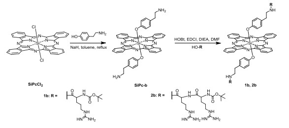
In this work, we designed four axially substituted SiPcs (1a, 2a, 1b, and 2b) through different connection ways and different number of arginine. 1a and 2a were respectively modified with Cys-Arg and Cys-Arg-Arg by using 3-(maleimido)propanoic acid as a linker, which was conjugated with the SiPc core through an ester bond (Scheme 1a). The other two SiPcs (1b and 2b) were modified with N terminal Boc-protected arginine and di-arginine peptide through an ether bond of phenoxyl group to link with the SiPc core (Scheme 1b). As shown in Scheme 1a, the maleimide-modified SiPc-a was synthesized through a nucleophilic substituted reaction of 3-(maleimido)propanoic acid and SiPcCl2, and then SiPc-a and Cys-Arg (or Cys-Arg-Arg) went through the Michael Addition reaction in the presence of TEA to obtain the corresponding final products 1a and 2a. Here the cysteine (Cys) was induced as the functional group for the coupling of arginine or di-arginine[35]. For the preparation of ether-modified 1b and 2b, SiPc-b which was synthesized by previous method[29] was coupled with N-Boc-L-Arg or N-Boc-Arg-Arg dipeptide respectively by using HOBt, EDCI as the activating agents in the presence of DIEA. All the compounds were purified by chromatographic methods, and the final products were characterized by HRMS. The purity was determined by HPLC analysis (≥ 95%).
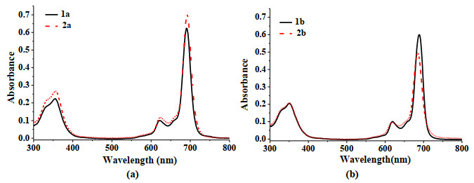
The spectroscopic properties of 1a, 2a, 1b and 2b were recorded in DMF, and the data are summarized in Table 1. As shown in Fig. S1, all the SiPcs showed a B-band at 355~360 nm, a vibronic band at 611~617 nm, and an intense and sharp Q-band at 680~686 nm, suggesting that they were nonaggregated in DMF. Excited at 610 nm, all SiPcs exhibited fluorescence emission at 686~696 nm with a fluorescence quantum yield (ФF) of 0.09~0.35 using unsubstituted ZnPc as a reference (ФF = 0.28)[31]. 1a and 2a showed similar spectral properties, so did 1b and 2b. The results indicate that the number of arginine did not affect their spectral properties obviously. However, it could be found that both in absorption and fluorescence spectra, the Q bands of the ester-linked SiPcs (1a and 2a) slightly red-shifted (4~10 nm) in contrast to that of ether-linked analogues (1b and 2b), as a result of the different electronic effect of the electron-withdrawing ester and electron-donating ether linkers, as previously reported by us[20]. Moreover, the fluorescence quantum yields of 1a and 2a were significantly higher than that of 1b and 2b also due to the different electronic effects of the linkers, indicating that linking the electron-donating group at the central atom of phthalocyanine core would quench the excited phthalocyanine, leading to the quenching of fluorescence.
 DownLoad:
CSV
DownLoad:
CSV
| SiPcs | λmax (nm) | λem (nm)a | Stoke shift (nm) | ε×105 (M-1·cm-1) | ФF b | Ф△c | |
| 1a | 684 | 696 | 12 | 1.81 | 0.35 | 0.33 | |
| 2a | 686 | 694 | 8 | 2.24 | 0.29 | 0.26 | |
| 1b | 680 | 686 | 6 | 2.01 | 0.09 | 0.10 | |
| 2b | 681 | 686 | 5 | 2.02 | 0.10 | 0.10 | |
| aExcited at 610 nm. bUsing ZnPc in DMF as the reference (ФF = 0.28)[31]. cDetermined by using DPBF as the chemical quencher, and ZnPc in DMF as the reference (Ф△ 0.56)[33]. |
|||||||
Using 1, 3-diphenylisobenzofuran (DPBF) as the scavenger, the singlet oxygen quantum yields (Ф△) of all the SiPcs in DMF were determined according to the previous method[32] with the singlet oxygen quantum yields (Ф△) ranging from 0.10 to 0.33 (Table 1). Similar to the results of fluorescence emission, 1b and 2b showed significantly lower singlet oxygen quantum yields relative to 1a and 2a.
The absorption and fluorescence spectra of all the SiPcs were also measured in aqueous solutions to explore their aggregation behavior. As shown in Fig. 1, all the SiPcs remained a sharp and strong Q band in H2O, suggesting they were basically in the form of a monomer in H2O. The result demonstrated that the axially substituted hydrophilic arginine or oligoarginine could reduce the aggregation of phthalocyanine effectively due to the steric effect as well as decline of hydrophobic interaction. Similar to that in DMF, 1b and 2b exhibited obviously weaker fluorescence emission than 1a and 2a, mainly due to the quenching of electron-donated ether linker (Fig. S2). However, in RPMI 1640 cell culture medium, some of them became aggregated in different degree even in the presence of 1% surfactant Cremophor EL™ (Fig. S3). The aggregation should be attributed to the effect of salt. The di-arginine substituted SiPcs (2a and 2b) were less aggregated relative to the analogues substituted with mono-arginine (1a and 1b), revealing that the arginine could contribute to the diminution of aggregation.
The photodynamic activities of 1a, 2a, 1b, and 2b against HepG2 human hepatocarcinoma cells were investigated by MTT assay. The IC50 values, defined the concentrations leading to 50% cell death upon irradiation, are summarized in Table 2. Fig. 2a and 2b show the dose-dependent survival curves with and without irradiation. All the SiPcs showed no cytotoxicity against HepG2 cell in dark at the detected concentrations. However, they have obvious photocytotoxicity against HepG2 cell upon irradiation with the trend of 2b > 1b > 2a > 1a, and 2b was the most potent with the IC50 value of 0.08 μM, which is greatly higher than the well-known anticancer photosensitizer chlorin e6 (IC50 = 0.75 μM) under the same condition[14]. Obviously, 1b and 2b exhibit higher photocytoxicity than the corresponding 1a and 2a. Moreover, the diarginine-modified SiPcs (2a and 2b) are more potent compared to the mono-arginine modified analogues (1a and 1b).
DownLoad:
CSV
| SiPc | HepG2 | Hela | SiPc | HepG2 | Hela | |
| 1a | 1.07 ± 0.15 | 1.43 ± 0.14 | 1b | 0.14 ± 0.01 | 0.09 ± 0.01 | |
| 2a | 0.38 ± 0.01 | 0.66 ± 0.09 | 2b | 0.08 ± 0.01 | 0.12 ± 0.01 |
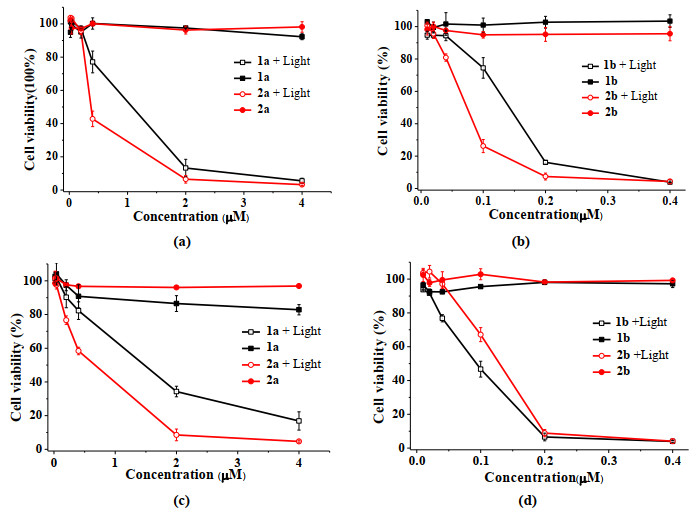
In addition, the photodynamic activities of these SiPcs against Hela human cervical cancer cells were also investigated (Table 2, Figs. 2c and 2d). Similarly, no obviously dark cytotoxicity against Hela cells was observed for all the SiPcs. The photocytotoxicity of all the SiPcs (except 1b) was slightly diminished in contrast to that against HepG2 cells, with the trend of 2b ≈ 1b > 2a > 1a. 1b and 2b still exhibited significantly higher photocytotoxicity than 1a and 2a.
Generally, the SiPcs (1b and 2b) modified with N terminal Boc-protected arginine and di-arginine showed much higher photocytotoxicity than the analogues (1a and 2a) modified with Cys-Arg and Cys-Arg-Arg for both cell lines. The difference of photocytotoxicity between 1a and 2a is significant, while for 1b and 2b, the difference is smaller.
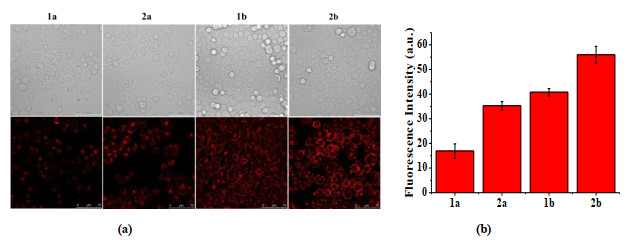
The cellular uptake of these SiPcs on HepG2 cells was further examined by confocal laser scanning microscopy (CLSM) to account for the different photodynamic activities. As shown in Fig. 3, 2b exhibits the strongest intracellular intensity, and the order of intracellular fluorescence intensity was consistent with the result of photocytotoxicities. Similarly, the intracellular fluorescence intensity was strengthened with the increase of arginine number, indicating that the arginine was conducive to the enhancement of cellular uptake of the SiPcs.
The damage of plasma membrane is a major contributing factor in overall PDT efficacy[36-39]. The plasma membrane localization of these SiPcs was further investigated by CLSM. The fluorescence images of these SiPcs on HepG2 cells were taken at 0.5 and 2.0 h. As displayed in Fig. 4a and 4b, for 1b and 2b, the strong fluorescence signal could be clearly observed mainly in the plasma membrane at both incubation time, while 1a and 2a majorly entered the cells with little fluorescence signal in the plasma membrane. In addition, to demonstrate the universality of the plasma membrane-targeting ability of 1b and 2b, the plasma membrane localization of these SiPcs was also tested on Hela cells. With similar result to that on HepG2 cells, 1b and 2b mainly accumulated in plasma membrane (Fig. S4).
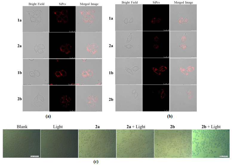
To further confirm the plasma membrane localization and its photodamage effect, 2a and 2b were chosen to detect the PDT-induced damage on the plasma membrane of HepG2 cells by inverted microscope using trypan blue staining. As shown in Fig. 4c, only the cells treated with 2b upon irradiation were stained with trypan blue, suggesting 2b could localize in the plasma membrane and induce the destruction.
The results demonstrated that the higher cellular uptake and plasma membrane localization ability of 1b and 2b should be the major responsibility for their significantly higher in vitro photodynamic activities relative to 1a and 2a. Interestingly, both a and b series of SiPcs contained arginine or diarginine, but they have obviously different plasma membrane localization ability and cellular uptake. The reason is still elusive at this stage. We presume that for 1a and 2a, which had one more cysteine residue on the substituent compared to 1b and 2b, the guanidine group of the arginine would electrostatically interact with the carboxyl group of the cysteine, resulting in the decrease of the affinity of arginine to the plasma membrane.
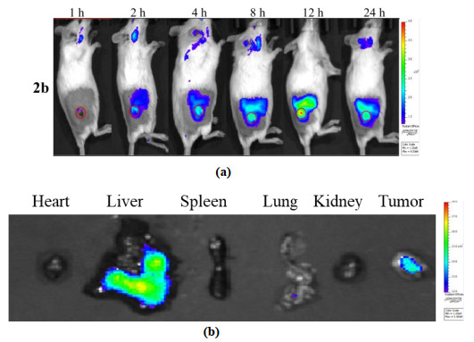
The in vivo fluorescence imaging was also performed to demonstrate the biological distribution and tumor-targeting of these SiPcs. The ICR (Institute of Cancer Research) mice bearing H22 tumors were intravenously injected with these SiPcs (100 μΜ, 100 μL), respectively, and monitored by Caliper Life Sciences IVIS SPECTRUM for 24 h. As shown in Fig. 5a, for the mice treated with 2b, the fluorescence signal could be observed mostly in tumor tissue after 2 h, and the signal was continuously enhanced until 12 h. After 24 h post-injection, the mice were sacrificed and then the fluorescence images of the tumor and major organs, including the heart, liver, spleen, lung, and kidney, were taken. As displayed in Fig. 5b, 2b mainly accumulated in tumor tissue and liver, and no fluorescence signal was observed on the other organs. However, the other SiPcs (1a, 2a, and 1b) did not show obvious tumor-targeting. As shown in Fig. S5, the fluorescence signal was basically distributed in the whole body after administration for a while. The results indicated that among these SiPcs, 2b exhibited the highest tumor-targeting. In addition, we could find that these compounds were metabolized in different rates. After 24 h postinjection, the mice treated with 1a showed weak fluorescence signal on the body, while those treated with 1b remained very strong fluorescence.
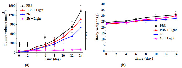
With the most potent photodynamic activity and the highest tumor-targeting, 2b was chosen to further study the in vivo PDT effect. The H22 tumor-bearing mice were treated with 2b by intravenous injection, and then irradiated with laser. The mice totally underwent PDT-treatment for three times. The groups treated with PBS with and without irradiation and that treated with 2b without irradiation were used as controls. Fig. 6a shows the tumor growth curves of the mice under different conditions over 14 days. For the treatment group (2b + light), the tumor size hardly changed over the test time, while for the other control groups, the tumor size continuously grew up over time, suggesting 2b could inhibit tumor growth effectively with the tumor inhibition rate up to 93%. In addition, the body weight of these ICR mice slightly increased over 14 days, indicating negligible systematic toxicity of 2b (Fig. 6b).
In summary, we have synthesized and characterized a series of novel SiPcs (1a, 2a, 1b, and 2b) axially modified with arginine or arginine-containing oligopeptides (Arg-Arg, Cys-Arg, Cys-Arg-Arg) through ester or ether linkers. The ester-linked SiPcs (1a and 2a) show red-shifted Q band, higher fluorescence, and singlet oxygen generation efficiency relative to the ether-linked analogues (1b and 2b) due to different electronic effects. The induction of the arginine moieties could significantly improve their hydrophilicity and relieve the aggregation behavior in aqueous solutions. Moreover, 1b and 2b exhibit more potent photodynamic activity against HepG2 and Hela cells than the corresponding 1a and 2a analogues as a result of the higher cellular uptake and plasma membrane localization property. Finally, the in vivo fluorescence imaging suggests that among these compounds, 2b is the most specific toward H22 tumor-bearing ICR mice, and it shows efficient tumor growth inhibition with the tumor inhibition rate up to 93%.
Monro, S.; Colon, K. L.; Yin, H.; Roque, J.; Konda, P.; Gujar, S.; Thummel, R. P.; Lilge, L.; Cameron, C. G. Transition metal complexes and photodynamic therapy from a tumor-centered approach: challenges, opportunities, and highlights from the development of TLD1433. Chem. Rev. 2019, 119, 797−828. doi: 10.1021/acs.chemrev.8b00211
Bolze, F.; Jenni, S.; Sour, A.; Heitz, V. Molecular photosensitisers for two-photon photodynamic therapy. Chem. Commun. 2017, 53, 12857−12877. doi: 10.1039/C7CC06133A
Dennis, E. J. G. J. D.; Dai, F.; Jain, R. K. Photodynamic therapy for cancer. Nat. Rev. Cancer. 2003, 3, 380−387. doi: 10.1038/nrc1071
Agostinis, P.; Berg, K.; Cengel, K. A.; Foster, T. H.; Girotti, A. W.; Gollnick, S. O.; Stephen, M. H. D.; Michael, R. H.; Asta, J.; David, K.; Mladen, K.; Johan, M.; Pawel, M.; Dominika, N. M.; Jacques, P.; Brian, C. W.; Jakub, G. Photodynamic therapy of cancer: an update. CA. Cancer J. Clin. 2011, 61, 250−281. doi: 10.3322/caac.20114
Lan, G.; Ni, K.; Lin, W. Nanoscale metal-organic frameworks for phototherapy of cancer. Coord. Chem. Rev. 2019, 379, 65−81. doi: 10.1016/j.ccr.2017.09.007
Liu, Y.; Meng, X.; Bu, W. Upconversion-based photodynamic cancer therapy. Coord. Chem. Rev. 2019, 379, 82−98. doi: 10.1016/j.ccr.2017.09.006
Li, X.; Zheng, B. Y.; Peng, X. H.; Li, S. Z.; Ying, J. W.; Zhao, Y.; Huang, J. D. Yoon, J. Phthalocyanines as medicinal photosensitizers: developments in the last five years. Coord. Chem. Rev. 2019, 379, 147−160. doi: 10.1016/j.ccr.2017.08.003
Josefsen, L. B.; Boyle, R. W. Unique diagnostic and therapeutic roles of porphyrins and phthalocyanines in photodynamic therapy, imaging and theranostics. Theranostics 2012, 2, 916−966. doi: 10.7150/thno.4571
Liu, Q.; Pang, M. P.; Tan, S. H.; Wang, J.; Chen, Q. L.; Wang, K.; Wu, W. J.; Hong, Z. Y. Potent peptide-conjugated silicon phthalocyanines for tumor photodynamic therapy. J. Cancer. 2018, 9, 310−320. doi: 10.7150/jca.22362
Jia, Y. H.; Li, J. Y.; Chen, J. C.; Hu, P.; Jiang, L. G.; Chen, X. Y.; Huang, M. D.; Chen, Z.; Xu, P. Smart photosensitizer: tumor-triggered oncotherapy by self-assembly photodynamic nanodots. ACS Appl. Mater. Inter. 2018, 10, 15369−15380. doi: 10.1021/acsami.7b19058
Xu, P.; Jia, Y.; Yang, Y.; Chen, J.; Hu, P.; Chen, Z.; Huang, M. Photodynamic oncotherapy mediated by gonadotropin-releasing hormone receptors. J. Med. Chem. 2017, 60, 8667−8672. doi: 10.1021/acs.jmedchem.7b01216
Tang, F. X.; Li, H. C.; Ren, X. D.; Sun, Y.; Xie, W.; Wang, C. Y.; Zheng, B. Y.; Ke, M. R.; Huang, J. D. Preparation and antifungal properties of monosubstituted zinc(П) phthalocyanine-chitosan oligosaccharide conjugates and their quaternized derivatives. Dyes Pigments. 2018, 159, 439−448. doi: 10.1016/j.dyepig.2018.07.004
Singh, S.; Aggarwal, A.; Bhupathiraju, N. V.; Arianna, G.; Tiwari, K.; Drain, C. M. Glycosylated porphyrins, phthalocyanines, and other porphyrinoids for diagnostics and therapeutics. Chem. Rev. 2015, 115, 10261−10306. doi: 10.1021/acs.chemrev.5b00244
Zheng, B. Y.; Shen, X. M.; Zhao, D. M.; Cai, Y. B.; Ke, M. R.; Huang, J. D. Silicon(IV) phthalocyanines substituted axially with different nucleoside moieties. Effects of nucleoside type on the photosensitizing efficiencies and in vitro photodynamic activities. J. Photochem. Photobiol. B 2016, 159, 196−204. doi: 10.1016/j.jphotobiol.2016.03.055
Patel, P.; Patel, H. H.; Borland, E.; Gorun, S. M.; Sabatino, D. Chemically robust fluoroalkyl phthalocyanine-oligonucleotide bioconjugates and their GRP78 oncogene photocleavage activity. Chem. Commun. 2014, 50, 6309−6311. doi: 10.1039/C4CC00703D
Wan, D. H.; Zheng, B. Y.; Ke, M. R.; Duan, J. Y.; Zheng, Y. Q.; Yeh, C. K.; Huang, J. D. C-Phycocyanin as a tumour-associated macrophage-targeted photosensitiser and a vehicle of phthalocyanine for enhanced photodynamic therapy. Chem. Commun. 2017, 53, 4112−4115. doi: 10.1039/C6CC09541K
Chen, Z.; Xu, P.; Chen, J. C.; Chen, H. W.; Hu, P.; Chen, X. Y.; Lin, L.; Huang, Y. M.; Zheng, K.; Zhou, S. Y.; Li, R.; Chen, S.; Liu, J. P.; Huang, M. D. Zinc phthalocyanine conjugated with the amino-terminal fragment of urokinase for tumor-targeting photodynamic therapy. Acta. Biomater. 2014, 10, 4257−4268. doi: 10.1016/j.actbio.2014.06.026
Wang, A.; Gui, L.; Lu, S.; Zhou, L.; Zhou, J. H.; Wei, S. H. Tumor microenvironment-responsive charge reversal zinc phthalocyanines based on amino acids for photodynamic therapy. Dyes Pigments. 2016, 126, 239−250. doi: 10.1016/j.dyepig.2015.12.009
Serra, V. V.; Zamarron, A.; Faustino, M. A.; Cruz, M. C.; Blazquez, A.; Rodrigues, J. M. New porphyrin amino acid conjugates: synthesis and photodynamic effect in human epithelial cells. Bioorg. Med. Chem. 2010, 18, 6170−6178. doi: 10.1016/j.bmc.2010.06.030
Sun, Q.; Zheng, B. Y.; Zhang, Y. H.; Zhuang, J. J.; Ke, M. R.; Huang, J. D. Highly photocytotoxic silicon(IV) phthalocyanines axially modified with l-tyrosine derivatives: effects of mode of axial substituent connection and of formulation on photodynamic activity. Dyes Pigments 2017, 141, 521−529. doi: 10.1016/j.dyepig.2017.03.004
Gobbi, A.; Frenking, G. Y-Conjugated compounds: the equilibrium geometries and electronic structures of guanidine, guanidinium cation, urea, and 1, 1-diaminoethylene. J. Am. Chem. Soc. 1993, 115, 2362−2372. doi: 10.1021/ja00059a035
Ji, Y.; Zhao, J.; Chu, C. Biodegradable nanocomplex from hyaluronic acid and arginine based poly(ester amide)s as the delivery vehicles for improved photodynamic therapy of multidrug resistant tumor cells: an in vitro study of the performance of chlorin e6 photosensitizer. J. Biomed. Mater. Res. A 2017, 105, 1487−1499. doi: 10.1002/jbm.a.35982
Kanekura, K.; Harada, Y.; Fujimoto, M.; Yagi, T.; Hayamizu, Y.; Nagaoka, K.; Kuroda, M. Characterization of membrane penetration and cytotoxicity of C9orf72-encoding arginine-rich dipeptides. Sci. Rep. 2018, 8, 12740−11. doi: 10.1038/s41598-018-31096-z
Wu, J.; Han, H.; Jin, Q.; Li, Z. H.; Li, H.; Ji, J. Design and proof of programmed 5-aminolevulinic acid prodrug nanocarriers for targeted photodynamic cancer therapy. ACS Appl. Mater. Inter. 2017, 9, 14596−14605. doi: 10.1021/acsami.6b15853
Kawaguchi, Y.; Takeuchi, T.; Kuwata, K.; Chiba, J.; Hatanaka, Y.; Nakase, I. Syndecan-4 is a receptor for clathrin-mediated endocytosis of arginine-rich cell-penetrating peptides. Bioconjug. Chem. 2016, 27, 1119−1130. doi: 10.1021/acs.bioconjchem.6b00082
Luedtke, N. W.; Carmichael, P.; Tor, Y. Cellular uptake of aminoglycosides, guanidinoglycosides, and poly-arginine. J. Am. Chem. Soc. 2003, 125, 12374−12375. doi: 10.1021/ja0360135
Mitchell, D. J.; Kim, D. T.; Steinman, L.; Fathman, C. G.; Rothbard, J. B. Polyarginine enters cells more efficiently than other polycationic homopolymers. J. Peptide Res. 2000, 56, 318−325. doi: 10.1034/j.1399-3011.2000.00723.x
Rothbard, J. B.; Jessop, T. C.; Lewis, R. S.; Murray, B. A.; Wender, P. A. Role of membrane potential and hydrogen bonding in the mechanism of translocation of guanidinium-rich peptides into cells. J. Am. Chem. Soc. 2004, 126, 9506−9507. doi: 10.1021/ja0482536
Zheng, Y. W.; Chen, S. F.; Zheng, B. Y.; Ke, M. R.; Huang, J. D. A silicon(IV) phthalocyanine-folate conjugate as an efficient photosensitizer. Chem. Lett. 2014, 43, 1701−1703. doi: 10.1246/cl.140607
Han, J.; Sun, L.; Chu, Y.; Li, Z.; Huang, D.; Zhu, X.; Qian, H.; Huang, W. Design, synthesis, and biological activity of novel dicoumarol glucagon-like peptide 1 conjugates. J. Med. Chem. 2013, 56, 9955−9968. doi: 10.1021/jm4017448
Scalise, I.; Durantini, E. N. Synthesis, properties, and photodynamic inactivation of escherichia coli using a cationic and a noncharged Zn(II) pyridyloxyphthalocyanine derivatives. Bioorg. Med. Chem. 2005, 13, 3037−3045. doi: 10.1016/j.bmc.2005.01.063
Merkel, P. B.; Kearns, D. R. Comment regarding the rate constant for the reaction between 1, 3-diphenylisobenzofuran and singlet oxygen. J. Am. Chem. Soc. 1975, 97, 462−463. doi: 10.1021/ja00835a063
Maree, M. D.; Kuznetsova, N.; Nyokong, T.; Chen, R. X.; Kitty, K. K. H.; Naresh, K.; Huang, J. D. Silicon octaphenoxyphthalocyanines: photostability and singlet oxygen quantum yields. J. Photoch. Photobio. A 2001, 140, 117−125. doi: 10.1016/S1010-6030(01)00409-9
Zheng, B. Y.; Yang, X. Q.; Zhao, Y.; Zheng, Q. F.; Ke, M. R.; Lin, T. Synthesis and photodynamic activities of integrin-targeting silicon(IV) phthalocyanine-cRGD conjugates. Eur. J. Med. Chem. 2018, 155, 24−33. doi: 10.1016/j.ejmech.2018.05.039
Li, F.; Zhao, Y.; Mao, C.; Kong, Y.; Ming, X. RGD-modified albumin nanoconjugates for targeted delivery of a porphyrin photosensitizer. Mol. Pharm. 2017, 14, 2793−2804. doi: 10.1021/acs.molpharmaceut.7b00321
Mitsunaga, M.; Ogawa, M.; Kosaka, N.; Rosenblum, L. T.; Choyke, P. L.; Kobayashi, H. Cancer cell-selective in vivo near infrared photoimmunotherapy targeting specific membrane molecules. Nat. Med. 2011, 17, 1685−1691. doi: 10.1038/nm.2554
Bulina, M. E.; Chudakov, D. M.; Britanova, O. V.; Yanushevich, Y. G.; Staroverov, D. B.; Chepurnykh, T. V. A genetically encoded photosensitizer. Nat. Biotechnol. 2006, 24, 95−99. doi: 10.1038/nbt1175
Cheng, H.; Zheng, R. R.; Fan, G. L.; Fan, J. H.; Zhao, L. P.; Jiang, X. Y.; Yang, B.; Yu, X. Y.; Li, S. Y.; Zhang, X. Z. Mitochondria and plasma membrane dual-targeted chimeric peptide for single-agent synergistic photodynamic therapy. Biomaterials 2019, 188, 1−11. doi: 10.1016/j.biomaterials.2018.10.005
Li, S. Y.; Qiu, W. X.; Cheng, H.; Gao, F.; Cao, F. Y.; Zhang, X. Z. A versatile plasma membrane engineered cell vehicle for contact-cell-enhanced photodynamic therapy. Adv. Funct. Mater. 2017, 27, 1604916−11. doi: 10.1002/adfm.201604916

Figure 2 Photocytotoxicities of 1a, 2a, 1b, and 2b on (a, b) HepG2 cells and (c, d) Hela cells in the absence and presence of light (λ > 610 nm, 15 mW/cm2, 27 J/cm2). Values represent mean ± standard deviation of three separate experiments

Figure 3 (a) Bright field (top row) and intracellular fluorescence images (bottom row) of 1a, 2a, 1b, and 2b (all at 4 μM, with 1% Cremophor EL™) in HepG2 cells after incubation for 2 h. (b) Comparison of the corresponding intracellular relative fluorescence intensity of 1a, 2a, 1b, and 2b. Data are expressed as the mean ± standard deviation (number of cells = 20)

Figure 4 Bright field (left), intracellular fluorescence images (middle), and the corresponding superimposed images (right) of 1a, 2a, 1b, and 2b (at 4 μM, formulated with 1% Cremophor EL™) in HepG2 cells after incubation for (a) 0.5 h and (b) 2 h. Scale bar = 25 μm. (c) The inverted microscope images of HepG2 cells of blank, light, (b) 2a, and 2b (4 μM, formulated with 1% Cremophor EL™) groups with and without irradiation (9 J/cm2, (c) 15 mW/cm2 for 10 min) in the presence of trypan blue (4 mg/mL)

Figure 5 (a) Fluorescence images of tumor-bearing ICR mice after intravenous injection of 2b. The red circle indicates tumor site. (b) Ex vivo distribution of 2b in tumor and different organs of the ICR mice after 24 h post-injection

Figure 6 (a) In vivo PDT effect of 2b. The H22 tumor growth curves of ICR mice after different treatments. Illumination with laser light (λex = 685 nm, 15 mW/cm2 for 10 min) was applied for PDT. The arrows indicate PDT-treatment. Data are expressed as mean value ± standard deviation (n = 4) (**P < 0.01, compared with control). (b) Body weight changes of H22 tumor-bearing ICR mice after various treatments. Data are expressed as mean value ± standard deviation (n = 4)
Table 1. Photophysical and Photochemical Data of 1a, 2a, 1b, and 2b in DMF
| SiPcs | λmax (nm) | λem (nm)a | Stoke shift (nm) | ε×105 (M-1·cm-1) | ФF b | Ф△c | |
| 1a | 684 | 696 | 12 | 1.81 | 0.35 | 0.33 | |
| 2a | 686 | 694 | 8 | 2.24 | 0.29 | 0.26 | |
| 1b | 680 | 686 | 6 | 2.01 | 0.09 | 0.10 | |
| 2b | 681 | 686 | 5 | 2.02 | 0.10 | 0.10 | |
| aExcited at 610 nm. bUsing ZnPc in DMF as the reference (ФF = 0.28)[31]. cDetermined by using DPBF as the chemical quencher, and ZnPc in DMF as the reference (Ф△ 0.56)[33]. |
|||||||
 下载: 导出CSV
Table 2. IC50 Values of 1a, 2a, 1b, and 2b against HepG2 Cells and Hela Cells
| SiPc | HepG2 | Hela | SiPc | HepG2 | Hela | |
| 1a | 1.07 ± 0.15 | 1.43 ± 0.14 | 1b | 0.14 ± 0.01 | 0.09 ± 0.01 | |
| 2a | 0.38 ± 0.01 | 0.66 ± 0.09 | 2b | 0.08 ± 0.01 | 0.12 ± 0.01 |
下载: 导出CSV

 扫一扫看文章

扫一扫关注我们


 下载:
下载: