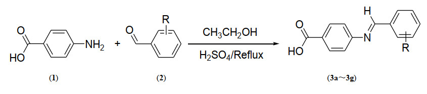
Scheme 1.
Synthetic scheme and structures of the synthesized 4-(benzylideneamino) benzoic acid derivatives (3a~3g)
Synthesis, Characterization, Biological and Docking Simulations of 4-(Benzylideneamino) Benzoic Acids
Aziz HAMID , Saeed AAMER , Jabeen FARUKH , Basit ABDUL , Zia Qureshi IRFAN , Aziz ABDUL , Haroon ATIF , Ur Rehman ASHFAQ

Diabetes mellitus (DM) is a group of life style-related, non-communicable, endocrinological and metabolic disorder with diverse etiologies[1]. DM is characterized by hyperglycemia resulting from defects in insulin secretion leading to incomplete metabolism of carbohydrates, lipids and proteins[2]. The prevalent type-II diabetes is a metabolic syndrome characterized by high blood glucose level because of hectic lifestyle. Type-I diabetes is resulted because of no insulin production and causes frequent urination, increased thirst and hunger[3]. Recent research suggests the loss of functional β-cell mass through apoptosis and advanced glycation end products as the leading cause of developing DM[4]. In this regard, nitrogen containing heterocycles particularly Schiff bases (SB) are revealed as potent antidiabetic agents[5-7].
SB serves as a promising source for the rational design of biologically relevant heterocycles including thiazolidinone[8], azetidone[9], oxadiazole, benzoxazoles[10], and quinolone[11]. SB contain azomethine linkages and display potent biological profile like anticancer[12], antituberculosis[13], antimicrobial[14], antipyretics[15], antiproliferative[16], and antidiabetic[17]. They serves as excellent chelating agents[10, 18]. The electrophilic carbon and nucleophilic nitrogen atoms interact with nucleophiles and electrophiles that lead to enzyme inhibition. Similarly, tumor cells selectively hydrolyze SB to serve as alkylating agents while the free amines simultaneously act as antimetabolites[10]. Rhodopsin is an azomethine pigment formed biochemically by the reaction of formyl group of cis-retinal with an amine group of an Apo protein opsin. The azomethine linkages of pyridoxal phosphate coenzyme play a pivotal role in the metabolic pathways of biologically essential amino acids[16, 19, 20]. Azomethine linkages serve as useful substrates to produce conducting materials as well as polymers[21, 22]. Reactive oxygen and nitrogen species cause oxidative stresses and damage cellular structures. Azomethine linkages provide protection against oxidative damage and consequently help to prevent DM[23, 24]. In this regard, azomethine linkages of isatin derivatives are reported as potent in vitro antiglycation agents[25]. Similarly, Vanco et al. reports the in vivo antidiabetic potential of N-salicylidene-β-alanine azomethine linkages[26].
Thus keeping in view the harmful effects of DM and the potent antidiabetic potential of amino-based heterocycles, there is an utmost need for the design and synthesis of SB. In this regard, the present paper reports the facile synthesis, spectroscopic characterization, biological and computational evaluation of 4-(benzylideneamino) benzoic acid derivatives (3a~3g).
All the chemicals used were obtained from commercial suppliers. NMR spectra were recorded using Bruker NMR spectrometer at 300 MHz (1H) as well as 75 MHz (13C). NMR chemical shifts are expressed in ppm scale (δ) relative to the reference standard TMS and DMSO-d6 as a solvent. The coupling constants J were measured in Hertz. Multiplicities in 1H NMR are reported as broad (b), singlet (s), doublet (d), triplet (t) and multiplet (m). FT-IR spectra were recorded on the Vertex 70 Bruker apparatus. Elemental analysis (CHNS) was performed to determine the percentage of each element. The reaction was monitored by TLC on 2.0cm × 5.0cm aluminium sheets (silica gel, 60F254 Merck).
The syntheses of 4-(benzylideneamino) benzoic acid derivatives (3a~3g) were performed as reported elsewhere[27]. Briefly, substituted aldehyde (0.1 mol) was completely dissolved in dry distilled ethanol (5.0 mL) at reflux in a 250 mL round bottom flask containing a drop of concentrated sulphuric acid. A solution of 4-aminobenzoic acid (0.1 mol) prepared in dry distilled ethanol (5.0 mL) was added to the reaction mixture and the reaction was refluxed for next 5 hours (hrs). During the reaction, TLC was constantly used to monitor the progress of the reaction. The reaction mixture was cooled to room temperature, which resulted in the formation of solid precipitates of the target products.
Yield: 75%; m.p: 248 ℃; color: white; FT-IR (ATR, cm-1): 3000, 2913(sp2 C–H stretch), 2954, 2855 (sp3 C–H stretch), 1678(COO-, asymmetric), 1673, 1567, 1420(C=C, aromatic), 1593(HC=N), 1420 (COO- symmetric), 1284(C–N); 1H NMR (DMSO-d6), 11.08 (b, s, 1H, COOH), 8.50 (d, 2H, J = 8.20 Hz, Ar), 8.48 (s, 1H, HC=N), 7.76 (d, 2H, J = 8.20 Hz, Ar), 7.63 (d, 1H, J = 7.92 Hz, Ar), 7.01 (d, 1H, J = 7.92 Hz, Ar); 6.94 (s, 1H, Ar), 3.72 (s, 3H, OCH3); 13C NMR (DMSO-d6), 169.84 (C=O), 160.22 (HC=N), 158.50 (Ar), 152.10 (Ar), 148.20 (Ar), 132.56 (Ar), 130.70 (Ar), 129.80 (Ar), 129.10 (Ar), 128.60 (Ar), 124.20 (Ar), 122.84 (Ar), 52.78 (O–C). Anal. Calcd. (%) for C15H13NO4: C, 66.41; H, 4.83; N, 5.16. Found (%): C, 66.35; H, 4.88; N, 5.21.
Yield: 70%; m.p: 269 ℃; color: yellow; FT-IR (ATR, cm-1): 2970(sp2 C–H stretch), 1679(COO-, asymmetric), 1620, 1568, 1496(C=C, aromatic), 1597(HC=N), 1428 (COO- symmetric), 1285(C–N); 1H NMR (DMSO-d6), 11.13 (b, s, 1H, COOH), 8.25 (d, 2H, J = 8.22 Hz, Ar), 8.20 (d, 2H, J = 8.22 Hz, Ar), 8.16 (s, 1H, HC=N), 7.69 (d, 2H, J = 8.10 Hz, Ar), 7.12 (t, 1H, J = 8.0 Hz, Ar), 6.83 (t, 1H, J = 8.0 Hz, Ar); 13C NMR (DMSO-d6), 170.20 (C=O), 158.46 (HC=N), 158.43 (Ar), 150.10 (Ar), 141.45 (Ar), 139.87 (Ar), 128.56 (Ar), 127.34 (Ar), 125.63 (Ar), 122.10 (Ar), 121.30 (Ar). Anal. Calcd. (%) for C14H11NO3: C, 69.70; H, 4.60; N, 5.81. Found (%): C, 69.79; H, 4.51; N, 5.80.
Yield: 75%; m.p: 235 ℃; color: white; FT-IR (ATR cm-1): 3033, 2981 (sp2 C–H stretch), 2943, 2876 (sp3 C–H stretch), 1681(COO-, asymmetric), 1660, 1560, 1440 (C=C, aromatic), 1593(HC=N), 1422(COO- symmetric), 1284(C–N); 1H NMR (DMSO-d6), 11.28 (b, s, 1H, COOH), 8.58 (d, 2H, J = 8.21 Hz, Ar), 8.38 (s, 1H, HC=N), 7.69 (d, 2H, J = 8.21 Hz, Ar), 7.68 (d, 2H, J = 8.15 Hz, Ar), 7.01 (d, 2H, J = 8.15 Hz, Ar); 2.29 (s, 3H, CH3); 13C NMR (DMSO-d6), 168.40 (C=O), 159.21 (HC=N), 157.50 (Ar), 142.20 (Ar), 132.10 (Ar), 130.40 (Ar), 129.20 (Ar), 128.50 (Ar), 128.10 (Ar), 122.40 (Ar), 25.50 (C–H). Anal. Calcd. (%) for C15H13NO2: C, 75.30; H, 5.48; N, 5.85. Found (%): C, 75.35; H, 5.43; N, 5.80.
Yield: 85%; m.p: 290 ℃; color: red; FT-IR (ATR, cm-1): 2970(sp2 C–H stretch), 1670(COO-, asymmetric), 1632, 1599, 1514(C=C, aromatic), 1589(HC=N), 1426(COO- symmetric), 1285(C–N); 1H NMR (DMSO-d6), 11.13 (b, s, 1H, COOH), 8.63 (s, 1H, HC=N), 8.22 (d, 2H, J = 8.31 Hz, Ar), 7.55 (d, 2H, J = 8.25 Hz, Ar), 8.35 (d, 2H, J = 8.31 Hz, Ar), 7.15 (d, 2H, J = 8.25 Hz, Ar); 13C NMR (DMSO-d6), 170.20 (C=O), 158.46 (HC=N), 158.43 (Ar), 150.10 (Ar), 141.45 (Ar), 139.87 (Ar), 128.56 (Ar), 127.34 (Ar), 125.63 (Ar), 121.30 (Ar). Anal. Calcd. (%) for C14H10N2O4: C, 62.22; H, 3.73; N, 10.37. Found (%): C, 62.17; H, 3.75; N, 10.40.
Yield: 80%; m.p: 176 ℃; color: yellow; FT-IR (ATR, cm-1): 3013, 2980(sp2 C–H stretch), 1678(COO-, asymmetric), 1625, 1588, 1489(C=C, aromatic), 1425(COO- symmetric), 1588 (HC=N), 1300 (C–N); 1H NMR (DMSO-d6), 11.10 (b, s, 1H, COOH), 8.56 (s, 1H, HC=N), 8.20 (d, 2H, J = 8.23 Hz, Ar), 7.80 (d, 2H, J = 8.13 Hz, Ar), 7.50 (d, 2H, J = 8.23 Hz, Ar), 7.15 (d, 2H, J = 8.13Hz, Ar); 13C NMR (DMSO-d6), 170.23 (C=O), 158.43 (HC=N), 158.31 (Ar), 140.16 (Ar), 131.45 (Ar), 129.67 (Ar), 128.60 (Ar), 127.20 (Ar), 126.30 (Ar), 125.12 (Ar). Anal. Calcd. (%) for C14H10ClNO2: C, 64.75; H, 3.88; N, 5.39. Found (%): C, 64.68; H, 3.93; N, 5.56.
Yield: 75%; m.p: 180 ℃; color: white; FT-IR (ATR cm-1): 3089, 2954(sp2 C–H stretch), 2950, 2860(sp3 C–H stretch), 1677(COO-, asymmetric), 1670, 1565, 1425(C=C, aromatic), 1589(HC=N), 1429 (COO- symmetric), 1284(C–N); 1H NMR (DMSO-d6), 11.08 (b, s, 1H, COOH), 8.50 (d, 2H, J = 8.30 Hz, Ar), 8.48 (s, 1H, HC=N), 7.76 (d, 2H, J = 8.30 Hz, Ar), 7.63 (d, 2H, J = 8.20 Hz, Ar), 7.01 (t, 1H, J = 8.25 Hz, Ar), 3.72 (s, 3H, OCH3); 13C NMR (DMSO-d6), 169.84 (C=O), 160.22 (HC=N), 158.50 (Ar), 152.10 (Ar), 148.20 (Ar), 132.56 (Ar), 130.70 (Ar), 129.80 (Ar), 129.10 (Ar), 128.60 (Ar), 124.20 (Ar), 122.84 (Ar), 52.78 (O–C). Anal. Calcd. (%) for C15H13NO4: C, 66.41; H, 4.83; N, 5.16; Found: C, 66.35; H, 4.87; N, 5.22.
Yield: 70%; m.p: 204 ℃; color: yellow; FT-IR (ATR, cm-1): 3000(sp2 C–H stretch), 1678(COO-, asymmetric), 1625, 1588, 1489(C=C, aromatic), 1581 (HC=N), 1428(COO- symmetric), 1293(C–N); 1H NMR (DMSO-d6), 11.10 (b, s, 1H, COOH), 8.52 (d, 2H, J = 8.21 Hz, Ar), 8.40 (s, 1H, HC=N), 7.74 (d, 2H, J = 8.21 Hz, Ar), 7.58 (d, 2H, J = 7.90 Hz, Ar), 7.08 (d, 2H, J = 7.90 Hz, Ar); 13C NMR (DMSO-d6), 170.20 (C=O), 158.35 (HC=N), 158.50 (Ar), 140.16 (Ar), 131.43 (Ar), 129.76 (Ar), 128.65 (Ar), 126.31 (Ar), 125.20 (Ar), 123.12 (Ar). Anal. Calcd. (%) for C14H10FNO2: C, 69.13; H, 4.14; N, 5.76. Found: C, 69.10; H, 4.18; N, 5.49.
For this purpose, thirty-six BALB/c healthy adult male mice (average weight = 33.8±0.8 gm) were procured from the National Institute of Health Islamabad. Four animals were housed per cage and had free access to standard diet of rodents and drinking water. Standard temperature of 28 ℃ and 12 hL: 12 hD photoperiod were maintained throughout. Diabetic models were made through single intraperitoneal injection (i.p.) dose (150 mg/kg b.w) of Alloxan monohydrate[17, 28]. The animals were checked for diabetes after 24 hrs of Alloxan monohydrate administered to 12 hrs fasted mice. Diabetic level was achieved at fasting plasma glucose concentration > 200 mg/dL. For antidiabetic screening, a total of nine experimental groups were constituted. Seven groups were treated with seven different derivatives at the dose of 5 mg/kg b.w dissolved in DMSO. The remaining two groups, positive and negative control groups, were treated with Glibenclamide (10 mg/kg b.w in DMSO) and DMSO, respectively. The screening derivatives were intraperitoneally administered through a single injection on day 2. Blood was obtained from caudal vein at nine different times; −1 hrs (pre-alloxan), 0 hrs (postalloxan) and 1~7 hrs after treatment with doses. Plasma glucose level was determined with dextrostix strips using glucometer. Serum triglycerides, cholesterol, alanine amino transferase (ALT) and aspartate amino transferase (AST) levels were estimated through commercially available kits using the standard calorimetry procedure[29].
Antibacterial assay of the derivatives (3a~3g) was determined by agar disc diffusion method[30]. Briefly, two gram-positive (Staphylococcus aureus and Streptococcus pneumoniae) and three gram-negative (Escherichia coli, Enterobacter aerogenes and Klebsiella pneumoniae) were cultured in a nutrient broth for 24 hrs at 37 ℃. The cultured strains were used as inoculums (1%) and were added to the nutrient agar medium at 45 ℃, poured into sterile Petri plates and allowed to solidify. 5 mL of the derivative (200 mg/mL, final concentration) was poured on sterile filter paper discs (4 mm) and placed on nutrient ager plates, respectively. Amikacin and DMSO were used as positive and negative controls, respectively. The assay was performed in triplicate and the plates were incubated at 37 ℃ for 24~48 hrs. The antibacterial potential was determined by measuring the diameter of zones of inhibition (ZOI, mm) using Vernier caliper.
Molecular operating environment (MOE)[31] was used for MD studies against antibacterial target E. coli 16S-rRNA A-site. First, 3D structures were generated using the builder tool executed in MOE package. Next, the derivatives were protonated and energy-minimized using the default parameters of MOE (gradient: 0.05, force field: MMFF94X). 3D structure of the target protein was retrieved from protein databank (PDB code 1j7t). The water molecules were removed and 3D protonation was carried out. Afterwards, the protein was energy-minimized using the MOE package. For docking studies, default parameters of the MOE package i.e., placement: triangle matcher, rescoring 1: London dG, refinement: forcefield, and rescoring 2: GBVI/WSA were used. For each ligand, ten conformations could be fashioned and the top-ranked conformations based on docking score were selected for additional analysis.
For MD simulations, all-atom simulations and reasonable analysis procedures were conducted in by AMBER18 software package. The LEaP module was used to add hydrogen atoms to both crystallographic structures. Counter-ions (Na+ and Cl−) were added to maintain the system neutrality. All systems were solvated in a truncated octahedral box of TIP3P water model with 10 Å buffer. Particle Mesh Ewald (PME) method was used to treat long-range electrostatic interactions, and ff12SB force field was used for all simulations. All the bonds involving hydrogen atoms were constrained with SHAKE algorithm. PMEMD of CUDA version was used to accelerate all the MD simulations. Steepest descent method was used to minimize the solvated systems for 20000-step, then 400 ps heating, and 200 ps equilibration in the NVT ensemble. The default parameters of the Langevin thermostat method were used for the heating and equilibration runs with a time step of 2 fs, and for the production runs under NPT ensemble at 298 K with a time step of 2 fs in Berendsen's thermostat and barostat.
One-way analysis of variance (ANOVA) was used for data analysis using Sigma Plot (Version 12.0), Post hoc Tukey-Kramer test, where the test for normality failed ANOVA on ranks test was applied. Statistically significant difference was considered where p < 0.05. Data are presented as line or bar graphs Graph-Pad Prism (Version 5.0).
The 4-(benzylideneamino) benzoic acid derivatives (3a~3g) were synthesized by dissolving substituted benzaldehydes in dry distilled ethanol containing a drop of concentrated sulphuric acid at reflux. After an hr, ethanolic solution of 4-aminobenzoc acid was added and the reaction mixture was again refluxed for another 5 hrs (scheme 1). The reaction progress was monitored by TLC in a (chloroform: methanol, 7:3) solvent system. After 5 hrs, the reaction was stopped and cooled to room temperature, resulting in the formation of solid precipitates which were filtered and recrystallized from a mixture of ethanol and DCM at room temperature[27].
 DownLoad:
CSV
DownLoad:
CSV
| S. No. | Compound | R | S. No. | Compound | R | |
| 1 | 3a | 3-OCH3, 4-OH | 5 | 3e | 4-Cl | |
| 2 | 3b | 2-OH | 6 | 3f | 3-OH, 4-OCH3, | |
| 3 | 3c | 4-CH3 | 7 | 3g | F | |
| 4 | 3d | 4-NO2 | … | … | … |
Structure assignments of the synthesized 4-(benzylideneamino) benzoic acid derivatives (3a~3g) were based on their spectroscopic data (FT-IR, 1H NMR, 13C NMR) and elemental analysis (CHNS). The derivatives (3a~3g) gave satisfactory C, H, N and S analyses data highly aligned with their structural formulae. FT-IR spectra of the derivatives (3a~3g) display absorption peaks in the range of 2900~3100 cm-1 being assigned to the sp2 hybridized CH stretch. The COOH group protons appear in the 1H NMR spectra as broader signals in the range of 11.0~11.28 ppm. FT-IR spectra of the derivatives (3a~3g) display peaks in the range of 1581~1597cm-1 for the subsequent azomethine linkages (HC=N) which are evident as sharp singlets at 8.16~8.63 and 158.31~160.22 ppm in their 1H and 13C NMR spectra[28].
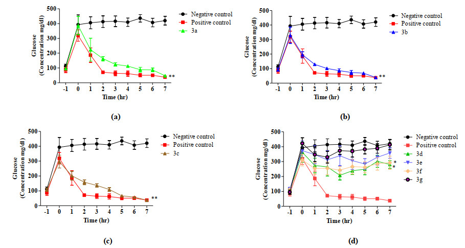
When mice were treated with the derivatives (3a~3g), the concentration of glucose was altered noticeably (F = 43.986, P < 0.001). 3a, 3b and 3c considerably (P < 0.001, Fig. 1a~1c) while 3d and 3f slightly but significantly decreased the glucose concentrations compared to the negative control (P < 0.05). In contrast, 3f and 3g did not show any significance effect on the glucose level (P > 0.05), (Fig. 1d).
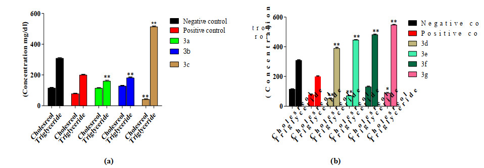
Significant alteration was found in total cholesterol after treatment of mice (F = 53.973, P < 0.001). 3c, 3d and 3e substantially decreased the cholesterol level by comparison with both positive and negative controls (P < 0.001 and P > 0.05). In contrast, 3b and 3f caused an increase as compared to both the controls (P < 0.001 and P < 0.05), whereas 3a and 3g increased the cholesterol level compared to positive control and decreased versus negative control (P < 0.001 and P < 0.05) (Figs. 2a and 2b). Serum triglyceride level also revealed significant alterations (F = 916.414, P < 0.001). The serum triglyceride level of mice decreased with 3a and 3b while increased with 3c~3g (P < 0.001 and P < 0.05) (Figs. 2a and 2b).
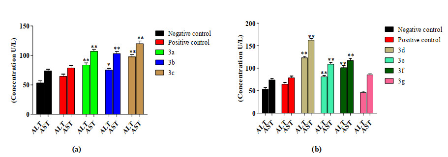
The ALT and AST levels of blood serum were also affected (F = 43.894, P < 0.001); i.e. their levels rose with 3a~3e and 3f (P < 0.001 and P < 0.05) while 3g had no effect (P > 0.05, Figs. 3a and 3b).
To conclude, the derivatives (3a~3d, 3f) showed glucose lowering effect compared to the reference drug Glibanclamide and the remaining derivatives 3e and 3g were found to be lacking antihyperglycemic activity. Likewise, the biochemical parameters showed significant alterations upon treatments. 3a, 3c, 3e and 3g showed high level of anticholesterol potency, while only 3a and 3b caused a decrease in the triglycerides level. 3c~3g increased the triglycerides level. The results obtained are consistent with the literature, indicating the metabolic alterations of mice when the derivatives were administered[17, 28, 29]. Except 3g, all the derivatives increased the ALT and AST levels. These results obtained show similarity with a diphenyl diselenide, diphenyl ditelluride and Ebselen caused potential renal and hepatic toxicity in rats and mice as reported elsewhere[29]. Thus, based on the results obtained, it can be concluded that the derivatives (3a~3g) are also hepatotoxic when administered to mice at the dose of 5 mg/kg.
Antibacterial potential of the derivatives (3a~3g) was determined from agar disc diffusion method. The derivatives (3a~3g) were employed in different zones and the results in terms of their ZOI were determined (Table 2). The derivative 3a showed higher antibacterial potential against all the screened strains. The antibacterial potential was found to be higher than the positive control Amikacin against the Enterobacter aerogenes. The derivative 3d showed moderate potential against all the bacterial strains. Similarly, the derivative 3e showed good antibacterial activity against all the screened strains except Streptococcus pneumoniae, where no activity was observed. Unlike 3e, 3b showed antibacterial activity only against Staphylococcus aureus and Enterobacter aerogenes. The remaining derivatives were found to be less potent against the screened bacterial strains[30].
DownLoad:
CSV
| S. No. | Compound | Zone of inhibition (mm) at 200 (mg/mL) | ||||
| E. coli | K. pneumoniae | S. aureus | S. pneumoniae | E. aerogenes | ||
| 1 | Amikacin | 19 | 19 | 30 | 20 | 22 |
| 2 | DMSO | 0 | 0 | 0 | 0 | 0 |
| 3 | 3a | 17 | 16 | 14 | 12 | 24 |
| 4 | 3b | 0 | 0 | 9 | 0 | 16 |
| 5 | 3c | 7 | 0 | 0 | 0 | 0 |
| 6 | 3d | 14 | 12 | 16 | 15 | 18 |
| 7 | 3e | 13 | 10 | 7 | 0 | 9 |
| 8 | 3f | 9 | 0 | 0 | 10 | 9 |
| 9 | 3g | 0 | 0 | 9 | 0 | 0 |
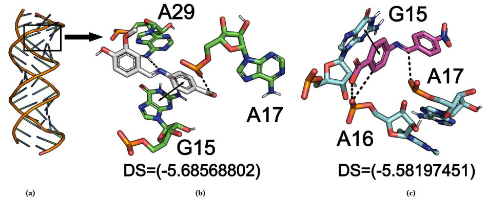
Binding modes of the derivatives (3a~3g) against antibacterial target were evaluated from in silico analysis. Crystal structure solved by Eric Westhof group[32] at 2.5 Å resolution (PDB code 1j7t) which includes the antibiotic docked into the eubacterial ribosomal decoding A site was chosen for antibacterial assay. MD results revealed the most potent derivatives (3a, 3d) possess the best activity against the target enzymes both in silico and experimentally. In case of (3a, 3d), the crystal structure revealed each RNA, and the fragment contains two A sites inserted between WatsonCrick pairs. 3a and 3d interact in an enlarged deep groove created by two bulging and one unpaired adenines (Fig. 4a) and adopted favorable interactions. The receptor-ligand interaction (RLI) profile for 3d revealed a typical interaction (A17) with target and some other, i.e., G15 and A16, respectively (Fig. 4c). 3a adopted total three interactions: π-stacking with the 1, 6-dihydropyrimidine ring of G15, with the (O) of OP3 of A17 and with the NH2 of A29 (Fig. 4b). The high potency of 3a over others might be due to the new methoxide group with OH over benzene compared with 3d, which only has the 1-nitrobenzene group. Even though in both cases, the substituted group belongs to deactivating category, it withdraws the electron from benzene ring and further triggers the partial positive charge at benzene ring, and this effect further enhances the potency of other atoms to adopt interactions with other residues to gain stability and enhance the activity at a different level. These results delineate that the molecules possessing double deactivating group at benzene ring have the tendency to trigger the partial positive charge over benzene ring, thus further activating other atoms to adopt favorable interactions with crucial residues and subsequently enhancing the corresponding activity.
To further validate the results of docking, MD simulation was performed[33, 34]. A total of 20ns simulations were performed in order to categorize the internal motion and interaction of the derivatives (3a, 3d). The root-meansquare-deviation (RMSd) was calculated to validate the stability of the simulations, which was a key parameter for evaluating the equilibrium of MD trajectories for the primary chain atoms of the protein system along the MD simulations. The amplitude of the RMSd curve fluctuation is negatively correlated with the protein's stability: the lower the fluctuation, the more stable the protein structure. The RMSd value of the backbone atoms for both derivatives was calculated and their results are shown (Fig. 5). The results indicate the RMSd value of 3a as compared to 3d for antibacterial assay as shown in Fig. 5a. Initially, the curve gradually increases (15 ns) and is highly fluctuated but becomes stable after 20 ns. The results show less fluctuation, which indicate that the trajectories generated during 20 ns are stable. Therefore, the following analysis was obtained from the 20ns trajectories, which shows that 3a strongly inhibited the target (antibacterial) and bonded explicitly to the binding site. The derivative 3d is loosely bound and inhibits the activity and hence is considered as a second inhibitor.
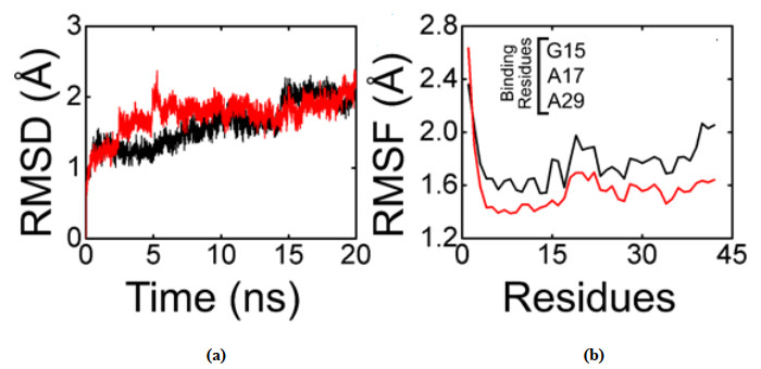
To determine how the derivatives (3a, 3d) influence the dynamics of the side chain of the target protein residues, the residues fluctuation in terms of root-mean-square-fluctuation (RMSf) was analyzed. The results of RMSf were like the RMSd value. 3a shows strong binding with the active site residues (G15, A17, and A29), so the fluctuation is higher in the binding site and hence shows the best activity against antibacterial target (Fig. 5b), while in comparison to the residue's fluctuation with 3d, fewer residues fluctuation was observed, which might indicate the lesser activity and interaction within the active pocket (Fig. 5b).
The current research paper presents the successful synthesis, characterization, biological and computational analysis of 4-(benzylideneamino) benzoic acid derivatives (3a~3g). The derivatives (3a~3c) possess strong in vivo antidiabetic activity and may be processed as potent antidiabetic candidate while the remaining derivatives (3d~3g) lack antidiabetic response. The derivatives (3a~3g) were found to be hepatotoxic at the present dose level and cause metabolic abnormality. Further detailed studies, therefore, are required to check the efficacy and safety of the derivatives (3a~3g) at different dose levels. They have minimum side effects, and will be less toxic and can be processed as alternative drug candidates for diabetes other than the currently available drugs. MD simulations of the derivatives (3a~3g) against RNA provided further insight into the mode of action, mechanism of the inhibition and inhibitory potential of the drug-like molecules. Derivatives 3d and 3a exhibited better inhibitory potential against both the targets. These ligands stabilize themselves inside the active site through tremendous electrostatic and hydrophobic interactions. MD simulations further ascertained the interaction of these molecules with both the targets.
Upadhyay, R. K. Anti-arthritic potential of plant natural products; its use in joint pain medications and anti-inflammatory drug formulations. Int. J. Green Pharm. 2016, 10, S120−S130.
Puranik, N. V.; Puntambekar, H. M.; Srivastava, P. Antidiabetic potential and enzyme kinetics of benzothiazole derivatives and their non-bonded interactions with α-glucosidase and α-amylase. Med. Chem. Res. 2016, 25, 805−816. doi: 10.1007/s00044-016-1520-3
Gin, H.; Rigalleau, V. Post-prandial hyperglycemia. Post-prandial hyperglycemia and diabetes. Diabetes. Metab. 2000, 26, 265−272.
American Diabetes Association. Diagnosis and classification of diabetes mellitus. Diabetes Care 2014, S81−90.
Schiff, H. The syntheses and characterization of Schiff base. Ann. Chem. Suppl 3 1864, 343−349.
da Silva, C. M.; da Silva, D. L.; Modolo, L. V.; Alves, R. B.; de Resende, M. A.; Martins, C. V.; de Fátima, Â. Schiff bases: a short review of their antimicrobial activities. J. Adv. Res. 2011, 2, 1−8. doi: 10.1016/j.jare.2010.05.004
Chakraborti, A. K.; Bhagat, S.; Rudrawar, S. Magnesium perchlorate as an efficient catalyst for the synthesis of imines and phenylhydrazones. Tetrahedron Lett. 2004, 45, 7641−7644. doi: 10.1016/j.tetlet.2004.08.097
Küçükgüzel, G.; Kocatepe, A.; De Clercq, E.; Şahin, F.; Güllüce, M. Synthesis and biological activity of 4-thiazolidinones, thiosemicarbazides derived from diflunisal hydrazide. Eur. J. Med. 2006, 41, 353−359. doi: 10.1016/j.ejmech.2005.11.005
Kalsi, R.; Shrimali, M.; Bhalla, T. N.; Barthwal, J. P. Synthesis and anti-inflammatory activity of indolyl azetidinones. Indian J. Pharm. Sci. 1990, 52, 129−134.
Hameed, A.; al-Rashida, M.; Uroos, M.; Abid, A. S.; Khan, K. M. Schiff bases in medicinal chemistry: a patent review (2010-2015). Expert Opin. Ther. Pat. 2017, 27, 63−79. doi: 10.1080/13543776.2017.1252752
Hu, G. Q.; Wu, X. K.; Wang, G. Q.; Duan, N. N.; Wen, X. Y.; Cao, T. Y.; Jun, Y.; Wei, W.; Xie, S. Q.; Huang, W. L. Synthesis and antitumor and antibacterial evaluation of fluoro-quinolone derivatives (III): mono- and bis-Schiff-bases. Chin. Chem. Lett. 2012, 23, 515−517. doi: 10.1016/j.cclet.2012.01.029
Ren, S.; Wang, R.; Komatsu, K.; Bonaz-Krause, P.; Zyrianov, Y.; McKenna, C. E.; Csipke, C.; Tokes, Z. A.; Lien, E. J. Synthesis, biological evaluation, and quantitative structure-activity relationship analysis of new Schiff bases of hydroxysemicarbazide as potential antitumor agents. J. Med. Chem. 2002, 45, 410−419. doi: 10.1021/jm010252q
Hearn, M. J.; Cynamon, M. H. Design and synthesis of antituberculars: preparation and evaluation against mycobacterium tuberculosis of an isoniazid Schiff base. J. Antimicrob. Chemother. 2004, 53, 185−191. doi: 10.1093/jac/dkh041
Silva, C. D.; Silva, D. D.; Modolo, L. V.; Alves, R. B.; Resende, M. D.; Martins, C. V.; Fatima, A. D. Schiff bases: a short review of their antimicrobial activities. J. Adv. Res. 2011, 2, 1−8. doi: 10.1016/j.jare.2010.05.004
Murtaza, S.; Akhtar, M. S.; Kanwal, F.; Abbas, A.; Ashiq, S.; Shamim, S. Synthesis and biological evaluation of schiff bases of 4-aminophenazone as an anti-inflammatory, analgesic and antipyretic agent. J. Saudi Chem. Soc. 2017, 21, S359−S372. doi: 10.1016/j.jscs.2014.04.003
Sztanke, K.; Maziarka, A.; Osinka, A.; Sztanke, M. An insight into synthetic Schiff bases revealing antiproliferative activities in vitro. Bioorg. Med. Chem. 2013, 21, 3648−3666. doi: 10.1016/j.bmc.2013.04.037
Rauf, A.; Shah, A.; Munawar, K. S.; Khan, A. A.; Abbasi, R.; Yameen, M. A.; Khan, A. M.; Khan, A. R.; Qureshi, I. Z.; Kraatz, H. B. Synthesis, spectroscopic characterization, DFT optimization and biological activities of Schiff bases and their metal(II) complexes. J. Mol. Struct. 2017, 1145, 132−140. doi: 10.1016/j.molstruc.2017.05.098
Zhao, F.; Liu, Z. Q. The protective effect of hydroxyl-substituted Schiff bases on the radical-induced oxidation of DNA. J. Phys. Org. Chem. 2009, 22, 791−798. doi: 10.1002/poc.1517
Heck, M.; Schädel, S. A.; Maretzki, D.; Bartl, F. J.; Ritter, E.; Palczewski, K.; Hofmann, K. P. Signaling states of rhodopsin formation of the storage form, metarhodopsin III, from active metarhodopsin II. J. Biol. Chem. 2003, 278, 3162−3169. doi: 10.1074/jbc.M209675200
Soderberg, T. Organic Chemistry with a Biological Emphasis Volume I. Chemistry Publications 2016.
Naderi, E.; Ehteshamzadeh, M.; Jafari, A. H.; Hosseini, M. G. Effect of carbon steel microstructure and molecular structure of two new Schiff base compounds on inhibition performance in 1M HCl solution by DC, SEM and XRD studies. Mater. Chem. Phys. 2010, 120, 134−141. doi: 10.1016/j.matchemphys.2009.10.036
Khuhawar, M. Y.; Mughal, M. A.; Channar, A. H. Synthesis and characterization of some new Schiff base polymers. Eur. Polym. J. 2004, 40, 805−809. doi: 10.1016/j.eurpolymj.2003.11.020
Rakesh, K. P.; Manukumar, H. M.; Gowda, D. C. Schiff's bases of quinazolinone derivatives: synthesis and SAR studies of a novel series of potential anti-inflammatory and antioxidants. Bioorg. Med. Chem. Lett. 2015, 25, 1072−1077. doi: 10.1016/j.bmcl.2015.01.010
Kumar, D.; Rawat, D. S. Synthesis and antioxidant activity of thymol and carvacrol based Schiff bases. Bioorg. Med. Chem. 2013, 23, 641−645. doi: 10.1016/j.bmcl.2012.12.001
Khan, K. M.; Khan, M.; Ali, M.; Taha, M.; Rasheed, S.; Perveen, S.; Choudhary, M. I. Synthesis of bis-Schiff bases of isatins and their antiglycation activity. Bioorg. Med. Chem. 2009, 17, 7795−7801. doi: 10.1016/j.bmc.2009.09.028
Vančo, J.; Marek, J.; Trávníček, Z.; Račanská, E.; Muselík, J.; Švajlenová, O. G. Synthesis, structural characterization, antiradical and antidiabetic activities of copper(II) and zinc(II) Schiff base complexes derived from salicylaldehyde and β-alanine. J. Inorg. Biochem. 2008, 102, 595−605. doi: 10.1016/j.jinorgbio.2007.10.003
Kia, R.; Kargar, H. Crystal structure, spectroscopic characterization, and computational study of two new 4-aminobenzoic acid derived Schiff base ligands. Sci. Iran. Trans. C, Chem. Chem. Eng. 2015, 22, 2298−2308.
Rauf, A.; Shah, A.; Khan, A. A.; Shah, A. H.; Abbasi, R.; Qureshi, I. Z.; Ali, S. Synthesis, pH dependent photometric and electrochemical investigation, redox mechanism and biological applications of novel Schiff base and its metallic derivatives. Spectrochim. Acta Part A: Mol. Biomol. Spectr. 2017, 176, 155−167. doi: 10.1016/j.saa.2017.01.018
Meotti, F. C.; Borges, V. C.; Zeni, G.; Rocha, J. B.; Nogueira, C. W. Potential renal and hepatic toxicity of diphenyl diselenide, diphenyl ditelluride and Ebselen for rats and mice. Toxicol. Lett. 2003, 143, 9−16. doi: 10.1016/S0378-4274(03)00090-0
Mehmood, S.; Rehman, M. A.; Ismail, H.; Mirza, B.; Bhatti, A. S. Significance of postgrowth processing of ZnO nanostructures on antibacterial activity against gram-positive and gram-negative bacteria. Int. J. Nanomed. 2015, 10, 4521−4533.
Molecular Operating Environment (MOE), 2016.08; Chemical Computing Group Inc., 1010 Sherbrooke St. West, Suite #910, Montreal, QC, Canada, H3A 2R7 2016.
Vicens, Q.; Westhof, E. Crystal structure of paromomycin docked into the eubacterial ribosomal decoding A site. Structure 2001, 9, 647−658. doi: 10.1016/S0969-2126(01)00629-3
Rehman, A. U.; Khan, M. T.; Liu, H.; Wadood, A.; Malik, S. I.; Chen, H. F. Exploring the pyrazinamide drug resistance mechanism of clinical mutants T370p and W403g in ribosomal protein S1 of mycobacterium tuberculosis. J. Chem. Inf. Model. 2019, 59, 1584−1597. doi: 10.1021/acs.jcim.8b00956
Rehman, A. U.; Rafiq, H.; Rahman, M. U.; Li, J.; Liu, H.; Luo, S.; Arshad, T.; Wadood, A.; Chen, H. F. Gain-of-function SHP2 E76Q mutant recusing autoinhibition mechanism associated with juvenile myelomonocytic leukemia. J. Chem. Inf. Model. 2019, 59, 3229−3239. doi: 10.1021/acs.jcim.9b00353

Scheme 1 Synthetic scheme and structures of the synthesized 4-(benzylideneamino) benzoic acid derivatives (3a~3g)

Figure 1 Glucose concentration of mice altered significantly when treated with (3a~3g). Data are presented as mean ± SE, *P < 0.05; **P < 0.001

Figure 2 Cholesterol and triglyceride levels of mice altered significantly when administered with (3a~3g). Data are presented as mean ± SE, *P < 0.05 and **P < 0.001

Figure 3 Concentration of ALT and AST of mice showed significant alteration after treatment with (3a~3g). Data are presented as mean ± SE, *P < 0.05 and **P < 0.001

Figure 4 RLI profile for the potent derivatives (3a, 3d) against antibacterial targets. (a) Surface representation of antibacterial target (PDB ID 1j7t). Binding mode of the high potent molecule (b) for 3a and (c) for 3d

Figure 5 (a) Superposed RMSd graphs for 3a (black) and 3d (red) against the anti-bacterial target. The superposed RMSf graphs (b) for 3a (black) and 3d (red) against antibacterial target
Table 1. Structures of the Synthesized 4-(Benzylideneamino) Benzoic Acid Derivatives (3a~3g)
| S. No. | Compound | R | S. No. | Compound | R | |
| 1 | 3a | 3-OCH3, 4-OH | 5 | 3e | 4-Cl | |
| 2 | 3b | 2-OH | 6 | 3f | 3-OH, 4-OCH3, | |
| 3 | 3c | 4-CH3 | 7 | 3g | F | |
| 4 | 3d | 4-NO2 | … | … | … |
 下载: 导出CSV
Table 2. Antibacterial Assay Results of the Screened 4-(Benzylideneamino) Benzoic Acid Derivatives (3a~3g)
| S. No. | Compound | Zone of inhibition (mm) at 200 (mg/mL) | ||||
| E. coli | K. pneumoniae | S. aureus | S. pneumoniae | E. aerogenes | ||
| 1 | Amikacin | 19 | 19 | 30 | 20 | 22 |
| 2 | DMSO | 0 | 0 | 0 | 0 | 0 |
| 3 | 3a | 17 | 16 | 14 | 12 | 24 |
| 4 | 3b | 0 | 0 | 9 | 0 | 16 |
| 5 | 3c | 7 | 0 | 0 | 0 | 0 |
| 6 | 3d | 14 | 12 | 16 | 15 | 18 |
| 7 | 3e | 13 | 10 | 7 | 0 | 9 |
| 8 | 3f | 9 | 0 | 0 | 10 | 9 |
| 9 | 3g | 0 | 0 | 9 | 0 | 0 |
下载: 导出CSV

 扫一扫看文章

扫一扫关注我们
 下载:
下载: