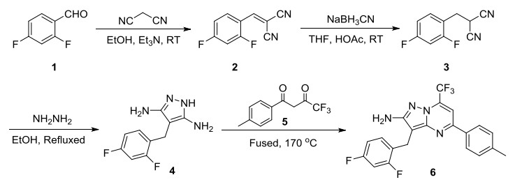
Scheme 1.
Synthetic route of the title compound

Cancer is a life-threatening disease and remains a major health problem around the globe. It is the second most prevalent disease after cardiovascular disease[1]. Despite the efforts to discover and develop small molecule anticancer drugs in the last decade, the development of new antitumor agents with improved tumor selectivity, efficiency, and safety remains desirable[2, 3].
In recent years, heterocycles have attracted the attention of many scientists because of their important utility in medicinal chemistry[4, 5]. As an extension of our work on the development of novel potent antitumor agents, we noticed that compounds which contain a pyrazolo[1, 5-a]pyrimidine framework displayed a multitude of biological activities, including antitumor[6], antibacterial[7], analgesics[8] and anti-inflammatory[9]. Many of pyrazolo[1, 5-a]pyrimidine derivatives are already being marketed or are under clinical/preclinical studies. Anticancer agent dinaciclib is one of approved drugs containing a pyrazolo[1, 5-a]pyrimidine core[10].
As an continuation of our research program on the synthesis of heterocycles which may serve as leads for novel antitumor agents[11-15], we now report the synthesis of a new pyrazolo[1, 5-a]pyrimidine, 3-(2, 4-difluorobenzyl)-5-(p-tolyl)-7-(trifluoromethyl)pyrazolo[1, 5-a]pyrimidin-2-amine (6), by a four-step synthesis method. The synthetic procedure for the title compound is shown in Scheme 1. In order to determine the molecular structure and spatial absolute conformation, the single crystal was grown and tested by X-ray single-crystal diffraction. Furthermore, the biological tests suggested the compound displayed distinct effective inhibition on the proliferation of MKN45, HT-29 and K562 human cancer cell lines.
Unless specified otherwise, all starting materials and reagents were obtained from commercial supplies without further purification. All melting points (℃) were taken on a Beijing Taike X-4 microscopy melting point apparatus and uncorrected. 1H NMR spectra were recorded on a Bruker Biospin 600 MHz instrument using TMS as the internal standard. Mass spectra were recorded on a Waters Quattro micro API mass spectrometer (ESI, direct injection). All chemical shifts were reported in ppm. IR spectra were recorded as KBr pellets on a Perkin-Elmer Spectrum one FT-IR spectrometer. Crystal data were obtained on a Bruker P4 X-diffractometer.
To the suspension of malononitrile (2.00 g, 30.27 mmol) and 2, 4-difluorobenzaldehyde (5.16 g, 36.33 mmol) in EtOH (30 mL) was added Et3N (0.20 mL), and the mixture was stirred at room temperature for 1.0 hour. The resultant precipitate was filtered, washed with EtOH and dried under vacuum to give 4.98 g of 2-(2, 4-difluorobenzylidene)malononitrile as yellow solid. Yield: 86.51%. m.p. 150~1152 ℃; 1H NMR (600 MHz, CDCl3) δ 8.35 (m, 1H), 8.01 (s, 1H), 7.14~7.05 (m, 1H), 7.05~6.92 (m, 1H); MS(ESI) m/z(%): 191.1[M+H]+.
2-(2, 4-Difluorobenzylidene)malononitrile (3.80 g, 19.98 mmol) was dissolved in 30 mL of THF. To the mixture, a solution of acetic acid (2.40 mL) and sodium cyanoborohydride (1.51 g, 24.00 mmol) in THF (20 mL) was added. After six hours of stirring at ambient temperature, THF was evaporated under reduced pressure and water (50 mL) was added to the residue. The precipitated solid was collected by filtration and recrystallized from ethyl acetate to obtain 3.12 g of 2-(2, 4-difluorobenzyl)malononitrile as a pale yellow solid in 81.0% yield. m.p. 120~122 ℃; 1H NMR (600 MHz, CDCl3) δ 7.40~7.30 (m, 1H), 7.01~6.86 (m, 2H), 3.97 (t, J = 7.4 Hz, 1H), 3.34 (d, J = 7.4 Hz, 2H); MS(ESI) m/z(%): 191.1[M-H]+.
A mixture of 2-(2, 4-difluorobenzyl)malononitrile (3.00 g, 15.61 mmol) and 80% hydrazine monohydrate (1.20 g, 19.18 mmol) in EtOH (20 mL) was heated at 80 ℃ for 5 hours. After the reaction was completed (TLC), most of the solvent was evaporated under reduced pressure when white solid appeared. After cooling to room temperature, the resulting precipitate was filtered off, washed with water, and dried under vacuum to afford 4-(2, 4-difluorobenzyl)-1H-pyrazole-3, 5-diamine (2.30 g) as a yellow solid, yield: 65.70%. m.p.: 158~160 ℃. IR (KBr, cm-1): 3422 (-NH2, middle), 3372 (-NH2, middle), 3194 (-NH-, middle), 2913, 1620 (Ar, strong), 1604 (Ar, strong), 1539, 1503 (Ar, strong), 1438, 1424, 1273, 1139, 1093, 967, 852, 729; 1H NMR (600 MHz, DMSO-d6) δ 10.26 (br, 1H), 7.23~7.07 (m, 2H), 7.02~6.91 (m, 1H), 4.32 (s, 4H), 3.50 (s, 2H); MS(ESI) m/z(%): 225.1[M+H]+.
A mixture of 4-(2, 4-difluorobenzyl)-1H-pyrazole-3, 5-diamine (0.50 g, 2.23 mmol) and 4, 4, 4-trifluoro-1-(p-tolyl)butane-1, 3-dione (0.57 g, 2.48 mmol) in a 25 mL flask was heated at 170 ℃ for 1.5 h, allowing the elimination of water evolved. After cooling to room temperature, the solid in the flask was recrystallized from methanol to afford 0.63 g the title compound as a yellow solid in 67.53% yield. m.p. 209~211 ℃; 1H NMR (600 MHz, CDCl3) δ 8.00 (d, J = 8.4 Hz, 2H), 7.32 (d, J = 8.4 Hz, 4H), 6.80 (m, 2H), 4.30 (s, 2H), 4.08 (s, 2H), 2.44 (s, 3H); MS(ESI) m/z(%): 419.1[M+H]+; Anal. Calcd. for C21H15F5N4: C, 60.29; H, 3.61; N, 13.39. Found: C, 60.24; H, 3.65; N, 13.47.
The yellow powder of the title compound was dissolved in ethanol/ethyl acetate/tetrahydrofuran (5/2/3 by V/V/V). After slowly evaporating the solvents for several days, some single crystals suitable for X-ray analysis were obtained. A yellow crystal (C21H15F5O4) with dimensions of 0.25mm × 0.23mm × 0.20mm was selected for data collection which was performed on a Bruker APEX-II CCD automatic diffractometer with a graphite-monochromatic MoKa radiation (λ = 0.71073 Å) by using the φ and ω-scan mode at 296(2) K. A total of 6635 reflections were collected in the range of 1.25 < θ < 25.00° (index ranges: –6 < h < 6, –12 < k < 12, –19 < l < 19) and 3318 were independent (Rint = 0.030), of which 2573 observed reflections with I > 2σ(I) were used in the structure determination and refinements. The structure was solved by direct methods with SHELXS-97 program[16] and expanded by Fourier technique. The non-hydrogen atoms were refined anisotropically. The hydrogen atoms bound to carbon were determined with theoretical calculations and those attached to nitrogen and oxygen were determined with successive difference Fourier syntheses. The structure was refined by full-matrix leastsquares techniques on F2 with SHELXL-2014/7[17]. The final refinement gave R = 0.0526 and wR = 0.1731 (w = 1/[σ2(Fo2) + (0.1000P)2 + 0.2575P], where P = (Fo2 + 2Fc2)/3). S = 1.081, (Δ/σ)max = 0.000, (Δρ)max = 0.453 and (Δρ)min = –0.441 e/Å3. Geometric parameters of title compound are listed in Table 1.
 DownLoad:
CSV
DownLoad:
CSV
| Bond | Dist. (Å) | Angle | (º) | |
| C(1)–C(2) | 1.386(3) | C(2)–C(1)–C(7) | 122.1(2) | |
| C(1)–C(7) | 1.502(3) | C(3)–C(2)–C(1) | 124.1(3) | |
| C(7)–C(9) | 1.499(3) | C(9)–C(7)–C(1) | 113.42(18) | |
| C(8)–N(4) | 1.347(3) | N(4)–C(8)–N(1) | 122.14(19) | |
| C(8)–C(9) | 1.377(3) | C(8)–C(9)–C(10) | 104.70(19) | |
| C(8)–N(1) | 1.409(3) | C(10)–C(9)–C(7) | 129.9(2) | |
| C(9)–C(10) | 1.412(3) | N(2)–C(10)–N(3) | 120.0(2) | |
| C(10)–N(2) | 1.347(3)) | N(1)–C(11)–C(12) | 118.96(19) | |
| C(10)–N(3) | 1.364(3) | N(4)–C(13)–C(12) | 121.1(2) | |
| C(11)–N(1) | 1.352(3) | C(12)–C(13)–C(15) | 121.4(2) | |
| C(11)–C(12) | 1.356(3) | C(20)–C(15)–C(13) | 122.6(2) | |
| C(11)–C(14) | 1.508(3) | C(17)–C(18)–C(21) | 121.5(2) | |
| C(12)–C(13) | 1.423(3) | N(2)–N(1)–C(8) | 111.87(17) | |
| C(13)–N(4) | 1.321(3) | C(13)–N(4)–C(8) | 118.38(18) | |
| C(13)–C(15) | 1.487(3) | Torsion angle | (º) | |
| C(15)–C(16) | 1.386(3) | C(7)–C(1)–C(2)–C(3) | 178.5(2) | |
| C(18)–C(21) | 1.511(3) | C(7)–C(9)–C(10)–N(3) | 2.2(4) | |
| C(19)–C(20) | 1.384(4) | N(4)–C(8)–N(1)–C(11) | 0.9(3) | |
| N(1)–N(2) | 1.363(2) | N(1)–C(8)–N(4)–C(13) | –0.5(3) |
The named compound 6 was evaluated for its in vitro antiproliferative activity against three cancer cell lines (human gastric cancer cell line MKN45, human colon cancer cell line HT-29 and human leukemic cell line K562) by the MTT-based assay using sorafenib tosylate as a positive control (Table 2). Cells were grown in 96-well culture plates. The tested compounds of various concentrations were added into the plates at 37 ℃ with 5% CO2. After 72 h treatment, the medium was removed. Cells were treated with 20 μL fresh MTT solution for 3~4 h at 37 ℃. The medium was replaced by 150 μL dimethyl sulfoxide and the absorbance was measured on a microplate reader at 490 nm.
DownLoad:
CSV
| Compound | IC50 ± SD (μmol·L-1) | ||
| MKN45 | HT29 | K562 | |
| The title compound | 5.57 ± 0.46 | 7.72 ± 0.58 | 0.83 ± 0.07 |
| Sorafenib tosylate | 4.26 ± 0.15 | 4.50 ± 0.26 | 2.18 ± 0.14 |
| aTest MTT colourimetric assay in MKN45, HT29 and K562 human cancer cell lines. Each experiment was carried out in triplicate | |||
Biological activity determination results indicated that the title compound showed remarkable antiproliferative activity against MKN45, HT-29 and K562 cell lines. The title compound 6 showed remarkable antiproliferative activity against K562 cell line with IC50 values of 0.83 μM, and thus it was 2.6-fold more potent than sorafenib tosylate. However, the title compound was slightly less potent than sorafenib tosylate in MKN45 and HT-29 cell lines. The IC50s are reported in Table 2. Further structure optimization may result in more active anticancer compounds.
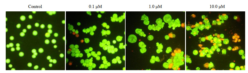
The title compound 6 was further confirmed by an AO/EB staining assay to assess the apoptosis-induction ability. AO/EB staining were performed to observe the morphologic changes in the treatment of the title compound by fluorescene microscopy. K562 cells were added to a final concentration of 1 × 10⁶ cells/mL in a 24-well plate, and the plate was incubated for 24 h. Cells were treated with the title compound at different concentrations. After being cultured for 48 h, cells were collected, washed with PBS stored at 4 ℃, and then dual fluorescent staining solution (1 μL) containing 100 μg/mL AO and 100 μg/mL EB was added to each suspension for 10 min, and then covered with a coverslip. The morphology of apoptotic cells was examined using fluorescent microscope.
Fig. 1 shows that the title compound (0.1, 1.0 and 10.0 µM) induced morphological changes and characteristic of apoptosis. The morphological changes such as cell volume shrinkage, membrane blebbing, chromatin condensation and apoptotic body formation (bright green nucleus with condensed chromatin and condensed orange chromatin means early apoptosis cells and last apoptosis cells, respectively). As a comparison, the untreated control cells showed normal morphology and stained in green. These data clearly demonstrated that the ability of the title compound to induce apoptosis was related to the concentrations.
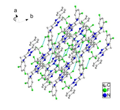
The synthesis route of the title compound by four steps with good yield was depicted in Scheme 1. The elemental analysis, IR, 1H NMR and X-ray diffraction data for the product are in good agreement with the structure of the title compound. The crystal structure of the target compound 6 was confirmed by X-ray diffraction analysis. The molecular structure and crystal packing picture are drawn by the Diamond program[18]. According to X-ray analysis, compound 6 crystallizes in triclinic system, space group P
In addition, preliminary bioassay indicated that compound 6 shows remarkable antiproliferative activity against MKN45, HT-29 and K562 cell lines with IC50 values of 5.57, 7.72 and 0.83 μM, respectively (Table 3). AO/EB staining of the title compound in K562 cells suggested that it induced dosedependent cell death in K562 cancer cells via apoptosis. Further studies on structural optimization and biological activities about these derivatives are still underway in our laboratory and will be reported in the future.
Kamel, M. M.; Megally Abdo, N. Y. Synthesis of novel 1, 2, 4-triazoles, triazolothiadiazines and triazolothiadiazoles as potential anticancer agents. Eur. J. Med. Chem. 2014, 86, 75–80. doi: 10.1016/j.ejmech.2014.08.047
You, W. K.; Sennino, B.; Williamson, C. W.; Falcón, B.; Hashizume, H.; Yao, L. C.; Aftab, D. T.; McDonald, D. M. VEGF and c-met blockade amplify angiogenesis inhibition in pancreatic islet. Cancer Res. 2011, 71, 4758–768.
Liu, J.; Hao, X. C.; Shi, D.; Ye, C.; Yang, W. Synthesis of novel 1-arylpyrazolo[3, 4-b][1, 5]benzodiazepine derivatives. Journal of Liaoning University (Natural Sciences Edition) 2018, 45, 244–248.
Hoelder, S.; Clarke, P. A.; Workman, P. Discovery of small molecule cancer drugs: successes, challenges and opportunities. Mol. Oncol. 2012, 6, 155–176. doi: 10.1016/j.molonc.2012.02.004
Liu, X. H.; Zhai, Z. W.; Xu, X. Y.; Yang, M. Y.; Sun, Z. H.; Weng, J. Q.; Tan, C. X.; Chen, J. Facile and efficient synthesis and biological activity determination of novel 1, 2, 4-triazolo[4, 3-a]pyridin-3(2H)-one derivatives via microwave irradiation. Bioorg. Med. Chem. Lett. 2015, 25, 5524–5228. doi: 10.1016/j.bmcl.2015.10.064
Shi, J. T.; Gong, Y. L.; Li, J.; Wang, Y.; Chen, Y.; Ding, S.; Liu, J. Synthesis, structure and biological activity of 2-[2-(4-fluorobenzylidene) hydrazinyl]-4-(1-methyl-1H-indol-3-yl)thieno[3, 2-d] pyrimidine. Chinese J. Struct. Chem. 2019, 38, 1810–1816.
Rivers, E. C.; Mancera, R. L. New anti-tuberculosis drugs in clinical trials with novel mechanisms of action. Drug Discov. Today 2008, 13, 1090–1098. doi: 10.1016/j.drudis.2008.09.004
Asghar, U.; Witkiewicz, A. K.; Turner, N. C.; Knudsen, E. S. The history and future of targeting cyclin-dependent kinases in cancer therapy. Nat. Rev. Drug Discov. 2015, 14, 130–146. doi: 10.1038/nrd4504
Gouda, M. A.; Berghot, M. A.; Shoeib, A. I. Synthesis and antimicrobial of new anthraquinone derivatives incorporating pyrazole moiety. Eur. J. Med. Chem. 2010, 45, 1843–1848. doi: 10.1016/j.ejmech.2010.01.021
Shaaban, M. R.; Saleh, T. S.; Mayhoub, A. S.; Mansour, A.; Farag, A. M. Synthesis and analgesic/anti-inflammatory evaluation of fused heterocyclic ring systems incorporating phenylsulfonyl moiety. Bioorg. Med. Chem. 2008, 16, 6344–6352. doi: 10.1016/j.bmc.2008.05.011
Auzzi, G.; Bruni, F.; Cecchi, L.; Costanzo, A.; Vettori, L. P.; Pirisino, R.; Corrias, M.; Ignesti, G.; Banchelli, G.; Raimondi, L. 2-Phenylpyrazolo[1, 5-a]pyrimidin-7-ones. A new class of nonsteroidal antiinflammatory drugs devoid of ulcerogenic activity. J. Med. Chem. 1983, 26, 1706–1709. doi: 10.1021/jm00366a009
Li, Y.; Gao, W.; Li, F.; Wang, J.; Zhang, J.; Yang, Y.; Zhang, S.; Yang, L. An in silico exploration of the interaction mechanism of pyrazolo[1, 5-a]pyrimidine type CDK2 inhibitors. Mol. Bio. Syst. 2013, 9, 2266–2281.
Lu, J. F.; Jin, L. X.; Ge, H. G.; Ji, X. H.; Guo, X. H.; Tian, G. H.; Song, J.; Jiang, M. Synthesis, crystal, computational study and biological activity of N-(1-(2, 4-dichlorophenyl)-1H-pyrazolo[3, 4-d]pyrimidin-4-yl)-4-(N, N-dipropylsulfamoyl)benzamide. Chinese J. Struct. Chem. 2017, 36, 1810–1816.
Ji, X. H.; Zhao, J.; Lu, J. F.; Jin, L. X.; Ge, H. G. Synthesis, crystal structure and biological activity of 1-(3-amino-4-morpholino-1H-indazole-1-carbonyl)-N-(4-fluorophenyl)cyclopropane-1-carboxamide. Chinese J. Struct. Chem. 2019, 38, 1889–1894.
Lu, J. F.; Huang, P.; Zhang, D.; Wang, Q.; Zheng, N.; Wu, R.; Liu, Q.; Jin, L. X.; Yu, X. H.; Ji, X. H.; Gao, Y. H.; Ge, H. G. 1-(3-Amino-4-morpholino-1H-indazole-1-carbonyl)-N-phenylcyclopropane-1-carboxamide: Design, synthesis, crystal structure, antitumor activity, DFT and Hirshfeld surface analysis. J. Mol. Struct. 2020, 1210, 127996–127999. doi: 10.1016/j.molstruc.2020.127996
Sheldrick, G. M. SHELXS-97, Program for the Solution of Crystal Structures. University of Göttingen, Germany 1997.
Sheldrick, G. M. SHELXL-2014/7, Crystal Structure Refinement with SHELXL. Acta Crystallographica C 2015, C71, 3-8.
Bergerhoff, G.; Berndt, M.; Brandenburg, K. Evaluation of crystallographic data with the program DIAMOND. J. Res. Natl. Inst. Stand. Technol. 1996, 101, 221–225. doi: 10.6028/jres.101.023

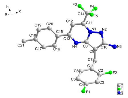
Figure 2 Structure of the title compound 6 with all non-H atom-labelling scheme and ellipsoids drawn at the 30% probability level
Table 1. Geometric Parameters of the Title Compound
| Bond | Dist. (Å) | Angle | (º) | |
| C(1)–C(2) | 1.386(3) | C(2)–C(1)–C(7) | 122.1(2) | |
| C(1)–C(7) | 1.502(3) | C(3)–C(2)–C(1) | 124.1(3) | |
| C(7)–C(9) | 1.499(3) | C(9)–C(7)–C(1) | 113.42(18) | |
| C(8)–N(4) | 1.347(3) | N(4)–C(8)–N(1) | 122.14(19) | |
| C(8)–C(9) | 1.377(3) | C(8)–C(9)–C(10) | 104.70(19) | |
| C(8)–N(1) | 1.409(3) | C(10)–C(9)–C(7) | 129.9(2) | |
| C(9)–C(10) | 1.412(3) | N(2)–C(10)–N(3) | 120.0(2) | |
| C(10)–N(2) | 1.347(3)) | N(1)–C(11)–C(12) | 118.96(19) | |
| C(10)–N(3) | 1.364(3) | N(4)–C(13)–C(12) | 121.1(2) | |
| C(11)–N(1) | 1.352(3) | C(12)–C(13)–C(15) | 121.4(2) | |
| C(11)–C(12) | 1.356(3) | C(20)–C(15)–C(13) | 122.6(2) | |
| C(11)–C(14) | 1.508(3) | C(17)–C(18)–C(21) | 121.5(2) | |
| C(12)–C(13) | 1.423(3) | N(2)–N(1)–C(8) | 111.87(17) | |
| C(13)–N(4) | 1.321(3) | C(13)–N(4)–C(8) | 118.38(18) | |
| C(13)–C(15) | 1.487(3) | Torsion angle | (º) | |
| C(15)–C(16) | 1.386(3) | C(7)–C(1)–C(2)–C(3) | 178.5(2) | |
| C(18)–C(21) | 1.511(3) | C(7)–C(9)–C(10)–N(3) | 2.2(4) | |
| C(19)–C(20) | 1.384(4) | N(4)–C(8)–N(1)–C(11) | 0.9(3) | |
| N(1)–N(2) | 1.363(2) | N(1)–C(8)–N(4)–C(13) | –0.5(3) |
 下载: 导出CSV
Table 2. In Vitro Anticancer Activity Testa of the Title Compound on the MKN45, HT29 and K562 Cell Lines
| Compound | IC50 ± SD (μmol·L-1) | ||
| MKN45 | HT29 | K562 | |
| The title compound | 5.57 ± 0.46 | 7.72 ± 0.58 | 0.83 ± 0.07 |
| Sorafenib tosylate | 4.26 ± 0.15 | 4.50 ± 0.26 | 2.18 ± 0.14 |
| aTest MTT colourimetric assay in MKN45, HT29 and K562 human cancer cell lines. Each experiment was carried out in triplicate | |||
下载: 导出CSV

 扫一扫看文章

扫一扫关注我们


 下载:
下载: