引用本文:
王涛, 马拉毛草, 马恒昌. 基于聚集诱导发光荧光探针的细胞成像研究进展[J]. 应用化学,
2018, 35(10): 1155-1165.
doi:
10.11944/j.issn.1000-0518.2018.10.170461

Citation: WANG Tao, MA Lamaocao, MA Hengchang. Research Progress on Cell Imaging Based on the Aggregation-induced Emission Fluorescent Probes[J]. Chinese Journal of Applied Chemistry, 2018, 35(10): 1155-1165. doi: 10.11944/j.issn.1000-0518.2018.10.170461
Citation: WANG Tao, MA Lamaocao, MA Hengchang. Research Progress on Cell Imaging Based on the Aggregation-induced Emission Fluorescent Probes[J]. Chinese Journal of Applied Chemistry, 2018, 35(10): 1155-1165. doi: 10.11944/j.issn.1000-0518.2018.10.170461
基于聚集诱导发光荧光探针的细胞成像研究进展
English
Research Progress on Cell Imaging Based on the Aggregation-induced Emission Fluorescent Probes
Abstract:
Fluorescent probes as a major discovery in the field of chemical sensing at the end of the 20th century have the advantages of simple synthesis, high sensitivity, good selectivity, short response time and high visualization. The combination of the fluorescent group with the aggregation-induced emission(AIE) characteristics and the biocompatible polymer makes the fluorescent materials have the characteristics of low toxicity, good light stability and good biocompatibility. In molecular, ion detection and cell imaging technology it has been widely studied and applied. This review summarizes the fluorescence probes for cytoplasmic imaging, cell membrane imaging, mitochondrial imaging, lysosomal imaging, lipid droplet imaging, nuclear imaging, nuclear and mitochondrial dual-targeting imaging and the prospects for their application.
-
Key words:
- fluorescence probe
- / chemical sensing
- / biological imaging
- / aggregation-induced emission
-
近年来,荧光材料在环境保护、分子识别、生物成像以及医学诊断等领域显示出重要的应用价值,受到材料和化学研究工作者广泛的重视[1-2]。其中,荧光探针是荧光分析方法的重要工具,拓展了荧光分析方法的应用范围,具有分析灵敏性高,操作简单,样品用量少,检测成本低,能对目标物进行实时原位监测等优点[3-5]。因此,设计和合成新的荧光探针,具有重要的意义。
2014年,诺贝尔化学奖颁发给了3位美国科学家Eric Betzig,William E Moemer和Stefan W)Hell,正是因为他们发现了超高分辨率荧光显微,实现了分子水平的荧光检测技术变革[6-8]。同样,唐本忠院士研究小组所定义的“聚集诱导发光现象”(Aggregation-Induced Emission, AIE)突破了传统荧光聚集猝灭的瓶颈, 也实现了一场关于荧光探针分子的变革[9-11]。生物体液及细胞液内基本成份为水,有机溶剂的含量几乎为零,传统的有机小分子荧光生色团具有聚集荧光淬灭(ACQ)效应,在水中容易发生聚集现象,使荧光猝灭,无法完成长期的成像及信号追踪[12]。AIE现象的独特性则能有效地避免这种不足,这是由于AIE材料在水中聚集和荧光的增强性质可以实现低浓度、大荧光信号及长期追踪[13-14]。可以说,AIE现象的发现为荧光材料领域提供了一个崭新的研究方向,具有这种性能的化合物作为荧光探针也被越来越多地应用于细胞成像等生物检测中,并取得了瞩目的研究成果[15-17]。据汤森路透与中国科学院文献情报中心联合发布的《2015研究前沿》报告,“聚集诱导发光化合物的合成、性质和用于细胞成像”在化学领域十大研究前沿中排名第二,属于重大热点前沿。
1. 荧光探针及其识别原理
1.1 荧光探针
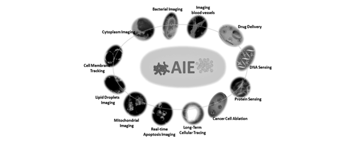
荧光探针是化学传感技术领域在20世纪末的一项重大发现,属于分子识别的范畴,而分子识别的基础——超分子化学,是重要的研究课题[18]。目前,荧光探针已广泛地应用于分子/离子检测、生物大分子检测,还可以检测包括温度、极性、粘度的动态变化[19-21]。人们可以直接观察宏观变化或者使用荧光光谱仪和显微镜、荧光显微镜、共聚焦显微镜等仪器获取检测到的相关信息,还可以实现生物活体和细胞及细胞器官的荧光成像[22]。荧光检测技术具有以下优点:对于分子/离子及生物分子检测具有灵敏度高、可视性强、操作简便、对细胞和生物体的损伤小、可观察亚细胞结构等。这些优点使其成为在临床分析、生物分析、环境监测、生命科学等领域不可缺少的检测手段[23-24],如图 1所示。
图 1
近几年来,随着荧光探针技术的发展科研人员对荧光探针有了更高的要求:更高的灵敏度;更高的选择性;更低的检测限;更快的分析速度;更智能的自动化程序;良好的生物相容性;更低的光毒性;更高的光稳定性;活体、微损或无损分析等[26-27]。
1.2 荧光探针的结构及识别原理
通常,荧光探针分子由荧光团(发光基团, Fluorophore)、识别基团(受体,Receptor)和连接臂(桥联基团)3个部分组成的。连接臂(桥联基团,Spacer)以共价键方式连接荧光团(发光基团, Fluorophore)和识别基团(受体,Receptor)。首先,识别基团(受体,Receptor)通过化学反应或者超分子弱作用力等方式与被检测物发生相互作用。其次,通过连接臂(桥联基团,Spacer)将识别基团的信号传递到荧光团。最后,信号在荧光团(发光基团, Fluorophore)处通过荧光变化(荧光的增强、淬灭以及光谱位移的变化等)的方式将信号表达出来。荧光探针分子具有非常大的潜力和应用价值,通过对分子的结构进行巧妙设计和改造,研究人员就可以设计出满足“特定需求”的探针分子。
按照光化学反应原理,荧光探针工作的机理大致有如下几种:光致电子转移(PET)[28]、分子内电荷转移(ICT)[29]、光诱导荧光共振能量转移(FRET)[30]、单体-激基缔合物(monomer/excimer)[31]、金属-配合体电荷转移(MLCT)[32]、激发态分子内质子转移(ESIPT)[33]和聚集诱导发光(AIE)[34-35]。基于以上的机理,不同类型的荧光探针被相继开发出来,具有成本低、体积较小、不受外界电磁场影响等优良特性,被广泛应用于环境监测、临床分析、生物分析等领域。
2. 荧光探针在生物成像方面的应用
荧光探针分子由于其合成简单、灵敏度高、选择性好、响应时间短、可直接观察等优点,在分子、离子检测和细胞成像技术中得到广泛的研究和应用。经过设计,当待测体系中的物质与识别基团发生作用时,引起探针共轭程度和电荷分布发生变化,致使荧光强度发生敏化或猝灭,从而实现检测目的。关于荧光探针的论述我们主要从探针使用的对象考虑,大体分为4种:离子探针、分子探针、刺激响应探针和生物成像探针。本文综述了荧光探针在生物成像方面的应用。
真核细胞含有细胞质膜、细胞核、线粒体、溶酶体、脂滴、高尔基体等细胞器。这些细胞器在细胞功能中起关键作用。细胞器及其形态或功能变化的可视化在临床分析或医学干预中具有巨大的潜力,提供关于细胞的细胞生物活性有价值的信息[36-37]。先进技术的发展对于形态或生物活性的可视化起着不可或缺的作用。
在众多成像模式中,荧光成像是可视化的强大工具之一。荧光成像(Fluorescent Imaging)技术是生物医学领域研究的重要手段,可用于研究目标分子的所在位置及浓度等,且这种方法具有无损伤、高特异性和灵敏度,以及能在细胞水平获得更高的分辨率等优势。随着荧光探针的多样化及相关仪器的改进,荧光成像技术已广泛用于分子、细胞及组织等不同层次的成像,即标记特殊离子,检测蛋白质、核酸和糖类等生物大分子,示踪活细胞的生物学行为和体内特殊器官或者肿瘤病灶成像等[38-42]。目前,常用的荧光探针有荧光蛋白(Fluorescent Proteins, FPs)、量子点(Quantum Dots, QDs)和有机小分子荧光探针[43-45]。细胞成像技术利用荧光探针标记细胞器或细胞器中的特异性包涵体,受益于先进光学显微镜,荧光的空间分辨率成像达到微米或甚至纳米级。此外,由于荧光造影剂的可用性及其实时操作的特性,非侵入性测试和具有成本效益的性能[46-47],荧光成像已被认为是生物系统中最强大的工具之一。所以对于特定细胞器的成像在生物学及医学领域有着巨大的意义。
2.1 细胞质染色
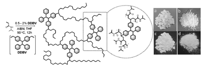
传统的荧光显影剂,特别是小分子荧光剂,很难长期存留在细胞内部,因此,很难将其应用于细胞传代研究。2016年,我们课题组[48]将四苯乙烯(TPE)与生物相容性的单体共聚得到了具有多重功能的AIE荧光材料P6(DDBV与AIBN、氮异丙基丙烯酰胺的共聚物)。如图 2所示,我们将此聚合物应用于Hela细胞的长期显影,取得了很好的效果。这种长期细胞示踪技术旨在能动态观察细胞的生物学特性,如形态变化、增殖、分化、迁移、侵袭和凋亡等过程。
图 2

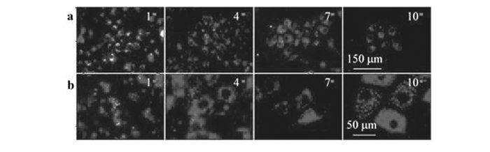
为了验证P6是否会被排出细胞,我们用含有P6(质量浓度为100 g/mL)的培养基培养A549细胞,实验结果如图 3所示,在每一代细胞内均能观测到很强的深蓝色的荧光。同时,由于显影剂P6良好的生物相容性,当细胞传至第10代时,在细胞内仍然有P6存在,并具有很强的荧光发射,因此该化合物可用作细胞示踪剂用于长期细胞示踪。研究充分说明聚合物也可以被细胞有效吞噬,并且通过聚合的方法还可以降低材料毒性,改善小分子探针水溶性和光稳定性差等缺点,实现优势互补。
图 3

2.2 细胞膜成像
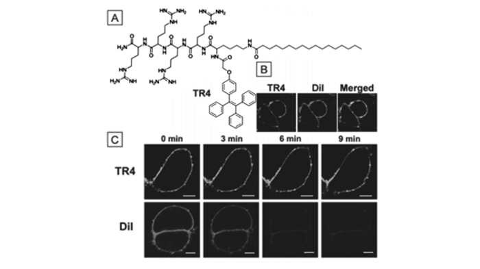
细胞膜由磷脂嵌入双层蛋白构成,它既使细胞维持稳定代谢的胞内环境,又能调节和选择物质进出细胞,同时,细胞膜也参与细胞识别和信号传导过程。所以细胞膜的重要性不言而喻[49]。2014年,梁新杰等[50]基于商业成熟的细胞膜绿色荧光探针(DIO)开发出了一种两亲性细胞膜染色剂TR4。如图 4所示,TR4染料由AIE部分(TPE)与带有带正电荷的亲水性四肽和棕榈酸合成,其中,TPE作为荧光基团,4个带正电的精氨酸单元带负电荷作为细胞膜靶向配体,棕榈酸中的长烷基链用于嵌入细胞膜。通过共聚焦成像可以看出,合成的TR4染料对细胞膜具有良好的靶向性,与商业上成熟的DIO染料染色部位完全重复。相对于DIO染料,合成的TR4染料具有良好的光稳定性,在紫外灯曝光9 min后依旧发出强荧光,DIO荧光几乎完全猝灭,该细胞膜染料有望取代现在工业上成熟的染色剂。
图 4
 图 4. TR4对细胞膜显影图片[50]Figure 4. TR4 on the cell membrane development diagram[50]
(A)Molecular structures of TR4; (B)Living MCF-7 cells were incubated with 50 μmol/L TR4 for 30 min and 10 μmol/L DiI in PBS solution for 10 min at 37 ℃. CLSM images of TR4(left, green, λex=405 nm), DiI(middle, red, λex=543 nm) and merged image of panels TR4 and DiI(right); (C)CLSM images of living MCF-7 cells treated with TR4(green) or DiI(red) with increasing scanning time(0~9 min). Scale bars are 10 μm
2.3 线粒体成像
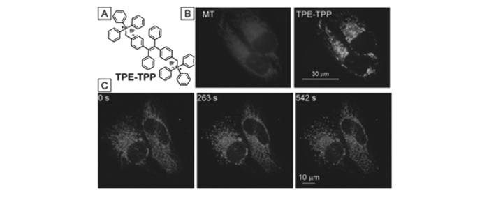
线粒体是一种半自主细胞器,有自己的DNA,可以独立地进行复制和表达。线粒体是碳水化合物、脂肪和氨基酸氧化产生能量三磷酸腺苷(ATP)的动力工作室[51]。同时,线粒体也参与许多生物过程,如细胞分化、凋亡等,具有调节细胞生长和细胞周期的能力。线粒体运动与其形态学高度相关,两个线粒体移动接近彼此,可能在融合后交换它们的内容。类似地,线粒体的分裂涉及线粒体区域分裂到相反的方向性[52]。因此,直接可视化线粒体动力学和荧光变化显微镜可以提供有价值的信息,了解涉及代谢和疾病的线粒体。但是线粒体的染料开发受限于其表面的电势,所以这仍是一个重要的研究课题。2012年,唐本忠等[53]开发出AIE型线粒体染色剂。如图 5所示,该分子由AIE基团TPE与线粒体靶向化合物TPP反应生成。TPE-TPP表现出了典型的AIE性质,且具有极小的尺寸,仅有144 nm,通过MTT法测得其生物毒性很低,这些条件为线粒体染色提供了良好的基础。TPE-TPP对线粒体有良好的靶向性,将该染料与商业通用线粒体染色剂MitoTracker Red做共染对比,证明了其对线粒体的选择性。相对于MitoTracker Red,TPE-TPP具有良好的光稳定性,可长期示踪线粒体,观察其形貌及变化过程。
图 5
 图 5. TPE-TPP对线粒体显影示意图[53]Figure 5. TPE-TPP schematic of mitochondria[53]
(A)Chemical structure of TPE-TPP; (B)Fluorescent images of CCCP(10 μmol/L) treated HeLa cells stained with MT(50 nmol/L) for 15 min and TPE-TPP(5 μmol/L) for 30 min. Excitation wavelength: 540~580 nm(for MT) and 330~385 nm(for TPE-TPP); (C)Fluorescent images of CCCP(20 μmol/L) treated living HeLa cells stained with TPE-TPP(5 μmol/L) with increasing scan time(shown in the upper left corner of the panel). Excitation wavelength:405 nm; emission filter:449~520 nm; irradiation time: 15.49 s/scan
2.4 溶酶体成像
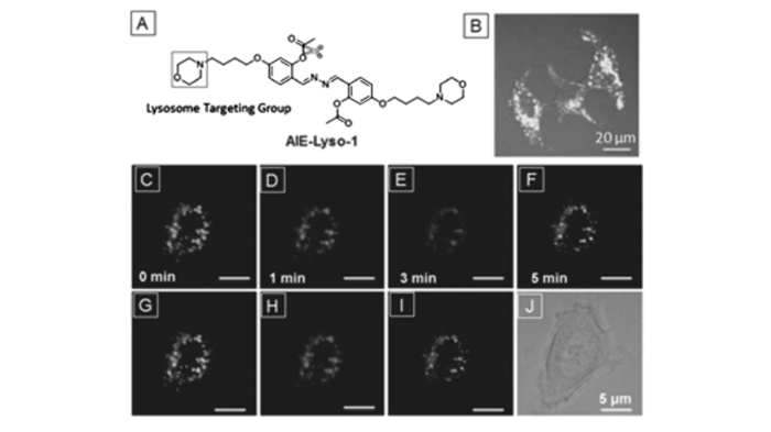
溶酶体是含有超过60种水解酶的真核细胞器。溶酶体有助于分解、吸收食物空泡转化为营养生物大分子或降解侵入病原体以保护细胞。同时,溶血自噬涉及细胞内失能的生物大分子或细胞器的过程。溶酶体的功能障碍或酶突变将导致严重的疾病,如白化病、苯丙酮尿症等。因此,监测溶酶体活性对于检测溶酶体相关疾病是有启发性的。荧光成像能够可视化它们的形态,映射它们的分布和跟踪它们在活细胞中的运动[54]。2014年,刘冰等[55]合成了第一个基于AIE特性的溶酶体染色剂AIE-LysoY。如图 6所示,分子中含有溶酶体配体吗啉和酯酶底物位点乙酰氧基。酯酶底物位点保证探针分子仅在溶酶体中有荧光发射,致使活性生物探针远离零反应性位点。最初,羟基被乙酰基保护,氢键断裂和N—N键自由旋转,导致荧光猝灭。当探针与人乳腺癌细胞(MCF-7)细胞孵育,被吗啉诱导的溶酶体可实现探针分子与人乳腺癌细胞(MCF-7)的特异性结合,其过程为酯酶水解保护乙酰基,产生的羟基中氢原子与氮原子作用,限制分子内旋转,发射出强荧光。通过与商业通用溶酶体染色剂Lyso Tracker Red共染,证明了其对溶酶体的良好的选择性。
图 6
 图 6. AIE-LysoY对溶酶体显影图片[55]Figure 6. Schematic representation of lysosomal development by AIE-LysoY[55]
(A)Design of probe AIE-Lyso-1 for specific detection of lysosomal esterase; (B)Confocal images of MCF-7 cells stained with 1.0 mmol/L AIE-Lyso-1 and 50 nmol/L LysoTracker Red. Confocal images of an MCF-7 cell stained with 1.0 mmol/L AIE-Lyso-1 and stimulated using 3 mmol/L chloroquine. Different pseudo-colors are used to illustrate the fuorescence images at different stimulation times of 0, 1, 3, and 5 min. (G~I)Merging images at two different times of 0 and 1 min(G), 1 and 3 min(H), 3 and 5 min(I), and the bright-field image(J). Scale bar:5 μm
2.5 脂滴成像
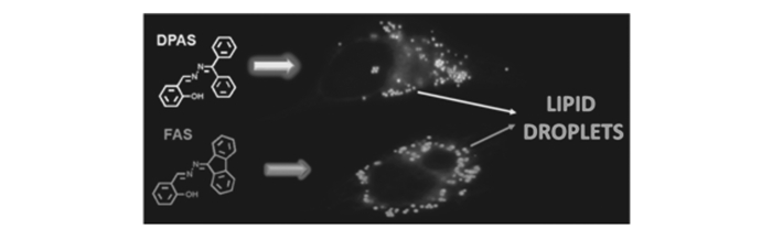
脂质液滴(LD)是富含脂质的细胞器,其主要作用是储存中性脂质的储库。自从17世纪发现以来的很长时间,LD仅被认为是脂肪库。随着研究的深入,LD中的一系列蛋白质被证明是高度动态的细胞器,其可以调节细胞内脂质储存和代谢[56]。它们还高度参与用于补充酰基甘油和胆固醇的膜转移。脂滴的活性或数量的异常是各种疾病的关键信号,例如脂肪肝疾病,Ⅱ型糖尿病和炎性肌病[57]。2016年,赵祖金等[58]报道了两种不同波长的脂滴探针芴酮水杨醛席夫碱(DPAS)和二苯甲酮水杨醛席夫碱(FAS)。DPAS和FAS均通过酮和水合肼之间的脱水反应制备,水杨醛席夫碱结构赋予二者AIE gens与激发态-分子内质子转移(ESIPT)属性。如图 7所示,由于ESIPT性质,DPAS和FAS的斯托克斯位移约为200 nm,优于商用BODIPY染料,对细胞内LD具有非常好的靶向性。与通用的BODIPY共染后,其重叠率分别为98%和97%,显示出了较高的LD靶向性。此外,该分子具有较好的光稳定性和pH稳定性。
图 7
2.6 细胞核成像
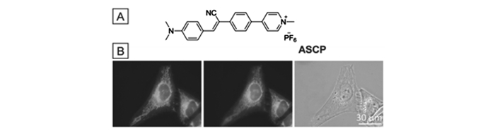
早在19世纪初细胞核就被发现是细胞中最大的细胞器,含有最多的遗传物质[59]并保持基因表达。在大多数真核细胞中,遗传物质是由长链DNA分子和核蛋白(如组蛋白)组成的染色体[60]。在基因表达过程中,最重要的程序之一是在核仁中rDNA转录到rRNA。之后,RNA与蛋白质组装以形成核糖体的前体,然后将其转运至细胞质[61]。目前,关于细胞核探针主要集中于2个位点,DNA与RNA[62]。2016年,唐本忠等[63]开发出一种分子内电荷转移(TICT)型细胞核染色剂聚集诱导发射特性的α-氰基二苯乙烯衍生物(ASCP)。ASCP分子由于存在TICI效应,所以具有发射波长随溶剂极性变化的特性,可根据需要调节不同的发射波长。该分子具有良好的生物相容性,可长期示踪细胞。如图 8所示,染料分子加入HeLa细胞后细胞核内发出强的红色荧光,与细胞核染色剂共染后也证明了其对于细胞核良好的选择性。
图 8

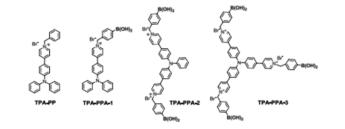
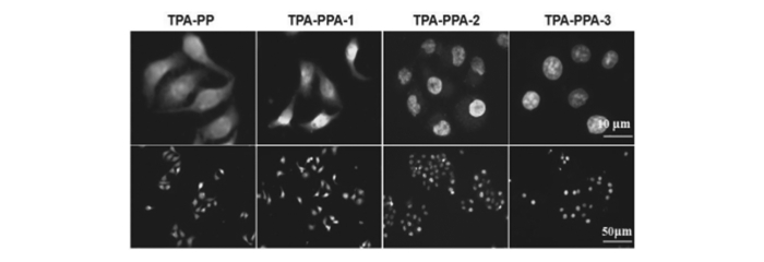
2017年,我们课题组以三苯胺为核使其接上不同取代数目的吡啶衍生物,用苄溴和对溴甲基苯硼酸进行修饰得到了4种化合物TPA(三苯胺)-PP(苄基吡啶盐)A(苯硼酸)-1、TPA-PPA-2、TPA-PPA-3和TPA-PP,化学结构以及细胞显影如图 9所示[64]。利用4种染料分子对HeGP-2进行了细胞染色实验,结果如图 10所示,我们观察到随着正离子数目的增加染色位置由细胞质转移到了细胞核, 并且TPAPPA-3对细胞核具有明显的靶向性。
图 9

图 10

2.7 细胞核和线粒体双靶向性的细胞成像
通过荧光探针在哺乳动物细胞中观察细胞核和线粒体是生物科学中重要的技术手段[65-66]。这是因为活体细胞中细胞器的可视化观察可以提供用于解决生物学问题的关键信息,可了解疾病机制,设计新的治疗方法。对于细胞核的可视化观察是癌症等相关疾病治疗的重要途径[67]。
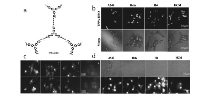
2016年,我们课题组设计合成了结构如图 11(a)所示的三苯胺三吡啶盐(TPPA-DBO)[68],我们在不同细胞中对其进行了细胞成像研究。观察发现其对细胞核具有很好的识别现象,结果如图 11(b)所示。在图 11(c)中观察到在染色并细胞繁殖一代后细胞核外围发生红色荧光,这是TPPA-DBO对线粒体进行了特定性的染色。
图 11
 图 11. 化合物TPPA-DBO化学结构式图(a);TPPA-DBO对A549、Hela、H4、HCM细胞系的细胞核染色(b);A549细胞中TPPA-DBO不同代数的染色(c);TPPA-DBO染色A549细胞一代后(d)照片[68]Figure 11. Chemical structure diagram of compound TPPA-DBO(a) and images of TPPA-DBO nuclei staining of A549, Hela, H4 and HCM(b), cell lines staining of different generations of TPPA-DBO in A549 cells(c), and TPPA-DBO stained A549 cells after the first-generation(d)[68]
目前,我们还不清楚其具体的机制,但是我们可以确认TPPA-DBO可以同时染色细胞核和线粒体,并且发出不同波长的荧光,可以实现对两种细胞器官的长期示踪。为了进一步验证,我们选用了不同的细胞系验证其现象,如图 11(d)所示,在不同的细胞系中TPPA-DBO对于细胞核和线粒体的双染色是完全成功的。从研究结果得到启示是:TPPA-DBO特异的细胞核靶向显影除了与探针化学结构有关外,还与通过聚合形成的多元正电荷不无关联。因此,通过对电荷的调控,有可能实现对不同细胞器官的靶向显影。我们课题组还在做进一步的研究和开发。
3. 结论与展望
本文主要综述具有AIE现象的荧光分子探针在生物成像方面的研究,分别对以下几方面做了综述:细胞膜成像、线粒体成像、溶酶体成像、脂滴成像、细胞核成像、细胞示踪、线粒体与细胞核双靶向性成像等。目前,常见的AIE探针分子是以三苯胺、四苯乙烯、噻咯衍生物、吡咯衍生物等为基本骨架,对其进行修饰得到了一系列不同荧光发射的AIE型探针分子。然而具有良好的生物相容性和水溶性的荧光探针分子是人们所期待的,目前所用到的方法是对得到的探针分子用生物大分子包埋或者用苯磺酸钠、PEG-2000等进行修饰来解决生物相容性以及水溶性的问题。但由于常见的荧光分子探针存在光稳定性差、细胞毒性大、探针分子较难合成等问题。因此,寻找发射波长较长、Stokes位移大、生物亲和性好、光稳定性好、廉价且无ACQ效应的荧光探针是推动荧光成像技术发展亟待解决的问题。AIE荧光分子探针在疾病检测与预防,以及如何长期进行细胞示踪方面有待进一步发展。
-
-
[1]
Shimizu, Yoshinori. Light Emitting Device Having a Nitride Compound Semiconductor and a Phosphor Containing a Garnet Fluorescent Material: US. Patent, 5998925A[P]. 1999-12-07.
-
[2]
Komoto, Satoshi. Semiconductor Light Emitting Device Including a Fluorescent Material: US Patent, 6340824A[P]. 2002-01-22.
-
[3]
Xu X, Ray R, Gu Y. Electrophoretic Analysis and Purification of Fluorescent Single-walled Carbon Nanotube Fragments[J]. J Am Chem Soc, 2004, 126(40): 12736-12737. doi: 10.1021/ja040082h
-
[4]
Shalon D, Smith S J, Brown P O. A DNA Microarray System for Analyzing Complex DNA Samples Using Two-color Fluorescent Probe Hybridization[J]. Genome Res, 1996, 6(7): 639-645. doi: 10.1101/gr.6.7.639
-
[5]
Semisotnov G V, Rodionova N A, Razgulyaev O I. Study of the "Molten Globule" Intermediate State in Protein Folding by a Hydrophobic Fluorescent Probe[J]. Biopolymers, 1991, 31(1): 119-128. doi: 10.1002/(ISSN)1097-0282
-
[6]
Betzig E, Patterson G H, Sougrat R. Imaging Intracellular Fluorescent Proteins at Nanometer Resolution[J]. Science, 2006, 313(5793): 1642-1645. doi: 10.1126/science.1127344
-
[7]
Kinkhabwala A, Yu Z, Fan S. Large Single-molecule Fluorescence Enhancements Produced by a Bowtie Nanoantenna[J]. Nat Photonics, 2009, 3(11): 654-657. doi: 10.1038/nphoton.2009.187
-
[8]
Eggeling C, Ringemann C. Direct Observation of the Nanoscale Dynamics of Membrane Lipids in a Living Cell[J]. Nature, 2009, 457(7233): 1159-1162. doi: 10.1038/nature07596
-
[9]
Luo J, Xie Z, Lam J W Y. Aggregation-induced Emission of 1-Methyl-1, 2, 3, 4, 5-Pentaphenylsilole[J]. Chem Commun, 2001, (18): 1740-1741. doi: 10.1039/b105159h
-
[10]
Hong Y, Lam J W Y, Tang B Z. Aggregation-induced Emission[J]. Chem Soc Rev, 2011, 40(11): 5361-5388. doi: 10.1039/c1cs15113d
-
[11]
Hong Y, Lam J W Y, Tang B Z. Aggregation-induced Emission:Phenomenon, Mechanism and Applications[J]. Chem Commun, 2009, (29): 4332-4353. doi: 10.1039/b904665h
-
[12]
Lakowicz J R. Topics in Fluorescence Spectroscopy Nonlinear and Two-Photon-Induced Fluorescence[J]. Springer Berlin, 2002, 6(1): 103-121.
-
[13]
Li M, Hong Y, Wang Z. Fabrication of Chitosan Nanoparticles with Aggregation-Induced Emission Characteristics and Their Applications in Long-Term Live Cell Imaging[J]. Macromol Rapid Commun, 2013, 34(9): 767-771. doi: 10.1002/marc.v34.9
-
[14]
Yan L, Zhang Y, Xu B. Fluorescent Nanoparticles Based on AIE Fluorogens for Bioimaging[J]. Nanoscale, 2016, 8(5): 2471-2487. doi: 10.1039/C5NR05051K
-
[15]
Ding D, Mao D, Li K. Precise and Long-term Tracking of Adipose-derived Stem Cells and Their Regenerative Capacity via Superb Bright and Stable Organic Nanodots[J]. ACS Nano, 2014, 8(12): 12620-12631. doi: 10.1021/nn505554y
-
[16]
Zhang X, Zhang X, Wang S. Surfactant Modification of Aggregation-induced Emission Material as Biocompatible Nanoparticles:Facile Preparation and Cell Imaging[J]. Nanoscale, 2013, 5(1): 147-150. doi: 10.1039/C2NR32698A
-
[17]
Shao A, Xie Y, Zhu S. Far-Red and Near-IR AIE-Active Fluorescent Organic Nanoprobes with Enhanced Tumor-Targeting Efficacy:Shape-Specific Effects[J]. Angew Chem Int Ed, 2015, 127(25): 7383-7388. doi: 10.1002/ange.v127.25
-
[18]
Lehn J M. 超分子化学: 概念和展望[M]. 沈兴海译. 北京: 北京大学出版社, 2002: 6-7.Lehn J M. Supramolecular Chemistry[M]. SHEN Xinghai, Trans. Beijing: Peking University Press, 2002: 6-7(in Chinese).
-
[19]
Arai S, Lee S C, Chang Y T. A Molecular Fluorescent Probe for Targeted Visualization of Temperature at the Endoplasmic Reticulum[J]. Sci Rep-UK, 2014, 4: 6701.
-
[20]
Zhang W, Liu W, Li P. Rapid-Response Fluorescent Probe for Hydrogen Peroxide in Living Cells Based on Increased Polarity of C B Bonds[J]. Anal Chem, 2015, 87(19): 9825-9828. doi: 10.1021/acs.analchem.5b02194
-
[21]
Liu T, Liu X, Spring D R. Quantitatively Mapping Cellular Viscosity with Detailed Organelle Information via a Designed PET Fluorescent Probe[J]. Sci Rep-UK, 2014, 4: 5418.
-
[22]
Cheng J, Ma X, Zhang Y. Optical Chemosensors Based on Transmetalation of Salen-based Schiff Base Complexes[J]. Inorg Chem, 2014, 53(6): 3210-3219. doi: 10.1021/ic5000815
-
[23]
Zhou Y, Zhang J F, Yoon J. Fluorescence and Colorimetric Chemosensors for Fluoride-Ion Detection[J]. Chem Rev, 2014, 114(10): 5511-5571. doi: 10.1021/cr400352m
-
[24]
Zhou X, Lee S, Xu Z. Recent Progress on the Development of Chemosensors for Gases[J]. Chem Rev, 2015, 115(15): 7944-8000. doi: 10.1021/cr500567r
-
[25]
Goswami S, Das S, Aich K. A Chemodosimeter for the Ratiometric Detection of Hydrazine Based on Return of ESIPT and Its Application in Live-cell Imaging[J]. Org Lett, 2013, 15(21): 5412-5415. doi: 10.1021/ol4026759
-
[26]
Yin J, Kwon Y, Kim D. Cyanine-based Fluorescent Probe for Highly Selective Detection of Glutathione in Cell Cultures and Live Mouse Tissues[J]. J Am Chem Soc, 2014, 136(14): 5351-5358. doi: 10.1021/ja412628z
-
[27]
Liu B, Wang J, Zhang G. Flavone-based ESIPT Ratiometric Chemodosimeter for Detection of Cysteine in Living Cells[J]. ACS Appl Mater Interfaces, 2014, 6(6): 4402-4407. doi: 10.1021/am500102s
-
[28]
Aigner D, Borisov S M, Fern ndez F J O. New Fluorescent pH Sensors Based on Covalently Linkable PET Rhodamines[J]. Talanta, 2012, 99: 194-201. doi: 10.1016/j.talanta.2012.05.039
-
[29]
Park B W, Philippe B, Gustafsson T. Enhanced Crystallinity in Organic Inorganic Lead Halide Perovskites on Mesoporous TiO2 via Disorder-Order Phase Transition[J]. Chem Mater, 2014, 2(15): 4466-4471.
-
[30]
Liu Z, He W, Guo Z. Metal Coordination in Photoluminescent Sensing[J]. Chem Soc Rev, 2013, 42(4): 1568-1600. doi: 10.1039/c2cs35363f
-
[31]
Xu Z, Yoon J, Spring D R. A Selective and Ratiometric Cu2+ Fluorescent Probe Based on Naphthalimide Excimer Monomer Switching[J]. Chem Commun, 2010, 46(15): 2563-2565. doi: 10.1039/c000441c
-
[32]
Wu J, Liu W, Ge J. New Sensing Mechanisms for Design of Fluorescent Chemosensors Emerging in Recent Years[J]. Chem Soc Rev, 2011, 40(7): 3483-3495. doi: 10.1039/c0cs00224k
-
[33]
Kim T I, Kang H J, Han G. A Highly Selective Fluorescent ESIPT Probe for the Dual Specificity Phosphatase MKP-6[J]. Chem Commun, 2009, (39): 5895-5897.
-
[34]
Mei J, Hong Y, Lam J W Y. Aggregation-Induced Emission:The Whole is More Brilliant Than the Parts[J]. Adv Mater, 2014, 26(31): 5429-5479. doi: 10.1002/adma.201401356
-
[35]
Chang Z F, Jing L M, Liu Y Y. Constructing Small Molecular AIE Luminophores Through a 2, 2-(2, 2-Diphenylethene-1, 1-diyl) Dithiophene Core and Peripheral Triphenylamine with Applications in Piezofluorochromism, Optical Waveguides, and Explosive Detection[J]. J Mater Chem C, 2016, 4(36): 8407-8415. doi: 10.1039/C6TC02395A
-
[36]
Miyanari Y, Atsuzawa K, Usuda N. The Lipid Droplet is an Important Organelle for Hepatitis C Virus Production[J]. Nat Cell Biol, 2007, 9(9): 1089-1097. doi: 10.1038/ncb1631
-
[37]
Berezney R. The Nuclear Matrix:A Heuristic Model for Investigating Genomic Organization and Function in the Cell Nucleus[J]. J Cell Biochem, 1991, 47(2): 109-123. doi: 10.1002/(ISSN)1097-4644
-
[38]
Vendrell M, Zhai D, Er J C. Combinatorial Strategies in Fluorescent Probe Development[J]. Chem Rev, 2012, 112(8): 4391-4420. doi: 10.1021/cr200355j
-
[39]
Xu W, Zeng Z, Jiang J H. Discerning the Chemistry in Individual Organelles with Small-Molecule Fluorescent Probes[J]. Angew Chem Int Ed, 2016, 55(44): 13658-13699. doi: 10.1002/anie.201510721
-
[40]
Li Y, Shao A, Wang Y. Tumor Bioimaging:Morphology-Tailoring of a Red AIEgen from Microsized Rods to Nanospheres for Tumor-Targeted Bioimaging[J]. Adv Mater, 2016, 28(16): 3224-3224. doi: 10.1002/adma.201670113
-
[41]
Li Y, Shao A, Wang Y. Morphology-Tailoring of a Red AIEgen from Microsized Rods to Nanospheres for Tumor-Targeted Bioimaging[J]. Adv Mater, 2016, 28(16): 3187-3193. doi: 10.1002/adma.201504782
-
[42]
Umezawa K, Yoshida M, Kamiya J M. Rational Design of Reversible Fluorescent Probes for Live-cell Imaging and Quantification of Fast Glutathione Dynamics[J]. Nat Chem, 2017, 9(3): 279-286. doi: 10.1038/nchem.2648
-
[43]
Ma H, Yang M, Zhang C. Aggregation-induced Emission(AIE)-active Fluorescent Probes with Multiple Binding Sites Toward ATP Sensing and Live Cell Imaging[J]. J Mater Chem B, 2017, 5(43): 8525-8531. doi: 10.1039/C7TB02399E
-
[44]
Pedram P, Mahani M, Torkzadeh-Mahani M. Cadmium Sulfide Quantum Dots Modified with the Human Transferrin Protein Siderophiline for Targeted Imaging of Breast Cancer Cells[J]. Microchim Acta, 2016, 183(1): 67-71. doi: 10.1007/s00604-015-1593-6
-
[45]
Sun L, Ding J, Xing W. Novel Strategy for Preparing Dual-modality Optical/PET Imaging Probes via Photo-click Chemistry[J]. Biol Chem, 2016, 27(5): 1200-1204.
-
[46]
Domaille D W, Que E L, Chang C J. Synthetic Fluorescent Sensors for Studying the Cell Biology of Metals[J]. Nat Chem Biol, 2008, 4(3): 168-175. doi: 10.1038/nchembio.69
-
[47]
Liang J, Li K, Liu B. Visual Sensing with Conjugated Polyelectrolytes[J]. Chem Soc, 2013, 4(4): 1377-1394.
-
[48]
Ma H, Qi C, Cheng C. AIE-active Tetraphenylethylene Cross-linked N-Isopropylacrylamide Polymer:A Long-term Fluorescent Cellular Tracker[J]. ACS Appl Mater Interfaces, 2016, 8(13): 8341-8348. doi: 10.1021/acsami.5b11091
-
[49]
Wang B, Zhu C, Liu L. Synthesis of a New Conjugated Polymer for Cell Membrane Imaging by Using an Intracellular Targeting Strategy[J]. Polym Chem-UK, 2013, 4(20): 5212-5215. doi: 10.1039/c3py00097d
-
[50]
Zhang C, Jin S, Yang K. Cell Membrane Tracker Based on Restriction of Intramolecular Rotation[J]. ACS Appl Mater Interfaces, 2014, 6(12): 8971-8975. doi: 10.1021/am5025897
-
[51]
Jouaville L S, Pinton P, Bastianutto C. Regulation of Mitochondrial ATP Synthesis by Calcium:Evidence for a Long-term Metabolic Priming[J]. PNAS, 1999, 96(24): 13807-13812. doi: 10.1073/pnas.96.24.13807
-
[52]
Suen D F, Norris K L, Youle R J. Mitochondrial Dynamics and Apoptosis[J]. Gene Dev, 2008, 22(12): 1577-1590. doi: 10.1101/gad.1658508
-
[53]
Leung C W T, Hong Y, Chen S. A Photostable AIE Luminogen for Specific Mitochondrial Imaging and Tracking[J]. J Am Chem Soc, 2013, 135(1): 62-65. doi: 10.1021/ja310324q
-
[54]
Van Meel E, Klumperman J. Imaging and Imagination:Understanding the Endo-lysosomal System[J]. Histo Chem Cell Biol, 2008, 129(3): 253-266. doi: 10.1007/s00418-008-0384-0
-
[55]
Gao M, Hu Q, Feng G. A Fluorescent Light-up Probe with "AIE+ ESIPT" Characteristics for Specific Detection of Lyssomal Esterase[J]. J Mater Chem B, 2014, 2(22): 3438-3442. doi: 10.1039/C4TB00345D
-
[56]
Martin S, Parton R G. Opinion:Lipid Droplets:A Unified View of a Dynamic Organelle[J]. Nat Rev Mol Cell Biol, 2006, 7(5): 373-378. doi: 10.1038/nrm1912
-
[57]
Alberti K G M M, Zimmet P, Shaw J. Metabolic Syndrome-A New World-wide Definition. A Consensus Statement from the International Diabetes Federation[J]. Diabetic Med, 2006, 23(5): 469-480. doi: 10.1111/dme.2006.23.issue-5
-
[58]
Wang Z, Gui C, Zhao E. Specific Fluorescence Probes for Lipid Droplets Based on Simple AIEgens[J]. ACS Appl Mater Interfaces, 2016, 8(16): 10193-10200. doi: 10.1021/acsami.6b01282
-
[59]
Kobayashi H, Ogawa M, Alford R. New Strategies for Fluorescent Probe Design in Medical Diagnostic Imaging[J]. Chem Rev, 2009, 110(5): 2620-2640.
-
[60]
Horobin R W, Stockert J C, Rashid-Doubell F. Fluorescent Cationic Probes for Nuclei of Living Cells:Why Are They Selective? A Quantitative Structure-Activity Relations Analysis[J]. Histochem Cell Biol, 2006, 126(2): 165-175. doi: 10.1007/s00418-006-0156-7
-
[61]
Collas P, Aleström P. Rapid Targeting of Plasmid DNA to Zebrafish Embryo Nuclei by the Nuclear Localization Signal of SV 40T Antigen[J]. Mol Mar Biol Biotechnol, 1997, 6(1): 48-58.
-
[62]
Wade W, Grabow , Luc J. RNA Self-Assembly and RNA Nanotechnology[J]. Acc Chem Res, 2014, 47: 1871-1880. doi: 10.1021/ar500076k
-
[63]
Chris Y Y, Kwok R T K, Tang B Z. A Photostable AIEgen for Nucleolus and Mitochondria Imaging with Organelle-specific Emission[J]. J Mater Chem B, 2016, 4(15): 2614-2619. doi: 10.1039/C6TB00319B
-
[64]
Ma H, Yang M, Zhang C. Aggregation-induced Emission(AIE)-active Fluorescent Probes with Multiple Binding Sites Toward ATP Sensing and Live Cell Imaging[J]. J Mater Chem B, 2017, 5(43): 8525-8531. doi: 10.1039/C7TB02399E
-
[65]
Haugland R P. Handbook of Fluorescent Probes and Research Products[M]. 9th Edn. Eugene OR: Molecular Probes, USA, 2002.
-
[66]
Giepmans B N, Adams S R, Ellisman M H. The Fluorescent Toolbox for Assessing Protein Location and Function[J]. Science, 2006, 312(5771): 217-224. doi: 10.1126/science.1124618
-
[67]
Ashkenazi A. Targeting Death and Decoy Receptors of the Tumour-necrosis Factor Superfamily[J]. Nat Rev Cancer, 2002, 2(6): 420-430. doi: 10.1038/nrc821
-
[68]
Ma H, Yang Z, Cao H. One Bioprobe:A Fluorescent and AIE-active Macromolecule; Two Targets:Nucleolus and Mitochondria with Long Term Tracking[J]. J Mater Chem B, 2017, 5(4): 655-660. doi: 10.1039/C6TB02844F
-
[1]
-
图 4 TR4对细胞膜显影图片[50]
Figure 4 TR4 on the cell membrane development diagram[50]
(A)Molecular structures of TR4; (B)Living MCF-7 cells were incubated with 50 μmol/L TR4 for 30 min and 10 μmol/L DiI in PBS solution for 10 min at 37 ℃. CLSM images of TR4(left, green, λex=405 nm), DiI(middle, red, λex=543 nm) and merged image of panels TR4 and DiI(right); (C)CLSM images of living MCF-7 cells treated with TR4(green) or DiI(red) with increasing scanning time(0~9 min). Scale bars are 10 μm
图 5 TPE-TPP对线粒体显影示意图[53]
Figure 5 TPE-TPP schematic of mitochondria[53]
(A)Chemical structure of TPE-TPP; (B)Fluorescent images of CCCP(10 μmol/L) treated HeLa cells stained with MT(50 nmol/L) for 15 min and TPE-TPP(5 μmol/L) for 30 min. Excitation wavelength: 540~580 nm(for MT) and 330~385 nm(for TPE-TPP); (C)Fluorescent images of CCCP(20 μmol/L) treated living HeLa cells stained with TPE-TPP(5 μmol/L) with increasing scan time(shown in the upper left corner of the panel). Excitation wavelength:405 nm; emission filter:449~520 nm; irradiation time: 15.49 s/scan
图 6 AIE-LysoY对溶酶体显影图片[55]
Figure 6 Schematic representation of lysosomal development by AIE-LysoY[55]
(A)Design of probe AIE-Lyso-1 for specific detection of lysosomal esterase; (B)Confocal images of MCF-7 cells stained with 1.0 mmol/L AIE-Lyso-1 and 50 nmol/L LysoTracker Red. Confocal images of an MCF-7 cell stained with 1.0 mmol/L AIE-Lyso-1 and stimulated using 3 mmol/L chloroquine. Different pseudo-colors are used to illustrate the fuorescence images at different stimulation times of 0, 1, 3, and 5 min. (G~I)Merging images at two different times of 0 and 1 min(G), 1 and 3 min(H), 3 and 5 min(I), and the bright-field image(J). Scale bar:5 μm
图 11 化合物TPPA-DBO化学结构式图(a);TPPA-DBO对A549、Hela、H4、HCM细胞系的细胞核染色(b);A549细胞中TPPA-DBO不同代数的染色(c);TPPA-DBO染色A549细胞一代后(d)照片[68]
Figure 11 Chemical structure diagram of compound TPPA-DBO(a) and images of TPPA-DBO nuclei staining of A549, Hela, H4 and HCM(b), cell lines staining of different generations of TPPA-DBO in A549 cells(c), and TPPA-DBO stained A549 cells after the first-generation(d)[68]
-

 下载:
下载:

 下载:
下载:


 扫一扫看文章
计量
- PDF下载量: 18
- 文章访问数: 3197
- HTML全文浏览量: 1098
 下载:
下载:
