Figure 1.
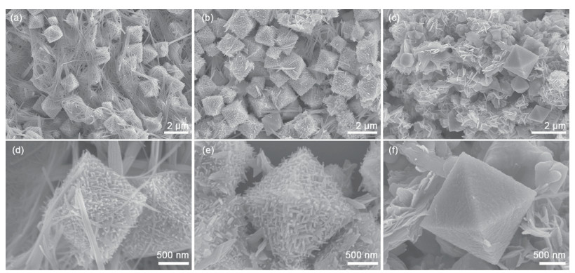
SEM images of Cu2O prepared at different reaction temperatures with 7.5 mL NaOH: (a, d) 10 ℃; (b, e) 20 ℃; (c, f) 30 ℃
High-sensitivity surface-enhanced Raman scattering detection and self-cleaning performance of organic pollutants based on a filter membrane sandwich structure
Huihui LIU , Baichuan ZHAO , Tingting ZHANG , Chuanhui WANG , Zhi WANG , Congyun ZHANG

As a class of additive brominated flame retardants, polybrominated diphenyl ethers (PBDEs) are widely used in electrical appliances, transportation, and the plastics and textiles industry[1-3]. Due to their environmental persistence, high lipophilicity, bioaccumulation, and high toxicity[4-7], PBDEs are of increasing concern in terms of their concentration in the environment and the hazards posed to organisms and human health[8]. The timely and sensitive monitoring of PBDEs and harmless treatment are identified as a vital approach to avoid the risk to human health and the ecological environment. Gas chromatography/mass spectrometry (GC/MS) and high-performance liquid chromatography (HPLC) are traditionally used as analytical methods for the detection of PBDEs[9-10]. However, the complicated operation protocols and expensive instrumentation have severely limited their application in the detection and effective removal of PBDEs[11].
Surface-enhanced Raman scattering (SERS) has garnered considerable attention for various applications such as analytical chemistry, diagnostics, food safety, environmental monitoring, and national security since it can detect and quantify target molecules with high sensitivity down to the single molecule level[12-17]. Exploration of novel substrates with high SERS performance and the potential to further eliminate organic pollutants is a high-priority task.
Among the currently available SERS substrates, many noble metal nanoparticles (NPs) and nanostructures remain the most widely used SERS-active materials due to their facile synthesis, tunable morphologies, and excellent chemical stability[18]. Their enhanced Raman activity is attributed to an electromagnetic mechanism (EM)[19], in which the generation of plasmonic "hot spots" plays a decisive role in achieving strong and reproducible signals from target analytes[20]. However, these materials still suffer from inherent limitations, including limited structural diversity[21], insufficient affinity toward many analytes, and suboptimal biocompatibility, all of which restrict their broader applicability in practical sensing scenarios[22].
Another critical limitation of conventional SERS substrates is their poor recyclability, which stems from the strong and often irreversible interactions between analyte molecules and metallic NPs. To overcome this drawback, recyclable SERS substrates have become the endeavor of many researchers, mainly based on integrating noble metal NPs with photocatalytically active semiconductor materials. Common semiconductor materials such as metal oxides (TiO2[23-24], ZnO[25], CuO[26], and W18O49[27]), two-dimensional semiconductor (such as g-C3N4[28-29], MoS2[30], and BP[31]), transition metal selenides[32], and phosphides[33] have been integrated with noble-metal NPs to construct photocatalytically recyclable SERS substrates. In such hybrid systems, recyclability arises from semiconductor-driven photocatalysis, in which photogenerated electron-hole pairs initiate oxidation or degradation of adsorbed analytes. Consequently, the semiconductor band gap critically dictates the excitation wavelength and overall photocatalytic efficiency. Among these materials, Cu2O possesses a moderate band gap of 2.0-2.2 eV, enabling effective absorption of both ultraviolet and visible light, including natural sunlight. This broader spectral responsiveness facilitates photocatalytic activation under mild illumination conditions[34]. In addition, Cu2O exhibits a high optical absorption coefficient and facet-dependent surface reactivity, both of which contribute to efficient charge separation and interfacial redox activity. These attributes collectively render Cu2O a particularly suitable candidate for constructing self-cleaning and recyclable SERS substrates[35-36].
In addition to plasmonic effects, the overall SERS performance is strongly influenced by the quantity, spatial distribution, and interfacial configuration of analyte molecules adsorbed on the substrate surface[37]. Improving the molecular adsorption properties of the substrate remains a demanding and important task. Graphene and its derivatives [graphene oxide (GO), reduced graphene oxide (rGO)] have attracted much attention due to their large specific surface area, extended π-conjugated framework, and abundant surface functional groups, which collectively enable efficient enrichment and adsorption of organic molecules through π-π stacking, electrostatic interactions, and hydrogen bonding. Moreover, the intrinsic fluorescence-quenching ability of graphene effectively suppresses background emission, thereby improving the Raman signal-to-noise ratio. These advantages have motivated the development of graphene-based hybrid SERS platforms. For example, a graphene nanomesh-Ag-ZnO hybrid fabricated through hydrothermal self-assembly followed by thermal annealing has been demonstrated as a highly sensitive SERS substrate due to its increased adsorption sites and synergistic charge-transfer pathways[38]. Similarly, rational design of three-dimensional MoS2@ Ag@rGO composites has shown that appropriate structural integration can simultaneously enhance molecular enrichment, Raman enhancement, and photocatalytic self-cleaning capability[39]. Collectively, these studies highlight the significant potential of graphene-based architectures for improving analyte adsorption and overall SERS performance.
Based on these considerations, a Cu2O-GO-AuNSts (AuNSts: gold nanostars) heterostructure as an ultrasensitive and recyclable SERS platform for the reliable detection and photocatalytic degradation of PBDEs was developed. In this design, the excellent photocatalytic performance of Cu2O, the LSPR characteristics of AuNSts, and the high electron mobility and adsorption capability of GO were synergistically integrated to construct a self-cleaning SERS substrate with high sensitivity, chemical stability, and reusability. Through controlled synthesis and structural optimization, anisotropic Cu2O with enhanced photocatalytic performance was incorporated to maximize charge-separation efficiency and interfacial reaction kinetics. The resulting semiconductor-GO-plasmonic metal heterostructure provides an effective pathway for coupling molecular enrichment, electromagnetic enhancement, and photocatalytic self-regeneration, thereby enabling high-performance SERS sensing and simultaneous pollutant degradation.
Gold(Ⅲ) chloride trihydrate (HAuCl4·3H2O), silver nitrate (AgNO3), sodium borohydride (NaBH4), 2, 2′, 4, 4′-tetrabromodiphenyl ether (BDE-47, ≥97%), and copper chloride (CuCl2) were purchased from Aldrich. Ascorbic acid (AA, ≥99%) and rhodamine 6G (R6G, fluorescent dye) were obtained from Alfa Aesar. Ethanol (97%), sodium hydroxide (NaOH), hydrazine hydrate (80%), and polyvinylpyrrolidone (PVP, Mw=10 000, K=27-32) were sourced from Tianjin Damao Chemical Reagent Factory. The GO aqueous solution (2.0 mg·mL-1) was purchased from Nanjing XFNANO Materials Tech. Co., Ltd. The filter membrane (FM, pore size: 0.22 μm) was purchased from Delv New Materia Tech. Co., Ltd. All chemicals used in the experiment were analytical grade without further purification. Ultrapure water (18.0 MΩ) was employed in all experiments.
At room temperature, a 0.01 mol·L-1 CuCl2 solution was prepared. Then, 0.36 g PVP powder was ultrasonically dispersed in 100 mL of CuCl2 solution. A hydrazine hydrate solution was prepared by diluting 80% hydrazine hydrate with ultrapure water in a volume ratio of 1∶31. Under continuous magnetic stirring at 20 ℃, 7.5 mL NaOH solution (1.5 mol·L-1) was mixed into 100 mL PVP/CuCl2 solution under the condition of magnetic agitation, and the solution was kept magnetically stirred for 10 min. Subsequently, 2 mL of the pre-configured diluted hydrazine hydrate solution was added to the solution, and the magnetic stirring was maintained for 30 min. The obtained reaction solution was centrifuged at 8 000 r·min-1 for 10 min to obtain a brick red precipitate. The resulting precipitate was centrifugally and ultrasonically washed with methanol and acetone solutions, respectively, followed by drying under vacuum at room temperature. To prevent oxidation, the as-prepared anisotropic Cu2O powder (35 mg) was stored in 50 mL of methanol for subsequent experiments.
To investigate the influence of synthesis parameters on morphology and physicochemical properties, the dosage of NaOH (5.0, 7.5, 10.0, and 15.0 mL) and the reaction temperature (10, 20, and 30 ℃) were systematically varied while maintaining other conditions constant.
The pre-synthesized 3 mL of Cu2O dispersion was ultrasonically dispersed homogeneously and vacuum-filtered onto a 0.2 μm FM using a Brinell's funnel in order to form a circular thin film filtration layer (named as Cu2O/FM) with a diameter of 3 cm. After natural drying, 300 μL of 0.02 mg·L-1 GO dispersion was immediately spin-coated on the FM-based Cu2O at 1 000 r·min-1 for 60 s and dried naturally to obtain the FM-based Cu2O-GO heterostructure (named as Cu2O-GO/FM).
AuNSts were prepared via the seed-mediated growth method. Firstly, a 0.05 mol·L-1 AgNO3 solution, a sodium citrate solution with a mass fraction of 5%, and a 1 mg·L-1 NaBH4 solution were prepared at room temperature. The AgNO3 solution (1 mL) and 1 mL of sodium citrate solution were added to 48 mL of ultrapure water under vigorous stirring for 5 min. Subsequently, 0.05 mL of NaBH4 solution was rapidly added to the reaction solution, which rapidly turned brown, and the magnetic stirring was maintained for 15 min. Subsequently, the reaction solution was centrifuged at 12 000 r·min-1 for 15 min, and the supernatant was collected for use as silver seed solution. Subsequently, 0.1 mL of the silver seed solution was added to 80 mL of 0.3 mmol·L-1 HAuCl4 under magnetic stirring, followed by the rapid addition of 1.5 mL of 1 mmol·L-1 AgNO3 and 0.5 mL of 0.1 mol·L-1 AA. The reaction was maintained under milder magnetic stirring for 2 min until the solution changed from colorless to blue-gray. The reaction solution was centrifuged at 4 000 r·min-1 for 15 min, and the centrifugate was concentrated to 500 μL to obtain the dispersion of AuNSts.
The dispersion of AuNSts (50 μL) was uniformly dropped on the surface of the pre-prepared Cu2O-GO heterostructures and spin-coated at 500 r·min-1 for 30 s. After completely drying in the dark, the FM-based Cu2O-GO-AuNSts sandwich structure (named as Cu2O-GO-AuNSts/FM) was obtained for subsequent applications.
The morphologies and structures of sandwich structures were investigated using field-emission scanning electron microscopy (SEM, Hitachi SU-8010) at an accelerating voltage of 10 kV and transmission electron microscopy (TEM, JEOL JEM-2100F) at an accelerating voltage of 200 kV. The crystal structure was investigated by high-resolution transmission electron microscopy (HRTEM) and X-ray diffractometer (Dandong Haoyuan DX-2700BH) equipped with Cu Kα radiation (λ=0.154 6 nm), a scan range of 5°-80°, a voltage of 40 kV, and a current of 40 mA. The surface defect and energy band parameter of the prepared samples were analyzed by X-ray photoelectron spectroscopy (XPS, Thermo Fisher Scientific Escalab250Xi) with Al Kα X-rays. The optical properties of the prepared hybrid systems were assessed by ultraviolet-visible (UV-Vis) absorption spectrum (Agilent Cary 5000). The photocatalytic activities of different synthesized samples were estimated by degrading BDE-47 molecules under UV-Vis light irradiation using a xenon lamp light source (PerfectLight PLS-SXE300, 300 W).
The photocatalytic activity of Cu2O-GO-AuNSts/FM was estimated by monitoring the degradation of BDE-47 molecules under UV-Vis light irradiation using a xenon lamp light source (PerfectLight PLS-SXE300, 300 W). Specifically, the prepared Cu2O-GO-AuNSts/FM was placed in 25 mL of 10-5 mol·L-1 BDE-47 in the dark at ambient temperature for 30 min to establish adsorption-desorption equilibrium before irradiation. When the photocatalytic reaction was running, 1 mL of the resultant dispersion solution subjected to light irradiation was collected at different intervals. A UV-Vis spectrometer was employed to monitor the residual concentration of BDE-47 in the reaction mixture. The photocatalytic activity evaluation of the hybrid systems targeting BDE-47 was carried out in a n-hexane solution under a nitrogen atmosphere.
To evaluate SERS performance, 50 μL of the probe molecule solution (R6G or BDE-47) at a certain mass concentration was dropped onto the Cu2O-GO-AuNSts/FM and kept undisturbed for 2 h to reach the adsorption-desorption equilibrium. The Raman spectra were acquired using a model confocal microscopy Raman spectrometer (Renishaw inVia) by using a 532 and 633 nm laser as the excitation source. The laser power was set at 1 mW for R6G detection and 5 mW for BDE-47 detection, with a ×50 objective lens and 10 s integration time for spectral collection.
The morphology of anisotropic octahedral Cu2O semiconductor materials was systematically investigated by controlling the reduction temperature (10, 20, and 30 ℃) and maintaining a constant NaOH dosage (7.5 mL) during in situ hydrazine hydrate reduction. As shown in Fig.1a, when the reaction temperature was maintained at 10 ℃, the Cu2O particles exhibited well-defined octahedral shapes with an average particle size of approximately 1.5 μm. Notably, these micro/nano-octahedra were intertwined with fibrous nanowires. These nanowire structures most likely arise from the low reaction temperature, which suppresses crystal growth kinetics and thereby favors the formation of fibrous intermediates during nucleation and subsequent growth. High-magnification imaging (Fig.1d) reveals that the surface of the octahedral Cu2O was covered with spinous protrusions, which substantially increase the specific surface area and are expected to enhance the photocatalytic activity by providing additional exposed active sites. When the reaction temperature rose to 20 ℃, the anisotropic octahedral Cu2O particles displayed improved uniformity and dispersion across the field of view (Fig.1b), accompanied by the disappearance of the fibrous nanofilaments observed at lower temperatures. This transition can be attributed to the accelerated nucleation and growth kinetics at elevated temperatures, which promote the formation of well-defined octahedral crystals. Under these conditions, the octahedral Cu2O retained its characteristic spinous surface architecture (Fig.1e), with uniformly distributed spines approximately 200 nm in length and densely decorating the crystal facets. These protrusions enrich the surface microtopography and may provide additional catalytically active sites, thereby facilitating enhanced photocatalytic interactions. Further increasing the reaction temperature to 30 ℃ leads to a pronounced loss of morphological uniformity, characterized by the emergence of non-faceted, aggregated structures that dominate the field of view (Fig.1c). A high-resolution SEM image (Fig.1f) revealed that the few remaining octahedral particles exhibited significantly smoother surfaces, with the spinous structures observed at lower temperatures largely diminished. This phenomenon is likely attributable to an excessively rapid reaction rate at 30 ℃, which disrupts the stable formation of spinous surface features and drives uncontrolled aggregation and fusion of Cu2O nuclei, ultimately giving rise to disordered, poorly defined morphologies. Collectively, these observations demonstrate that the reduction temperature critically influences the nucleation and growth dynamics of anisotropic octahedral Cu2O structures, thereby dictating their morphological features. Optimal formation of uniform, well-faceted octahedra with dense, spinous surface structures was achieved at 20 ℃, offering a favorable balance between growth kinetics and structural integrity for potential photocatalytic applications.
In addition to temperature effects, the evolution of Cu2O morphology is also strongly dependent on the concentration of the complexing agent NaOH. At 20 ℃, the addition of 5.0 mL NaOH yield well-defined octahedral particles with smooth facets, indicating that the alkalinity was sufficient to induce the formation of [Cu(OH)4]2- complexes while maintaining a relatively slow and controlled reduction process (Fig.2a). Increasing the NaOH dosage to 7.5 mL enhanced the OH- concentration, which intensifies anisotropic growth by facilitating the directional release of Cu+ from the [Cu(OH)4]2- complexes. Under these conditions, the octahedral surfaces become densely decorated with uniformly oriented spiny protrusions, producing the most ordered anisotropic architectures observed in this study (Fig.2b). However, further increasing the NaOH dosage to 10.0 mL disrupted this delicate balance. Excess hydroxide accelerates the reduction kinetics and induces uncontrolled nucleation, resulting in partial stacking of the spiny protrusions and even distortion of the octahedral framework (Fig.2c). When the NaOH dosage was 15.0 mL, this overgrowth phenomenon became more pronounced: the protrusions coalesced into thickened surface layers and gradually evolved into spherical aggregates, masking the underlying octahedral geometry (Fig.2d).
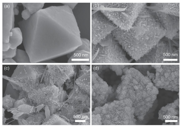
These results clearly indicate that NaOH plays a dual role in this system: (ⅰ) it regulates the formation of [Cu(OH)4]2- complexes, thereby controlling the availability of Cu+ species for reduction; and (ⅱ) it modulates the balance between nucleation and facet-selective growth. At moderate NaOH dosage (7.5 mL), these two effects are optimally balanced, producing anisotropic Cu2O with well-developed spiny octahedra. In contrast, insufficient or excessive NaOH addition shifts the system toward either smooth surfaces or uncontrolled overgrowth. Thus, precise tuning of the complexing agent concentration is crucial for achieving Cu2O architectures with desirable anisotropic features.
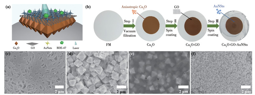
Under the optimized conditions (7.5 mL NaOH, 20 ℃), the Cu2O crystals displayed uniform spiny octahedral morphology, which was selected as the structural template for assembling the subsequent hybrid architecture. On this basis, a Cu2O-GO-AuNSts hybrid structure was constructed via a layer-by-layer assembly strategy, as illustrated in Fig.3a and 3b. In the first step, anisotropic octahedral Cu2O particles prepared under the optimized conditions were deposited onto a 0.2 μm pore-size FM substrate through vacuum filtration (Fig.3c). The SEM image revealed that the anisotropic octahedral Cu2O particles with an average size of 1.5-2.0 μm were uniformly distributed across the FM base (Fig.3d). These particles maintained well-defined octahedral morphology with distinct edges and smooth facets, ensuring consistent coverage and structural uniformity. Subsequently, GO nanosheets (0.02 mg·L-1) were dispersed and assembled onto the Cu2O-loaded membrane (Fig.3b, Step Ⅱ). SEM image (Fig.3e) confirmed that the lamellar GO sheets formed a continuous and uniform coating while still allowing the underlying Cu2O architectures to remain clearly visible. The optical transparency of the GO layer helps preserve light accessibility, which is advantageous for subsequent SERS applications. In the final step, AuNSts were synthesized via a seed-mediated growth method and uniformly deposited onto the Cu2O-GO composite membrane by spin coating (Fig.3b, Step Ⅲ). As shown in Fig.3f, AuNSts possessed well-defined branched nanostar morphologies, providing a high density of plasmonic "hot spots" crucial for strong LSPR and enhanced Raman scattering. AuNSts were densely and uniformly distributed throughout the substrate, a key factor for achieving high reproducibility and spatial uniformity in SERS performance.
The microstructure of the Cu2O particles and AuNSts was further examined (Fig.4). Low-magnification TEM images (Fig.4a, 4b) showed that the anisotropic Cu2O particles possessed well-defined bipyramidal octahedral geometries with an average size of 1.5 μm. The corresponding HRTEM image (Fig.4c) displayed clear lattice fringes with spacings of 0.213 and 0.151 nm, indexed to the (200) and (220) planes of Cu2O, confirming their high crystallinity. AuNSts exhibited characteristic multi-branched morphologies, as shown in Fig.4d, featuring ca. 80 nm cores surrounded by numerous sharp protrusions. The topography is expected to generate a high density of plasmonic "hot spots", favorable for SERS enhancement. The HRTEM image of AuNSts (Fig.4e) revealed lattice fringes with a spacing of 0.203 nm, corresponding to the (200) plane of face-centered cubic (fcc) Au, indicating the structural stability and preserved crystallinity of the nanostars after assembly.
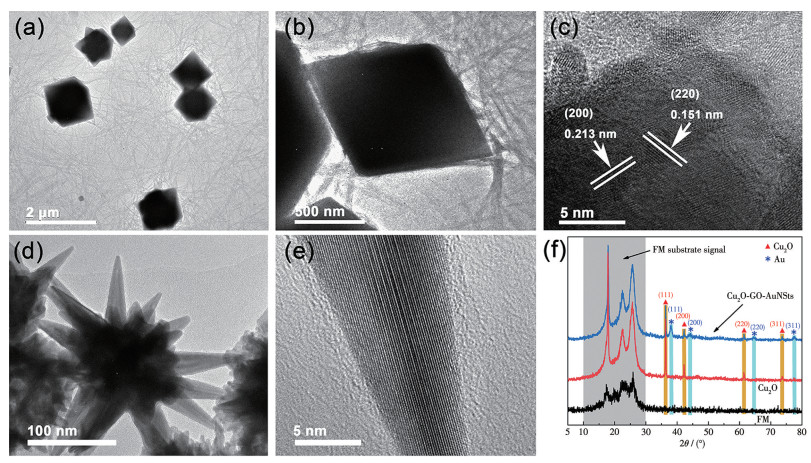
X-ray diffraction (XRD) analysis (Fig.4f) further verifies the successful construction of the Cu2O-GO-AuNSts sandwich architecture. The blank FM exhibited broad features between 10°-30°, characteristic of amorphous carbon fibers. Upon loading Cu2O, distinct diffraction peaks emerged at 2θ=36.3°, 42.4°, 61.5°, and 73.7°, assigned to the (111), (200), (220), and (311) planes of Cu2O, confirming the formation of the anisotropic octahedral phase. In addition, in the XRD pattern of Cu2O-GO-AuNSts/FM, apart from the diffraction peaks assigned to Cu2O, distinct diffraction peaks at 2θ=38.1°, 44.4°, 64.4°, and 77.5° were observed, which match the (111), (200), (220), and (311) planes of the fcc structure of Au NPs. The XRD patterns are fully consistent with the TEM/HRTEM observations, jointly demonstrating the successful sequential integration of Cu2O, GO, and AuNSts layers into the final sandwich structure.
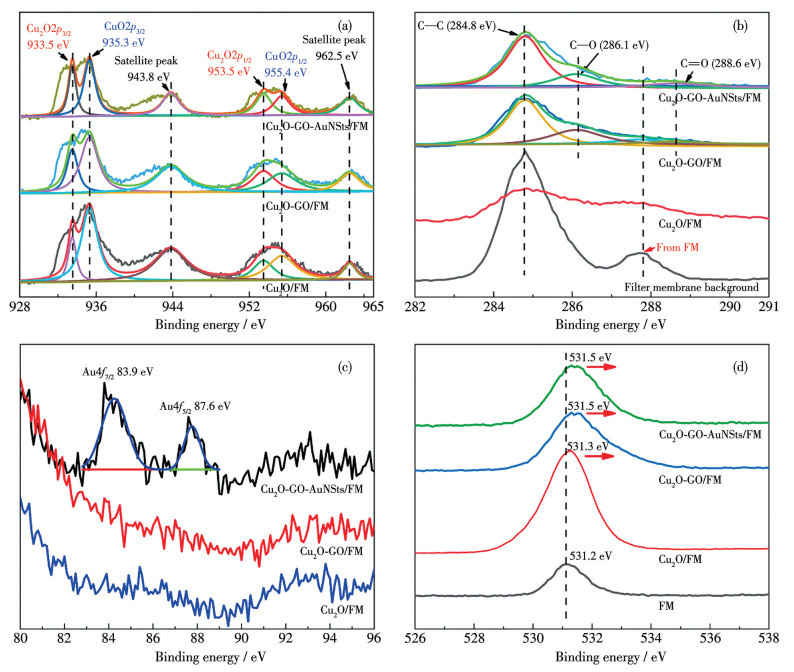
The interfacial characteristics and sequential assembly of Cu2O-GO-AuNSts/FM were further examined by high-resolution XPS (Fig.5). As shown in the Cu2p spectrum (Fig.5a), the prominent peaks at 933.5 eV (Cu2O, Cu2p3/2) and 953.5 eV (Cu2O, Cu2p1/2) confirm the presence of Cu+ species in Cu2O. Weak features at 935.3 eV (CuO, Cu2p3/2) and 955.4 eV (CuO, Cu2p1/2), accompanied by satellite peaks at 943.8 and 962.5 eV, indicate the coexistence of CuO in the hydrazine hydrate-reduced Cu2O, likely originating from partial oxidation during hydrazine hydrate reduction or surface re-oxidation upon exposure to air. Comparative analysis of the Cu2p spectra for Cu2O/FM, Cu2O-GO/FM, and Cu2O-GO-AuNSts/FM revealed negligible shifts in binding energies, suggesting Cu2O remained chemically intact during assembly and interacts with GO and AuNSts primarily through non-covalent forces rather than electronic coupling or alloying. The C1s spectra (Fig.5b) of the pristine FM are dominated by a C—C peak at 284.8 eV and a secondary component at 287.7 eV associated with carbonyl-containing species intrinsic to the membrane fibers. Upon GO integration, the C1s spectrum displayed additional peaks at 286.1 eV (C—O) and 288.6 eV (C=O), while retaining the substrate-related peak at 287.7 eV, confirming the successful incorporation of GO with its characteristic oxygenated functional groups. Notably, the Au4f spectra (Fig.5c) showed no detectable Au signals in the Cu2O/FM and Cu2O-GO/FM, whereas the Cu2O-GO-AuNSts/FM exhibited distinct doublet peaks at 83.9 eV (Au4f7/2) and 87.6 eV (Au4f5/2) with a spin-orbit splitting of 4.3 eV, characteristic of metallic Au0. This confirms the successful immobilization of AuNSts on GO-Cu2O/FM. As shown in O1s spectra (Fig.5d), FM showed a broad peak centered at 531.2 eV, attributed to oxygen in amorphous carbon structures. Following sequential assembly of Cu2O, GO, and AuNSts, the O1s peak remained singular but exhibited gradual, component-dependent shifts. Such subtle variations likely arise from changes in the local electronic environment associated with the layered architecture rather than the formation of new chemical states.
To identify the optimal excitation wavelength, Raman measurements of a 10-5 mol·L-1 R6G solution on the sandwich substrate were conducted at 532 and 633 nm. As shown in the obtained spectra (Fig.S1, Supporting information), significantly higher sensitivity for the characteristic Raman peaks was observed under 532 nm excitation. This result can be explained by the resonance effect: since 532 nm lies within the visible absorption band of R6G, the acquired signal is enhanced by surface-enhanced resonance Raman scattering (SERRS). Consequently, a 532 nm excitation source was employed in subsequent experiments.
The Cu2O-GO-AuNSts/FM sandwich substrate was fabricated through a pumping-filtering-spin coating sequence, and its SERS performance was systematically evaluated. As shown in Fig.6a, both FM and Cu2O/FM displayed negligible Raman signals. In contrast, the Cu2O-GO layer exhibited pronounced bands at 1 360 and 1 580 cm-1, corresponding to the D and G modes of GO, confirming the successful incorporation of GO nanosheets. Notably, Cu2O-GO-AuNSts/FM exhibited only the GO-derived D and G bands without any additional features, indicating that no extraneous impurities were introduced during the assembly process. R6G was subsequently employed as the probe molecule to investigate the SERS activity of Cu2O-GO-AuNSts/FM. As shown in Fig.6b, characteristic R6G vibrational fingerprints at 611, 773, 1 126, 1 183, 1 303, 1 360, 1 501, 1 573, and 1 647 cm-1 were observed, demonstrating that the engineered sandwich architecture generated strong electromagnetic enhancement. Remarkably, the SERS intensity decreased progressively with R6G concentration from 10-5 to 10-13 mol·L-1, yet the diagnostic peaks at 611 and 1 647 cm-1 remained detectable even at 10-13 mol·L-1. The experimental results close to single-molecule detection underscore the exceptional sensitivity of the Cu2O-GO-AuNSts/FM platform.
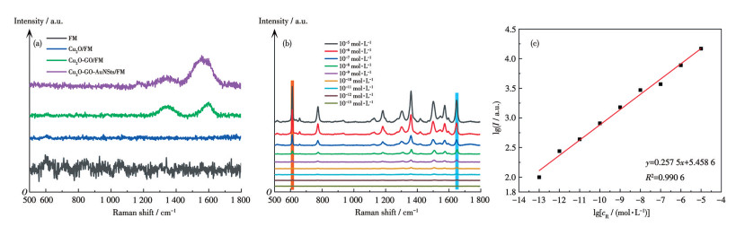
To quantify the sensing performance, the logarithmic SERS intensity (lg I) at 1 647 cm-1 was plotted against the logarithmic R6G concentration (lg cR, Fig. 6c). The resulting linear calibration curve exhibits excellent correlation (R2=0.99), revealing both the robustness and reliability of the sandwich substrate for quantitative detection. Collectively, these results demonstrate that Cu2O-GO-AuNSts/FM provides highly stable, ultrasensitive, and quantitatively reliable SERS responses, revealing significant potential for trace-level molecular detection in practical analytical applications.
To elucidate the function of each layer in the sandwich architecture, the SERS performance of Cu2O-AuNSts/FM, GO-AuNSts/FM, and Cu2O-GO/FM was evaluated using 10-5 mol·L-1 R6G as probe molecules (Fig.S2). GO-AuNSts/FM showed the highest signal intensity, surpassing that of Cu2O-AuNSts/FM and Cu2O-GO/FM. This comparison indicates that the LSPR associated with AuNSts was the primary origin of SERS enhancement. The presence of GO mainly improves the effective analyte availability at plasmonic hot spots (e.g., through adsorption/π-π interactions), thereby further amplifying the signal. In contrast, the chemical-mechanism contributions from Cu2O and GO appear minimal under the present conditions. Notably, Cu2O plays a distinct role in this system, serving predominantly as the photocatalytic component for substrate self-cleaning.
BDE-47, a representative PBDE congener, is widely recognized for its environmental persistence, bioaccumulative toxicity, and resistance to natural degradation. Monitoring and eliminating such POPs remains a major challenge in environmental analysis and remediation. To demonstrate the dual SERS-sensing and photocatalytic functions of Cu2O-GO-AuNSts/FM, BDE-47 was selected as a model pollutant. As shown in Fig.7a, the Raman spectra of BDE-47 from 10-4 to 10-7 mol·L-1 exhibited distinct vibrational fingerprints at 670, 800, 1 092, 1 143, 1 206, and 1 569 cm-1. These peaks remained detectable even at 10-7 mol·L-1, confirming the excellent trace-level SERS sensitivity of the sandwich substrate.
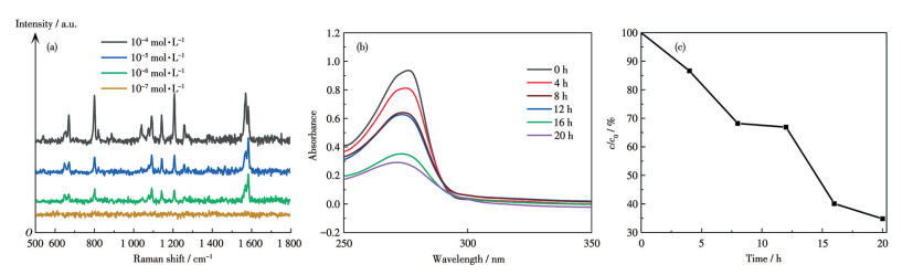
Fig. 7b illustrates the time-dependent UV-Vis absorption spectra of BDE-47 (10-5 mol·L-1) during photocatalytic degradation on the Cu2O-GO-AuNSts substrate under UV irradiation. The characteristic absorption band at 260-290 nm exhibited a gradual decrease and a slight blue shift under UV irradiation, indicating the continuous transformation of BDE-47 and partial cleavage of the aromatic ether structure. Quantitative analysis (Fig. 7c) showed that approximately 66% of BDE-47 was degraded after 20 h. Although the degradation efficiency is moderate and consistent with the well-known chemical stability of PBDE congeners, the result clearly demonstrates that Cu2O-GO-AuNSts/FM provides a measurable photocatalytic response toward a highly recalcitrant POP. More importantly, this partial degradation is sufficient to endow the substrate with a self-cleaning tendency, allowing gradual removal of adsorbed analytes following trace-level SERS detection. Such dual SERS-photocatalytic functionality highlights the potential of this platform as a versatile detection-remediation interface, which could be further optimized for practical environmental applications.
In this paper, anisotropic Cu2O particles were prepared under the optimized conditions of 20 ℃ and a NaOH dosage of 7.5 mL and subsequently integrated into Cu2O-GO-AuNSts/FM via a layer-by-layer spin-coating process. The resulting substrate exhibited exceptional SERS activity, enabling trace detection of R6G with a detection limit as low as 10-13 mol·L-1. Furthermore, the engineered sandwich structure allowed simultaneous SERS detection and photocatalytic degradation of BDE-47. The excellent trace SERS detection ability of the sandwich structure originates from the LSPR effect of Au NPs, the good molecular enrichment ability of GO, and the porous filter-membrane support. The photocatalytic degradation capability is attributed to the synergistic contributions of the high intrinsic photocatalytic activity of Cu2O, the suppressed electron-hole recombination resulting from Au-induced charge-transfer effects, and the enhanced pollutant adsorption enabled by GO. Collectively, these results demonstrate that the Cu2O-GO-AuNSts sandwich substrate offers a promising platform for in situ SERS analysis coupled with self-cleaning photocatalytic degradation of persistent organic pollutants.
BILL A, HAARMAN A, GASSER M, BöNI H, RöSSLEIN M, WäGER A P. Characterizing plastics from large household appliances: Brominated flame retardants, other additives, and density profiles[J]. Resour. Conserv. Recycl., 2022, 177: 105956 doi: 10.1016/j.resconrec.2021.105956
ESTILL C F, MAYER A C, CHEN I C, SLONE J, LAGUARDIA J M, JAYATILAKA N, OSPINA M, SjODIN A, CALAFAT M A. Biomarkers of organophosphate and polybrominated diphenyl ether (PBDE) flame retardants of American workers and associations with inhalation and dermal exposures[J]. Environ. Sci. Technol., 2024, 58(19): 8417-8431 doi: 10.1021/acs.est.3c09342
JIN M T, ZHANG S F, YE N X, ZHOU S S, XU Z Y. Distribution and source of and health risks associated with polybrominated diphenyl ethers in dust generated by public transportation[J]. Environ. Pollut., 2022, 309: 119700 doi: 10.1016/j.envpol.2022.119700
LA GUARDIA J M, MAINOR M T, LUELLEN R D, HARVEY E, HALE C R. Twenty years later: PBDEs in fish from U. S. sites with historically extreme contamination[J]. Chemosphere, 2024, 351: 141126 doi: 10.1016/j.chemosphere.2024.141126
LAKSHMINARASIMMAN N, GEWURTZ B S, PARKER J W, SMYTH A S. Quantifying the removal of polybrominated diphenyl ethers (PBDEs) in physical, chemical, and biological sludge treatment systems[J]. Chemosphere, 2024, 351: 141203 doi: 10.1016/j.chemosphere.2024.141203
BLOCH S, LEVEQUE L, HERTZ-PICCIOTTO I, PUSCHNER B, FRITSCHE E, KLOSE J, KRAMER N I, BOUCHARD M F, CHANDRASEKERA P C, VERNER M A. Using in vitro data to derive acceptable exposure levels: A case study on PBDE developmental neurotoxicity[J]. Environ. Int., 2024, 183: 108411 doi: 10.1016/j.envint.2023.108411
ABBASI G, LI L, BREIVIK K. Global historical stocks and emissions of PBDEs[J]. Environ. Sci. Technol., 2019, 53(11): 6330-6340 doi: 10.1021/acs.est.8b07032
XU W G, WANG X, CAI Z W. Analytical chemistry of the persistent organic pollutants identified in the Stockholm convention: A review[J]. Anal. Chim. Acta, 2013, 790: 1-13
GONZALEZ-GAGO A, MANUEL MARCHANTE-GAYON J, FERRERO M, ALONSO J I G. Synthesis of 81Br-labeled polybrominated diphenyl ethers and their characterization using GC(EI)MS and GC(ICP)MS[J]. Anal. Chem., 2010, 82(7): 2879-2887 doi: 10.1021/ac902889u
WEI L Y, ZHOU B, XIAO K, YANG B, YU G, LI J Y, ZHU C Z, ZHANG J M, DUAN H B. Highly efficient degradation of 2, 2′, 4, 4′-tetrabromodiphenyl ether through combining surfactant-assisted Zn0 reduction with subsequent Fenton oxidation[J]. J. Hazard. Mater., 2020, 385: 121551 doi: 10.1016/j.jhazmat.2019.121551
WANG R, TANG T, XIE J B, TAO X Q, HUANG K B, ZOU M Y, YIN H, DANG Z, LU G N. Debromination of polybrominated diphenyl ethers (PBDEs) and their conversion to polybrominated dibenzofurans (PBDFs) by UV light: Mechanisms and pathways[J]. J. Hazard. Mater., 2018, 354: 1-7
LIU J H, CHEN J H, JIA S J, WANG Y, WU D, WU Y N, LI G L. CRISPR/Cas12a triggered SERS and naked eye dual-mode biosensor for ultrasensitive and on-site detection of nucleic acid via cascade signal amplification[J]. Sens. Actuator. B‒Chem., 2024, 404: 135249 doi: 10.1016/j.snb.2023.135249
KIM M, HUH S, PARK H J, CHO H C, LEE M Y, JO S H, JUNG Y S. Surface-functionalized SERS platform for deep learning-assisted diagnosis of Alzheimer's disease[J]. Biosens. Bioelectron., 2024, 251: 116128 doi: 10.1016/j.bios.2024.116128
HUANG C, WANG Y H, WANG Y Q, WANG A, ZHOU Y D, JIN S Z, ZHANG F L. Quantitative analysis of trace analytes with highly sensitive SERS tags on hydrophobic interface[J]. ACS Appl. Nano Mater., 2024, 16(14): 18124-18133 doi: 10.1021/acsami.3c18980
HOU L W, XU X Y, WANG X L, WANG L, TIAN F C, XU Y. SERS sensing chip based on Ti3C2/nano-Au@MA for ultrasensitive amine gas detection[J]. J. Mater. Chem. A, 2024, 12(16): 9817-9829 doi: 10.1039/D4TA00429A
BAI H Y, WEN G Q, LIANG A H, JIANG Z L. Ti3C2@Pd nanocatalytic amplification-polypeptide SERS/RRS/Abs trimode biosensoring platformfor ultratrace trinitrotoluene[J]. Biosens. Bioelectron., 2022, 217: 114743 doi: 10.1016/j.bios.2022.114743
LIU W, WANG Z H, LIU Z P, CHEN J X, SHI Y J, HUANG L J, LIU Y, CUI S, HE Y. Utilizing an automated SERS-digital microfluidic system for high-throughput detection of explosives[J]. ACS Sens. 2023, 8(4): 1733-1741 doi: 10.1021/acssensors.3c00012
FU X Q, LI Z, ZHAO J R, YANG J, ZHU G X, LI G F, HUO P W. Coupling plasmon and catalytic-active hotspots of Au@Pt core-satellite nanoparticles for in-situ spectroscopic observation of plasmon-promoted decarboxylation[J]. J. Colloid Interface Sci., 2024, 676: 127-138 doi: 10.1016/j.jcis.2024.07.091
DING S Y, YOU E M, TIAN Z Q, MOSKOVITS M. Electromagnetic theories of surface-enhanced Raman spectroscopy[J]. Chem. Soc. Rev., 2017, 46(13): 4042-4076 doi: 10.1039/C7CS00238F
YANG X, SU D, YU X, ZENG P, LIANG H G, ZHANG G Z, SONG B X, JIANG S L. Hot spot engineering in hierarchical plasmonic nanostructures[J]. Small, 2023, 19(22): 2205659 doi: 10.1002/smll.202205659
JIA Q, OU X, LANGER M, SCHREIBER B, GRENZER J, SILES F P, RODRIGUEZ D R, HUANG K, YUAN Y, HEIDARIAN A, HÜBNER R, YOU T G, YU W J, LENZ K, LINDNER J, WANG X, FACSKO S. Ultra-dense planar metallic nanowire arrays with extremely large anisotropic optical and magnetic properties[J]. Nano Res., 2018, 11(7): 3519-3528 doi: 10.1007/s12274-017-1793-y
AMIN M U, ZHANG R Y, LI L W, YOU H J, FANG J X. Solution-based SERS detection of weak surficial affinity molecules using cysteamine-modified Au bipyramids[J]. Anal. Chem., 2021, 93(21): 7657-7664 doi: 10.1021/acs.analchem.1c00439
ZHAO X, WANG W Z, LIANG Y J, FU J L, ZHU M, SHI H L, LEI S J, TAO C J. Visible-light-driven charge transfer to significantly improve surface-enhanced Raman scattering (SERS) activity of self-cleaning TiO2/Au nanowire arrays as highly sensitive and recyclable SERS sensor[J]. Sens. Actuator. B‒Chem., 2019, 279: 313-319 doi: 10.1016/j.snb.2018.10.010
CHEN Z Y, SUN Y, ZHANG X N, SHEN Y, KHALIFA A M S, HUANG X W, SHI J Y, LI Z H, ZOU X B. Green and sustainable self-cleaning flexible SERS base: Utilized for cyclic-detection of residues on apple surface[J]. Food Chem., 2024, 441: 138345 doi: 10.1016/j.foodchem.2023.138345
QUAN Y N, YAO J C, YANG S, CHEN L, LIU Y, LANG J H, ZENG H Q, YANG J H, GAO M. Detect, remove and re-use: Sensing and degradation pesticides via 3D tilted ZMRs/Ag arrays[J]. J. Hazard. Mater., 2020, 391: 122222 doi: 10.1016/j.jhazmat.2020.122222
YANG Q, WANG J J, WU H R, QIN S X, PAN J Q, LI C R. Hierarchically rough CuO/Ag composite film with controlled morphology as recyclable SERS-active substrate[J]. Appl. Surf. Sci., 2022, 598: 153746 doi: 10.1016/j.apsusc.2022.153746
LI J, LIU L, CHE W Q, HUANG X Y, LIU X L, LOU Z Z, LI B J. A dual-functional plasmonic W18O49/rGO heterostructure for ultrasensitive SERS detection and in situ tracking of photocatalytic reactions[J]. Nanoscale, 2025, 17(44): 25884-25891 doi: 10.1039/D5NR03353E
LU Y, BI Z F, SHANG G Y. A nanocomposite of silver nanoparticles and porous g-C3N4 for recyclable SERS detection of trace fluorene[J]. ACS Appl. Nano Mater., 2024, 7(1): 466-475 doi: 10.1021/acsanm.3c04673
QU L L, GENG Z Q, WANG W, YANG K C, WANG W P, HAN C Q, YANG G H, VAJTAI R, LI D W, AJAYAN M P. Recyclable three-dimensional Ag nanorod arrays decorated with O-g-C3N4 for highly sensitive SERS sensing of organic pollutants[J]. J. Hazard. Mater., 2019, 379: 120823 doi: 10.1016/j.jhazmat.2019.120823
ZHAO X F, LIU C D, YU J, LI Z, LIU L, LI C H, XU S C, LI W F, MAN B Y, ZHANG C. Hydrophobic multiscale cavities for high-performance and self-cleaning surface-enhanced Raman spectroscopy (SERS) sensing[J]. Nanophotonics, 2020, 9(16): 4761-4773
MA J L, XU L X, ZHANG Y L, DONG L Y, GU C J, WEI G D, JIANG T. Multifunctional SERS chip mediated by black phosphorus@gold-silver nanocomposites inserted in bilayer membrane for in-situ detection and degradation of hazardous materials[J]. J. Colloid Interface Sci., 2022, 626: 787-802
SHAFI M, DUAN P Y, LIU W Y, ZHANG W J, ZHANG C, HU X X, LIU C, WALI S, JIANG S Z, ZHANG C, MAN B Y, LIUVV M. Recyclable surface-enhanced Raman spectroscopy (SERS) platform fabricated with Ag-decorated ZnSe nanowires and metamaterial[J]. Sens. Actuator. B‒Chem., 2023, 380: 133410
LI L, ZHANG L C, GOU L C, WEI S Q, HOU X D, WU L. Au nanoparticles decorated CoP nanowire array: A highly sensitive, anticorrosive, and recyclable surface-enhanced Raman scattering substrate[J]. Anal. Chem., 2023, 95(29): 11037-11046 doi: 10.1021/acs.analchem.3c01282
BHARADWAj S, PANDEY A, YAGCI B, OZGUZ V, QURESHI A. Graphene nano-mesh-Ag-ZnO hybrid paper for sensitive SERS sensing and self-cleaning of organic pollutants[J]. Chem. Eng. J., 2018, 336: 445-455
ZHOU S, WANG S, LI H, YAN X Z, SHENG J S, YANG H, BITTENCOURT C, SNYDERS R, LI W J. Interface coupling of octahedron Cu2O with reduced graphene oxide for enhanced photocatalytic and photoelectrochemical activity[J]. Opt. Mater., 2024, 156: 115981
ZHANG Y Y, ZHANG Z H, ZHANG Y, LI Y, YUAN Y. Shape-dependent synthesis and photocatalytic degradation by Cu2O nanocrystals: Kinetics and photocatalytic mechanism[J]. J. Colloid Interface Sci., 2023, 651: 117-127
WU H Y, LU Q Y, KIANPOOR Z, KANIKE C, XIA L Y, LIANG K, ZHANG X H. Surface-bound metal-organic framework microdomes and hybrids via a reacting microdroplet-driven approach: Enabling recyclable photocatalysis and real-time in-situ monitoring[J]. Chem. Eng. J., 2025, 519: 164839
WANG K F, LV M R, SI T, TANG X N, WANG H, CHEN Y Y, ZHOU T. Mechanism analysis of surface structure-regulated Cu2O in photocatalytic antibacterial process[J]. J. Hazard. Mater., 2024, 461: 132479
LIU J X, PAN M D, JIN J Z, LU Y S, FENG Z M, SUN T. Prepared three-dimensional flowerlike MoS2/Ag@rGO nanocomposite as a self-cleaning SERS substrate for the trace detection of 17β-estradiol in environmental water[J]. ACS Sustain. Chem. Eng., 2024, 12(2): 893-903

Figure 1 SEM images of Cu2O prepared at different reaction temperatures with 7.5 mL NaOH: (a, d) 10 ℃; (b, e) 20 ℃; (c, f) 30 ℃

Figure 2 SEM images of Cu2O prepared with different dosages of NaOH at 20 ℃: 5.0 mL (a); 7.5 mL (b); 10.0 mL (c); 15.0 mL (d)

Figure 3 (a) Schematic diagram of Cu2O-GO-AuNSts sandwich structure; (b) Assembly process of sandwich substrate; SEM image of (c) FM, (d) Cu2O/FM, (e) Cu2O-GO/FM, and (f) Cu2O-GO-AuNSts/FM

Figure 4 (a, b) TEM and (c) HRTEM images of Cu2O particles; (d) TEM and (e) HRTEM images of AuNSts; (f) XRD patterns of the samples

Figure 5 XPS high resolution spectra of (a) Cu2p, (b) C1s, (c) Au4f, and (d) O1s of Cu2O-GO-AuNSts/FM

Figure 6 (a) Raman signal spectra of the blanks of each layer of the Cu2O-GO-AuNSts/FM; (b) SERS detection limit spectra of the sandwich substrate for R6G molecules, and (c) corresponding lg I versus lg cR plot of the Cu2O-GO-AuNSts/FM substrate sandwich

 扫一扫看文章

扫一扫关注我们
 下载:
下载:
 下载:
下载:

 下载:
下载: