Table 1.
Crystal data and structure refinement parameters for 1
Citation:
Xuetian WANG, Jijiang WANG, Long TANG, Erlin YUE, Xiao WANG, Yuqi ZHANG. A three-dimensional zinc(Ⅱ) metal-organic framework based on nitrogen-containing ligands for the detection of 2,4-dinitrophenylhydrazine and tetracycline[J]. Chinese Journal of Inorganic Chemistry,
2026, 42(4): 861-871.
doi:
10.11862/CJIC.20250294

基于含氮配体构建的三维锌(Ⅱ)金属有机骨架用于2,4-二硝基苯肼和四环素检测
摘要:
采用溶剂热法合成1例金属有机骨架{[Zn(L)0.5(1,2,4,5-tpb)0.5]·DMF·3H2O}n (1),其中H4L=5,5′-(1,2-亚乙基)二(间苯二甲酸),1,2,4,5-tpb=1,2,4,5-四(吡啶-4-基)苯。单晶结构分析表明,L4-和1,2,4,5-tpb连接Zn(Ⅱ)形成二维层状结构,层与层之间通过1,2,4,5-tpb连接形成三维结构。1可作为高选择性荧光探针检测2,4-二硝基苯肼(DNP)和四环素(TET),检出限分别为0.013和0.31 μmol·L-1。1被成功用于延河水样中TET含量的测定。
English
A three-dimensional zinc(Ⅱ) metal-organic framework based on nitrogen-containing ligands for the detection of 2,4-dinitrophenylhydrazine and tetracycline
Abstract:
A metal-organic framework {[Zn(L)0.5(1,2,4,5-tpb)0.5]·DMF·3H2O}n (1) was synthesized by solvothermal reaction, where H4L=5,5′-(ethane-1,2-diyl)diisophthalic acid, and 1,2,4,5-tpb=1,2,4,5-tetra(pyridin-4-yl)benzene. The analysis of the single crystal structure indicates that L4- and 1,2,4,5-tpb are connected with Zn(Ⅱ) to form a 2D layered structure, and the layers are linked by 1,2,4,5-tpb to form a 3D structure. 1 can be used as a highly selective fluorescent probe for the detection of 2,4-dinitrophenylhydrazine (DNP) and tetracycline (TET), and the detection limits were 0.013 and 0.31 μmol·L-1, respectively. 1 was applied successfully to the determination of TET content in the Yanhe River water sample.
-
Key words:
- metal-organic frameworks
- / Zn(Ⅱ)
- / crystal structure
- / fluorescent sensing
-
0. Introduction
With the advancement of technology and industrial development, environmental issues are becoming increasingly severe. The misuse of antibiotics, pesticide residues, dye-containing wastewater, and other industrial discharges significantly contributes to environmental pollution and ecosystem degradation. 2,4-Dinitrophenylhydrazine (DNP), a phenylhydrazine derivative, is widely used in various applications. Its extensive utilization has led to inevitable environmental release, thereby entering the human body through the food chain and posing health risks[1]. Antibiotics, as a class of antimicrobial agents, play a critical role in clinical medicine, veterinary practice, and agricultural production[2-7], exerting substantial beneficial impacts on public health. However, in recent years, widespread antibiotic misuse has led to serious consequences, including the development of antimicrobial resistance and negative effects on ecosystem stability[8]. Tetracycline (TET), for instance, is globally consumed due to its cost-effectiveness and broad-spectrum antibacterial activity. Nevertheless, its extensive use has contributed to the emergence of drug-resistant bacteria and caused significant environmental and health problems[9]. Given the simultaneous presence of diverse pollutants like DNP and TET in the environment, it is imperative to develop a simple, rapid, efficient, and highly sensitive detection platform for both DNP and TET to mitigate their risks and support environmental and public health protection. Conventional detection methods primarily include chromatographic techniques, such as gas chromatography (GC)[10] and high-performance liquid chromatography (HPLC)[11], as well as electrochemical approaches like cyclic voltammetry[12]. Supplementary techniques include titration[13] and atomic absorption spectroscopy (AAS)[14]. Although these methods are highly reliable, they exhibit inherent limitations: (1) dependence on costly instrumentation, (2) time-consuming sample pretreatment protocols that hinder field deployment, (3) operational complexity coupled with constrained detection ranges and suboptimal sensitivity. To address these drawbacks, this study proposes an alternative detection strategy based on fluorescence spectroscopy. This approach has garnered significant attention in recent years due to its operational simplicity, cost-effectiveness, high sensitivity, rapid response, and straightforward signal quantification facilitated by fluorescence enhancement or quenching effects[15-16].
Metal-organic frameworks (MOFs) represent a class of crystalline, porous solids with tunable molecular architectures, which have generated substantial research interest due to their designer functionality and structural predictability[17-18]. MOFs are hybrid crystalline materials constructed from inorganic metal nodes interconnected by organic linkers, forming tunable 1D, 2D, or 3D architectures. This unique combination of inorganic rigidity and organic flexibility endows MOFs with exceptional characteristics, including permanent porosity, ultrahigh surface areas, and abundant metal active sites. These properties have enabled diverse applications, such as spanning chemical sensing[19-20], gas storage and separation21-23], and energy storage technologies[24]. Particularly, luminescent MOFs have emerged as powerful platforms for detecting metal ions, organic molecules, pesticides, nitroaromatic compounds (NACs), and antibiotics[25-27]. Among these, d10 transition metal-based MOFs often demonstrate superior luminescent properties, which can originate from diverse mechanisms, such as ligand-centered emission, metal-centered emission, metal-to-ligand charge transfer (MLCT), ligand-to-metal charge transfer (LMCT), and ligand-to-ligand charge transfer (LLCT), among others[28-30].
In this work, a new type of MOF {[Zn(L)0.5(1,2,4,5-tpb)0.5]·DMF·3H2O}n (1) was designed and synthesized through solvothermal reaction, which is composed of 5,5′-(ethane-1,2-diyl) diisophthalic acid (H4L), Zn(Ⅱ), and 1,2,4,5-tetra(pyridin-4-yl)benzene (1,2,4,5-tpb). We describe the synthesis, crystal structure, and fluorescence sensing characteristics of 1. It exhibits extremely high selectivity towards DNP and TET. Additionally, the fluorescence quenching mechanism of 1 was also investigated. Finally, 1 was successfully employed for the determination of the concentrations of TET in water samples collected from the Yanhe River.
1. Experimental
1.1 Reagents and instruments
All reagents and solvents were commercially available and used directly without further purification. The crystal data were collected on a Bruker SMART APEX-Ⅱ single crystal X-ray diffractometer. The C, H, and N elemental analyses were conducted with a PerkinElmer PE-2400 elemental analyzer. Powder X-ray diffraction (PXRD) patterns were recorded with a Bruker D8 ADVANCE diffractometer operating at 40 kV and 40 mA using Cu Kα radiation (λ=0.154 18 nm) at a scanning rate of 2 (°)·min-1 from 5° to 50°. The FTIR spectrum (400-4 000 cm-1) was recorded on a Bruker EQUINOX55 spectrophotometer. Thermal gravimetric analysis (TGA) was performed with a NETZSCH STA 449F3 thermal gravimetric analyzer in flowing nitrogen at a heating rate of 10 ℃·min-1. The UV-Vis spectra were measured using a UV-2700 spectrophotometer. The fluorescence experiment was conducted on the Hitachi F-7100 Fluorescence Spectrophotometer.
1.2 Synthesis of MOF 1
A mixture of Zn(NO3)2·6H2O (0.1 mmol, 0.029 7 g), H4L (0.05 mmol, 0.017 9 g), and 1,2,4,5-tpb (0.05 mmol, 0.019 3 g) in DMF (3 mL), H2O (3 mL), and HNO3 (0.15 mL, 6 mol·L-1) were added into a 10 mL glass bottle and heated at 95 ℃ for three days. Then the bottle was naturally cooled down to 25 ℃, and a silver-colored, sheet-like, transparent solid was obtained. Yield: 51% (based on Zn). Anal. Calcd. for C25H27N3O8Zn(%): C, 53.30; H, 4.80; N, 7.46. Found(%): C, 53.19; H, 4.97; N, 7.86. IR (KBr pellet, cm-1): 3 499 w, 2 925m, 1 617 s, 1 573 s, 1 497 s, 1 359 s, 1 257 s, 1 095 s, 910 s, 733 s, 570 s, 441 s, 424 s.
1.3 Crystal structure determination
A Zn(Ⅱ) MOF was obtained by solvothermal reaction. Crystals of 1 with regular shapes, transparent colors, and a size of 0.23 mm×0.16 mm×0.12 mm were selected for testing. The single crystal data of 1 were collected on the Bruker SMART APEX-Ⅱ diffractometer (Mo Kα radiation, λ=0.071 073 nm). Empirical absorption correction was performed by the SADABS program. The crystal structure was analyzed by the Olex2 1.3 program and refined by the full-matrix least-squares methods based on F2. All non-hydrogen atoms were refined anisotropically, and all of the hydrogens were geometrically placed in calculated positions. Crystallographic data for 1 are shown in Table 1. The selected bond lengths and bond angles are listed in Table 2.
Table 1

 下载:
导出CSV
下载:
导出CSV
Parameter 1 Parameter 1 Formula C25H27N3O8Zn V/nm3 5.052(5) Formula weight 562.86 Z 8 Crystal system Orthorhombic D/(g·cm-3) 1.48 Space group Pbcn F(000) 2 336 a/nm 1.879 4(11) Goodness⁃of⁃fit on F 2 1.057 b/nm 1.326 6(8) R1, wR2 [I>2σ(I)] 0.048 9, 0.107 6 c/nm 2.026 5(13) R1, wR2 (all data) 0.079 3, 0.121 3 Table 2
Table 2. Selected bond lengths (nm) and angles (°) for 1下载:
导出CSV
Zn1—O1 0.198 4(3) Zn1—O4A 0.198 7(3) Zn1—N1 0.204 0(3) Zn1—N2B 0.203 5(3) O1—Zn1—O4 99.34(11) O1—Zn1—N1 107.30(11) O1—Zn1—N2B 113.67(11) O4A—Zn1—N1 108.78(11) O4A—Zn1—N2B 106.50(11) N2B—Zn1—N1 119.30(12) Symmetry codes: A: x, -y, 1/2+z; B: x, -1+y, z. 2. Results and discussion
2.1 Crystal structure
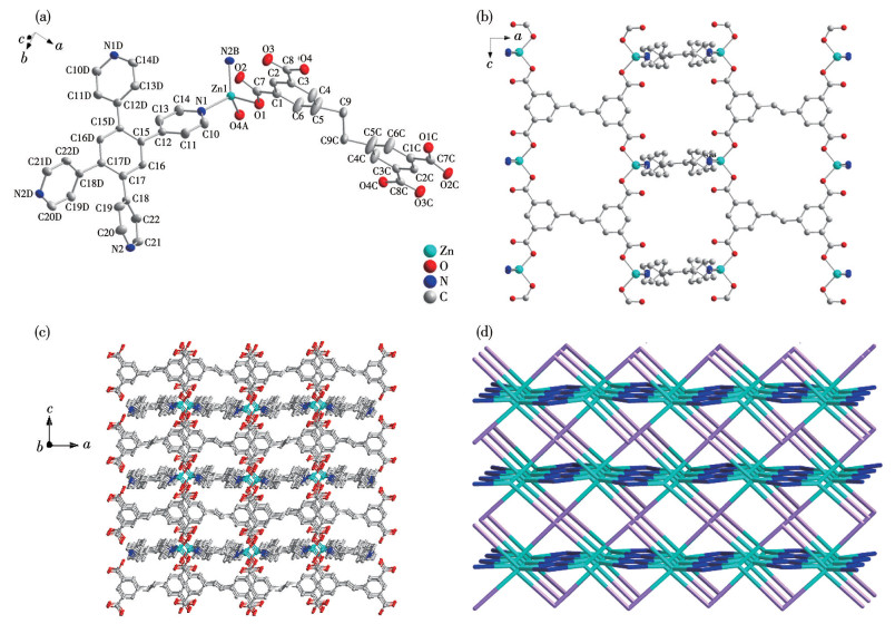
The asymmetric unit of 1 consists of one independent Zn(Ⅱ) ion, half a L4- ligand, half a 1,2,4,5-tpb ligand, one free DMF molecule, and three free water molecules. 1 belongs to the orthorhombic crystal system and the Pbcn space group. As shown in Fig.1a, the Zn1 ion adopts a tetra-coordinated mode. Zn1 is surrounded by two nitrogen atoms (N1, N2B) from the 1,2,4,5-tpb ligands and two oxygen atoms (O1, O4A) from the L4- ligands. The bond lengths of Zn1—O1 [0.198 4(3) nm], Zn1—O4A [0.198 7(3) nm], Zn1—N1 [0.204 0(3) nm], and Zn1—N2B [0.203 5(3) nm] all fall within the normal range (Table 2). The carboxyl group in 1 exhibits monodentate coordination, connecting Zn(Ⅱ) ions. As shown in Fig.1b, Zn(Ⅱ) ions are connected with the L4- and 1,2,4,5-tpb ligands in a horizontal plane, forming a 2D layered structure. As shown in Fig.1c, the layers are further connected through 1,2,4,5-tpb to form a 3D structure. From a topological perspective, the ligands and metal centers were simplified as nodes according to their connectivity, affording the topological network depicted in Fig.1d. Topological analysis reveals that this network exhibits a pts topology. Systematic calculation and analysis using ToposPro software ultimately classifies 1 as a binodal 4.4-c net. This framework is constructed from the interconnection of four-connected Zn(Ⅱ) metal nodes and four-connected organic ligands (L4- and 1,2,4,5-tpb). The point symbol for both types of nodes is (42.84).
Figure 1
 Figure 1. (a) Coordination environment of Zn(Ⅱ) in 1 with 50% ellipsoid probability; (b) 2D network structure of 1; (c) 3D framework of 1; (d) Underlying pts topological net of 1 (Zn: cyan, L4- ligand: pink, 1,2,4,5-tpb ligand: blue)
For clarity, all hydrogen atoms are omitted; Symmetry codes: A: x, -y, 1/2+z; b: x, -1+y, z; c: 2-x, -y, 1-z; d: 1-x, y, 3/2-z.
2.2 PXRD analysis
To study the purity of 1, PXRD patterns ranging from 5° to 50° were obtained at room temperature. The experimental results are shown in Fig.S1 (Supporting information). The experimental peak position of 1 was basically consistent with the simulated peak position, indicating that 1 is a pure phase and the crystal purity is relatively high. The skeleton of 1 remained largely unchanged after immersion in both acidic and basic aqueous solutions for 24 h, demonstrating its high resistance to acids and bases (Fig.S2).
2.3 Thermal stability
In order to investigate the thermal stability of 1, TGA was performed. As shown in Fig.S3, from room temperature to 100 ℃, the evaporation of unbound water was observed. From 110 to 270 ℃, the weight loss rate was approximately 12.73% (Calcd. 12.97%), corresponding to one DMF molecule. Further heating to around 480 ℃ led to the decomposition of the framework. The results of thermal stability analysis are consistent with those of structural analysis.
2.4 Infrared spectroscopic analysis
To further confirm the synthesis of the MOF material, 1 was subjected to infrared spectroscopy testing (Fig.S4). A strong and broad absorption peak was observed at 3 499 cm-1, attributed to the O—H stretching vibration of the carboxyl group, indicating the presence of non-deprotonated carboxyl groups in 1 and possibly coordinated water molecules. The absorption peak at 1 673 cm-1 corresponds to the asymmetric stretching vibration of the carboxylate group (νas), while the peak at 1 427 cm-1 is assigned to the symmetric stretching vibration (νs)[31]. Notably, the difference Δν (νas-νs) exceeded 200 cm-1, suggesting that the carboxyl groups in the ligand molecules adopt a monodentate coordination mode with the metal ions[32]. Additionally, the stretching vibration peak of the C—C bond appeared at approximately 1 359 cm-1, which is consistent with the crystal structure analysis of 1.
2.5 Photoluminescence property
Due to the excellent luminescence properties of d10 metal MOFs, the luminescence of 1 and H4L ligand was studied. The corresponding emission spectra are presented in Fig.S5. 1 had an emission peak at 467 nm when excited at 355 nm, and H4L had an emission peak at 400 nm when excited at 310 nm. Compared with the H4L ligand, the emission spectrum of 1 had a partial redshift, which may be caused by the electronic transition of the ligand, which enhances the structural stiffness, or due to the mutual coordination[33].
To further investigate the fluorescence of 1 in water, an accurately weighed 6 mg portion of the MOF powder was dispersed in 20 mL of distilled water. The resulting suspension was then subjected to ultrasonic treatment for 2 h to ensure a homogeneous dispersion. After sonication, the mixture was allowed to stand at room temperature for 24 h. The supernatant was collected (as shown in Fig.S5), and its emission maximum was observed at 415 nm. Compared with the solid-state luminescence, the emission of 1 experienced a blueshift, which might be due to the influence of solvent molecules on the luminescence.
2.6 NACs sensing
NACs are highly explosive and chemically toxic. They are often used in the synthesis of pesticides and the production of explosives. Due to their explosive properties, they pose a significant threat to the environment and human health[34-35]. In this work, a variety of NACs were selected for fluorescence detection, including 3-nitroaniline (3-NT), 2,4-dinitrophenylhydrazine (DNP), 4-nitrophenol (4-NP), 2-nitrophenol (2-NP), p-nitrobenzoic acid (PNBA), 4-nitrophenylhydrazine (4-NPH), o-nitrobenzaldehyde (ONBA), o-nitroaniline (ONT), trinitrophenol (TNP), and nitrobenzene (NB).
As shown in Fig.2a, it can be observed that in the presence of DNP, the fluorescence intensity reached the maximum quenching. The fluorescence intensities of other NACs changed relatively little when there was no DNP. However, when DNP was further added to other NACs, as shown in Fig.2b, the fluorescence intensities were significantly quenched, indicating that DNP detection by 1 has anti-interference properties. In order to conduct a detailed analysis and study of its extinction situation, a quantitative experiment was carried out. As shown in Fig.2c, as the concentration of DNP gradually increased (0-15 μmol·L-1), the fluorescence intensity of 1 showed a gradually decreasing trend. We plotted the relationship between I0/I-1 and cDNP based on the Stern-Volmer (SV) equation: I0/I=1+KSVcDNP, where I0 and I represent the fluorescence intensities of 1 before and after DNP addition, respectively, KSV is the quenching constant, and cDNP is the DNP concentration. As shown in Fig.2d, it was found that within the low concentration range, the two variables exhibited a good linear relationship. The detection limit was calculated with 3σ/k (σ: standard deviation, k: slope), and the detection limit of 1 to DNP was 0.013 μmol·L-1. Furthermore, we observed that the fluorescence intensity could be fully restored to its initial level, suggesting that 1 may function as a recyclable fluorescent sensor (Fig.2e). It was also noted that upon addition of 15 μmol·L-1 DNP, the luminescence intensity decreased rapidly and reached a stable plateau within 20 s (Fig.2f).
Figure 2
 Figure 2. (a) Luminescent intensity of 1 (10 mmol·L-1) in different NAC solution; (b) Anti-interference of DNP detection by 1 in the presence of other NACs; (c) Emission spectra of 1 with different concentrations of DNP; (d) SV plot of DNP in low concentration ranges; (e) Cycle stability of 1 for the detection of DNP (red square: 1, blue square: 1+DNP); (f) Response time on DNP detection by 1
2.7 Antibiotic sensing
In recent years, due to the excessive use of antibiotics, significant pollution has been caused in water systems and the environment. Therefore, we must find a simple and efficient method for antibiotic detection[36]. Therefore, we need to find a simple and effective method for detecting antibiotics. Due to the excellent luminescent properties of 1, in this work, ten kinds of antibiotics, including penicillin sodium (PEN), ornidazole (ORN), metronidazole (MET), lincomycin hydrochloride (LIN), gentamicin sulfate (ROX), chloramphenicol (CAP), azithromycin (AZM), cefixime (CEF), streptomycin sulfate (GEN), and TET, were selected for testing. The relevant information for these antibiotics, including their molecular structures and structural formula, is summarized in Table S1.
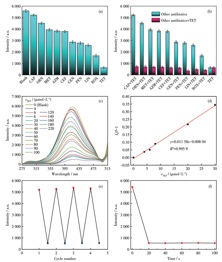
As shown in Fig.3a, it could be observed that in the presence of TET, the fluorescence intensity of 1 reached the maximum quenching, and the quenching efficiency was the highest. The fluorescence intensities of other antibiotics showed relatively small variations without TET. However, when TET was added to the solutions of other antibiotics, as shown in Fig.3b, the fluorescence intensities were significantly quenched, indicating that TET detection by 1 has anti-interference properties. In order to conduct a detailed analysis and study of its quenching situation, a quantitative experiment was carried out. As shown in Fig.3c, the TET concentration increased within a range of 0-220 μmol·L-1, and the fluorescence intensity of 1 gradually decreased. To investigate the relationship between TET concentration and fluorescence intensity, we plotted the relationship between I0/I-1 and cTET based on the SV equation, as shown in Fig.3d. The detection limit was calculated with 3σ/k, and it was 0.31 μmol·L-1. In addition, we observed that the fluorescence intensity could be fully restored to its initial level, suggesting that 1 may function as a recyclable fluorescent sensor (Fig.3e). It was also noted that upon the addition of 220 μmol·L-1 TET, the luminescence intensity decreased rapidly and reached a stable plateau within 20 s (Fig.3f).
Figure 3
 Figure 3. (a) Luminescent intensity of 1 (5 mmol·L-1) in different antibiotic solutions; (b) Anti-interference of TET detection by 1 in the presence of other antibiotics; (c) Emission spectra of 1 with different concentrations of TET; (d) SV plot of TET in low concentration ranges; (e) Cycle stability of 1 for the detection of TET (red square: 1, blue square: 1+TET); (f) Response time on TET detection by 1
2.8 Possible sensing mechanism
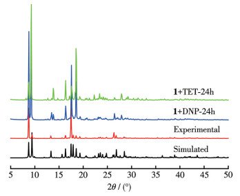
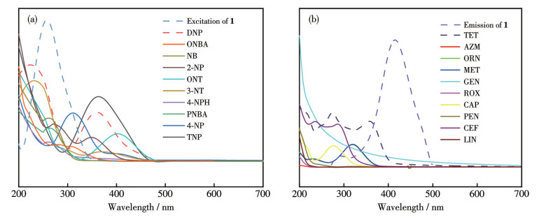
The mechanisms of fluorescence quenching reported are as follows: (1) the collapse and disintegration of the framework, (2) the mechanism of photoinduced electron transfer, (3) the mechanism of energy transfer, (4) the exchange of center metal ions, (5) the mechanism of energy competition and absorption[37]. To investigate the possible fluorescence quenching mechanism, we conducted a series of experiments. By analyzing the PXRD patterns of the 1 and synthesized samples after being immersed in DNP and TET solutions for 24 h, we found that the peak positions were almost the same as those of the crystal simulation, indicating that the quenching was not caused by the collapse of the framework (Fig.4). As shown in Fig.5a, the excitation spectrum of 1 overlapped with the UV-Vis absorption spectrum of DNP, and the overlapping portion was significant. This suggests that the reason for the fluorescence quenching of 1 may be due to energy competition and absorption. As shown in Fig.5b, the emission spectrum of 1 overlapped with the UV-Vis absorption spectrum of TET, which indicates that the reason for the fluorescence quenching of 1 by TET should be caused by resonance energy transfer.
Figure 4
 Figure 4. PXRD patterns of 1 and the as-synthesized samples immersing in DNP, TET solution for 24 h
Figure 5
 Figure 5. (a) UV-Vis absorption spectra of the NACs and the excitation spectrum of 1; (b) UV-Vis absorption spectra of the antibiotics and the emission spectrum of 1
2.9 Detection of TET in Yanhe River water
To prove the practicability of this method, TET was tested in Yanhe River water through the spiked recovery experiment. As shown in Table 3, the spiked recoveries at different concentrations were obtained, ranging from 98% to 102%. The relative standard deviation (RSD) values were 1.8%-3.1%, indicating the reliability and practicability of 1 to detect TET in real samples.
Table 3
Table 3. Recovery test of TET spiked in Yanhe River water samples下载:
导出CSV
cTET/(μmol·L-1) RSD*/% Recovery/% Spiked Detected 0 — 4 4.1 3.1 102 6 5.9 1.8 98 8 7.9 2.1 99 * n=3. 3. Conclusions
In summary, a metal-organic framework {[Zn(L)0.5(1,2,4,5-tpb)0.5]·DMF·3H2O}n (1) was synthesized under solvothermal conditions using Zn(Ⅱ) as the metal nodes, 5,5′-(ethane-1,2-diyl) diisophthalic acid (H4L) as the organic carboxylate ligand, and 1,2,4,5-tetra (pyridin-4-yl)benzene (1,2,4,5-tpb) as the nitrogen-containing auxiliary ligand. The structure of 1 was unambiguously determined by single-crystal X-ray diffraction, which revealed a 3D framework. Furthermore, the composition and stability were confirmed by thermogravimetric analysis and infrared spectroscopy. The solid and liquid fluorescence properties of 1 were investigated. The material exhibited significant fluorescence quenching responses toward 2,4-dinitrophenylhydrazine (DNP) and tetracycline (TET), with detection limits as low as 0.013 and 0.31 μmol·L-1, respectively. Mechanistic studies suggest that the quenching effect likely originates from competitive energy absorption (for DNP) and resonance energy transfer (for TET). Finally, the practical application of 1 was demonstrated through the detection of TET in real water samples collected from the Yanhe River, confirming its potential as a fluorescent sensor for environmental monitoring.
Supporting information is available at
http://www.wjhxxb.cn
-
-
[1]
WANG Q Y, WANG X H, WU Y W. Highly sensitive and selective fluorescence probe for 2,4-dinitrophenylhydrazine detection in wastewater using water-soluble CdTe QDs[J]. Photochem. Photobiol., 2019, 95(3): 895-900 doi: 10.1111/php.13084
-
[2]
LIU X G, TAO C L, YU H Q, CHEN B, LIU Z, ZHU G P, ZHAO Z J, SHI L, TANG B Z. A new luminescent metal-organic framework based on dicarboxyl-substituted tetraphenylethene for efficient detection of nitro-containing explosives and antibiotics in aqueous media[J]. J. Mater. Chem. C, 2018, 12: 2983-2988
-
[3]
MUTHARANI B, CHEN T W, CHEN S M, LIU X H. Reversibly switchable ruthenium hybrid thermo-responsive electrocatalyst-based voltammetric sensor for sensitive detection of sulfamethazine in milk samples[J]. Sens. Actuator B‒Chem., 2020, 316: 128103 doi: 10.1016/j.snb.2020.128103
-
[4]
LI C P, LONG W W, LEI Z, GUO L, XIE M J, LV J, ZHU X D. Anionic metal-organic framework as a unique turn-on fluorescent chemical sensor for ultra-sensitive detection of antibiotics[J]. Chem. Commun., 2020, 56: 12403-12406 doi: 10.1039/D0CC05175F
-
[5]
WALPER S A, GUILERMO L A, SAPSFORD K E, BROWN I C W, ROWLAND C E, BREGER J C, MEDINTZ I L. Detecting biothreat agents: From current diagnostics to developing sensor technologies[J]. ACS Sens., 2018, 3(10): 1894-2024 doi: 10.1021/acssensors.8b00420
-
[6]
LI F L, WANG X Y, SUN X, GUO Y M. Multiplex electrochemical aptasensor for detecting multiple antibiotics residues based on carbon fiber and mesoporous carbon-gold nanoparticles[J]. Sens. Actuator B‒Chem., 2018, 265: 217-226 doi: 10.1016/j.snb.2018.03.042
-
[7]
JAMPASA S, PUMMOREE J, SIANGPROH W, KHONGCHAREONPORN N, NGAMROJANAVANICH N, CHAILAPAKUL O, CHAIYO S. “Signal-On” electrochemical biosensor based on a competitive immunoassay format for the sensitive determination of oxytetracycline[J]. Sens. Actuator B‒Chem., 2020, 320: 128389 doi: 10.1016/j.snb.2020.128389
-
[8]
WANG X, WANG J J, TANG L, YUE E L, BAI C, WANG X, ZHANG Y Q, REN Y X, CHEN X L. Two different 2D Zn(Ⅱ) coordination polymers for highly selective detection of 2,4,6-trinitrophenol, tetracycline and fluazinam[J]. J. Mol. Struct., 2024, 1298: 137056
-
[9]
张欢, 王记江, 范广, 唐龙, 岳二林, 白超, 王潇, 张玉琦. 一种用于检测四环素和对硝基苯酚的高稳定性镉(Ⅱ)金属有机骨架[J]. 无机化学学报, 2014, 40(3): 646-654ZHANG H, WANG J J, FAN G, TANG L, YUE E L, BAI C, WANG X, ZHANG Y Q. A highly stable cadmium(Ⅱ) metal-organic framework for detecting tetracycline and p-nitrophenol[J]. Chinese J. Inorg. Chem., 2014, 40(3): 646-654
-
[10]
TAKEUCHI A, YAMAMOTO S, NARAI R, NISHIDA M, YASHIIKI M, SAKUI N, NAMERA A. Determination of dimethyl sulfoxide and dimethyl sulfone in urine by gas chromatography-mass spectrometry after preparation using 2,2-dimethoxypropane[J]. Biomed. Chromatogr., 2010, 24(5): 465-471 doi: 10.1002/bmc.1313
-
[11]
THUMM W, FREITAG D, KETTRUP A. Determination and quantification of dimethyl sulphoxide by HPLC[J]. Chromatographia, 1991, 32: 461-462 doi: 10.1007/BF02327979
-
[12]
LI B, DONG J P, ZHOU Z, WANG R, WANG L Y, ZANG S Q. Robust lanthanide metal-organic frameworks with “all-in-one” multifunction: Efficient gas adsorption and separation, tunable light emission and luminescence sensing[J]. J. Mater. Chem. C, 2021, 9(10): 3429-3439 doi: 10.1039/D0TC05707J
-
[13]
KUMAR A, HUR W, SEONG G H, CHAE P S. Sensitive detection of DMSO/DMF in water, human urine and blood plasma using novel 1,8-naphthalimide-based amphiphilic spectroscopic probes[J]. Dyes Pigment., 2021, 189: 109240 doi: 10.1016/j.dyepig.2021.109240
-
[14]
ANDERSEN J E T. A novel method for the filterless preconcentration of iron[J]. Analyst, 2005, 130(3): 385-390 doi: 10.1039/b412061b
-
[15]
ZHANG J F, ZHOU Y, YOOM J, KIM J S. Recent progress in fluorescent and colorimetric chemosensors for detection of precious metal ions (silver, gold and platinum ions)[J]. Chem. Soc. Rev., 2011, 40(7): 3416-3429 doi: 10.1039/c1cs15028f
-
[16]
YAO Y, LI N, ZHANG X, MACHUKI J O, YANG D Z, YU Y Y, LI J J, TANG D Q, TIAN J W, GAO F L. DNA-templated silver nanocluster/porphyrin/MnO2 platform for label-free intracellular Zn2+ imaging and fluorescence-/magnetic resonance imaging guided photodynamic therapy[J]. ACS Appl. Mater. Interfaces, 2019, 11(15): 13991-14003 doi: 10.1021/acsami.9b01530
-
[17]
YAGHI O M, O′KEFFE M, OCKWIG N W, CHAE, H K, EDDAOUDI M, KIM J. Reticular synthesis and the design of new materials[J]. Nature, 2003, 423: 705-714 doi: 10.1038/nature01650
-
[18]
BUTOVA V V, SOLDATOY M A, GUDA A A, LOMACHENKO K A, LAMBERTI C. Metal-organic frameworks: Structure, properties, methods of synthesis and characterization[J]. Russ. Chem. Rev., 2016, 85: 280-307 doi: 10.1070/RCR4554
-
[19]
DUTTA B, HAZRA A, DEY A, SINHA C, RAY P P, BANERJEE P, MIR M H. Construction of a succinate bridged Cd(Ⅱ)-based two-dimensional coordination polymer for efficient optoelectronic device fabrication and explosive sensing application[J]. Cryst. Growth Des., 2020, 20(2): 765-776 doi: 10.1021/acs.cgd.9b01181
-
[20]
WEN T, ZHANG D X, LIU J, LIN R, ZHANG J. A multifunctional helical Cu coordination polymer with mechanochromic, sensing and photocatalytic properties[J]. Chem. Commun., 2013, 49: 5600-5662
-
[21]
LIN J B, ZHANG J P, CHEN X M. Nonclassical active site for enhanced gas sorption in porous coordination polymer[J]. J. Am. Chem. Soc., 2010, 132: 6654-6656 doi: 10.1021/ja1009635
-
[22]
DUAN J G. JIN W Q, KITAGAWA S. Water-resistant porous coordination polymers for gas separation[J]. Coord. Chem. Rev., 2017, 332: 48-74 doi: 10.1016/j.ccr.2016.11.004
-
[23]
DUAN J G, HIGUCHI M, KRISHNA R, KIYONAGA T, TSUTSUMI Y, SATO Y, KUBOTA Y, TAKATA M, KITAGAWA S. High CO2/N2/O2/CO separation in a chemically robust porous coordination polymer with low binding energy[J]. Chem. Sci., 2014, 5(2): 660-666 doi: 10.1039/C3SC52177J
-
[24]
LUO X Z, ABAZARI R, TAHIR M, FAN W K, KUMAR A, KALHORIZADEH T, KIRILLOW A M, AMANI-GHADIM A R, CHEN J, ZHOU Y T. Trimetallic metal-organic frameworks and derived materials for environmental remediation and electrochemical energy storage and conversion[J]. Coord. Chem. Rev., 2022, 461: 214505 doi: 10.1016/j.ccr.2022.214505
-
[25]
陈小莉, 刘露, 商璐, 蔡苗, 崔华莉, 杨华, 王记江. 一种高灵敏、多响应的Zn-MOF荧光传感器对Fe3+、2,4,6-三硝基苯酚和奥硝唑的检测[J]. 无机化学学报, 2022, 38(4): 735-744CHEN X L, LIU L, SHANG L, CAI M, CUI H L, YANG H, WANG J J. A highly sensitive and multi-responsive Zn-MOF fluorescent sensor for detection of Fe3+, 2,4,6-trinitrophenol, and ornidazole[J]. Chinese J. Inorg. Chem., 2022, 38(4): 735-744
-
[26]
张龄文, 刘淑芹, 张佩佩, 倪爱云, 张建军. 一例基于5-((萘-1-基甲基)氨基)间苯二甲酸配体的Cd(Ⅱ)金属有机骨架的合成、晶体结构及对酸性氨基酸的检测[J]. 无机化学学报, 2022, 38(9): 1871-1877ZHANG L W, LIU S Q, ZHANG P P, NI A Y, ZHANG J J. Synthesis, crystal structure, and detection of acidic amino acids of a Cd(Ⅱ) metal-organic framework based on 5-((naphthalen-1-ylmethyl) amino) isophthalic acid[J]. Chinese J. Inorg. Chem., 2022, 38(9): 1871-1877
-
[27]
徐涵, 潘兆瑞, 亓昭鹏, 孙洁. 基于Ⅴ型配体的三个荧光Zn-MOF对水溶液中2,4,6-三硝基苯酚和Fe3+的荧光传感[J]. 无机化学学报, 2022, 38(12): 2479-2490XU H, PAN Z R, QI Z P, SUN J. Three luminescent Zn-MOFs based on Ⅴ-shaped ligands for fluorescence sensing of 2,4,6-trinitrophenol and Fe3+ in aqueous solution[J]. Chinese J. Inorg. Chem., 2022, 38(12): 2479-2490
-
[28]
WANG S Q, WANG L, ZHU Y M, SONG Y H. Fluorescent detection of S2- based on ZnMOF-74 and CuMOF-74[J]. Spectroc. Acta Pt. A‒Molec. Biomolec. Spectr., 2020, 236: 118327 doi: 10.1016/j.saa.2020.118327
-
[29]
FARAHANI Y D, SAFARIFARD V. Highly selective detection of Fe3+, Cd2+ and CH2Cl2 based on a fluorescent Zn-MOF with azine-decorated pores[J]. J. Solid State Chem., 2019, 275: 131-140 doi: 10.1016/j.jssc.2019.04.018
-
[30]
CHANDRASEKHAR P, MUKHOPADHYAY A, SAVITHA G, MOORTHY J N. Remarkably selective and enantiodifferentiating sensing of histidine by a fluorescent homochiral Zn-MOF based on pyrene-tetralactic acid[J]. Chem. Sci., 2016, 7: 3085-3091 doi: 10.1039/C5SC03839A
-
[31]
PAUL A G, NATHAN J P, PETER C B. Uranyl oxalate hydrates: Structures and IR spectra[J]. Z. Kristall., 2006, 221(4): 252-259
-
[32]
ZHANG Y H, SUN B, YAN X Q, PEI J, GENG X T, ZHANG Y, WANG Y Y, YAN J B. Synthesis, structure characterization and fluorescence studies of the complexes of europium with phenylglyoxylic acid, 1,10-phenanthroline and triphenyl phosphine oxide[J]. Spectrosc. Spectr. Anal., 2005, 25(12): 2034-2038
-
[33]
GOGIA A, MANDAL S K. A rational design and green synthesis of 3D metal organic frameworks containing a rigid heterocyclic nitrogen-rich dicarboxylate: Structural diversity, CO2 sorption and selective sensing of 2,4,6-TNP in water[J]. Dalton Trans., 2019, 48(7): 2388-2398 doi: 10.1039/C8DT04474K
-
[34]
LEE J Y, ROOT H D, ALI R, AN W, LYNCH V M, BAHRING S, KIM I S, SESSLER J L, PARK J S. Ratiometric turn-on fluorophore displacement ensembles for nitroaromatic explosives detection[J]. J. Am. Chem. Soc., 2020, 142(46): 19579-19587 doi: 10.1021/jacs.0c08106
-
[35]
MA Y X, LI H, PENG S, WANG L Y. Highly selective and sensitive fluorescent paper sensor for nitroaromatic explosive detection[J]. Anal. Chem., 2012, 84(19): 8415-8421 doi: 10.1021/ac302138c
-
[36]
WANG L B, WANG J J, YUE E L, LI J F, TANG L, BAI C, WANG X, HOU X Y, ZHANG Y Q. Information encryption, highly sensitive detection of nitrobenzene, tetracycline based on a stable luminescent Cd-MOF[J]. Spectroc. Acta Pt. A‒Molec. Biomolec. Spectr., 2022, 269: 120752 doi: 10.1016/j.saa.2021.120752
-
[37]
ZHAO Y F, ZENG H, ZHU X W, LU W G, LI D. Metal-organic frameworks as photoluminescent biosensing platforms: Mechanisms and applications[J]. Chem. Soc. Rev., 2021, 50: 4484-4513 doi: 10.1039/D0CS00955E
-
[1]
-
Figure 1 (a) Coordination environment of Zn(Ⅱ) in 1 with 50% ellipsoid probability; (b) 2D network structure of 1; (c) 3D framework of 1; (d) Underlying pts topological net of 1 (Zn: cyan, L4- ligand: pink, 1,2,4,5-tpb ligand: blue)
For clarity, all hydrogen atoms are omitted; Symmetry codes: A: x, -y, 1/2+z; b: x, -1+y, z; c: 2-x, -y, 1-z; d: 1-x, y, 3/2-z.
Figure 2 (a) Luminescent intensity of 1 (10 mmol·L-1) in different NAC solution; (b) Anti-interference of DNP detection by 1 in the presence of other NACs; (c) Emission spectra of 1 with different concentrations of DNP; (d) SV plot of DNP in low concentration ranges; (e) Cycle stability of 1 for the detection of DNP (red square: 1, blue square: 1+DNP); (f) Response time on DNP detection by 1
Figure 3 (a) Luminescent intensity of 1 (5 mmol·L-1) in different antibiotic solutions; (b) Anti-interference of TET detection by 1 in the presence of other antibiotics; (c) Emission spectra of 1 with different concentrations of TET; (d) SV plot of TET in low concentration ranges; (e) Cycle stability of 1 for the detection of TET (red square: 1, blue square: 1+TET); (f) Response time on TET detection by 1
Figure 4 PXRD patterns of 1 and the as-synthesized samples immersing in DNP, TET solution for 24 h
Figure 5 (a) UV-Vis absorption spectra of the NACs and the excitation spectrum of 1; (b) UV-Vis absorption spectra of the antibiotics and the emission spectrum of 1
Table 1. Crystal data and structure refinement parameters for 1
Parameter 1 Parameter 1 Formula C25H27N3O8Zn V/nm3 5.052(5) Formula weight 562.86 Z 8 Crystal system Orthorhombic D/(g·cm-3) 1.48 Space group Pbcn F(000) 2 336 a/nm 1.879 4(11) Goodness⁃of⁃fit on F 2 1.057 b/nm 1.326 6(8) R1, wR2 [I>2σ(I)] 0.048 9, 0.107 6 c/nm 2.026 5(13) R1, wR2 (all data) 0.079 3, 0.121 3  下载: 导出CSV
Table 2. Selected bond lengths (nm) and angles (°) for 1
Zn1—O1 0.198 4(3) Zn1—O4A 0.198 7(3) Zn1—N1 0.204 0(3) Zn1—N2B 0.203 5(3) O1—Zn1—O4 99.34(11) O1—Zn1—N1 107.30(11) O1—Zn1—N2B 113.67(11) O4A—Zn1—N1 108.78(11) O4A—Zn1—N2B 106.50(11) N2B—Zn1—N1 119.30(12) Symmetry codes: A: x, -y, 1/2+z; B: x, -1+y, z.
下载: 导出CSV
Table 3. Recovery test of TET spiked in Yanhe River water samples
cTET/(μmol·L-1) RSD*/% Recovery/% Spiked Detected 0 — 4 4.1 3.1 102 6 5.9 1.8 98 8 7.9 2.1 99 * n=3.
下载: 导出CSV
-


 扫一扫看文章
计量
- PDF下载量: 0
- 文章访问数: 396
- HTML全文浏览量: 56
 下载:
下载:
