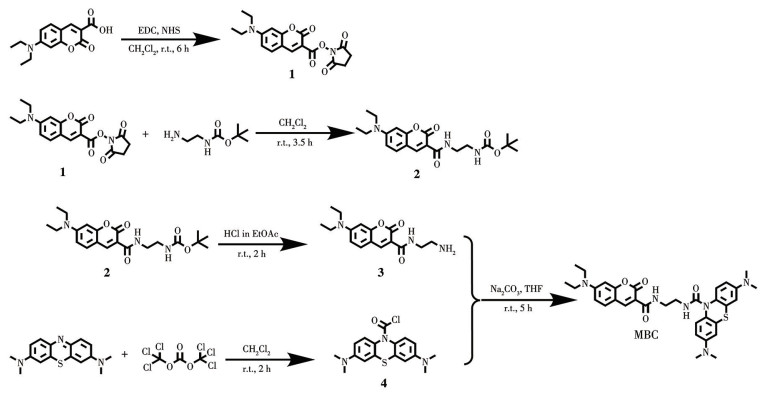
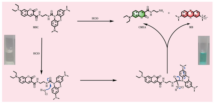
Scheme 1.
Synthesis of MBC
A self-calibrating fluorescent probe for the selective detection and bioimaging of HClO
Wei GAO , Meiqi SONG , Xuan REN , Jianliang BAI , Jing SU , Jianlong MA , Zhijun WANG

Reactive oxygen species (ROS) are integral to a variety of physiological and biochemical processes, including anti-inflammatory regulation, pathogen response, and mediation of biological signal transduction[1-2]. Hypochlorous acid (HClO), a prototypical ROS[3], is typically generated in vivo via myeloperoxidase-catalyzed oxidation of chloride ions (Cl-) by hydrogen peroxide (H2O2)[4-6]. HClO exhibits a dual nature in vivo ‒It effectively eliminates invading pathogens and bacteria owing to its potent oxidizing properties[4, 7-9] and plays a crucial role in immune defense and inflammation[6, 10]. However, excessive HClO levels can lead to various diseases, such as obesity, rheumatoid arthritis[8, 11], diabetes[12], neurodegenerative disorders[4, 8, 13-14], atherosclerosis[6, 15], liver injury[5, 15], pulmonary fibrosis, and even cancer[7, 14-15]. Therefore, developing a practical chemical tool for in situ HClO labeling that can provide valuable insights into the pathological functions of HClO is crucial.
Scientists have used a variety of analytical techniques to detect HClO, including colorimetry, mass spectrometry, electrochemistry, polarography, and fluorescence imaging. Among these techniques, fluorescence imaging utilizing fluorescent probes has demonstrated significant advantages in terms of sensitivity, simplicity, response time, and spatiotemporal resolution[3]. These inherent advantages make it suitable not only for in vitro analysis but also for in vivo imaging studies. A common strategy for designing HClO fluorescent probes involves linking an HClO reactive moiety to an organic fluorophore.
Some of the major studies involving HClO include oxidation of o-(p-)quinone to o-(p-)benzoquinone by HClO, oxidation of diacylhydrazine by ClO-, conversion of an aniline group to a nitro group, HClO-mediated intramolecular oxidation leading to ring formation or ring-opening, partial oxidation of thiourea to imidazoline by HClO, oxidative deprotection of thioheterocycles and sulfation of thioethers by HClO, oxidation of tolyl sulfides or selenides, oxidation of transition metal complexes, and oxidative cleavage of C=C, C=S, and C=N bonds by HClO[1-6, 8-10, 12-14, 16-30].
In this study, we developed a dual-wavelength emitting fluorescent probe, N-(2-(7-(diethylamino)-2-oxo-2H-chromene-3-carboxamido)ethyl)-3, 7-bis(dimethylamino)-10H-phenothiazine-10-carboxamide (MBC), for detecting HClO. The molecular structure of MBC contains methylene blue (MB) and coumarin fluorophores. These fluorophores emit distinct fluorescence signals upon excitation at specific wavelengths of light, with non‑overlapping emission spectra. The two fluorophores are covalently linked via a carbonyl group, which effectively quenches their fluorescence. On exposing MBC to HClO, HClO induces oxidative cleavage of the carbonyl linkage owing to its strong oxidizing property. This leads to the release of MB and coumarin, generating red and green fluorescence, respectively. Through dual-fluorescence calibration and localization without mutual interference, the probe exhibited enhanced accuracy and anti-interference performance in biomarker detection and analytical applications.
Sodium hypochlorite (NaClO), hydrogen peroxide (H2O2), potassium superoxide (KO2), tert-butyl hydroperoxide (TBHP), hydrochloric acid (HCl), sodium hydroxide (NaOH), sodium nitrate (NaNO3), and ferrous sulfate heptahydrate (FeSO4·7H2O) were procured from Sinopharm Chemical Reagent Group. Other reagents (analytical grade), including dimethyl sulfoxide (DMSO), sodium perchlorate (NaClO4), and sodium thiocyanate (NaSCN), were obtained from AladdinReagent (Shanghai) Co., Ltd. Organic solvents were obtained from Anhui Zesheng Technology Co., Ltd. Ultrapure water was produced using a Millipore Direct-Q system.
Ultraviolet-visible (UV-Vis) absorption spectroscopy was performed with a UV-2600 spectrometer. Fluorescence spectra were collected on an FLSP920 spectrometer, and fluorescence quantum yield was measured with an RF-5301PC spectrometer. pH measurements were performed with a PHB-4 pH meter (Thunder Magnetic). NMR spectra were recorded on a Varian INOVA 400 MHz superconducting NMR spectrometer. High-resolution mass spectra were collected on a Bruker microTOF-Q Ⅱ quadrupole-time-of-flight mass spectrometer. Liquid chromatography‑tandem mass spectrometry (LC-MS/MS) was performed with an Agilent 1100 series LC/MS system. Confocal microscopy was performed with an Olympus FV1200 laser scanning confocal microscope designed for live cell imaging.
MBC comprises a MB component (which emits red fluorescence), a diethylaminocoumarin component (which emits green fluorescence), and an ester‑ethylenediamine linkage that can be cleaved by HClO. Details regarding the characterization of MBC are provided in the Supporting Information. The synthetic routes for MBC are illustrated in Scheme 1.
First, 7-(diethylamino)coumarin-3-carboxylic acid (1.31 g, 5.0 mmol) was dissolved in 15 mL of anhydrous dichloromethane. Subsequently, 1-(3-dimethylaminopropyl)‑3‑ethylcarbodiimide hydrochloride (EDC, 4.8 g, 21.0 mmol) and N-hydroxysuccinimide (NHS, 2.59 g, 22.5 mmol) were added to the solution, followed by stirring at room temperature for 6 h. Upon completion of the reaction, the solvent was removed under reduced pressure. The residue was re-dissolved in 50 mL of dichloromethane and washed with saturated sodium bicarbonate (20 mL×3) and saturated brine (20 mL×3). The organic layer was dried over anhydrous sodium sulfate and concentrated under reduced pressure to obtain a yellow powder, which was named compound 1 (1.13 g). The product was used directly without purification.
Next, compound 1 (0.15 g, 0.419 mmol) was placed in a round-bottom flask and dissolved in 15 mL of dichloromethane. N-tert-butoxycarbonyl-1, 2-ethanediamine (0.30 g, 1.8 mmol) and N, N-diisopropylethylamine (0.20 g, 2 mmol) were added to the mixture, and the resulting solution was stirred at room temperature for 13 h. After the reaction was complete, the solvent was removed by vacuum distillation. The residue was re-dissolved in 15 mL of dichloromethane and washed successively with saturated ammonium chloride solution (20 mL×3), saturated sodium carbonate solution (20 mL×3), and saturated brine (20 mL×3). The organic phase was concentrated under reduced pressure, and the crude product was purified by column chromatography with CH2Cl2/MeOH (15∶1,V: V) solution as the eluent to yield 0.393 6 g of a light-yellow solid, which was named compound 2. The 1H NMR analysis (400 MHz, chloroform-d) details were as follows: δ 8.97 (t, J=6.1 Hz, 1H), 8.69 (s, 1H), 7.43 (d, J=8.9 Hz, 1H), 6.65 (dd, J=9.0, 2.3 Hz, 1H), 6.50 (d, J=2.3 Hz, 1H), 5.11 (t, J=5.8 Hz, 1H), 3.56 (q, J=6.0 Hz, 2H), 3.46 (q, J=7.2 Hz, 4H), 3.36 (q, J=6.0 Hz, 2H), 1.44 (s, 9H), and 1.24 (t, J=7.1 Hz, 6H).
Compound 2 (0.39 g, 0.976 mmol) was dissolved in 10 mL of a 4 mol·L-1 hydrochloric acid solution prepared using ethyl acetate (EtOAc) as the solvent, and the resulting solution was stirred at room temperature until a white precipitate (compound 3) was obtained. Compound 3 was used directly without further purification.
MB (1.69 g, 5.00 mmol) was dissolved in 25 mL of dichloromethane and transferred to a 100 mL round-bottom flask. An aqueous sodium hydrosulfide solution (1.04 g, 6.00 mmol) in 25 mL of water was then slowly added to the reaction mixture under continuous stirring. The temperature of the system was gradually increased to 40 ℃, and stirring was maintained for 1 h. The reaction mixture was then cooled by placing it in an ice-water bath. Subsequently, a solution of triphosgene (1.78 g, 6.00 mmol) in 25 mL of dichloromethane was added dropwise over 1 h. The resulting product was extracted with dichloromethane (20 mL×2) and washed with saturated brine (20 mL×3). The organic phase was dried over anhydrous sodium sulfate and then concentrated under reduced pressure. The crude product was purified by column chromatography using CH2Cl2/petrol ether (5∶1,V/V) solution as the eluent to obtain a white solid, which was named compound 4. Owing to instability, compound 4 was utilized directly in subsequent reactions without additional isolation.
A 50 mL round-bottomed flask was charged with a solution of compound 4 (0.03 g, 0.09 mmol) and 5 (0.03 mg, 0.09 mmol) in anhydrous tetrahydrofuran (THF, 5 mL). Subsequently, sodium carbonate (0.02 mg, 0.179 mmol) was added, and the mixture was stirred at room temperature for 5 h. The reaction solvent was evaporated under reduced pressure, and the crude product was purified by column chromatography using dichloromethane/methanol (20∶1,V/V) as the eluent to yield 25 mg of MBC. The 1H NMR (400 MHz, chloroform-d) analysis details were as follows: δ 8.93 (t, J=6.1 Hz, 1H), 8.65 (s, 1H), 7.40 (dd, J=14.1, 8.8 Hz, 3H), 6.69-6.63 (m, 3H), 6.61 (dd, J=8.8, 2.8 Hz, 2H), 6.51 (d, J=2.4 Hz, 1H), 5.36 (t, J=5.6 Hz, 1H), 3.58 (q, J=6.1 Hz, 2H), 3.45 (p, J=6.5, 5.7 Hz, 6H), 2.90 (s, 12H), and 1.24 (t, J=7.1 Hz, 6H). MS (electrospray ionization, ESI) spectra were also collected‒m/z calculated for C33H38N6O4S: 614.267 5; Found: 615.210 3 [M+H]+.
ROS were prepared by methods adapted from established protocols. In this study, commercially available NaClO was used as a substitute for HClO. TBHP and H2O2 were diluted to their respective working concentrations using commercially available reagents. Sodium nitrite (NaNO2) was dissolved in ultrapurewater to generate NO2- ions. Hydroxyl radicals (·OH) were generated via the Fenton reaction by reacting Fe2+ with 10 equivalents of H2O2. Superoxide anions (·O2-) were prepared by dissolving KO2 in anhydrous DMSO, followed by ultrasonic treatment. Singlet oxygen (1O2) was produced by reacting H2O2 with 10 equivalents of HClO. Nitric oxide (NO) was generated by adding sodium nitroprusside to degassed ultrapure water under nitrogen, followed by vigorous stirring for 30 min. All ROS solutions were freshly prepared and used immediately. Other required ions were prepared with ultrapure water.
All spectral measurements were performed in 10.0 mmol·L-1 phosphate-buffered saline (PBS, pH 7.4). Fluorescence responses of 5 μmol·L-1 MBC to different species in PBS were collected in a 1 cm pathlength cuvette in two channels (blue: excitation wavelength of 405 nm, red: excitation wavelength of 620 nm). The resulting mixtures were incubated at 25 ℃ for 3 min before fluorescence spectroscopy analysis. Fluorescence spectra were recorded in two wavelength ranges: excitation at 405 nm with emission in the 430-620 nm range and excitation at 620 nm with emission in the 640-800 nm range.
HepG2 cells were cultured in Dulbecco′s Modified Eagle Medium supplemented with a volume fraction of 10% fetal bovine serum, 100 U·mL-1 penicillin, and 100 μg·mL-1 streptomycin and maintained in a humidified incubator at 37 ℃ with a volume fraction of 5% CO2. The cells were seeded in 96-well plates at a density of 8 000 cells per well and cultured for 24 h to facilitate cellular adherence. Subsequently, the cells were exposed to various concentrations of MBC (0, 2.0, 4.0, 6.0, 8.0, and 10.0 μmol·L-1) for an additional 24 h. After treatment, the medium was carefully aspirated, and 110 μL of fresh medium containing 10 μL of cell counting kit-8 (CCK-8) reagent was added to each well under light-protected conditions. The plates were then incubated for 2 h, and absorbance was measured at 450 nm using a microplate reader to evaluate cytotoxicity.
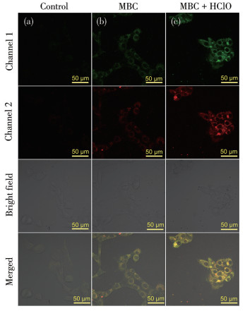
For the imaging experiment, HepG2 cells at a density of 30 000 cells per well in laser confocal culture dishes (diameter: 35 mm; glass bottom diameter: 10 mm) were incubated with 5.0 μmol·L-1 MBC at 37 ℃ for 30 min before laser confocal imaging. To determine the effect of exogenous HClO, HepG2 cells pre‑ incubated with 5.0 μmol·L-1 MBC were washed thoroughly and then incubated with 20.0 μmol·L-1 HClO for 30 min. Subsequently, the cells were subjected to laser confocal imaging. All cells were washed three times with sterile PBS and imaged using an Olympus FV1200 laser confocal microscope (green channel: 488 nm of excitation wavelength, 490-540 nm of emission collection range; red channel: 633 nm of excitation wavelength, 650-700 nm of emission collection range).
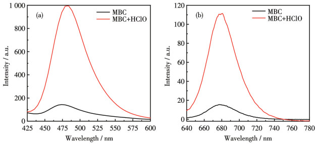
To determine the performance of MBC, first, its fluorescence response was investigated using 10.0 mmol·L-1 PBS at pH 7.4 by adding three equivalents of HClO. As shown in Fig. 1, MBC (5.0 μmol·L-1) exhibited minimal fluorescence in the absence of HClO. Upon the addition of three equivalents of HClO, MBC demonstrated distinct dual fluorescence. Specifically, under excitation at 405 nm, the fluorescence intensity in the 440-600 nm range was significantly enhanced, peaking at 480 nm. Moreover, when excited at 620 nm, MBC emitted visible fluorescence in the 640-740 nm range, peaking at 678 nm. Subsequently, a kinetic study was conducted to investigate the response of MBC to HClO by monitoring the changes in the fluorescence intensity at 480 nm (λex=405 nm) with time. As illustrated in Fig.S4, following the introduction of HClO, the fluorescence signal intensity rapidly increased, reaching a maximum within 90 s. These findings confirmed the potential of MBC for rapid and sensitive detection of HClO.
To investigate the selectivity of MBC, we examined the fluorescence spectral changes of 5.0 μmol·L-1 MBC in the presence of various reactive species and ions (Fig. 2). Specifically, we used different ROS (15.0 μmol·L-1): HClO, H2O2, NO, 1O2, and ·OH. In addition, we evaluated the effects of anions (0.1 μmol·L-1) such as NO2-, ClO4-, SO32-, Cl-, SO42-, NO3-, and PO43-, along with 0.1 mmol·L-1 cations and biological thiols, including K+, Na+, Fe2+, Fe3+, Mg2+, cysteine (Cys), and glutathione (GSH). The results indicated that uponexcitation at both 405 and 620 nm, only HClO caused a significant increase in the fluorescence signal of MBC. The other ROS, cations, anions, and biological thiols, commonly found in body fluids, did not induce any observable changes in the fluorescence of the probe. These findings demonstrated the high selectivity of MBC toward HClO.
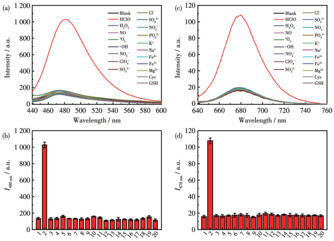
1: Blank; 2: HClO; 3: H2O2; 4: NO; 5: 1O2; 6: ·OH; 7: NO2-; 8: ClO4-; 9: SO32-; 10: Cl-; 11: SO42-; 12: NO3-; 13: PO43-; 14: K+; 15: Na+; 16: Fe2+; 17: Fe3+; 18: Mg2+; 19: Cys; 20: GSH.
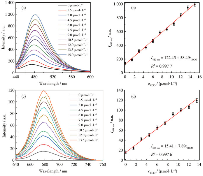
Moreover, we systematically evaluated the analytical performance of MBC using standard HClO solutions with varying concentrations in the aforementioned experiments. The experimental results are presented in Fig. 3. As shown, the fluorescence signal intensity gradually enhanced with increasing concentration of HClO. A strong linear relationship was observed between the fluorescence intensity at 480 nm (I480 nm) and HClO concentration (cHClO) in the 0-15.0 μmol·L-1 range. The following linear equation was used: I480 nm=122.45+58.49cHClO; the correlation coefficient (R2) was determined to be 0.997 7. The standard deviation (σ) of the blank was 0.49. With this information, the HClO detection limit for MBC was calculated to be 25.13 nmol·L-1. The fluorescence signal intensity at 678 nm (λex=620 nm) also increased, and the relationship between the intensity (I678 nm) and cHClO was found to be linear. The following linear equation was used: I678 nm=15.41+7.89cHClO; R2 was determined to be 0.997 6. Moreover, σ of the blank was 0.083, and the detection limit at this wavelength was determined to be 31.55 nmol·L-1. Thus, the detection limits derived from the fluorescence signals at both wavelengths (480 and 678 nm) were consistent.
The principle of sensing HClO by MBC for the optical changes is depicted in Scheme 2. With the addition of HClO, the free MBC undergoes a nucleophilic addition reaction through the process described in Scheme 2. The cleavage of the HClO-responsive site generates MB and coumarin ethylenediamine (CMEA) fluorophores, which, as two independent strong conjugated systems and induce fluorescence enhancement. To further investigate the sensing mechanism of MBC, the mass spectrometry analysis indicated corroborative evidence for the formation of MBC conjugate at m/z=326.518 5 (Obsd.) ([CMEA+Na]+, Calcd. 326.351 2 for C16H21N3NaO3+) and at m/z=285.102 4 (Obsd.) ([MB+H]+, Calcd. 285.121 6 for C16H18N3S+), implying the addition product (Fig.S5).
The aforementioned experiments demonstrated the potential of MBC for rapid and effective in vitro HClO detection. To evaluate the practical applicability of the synthesized probe, we conducted cellular studies. Initially, the cytotoxicity of MBC was assessedusing the CCK-8 assay. After incubation for 24 h with varying concentrations of MBC (0, 2.0, 4.0, 6.0, 8.0, and 10.0 μmol·L-1), HepG2 cells exhibited a survival rate exceeding 80% (Fig.S6), suggesting minimal cytotoxicity of MBC and its potential for practical applications.
Subsequently, we evaluated the applicability of MBC in detecting exogenous HClO in cells. As shown in Fig. 4, untreated HepG2 cells did not exhibit any fluorescence in the green channel or red channel. Following incubation of HepG2 cells for 30 min with 5.0 μmol·L-1 MBC at 37 ℃, a weak fluorescence signal was observed in both channels. This indicated that MBC possessed a certain degree of cell membrane permeability. To assess the capability of MBC in detecting intracellular HClO in living cells, we performed anexperiment wherein HepG2 cells were pre-incubated with fresh medium containing 15.0 μmol·L-1 HClO for 30 min. Subsequently, the cells were treated with 5.0 μmol·L-1 MBC for an additional 30 min before imaging. As shown in Fig. 4, significantly enhanced fluorescence was observed in both channels. These experimental results demonstrate that MBC can be effectively used for exogenous HClO detection in cells and biological applications.
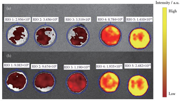
To verify the rapid and convenient detection of HClO in water samples, we developed a colorimetric test strip based on MBC. Typically, a circular filter paper with a 1 cm diameter was immersed in 4 mL of 0.1 μmol·L-1 MBC solution, followed by natural drying in a well-ventilated environment. To evaluate the performance, hypochlorite solutions with varying concentrations (25, 50, 75, and 100 μmol·L-1) were applied to the prepared strips. Subsequently, fluorescence imaging analysis was performed under laser irradiation with an in vivo optical imaging system after a 1-minute incubation period. As shown in Fig. 5, with increasing concentrations of ClO-, a distinct fluorescence intensity gradient was observed in both green and red channels, which confirmed the applicability of the developed colorimetric test strip in rapid semi-quantitative ClO- analysis. Thus, this simple and effective method could be used for water quality monitoring.
Green channel: excitation wavelength=420 nm, emission wavelength of green fluorescent protein (GFP) dye; Red channel: excitationwavelength=620 nm, emission wavelength of Cyanine5.5 (Cy5.5) dye.
In this study, we developed a near-infrared HClO fluorescence probe with dual-channel self-calibration capability. The probe featured a green-light coumarin fluorophore and a near-infrared methylene blue fluorophore integrated via a cleavable linkage that responded to HClO, allowing for dual-channel HClO monitoring in aqueous solutions. Utilizing this probe, we performed dual-channel detection of both endogenous and exogenous HClO in HepG2 cells. The probe exhibited consistent sensitivity and detection limits for HClO in both 480 and 620 nm channels. Our study demonstrates that the probe enables accurate detection of HClO in complex systems while effectively mitigating the interference from background fluorescence, inherent in single-channel detection.
SHANGGUAN L A, WANG J, QIAN X, WU Y Q, LIU Y. Mitochondria-targeted ratiometric chemodosimeter to detect hypochlorite acid for monitoring the drug-damaged liver and kidney[J]. Anal. Chem., 2022, 94: 11881-11888. doi: 10.1021/acs.analchem.2c02431
ZHAO G, LV C C, YANG X K, ZHAO X L, XIE F W. Levonorgestrel protected Au10 cluster for hypochlorite sensing in living organisms[J]. Anal. Chim. Acta, 2024, 1320: 343033. doi: 10.1016/j.aca.2024.343033
XIE J H, XING H Y, MENG L, ZHAO Y, ZENG Q, XIAO Q, LI N B, XUE P, LUO H Q. Engineering HClO ratiometric fluorescent probe by inducing molecular aggregation to suppress TICT formation for monitoring drug-induced liver injury[J]. Anal. Chem., 2024, 97(1): 220-228.
MA J T, KONG X T, ZHAO M T, JIAO Z L, XIE H, SI J, LI H, ZHANG Z X. A dual-functional NIR fluorescence probe for detecting hypochlorous acid and bisulfite in biosystem[J]. Anal. Chim. Acta, 2024, 1320: 342993. doi: 10.1016/j.aca.2024.342993
FORTIBUI M M, PARK C, KIM N Y, KIM T H, LEE M H. Dual-emissive detection of ATP and hypochlorite ions for monitoring inflammation-driven liver injury in vitro and in vivo[J]. Anal. Chem., 2024, 96(23): 9408-9415. doi: 10.1021/acs.analchem.4c00270
YAO L L, SONG H J, YIN C X, HUO F J. An ICT-switched fluorescent probe for visualizing lipid and HClO in lipid droplets during ferroptosis[J]. Chem. Commun., 2024, 60(7): 835-838. doi: 10.1039/D3CC05679A
SHAO K Y, GUO L J, ZHONG Y P, ZHANG L L, LU Z T, WANG D. Carbon quantum dots for rapid and ratiometric fluorescence determination of hypochlorite[J]. ACS Appl. Nano Mater., 2024, 7(8): 8645-8654. doi: 10.1021/acsanm.3c06131
WANG X, WANG H, DUAN J, SUN Q, ZHANG C L, XU L, LIU Z P. Phenothiazine-hemicyanine hybrid as a near-infrared fluorescent probe for ratiometric imaging of hypochlorite in vivo[J]. Sens. Actuators B-Chem., 2024, 407: 135453. doi: 10.1016/j.snb.2024.135453
CHAO M Z, ZHANG H T, HU Q F, MA S H, CUI X B, YU X. AIE-based fluorescent probe designed with xanthone as a π-bridge for detecting of ClO- in pericarp and living cells[J]. Spectroc. Acta Pt. A-Molec. Biomolec. Spectr., 2025, 324: 124984. doi: 10.1016/j.saa.2024.124984
NI J X, YU L C, WANG Y X, YANG T, BAI Y Q, ZHENG B W, LIANG M S, YE X X, QUAN Y Y, LIN F F. Win-win integration: A mitochondria targeted AIE photosensitizer for hypochlorite detection and type Ⅰ & type Ⅱ photodynamic therapy[J]. Anal. Chim. Acta, 2024, 1320: 343035. doi: 10.1016/j.aca.2024.343035
GU Y, ZHENG X, CHEN Z, TENG R M, ZHANG Y H, LI H, DING C P, HUANG Y J. Fluorescent-colorimetric dual signal ratio sensor with AuNRs@UCNPs superstructure nanoprobe for accurate hypo-chlorite detection[J]. Sens. Actuators B-Chem., 2024, 419: 136384. doi: 10.1016/j.snb.2024.136384
ZHANG Q L, ZHOU X, YU Q, SHAN X F, ZHANG Q, ZHANG Z, SHEN LY, REDSHAW C, XU H, ZHU B X. 2-Hydroxynaphthalenyl hemicarbocyanine dyes: Hypochlorite response and UV-catalysed induction of the formation of chlorinated hemicarbocyanine-like naphtho[2, 1-b] furans[J]. Sens. Actuators B-Chem., 2025, 426: 137105. doi: 10.1016/j.snb.2024.137105
LIU Y P, GUI Z R, ZHANG Z W, WANG S K, LANG W, LIU Y Z, CAO Q Y. A phenylphenthiazide anchored Tb(Ⅲ)-cyclen complex for fluorescent turn-on sensing of ClO-[J]. Chin. Chem. Lett., 2025, 36(2): 109769. doi: 10.1016/j.cclet.2024.109769
LIAO C, SHAN F, ZHU Y, MI H D, LIU Y H, SONG Q, WANG C F, WANG L Y, WANG Z Y. Self-ratiometric photoluminescence carbonized polymer dots with Junas structure for hypochlorite selective sensing[J]. Sens. Actuators B-Chem., 2024, 420: 136453. doi: 10.1016/j.snb.2024.136453
YANG J Y, CHEN Z M, YANG Y X, ZHENG B B, ZHU Y, WU F P, XIONG H. Visualization of endogenous hypochlorite in drug-induced liver injury mice via a bioluminescent probe combined with firefly luciferase mRNA-loaded lipid nanoparticles[J]. Anal. Chem., 2024, 96(18): 6978-6985. doi: 10.1021/acs.analchem.4c00008
KIM J, KIM Y. A water-soluble sulfonate-BODIPY based fluorescent probe for selective detection of HOCl/OCl- in aqueous media[J]. Analyst, 2014, 139(12): 2986-2989. doi: 10.1039/C4AN00466C
HU J J, WONG N K, GU Q S, BAI X Y, YE S, YANG D. HKOCl-2 series of green BODIPY-based fluorescent probes for hypochlorous acid detection and imaging in live cells[J]. Org. Lett., 2014, 16(13): 3544-3547. doi: 10.1021/ol501496n
HU J J, WONG N K, LU M Y, CHEN X M, YE S, ZHAO A Q, GAO P, KAO R Y T, SHEN J G, YANG D. HKOCl-3: A fluorescent hypochlorous acid probe for live-cell and in vivo imaging and quantitative application in flow cytometry and a 96-well microplate assay[J]. Chem. Sci., 2016, 7(3): 2094-2099. doi: 10.1039/C5SC03855C
WANG Y T, CHENG X Y, YIN N, WANG M X, QIN G X, TANG J L, ZHANG Y L, XU Q L. A hypochlorite-activated theranostic prodrug for selective imaging of high myeloperoxidase expression acute myeloid leukemia cells and drug release[J]. Anal. Chem., 2024, 30: 12238-12245.
ZHU B C, WU L, ZHANG M, WANG Y W, LIU C Y, WANG Z K, DUAN Q X, JIA P. A highly specific and ultrasensitive near-infrared fluorescent probe for imaging basal hypochlorite in the mitochondria of living cells[J]. Biosens. Bioelectron., 2018, 107: 218-223. doi: 10.1016/j.bios.2018.02.023
YUAN Q, ZHAO Z M, ZHANG Y R, SU L, MIAO J Y, ZHAO B X. A lysosome-targeted ratiometric fluorescent probe for detection of hypochlorous acid in living cells[J]. Sens. Actuators B-Chem., 2017, 247: 736-741. doi: 10.1016/j.snb.2017.03.049
GUI L, YAN J, ZHAO J, WANG S Y, JI Y Y, LIU J, WU J S, YUAN K, LIU H, DENG D W, YUAN Z W. Hypochlorite activatable ratiometric fluorescent probe based on endoplasmic reticulum stress for imaging of atherosclerosis[J]. Biosens. Bioelectron., 2023, 240: 115660. doi: 10.1016/j.bios.2023.115660
ZHAO W Q, ZHANG S T, YAN J L, XU P Y, LI B, ZHANG Y M, LI J L, WU S P. A dual-emission fluorescent probe for simultaneous detection of singlet oxygen and hypochlorous acid in lipid droplets[J]. Sens. Actuators B-Chem., 2024, 412: 135813. doi: 10.1016/j.snb.2024.135813
ZHANG P S, WANG H, HONG Y X, YU M L, ZENG R J, LONG Y F, CHEN J. Selective visualization of endogenous hypochlorous acid in zebrafish during lipopolysaccharide-induced acute liver injury using a polymer micelles-based ratiometric fluorescent probe[J]. Biosens. Bioelectron., 2018, 99: 318-324. doi: 10.1016/j.bios.2017.08.001
YUAN L, WANG L, AGRAWALLA B K, PARK S J, ZHU H, SIVARAMAN B, PENG J J, XU Q H, CHANG Y T. Development of targetable two-photon fluorescent probes to image hypochlorous acid in mitochondria and lysosome in live cell and inflamed mouse model[J]. J. Am. Chem. Soc., 2015, 137(18): 5930-5938. doi: 10.1021/jacs.5b00042
HE Q G, GUO T, LAN M H, ZHAO S J, HAN S H, YAO C Y, SONG X Z. Dual-ratiometric fluorescent probes for monitoring ClO- and polarity dynamics in ferroptosis[J]. Sens. Actuators B ‒ Chem., 2024, 415: 136030. doi: 10.1016/j.snb.2024.136030
BI W, ZHAO X Y, YANG X J, YUAN X S, LIN Y F, XU K M, LIU L, ZENG H Y, DU G B, ZHANG L P. Ratiometric fluorescent probe with AIE characteristics for hypochlorite detection and biological imaging[J]. Spectroc. Acta Pt. A ‒ Molec. Biomolec. Spectr., 2024, 323: 124904. doi: 10.1016/j.saa.2024.124904
SUN M T, YU H, ZHU H J, MA F, ZHANG S, HUANG D J, WANG S H. Oxidative cleavage-based near-infrared fluorescent probe for hypochlorous acid detection and myeloperoxidase activity evaluation[J]. Anal. Chem., 2014, 86(1): 671-677. doi: 10.1021/ac403603r
XU Q, HEO C H, KIM J A, LEE H S, HU Y, KIM D, SWAMY K M K, KIM G, NAM S J, KIM H M, YOON J. A selective imidazoline-2-thione-bearing two-photon fluorescent probe for hypochlorous acid in mitochondria[J]. Anal. Chem., 2016, 88(12): 6615-6620. doi: 10.1021/acs.analchem.6b01738
YE Q, XU K, MENG Z Y, LI X Y, WANG Z L, WANG S F. Construction of a novel isopinocamphone-based fluorescent probe for rapid and specific detection of ClO- and its application in environmental analysis and biological imaging[J]. New J. Chem., 2024, 48(32): 14182-14190. doi: 10.1039/D4NJ02375G

Figure 1 Intensity changes in the fluorescence spectra of MBC (5.0 μmol·L-1) upon addition of HClO (15.0 μmol·L-1): (a) excitation at 405 nm and (b) excitation at 620 nm

Figure 2 Fluorescence spectra of MBC (5.0 μmol·L-1) in the presence of various analytes upon excitation at wavelengths(a) 405 nm and (c) 620 nm, and the corresponding fluorescence intensities at emission wavelengths (b) 480 nmand (d) 678 nm
1: Blank; 2: HClO; 3: H2O2; 4: NO; 5: 1O2; 6: ·OH; 7: NO2-; 8: ClO4-; 9: SO32-; 10: Cl-; 11: SO42-; 12: NO3-; 13: PO43-; 14: K+; 15: Na+; 16: Fe2+; 17: Fe3+; 18: Mg2+; 19: Cys; 20: GSH.

Figure 3 (a) Fluorescence spectra of MBC upon excitation at 405 nm varying with cHClO; (b) Linear relationship between I480 nm and cHClO; (c) Fluorescence spectra of MBC upon excitation at 620 nm varying with cHClO; (d) Linear relationship between I678 nm and cHClO

Figure 4 Fluorescence imaging of HepG2 cells: (a) untreatedcells; (b) cells treated with 5.0 μmol·L-1 MBC for 30 min; (c) cells pre-incubated with 15.0 μmol·L-1 HClO for 30 min followed by treatment with 5.0 μmol·L-1 MBC for an additional 30 min

Figure 5 Fluorescence imaging of (a) green and (b) red fluorescence channels with the fluorescence imaging system underdifferent concentrations of hypochlorite solutions
Green channel: excitation wavelength=420 nm, emission wavelength of green fluorescent protein (GFP) dye; Red channel: excitationwavelength=620 nm, emission wavelength of Cyanine5.5 (Cy5.5) dye.

 扫一扫看文章

扫一扫关注我们
 下载:
下载:

 下载:
下载:
