
Scheme 1.
Structure of H3L
Synthesis, crystal structure, and DNA-binding of binuclear lanthanide complexes based on a multidentate Schiff base ligand
Xiaofen GUAN , Yating LIU , Jia LI , Yiwen HU , Haiyuan DING , Yuanjing SHI , Zhiqiang WANG , Wenmin WANG

Metal - organic frameworks (MOFs) are effective molecular devices used in fields like gas separation[1-2], water purification[3-5], industrial catalysis[6-11], biomedicine[12-14], and so on[15-16]. Over the past decades, the MOFs that can interact with DNA have been intensively studied[17-19]. The interaction between DNA and metal complexes is an important fundamental issue in life sciences which is related to the replication and transcription of DNA in vivo, mutation of genes and related variations of species in character, action mechanisms of some DNA - targeted drugs, origins of some diseases, action mechanisms of some synthetic chemical nucleases, etc. Hence, the research on the action mode of metal complexes on DNA is expected to promote the development of molecular biology and pharmacology[20-22].
Compared to conventional drugs, MOF-based antibacterial agents have many advantages. The active sites of metal ions in the MOFs are effectively wrapped by the organic ligands and evenly distributed across the overall material, enabling a sustained release of metal ions to avoid the toxicity caused by a burst release of metal ions. In the 1960s, Rosenberg et al.first investigated platinum anticancer drugs, and they discovered that platinum complexes can inhibit cell division, which laid the foundation for the research of such chemotherapeutic agents[23-24]. Since then, scientists have begun to explore MOF - based anticancer drugs. In 2014, Das et al. demonstrated that triply phenoxo bridged Eu(Ⅲ) and Sm(Ⅲ) complexes with 2, 6-diformyl- 4-methylphenoldi(benzoylhydrazone) exhibited anti - mycobacterial activity[25]. In 2022, Keshavarzian et al. presented the interaction between the heterodinuclear Cu-Gd (3d-4f) complex and CT-DNA intercalation action[26]. In 2022, Kundu et al. reported that the pyrene-based fluorescent Ru(Ⅱ)-arene complexes exhibit strong interactions with CT - DNA[27], and so forth.Therefore, it is significant to search for metal complexes as potential antibacterial agents.
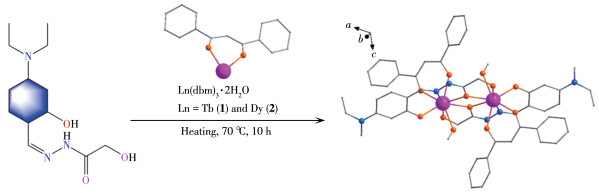
Schiff bases are well-known pharmacophores for a wide range of biological activities that interfere with normal cellular processes by forming intramolecular hydrogen bonds between the imine group and the active center of the cellular components[28-29]. Therefore, it is conceived that combining these biologically active Schiff base ligands with the metals can invoke a synergistic effect that could be used to design effective metallodrugs for chemotherapy. In continuation of our previous effort for generating lanthanide - based complexes, we designed and constructed a Schiff base ligand (Scheme 1) hydroxy-acetic acid(4-diethylamino-2 - hydroxy - benzylidene) - hydrazide (H3L). And then it was introduced into lanthanide metals to generate two new binuclear lanthanide (Ln2) complexes with the general formula [Ln2(dbm)2(HL)2(CH3OH)2]·4CH3OH, where Ln=Tb (1) and Dy (2), and Hdbm=dibenzoyl-methane. The interaction of the complexes with DNA was studied using UV -Vis spectra, fluorescence spectra, and electrochemical analysis methods.
Reagents included Ln(dbm)·2H2O (Ln=Tb or Dy)(Energy Chemical), Hdbm (AR, Aladdin), hydroxyacetohydrazide (Energy Chemical), 4 - (diethylamino)salicylaldehyde (Energy Chemical), CH3OH (AR, Kermel), anhydrous CH3CN (AR, Kermel), CH2Cl2 (AR, Kermel), N, N-dimethylformamide (DMF, AR, Kermel), ethidium bromide (EB, AR, Kermel). Deionized water (DI water) was produced by the Milli-Q water purification system.All chemicals and solvents were used as received without further purification. CT - DNA was obtained from the Shanghai Institute of Materia Medica.
Elemental analyses for C, H, and N were performed on a Perkin-Elmer 240 CHN elemental analyzer. Powder X -ray diffraction (PXRD) data were examined on a Rigaku Ultima Ⅳ instrument with Cu Kα radiation (λ=0.154 056 nm) and a scan speed of 5 (°)·min-1 in a 2θ range of 5° - 50°. The operating voltage and current were 40 kV and 25 mA, respectively. Thermogravimetric analysis (TGA) measurements were obtained in a nitrogen atmosphere on a Labsys NETZSCH TG 209 Setaram apparatus from 30 to 800 ℃ with a heating rate of 10 ℃·min-1. Absorption spectra were performed on a TU-1950 UV-Vis spectrophotometer and emission spectra were recorded on an F - 320 fluorescence spectrophotometer. Cyclic voltammetry (CV) was performed with Chenhua CHI750E Electrochemical Workstation. 1H and 13C NMR spectra were recorded at 600 MHz in DMSO using JEOL-JNM-ECZ600R.
The polydentate Schiff base ligand H3L is prepared in a simple aldimine condensation reaction of hydroxyacetohydrazide (20 mmol) with 4-(diethylamino) salicylaldehyde (20 mmol) in 10 mL methanol (Scheme 2). The mixture was refluxed for 10 h. Then it was cooled to room temperature. Afterward, a pale - yellow precipitate was obtained and washed carefully with methanol. The product was dried in a vacuum. Yield: 68%. Anal. Calcd. for C13H16N3O3(%): C, 59.54; H, 6.11; N, 16.03. Found(%): C, 59.58; H, 6.14; N, 16.08.IR(cm-1): 3 317(s), 3 175(w), 2 963(m), 2 926(m), 2 896(w), 2 866(w), 1 659(m), 1 624(s), 1 604(w), 1 557(m), 1 523(m), 1 505(m), 1 479(m), 1 438(w), 1 400(m), 1 368(w), 1 348(s), 1 307(w), 1 282(w), 1 245(s), 1 218(m), 1 197(m), 1 126(s), 1 100(s), 1 074(w), 1 015(m), 968(m), 904(m), 858(m), 819(s), 783(s), 727(m), 705(w), 666(m), 642(m), 560(m), 550(w), 520(w), 445(w), 430(m), 419(w) (Fig. S1, Supporting information). 1HNMR (600 MHz, DMSO): δ 11.43 (s), 11.12 (s), 8.34(s), 7.04 (d, J=8.7 Hz), 6.19 (dd, J=8.7, 2.4 Hz), 6.07(d, J=2.3 Hz), 3.98 (d, J=5.9 Hz), 3.36-3.29 (m), 1.14-1.07 (m) (Fig. S2). 13C NMR (600 MHz, DMSO): δ 167.33 (s), 159.67 (s), 150.14 (s), 149.86 (s), 131.69(s), 106.32 (s), 103.32 (s), 97.53 (s), 61.21 (s), 43.82(s), 12.44 (s) (Fig.S3).
Complexes 1 and 2 were prepared under the same conditions. Ln(dbm)3·2H2O (0.025 mmol), H3L (0.025mmol), CH3OH (8 mL), CH3CN(2 mL), DMF(0.5 mL), and CH2Cl2 (2 mL) were enclosed in a 15 mL vial by stirring for 5-10 min. The vial was sealed and heated at 70 ℃ for 10 h, then the temperature was dropped to room temperature at a rate of about 5 ℃·h-1. Yellow block crystals of the products were obtained. The synthesis diagram of complexes 1 and 2 is shown in Scheme 3.
Complex 1: Yield based on Tb: 48%. Anal. Calcd.for C62H80Tb2N6O16(%): C, 50.16; H, 5.39; N, 5.66.Found(%): C, 50.31; H, 5.43; N, 5.63.
Complex 2: Yield based on Dy: 52%. Anal. Calcd.for C62H80Dy2N6O16(%): C, 49.92; H, 5.37; N, 5.64.Found(%): C, 49.95; H, 5.39; N, 5.61.
Single crystal X-ray diffraction data of complexes 1 and 2 were collected on a computer-controlled Rigaku Saturn CCD area detector diffractometer, equipped with confocal monochromatized Mo Kα radiation with a radiation wavelength of 0.071 073 nm using the ω-φ scan technique. The structures were solved by direct methods and refined with a full - matrix least-squares technique based on F2 using the SHELXS-2016 and SHELXL-2016 programs. Anisotropic thermal parameters were assigned to all non - hydrogen atoms. The crystals were measured as soon as they were isolated from the solution, thus there were some disordered solvent molecules. Details of the crystal parameters, data collection, and refinements for 1 and 2 are summarized in Table 1. Selected bond lengths and angles of complexes 1 and 2 are given in Table S1 and S2.
 下载:
导出CSV
下载:
导出CSV
| Parameter | 1 | 2 |
| Formula | C62H80Tb2N6O16 | C62H80Dy2N6O16 |
| Formula weight | 1 483.16 | 1 490.32 |
| T/K | 150.0 | 150.0 |
| Crystal system | Monoclinic | Monoclinic |
| Space group | P21/c | P21/c |
| a/nm | 1.509 26(7) | 1.509 58(12) |
| b/nm | 1.210 40(6) | 1.211 74(8) |
| c/nm | 1.882 85(7) | 1.883 37(12) |
| β/(°) | 112.499 0(16) | 112.426(2) |
| V/nm3 | 3.177 8(2) | 3.184 6(4) |
| Z | 2 | 2 |
| Dc/(g·cm-3) | 1.550 | 1.554 |
| μ/mm-1 | 2.278 | 2.399 |
| θ range/(°) | 2.228-26.057 | 2.226-26.404 |
| F(000) | 1 504 | 1 508 |
| Reflection collected | 43 526 | 51 540 |
| Unique reflection | 6 253 | 6 514 |
| Rint | 0.067 5 | 0.061 1 |
| GOF(F2) | 1.096 | 1.094 |
| R1, wR2[I>2σ(I)] | 0.039 8, 0.087 1 | 0.033 2, 0.091 7 |
| R1, wR2 (all data) | 0.053 6, 0.094 1 | 0.043 3, 0.104 0 |
The CT-DNA mother liquor (3 mL, 2 μg·mL-1) was determined by the absorbance at A260/A280 by UV spectrophotometer. Tris-HCl/NaCl buffer solution (500 mL, 5 mmol·L-1, pH=7.25) was obtained through Tris (0.302 5 g), HCl (6 mol·L-1, 0.55 mL), NaCl (0.146 3 g), and distilled water. Complexes 1 and 2 and ligand H3L were dissolved in Tris-HCl/NaCl buffer solution to give a final concentration of 17 μmol·L-1.
At room temperature, the concentrations of complexes 1 and 2 and ligand H3L were kept constant and the CT - DNA concentration was adjusted for UV - Vis spectrophotometry in a wavelength range of 190 - 450nm. Tris-HCl/NaCl buffer solution was used as a reference solution, and CT-DNA solution was used as a control. The same amount of CT - DNA solution (10 μL) was dropped in turn, and the absorbance of the solution was tested after 30 min of reaction.
In the fluorescence quenching experiment, the initial mass concentrations of EB and CT-DNA were 0.06mg·mL-1 and 2 μg·mL-1 (8.4 μmol·L-1), respectively.They were mixed in equal volumes and left in the dark for 12 h before use. The specific test process was as follows: 3 mL mixed solution of EB-DNA was added into the sample pool, and the fluorescence emission spectra were measured in a range of 550-700 nm at the excitation wavelength of 270 nm. Then, the same volume of the compound (H3L, 1, 2) solution (17 μmol·L-1, 10μL) was added to the EB-DNA system in turn. After each addition, the mixed solution was shaken well and reacted for 5 min at room temperature, and then the emission spectrum was determined.
In CV measurement, the three - electrode system was used. The working electrode was a glassy carbon electrode, the reference electrode was a saturated calomel electrode, and the counter electrode was a platinum wire electrode. The scanning potential range was from -0.15 to 0.8 V, the scanning speed was 0.1 mV·s-1, the sample interval was 0.001 V, and the standing time was 2 s. 17 μmol·L-1 compounds (complexes 1 and 2 and ligand H3L) and 2 μg·mL-1 DNA were used for the experiment.
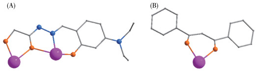
The single - crystal X - ray analysis technique was employed to indisputably determine the structures of complexes 1 and 2. The complexes crystallize in the monoclinic space group P21/c. The X - ray crystallographic data of 1 and 2 are given in Table 1. Given the similarity of the molecular structures of 1 and 2, we describe the structure of [Tb2(dbm)2(HL)2(CH3OH)2]·4CH3OH (1) as a representative example. As shown in Fig. 1. The molecular structure of 1 mainly contains two Tb(Ⅲ) ions, two dbm- ions, two HL2- ions, two CH3OH molecules, and four free methanol molecules. Two HL2- ligands are involved in holding the dimeric terbium unit together in 1 through two bridging phenolate oxygen atoms [Tb—O2 0.241 2(3) nm, Tb—O2a 0.238 6(3) nm; Tb—O—Tba 115.30(13)°] (Fig. 2A).While one arm of the ligand (—OMe) binds to one Tb3+[Tb1—O1a 0.243 4(4) nm], the other arm of the ligand (N) holds the other Tb3+ [Tb1a—N2a 0.243 0(4) nm, Tb1a—O3a 0.220 0(3) nm]. Finally, each Tb3+ has a unidentate methanol ligand [Tb(1) —O(6) 0.240 8(3)nm] and dbm- ligand [Tb1—O4 0.234 8(3) nm, Tb1—O5 0.235 1(3) nm]. Thus, each Tb3+ ion is eight-coordinated (seven O atoms and one N atom), as shown in Fig. 2B. The coordination mode of HL2- and dbm- is shown in Fig. 3A and 3B. Each HL2- connected two central Tb3+ ions, and two Tb(Ⅲ) ions are bridged by two μ2-O atoms (O2 and O2a) of two HL2- ligands, leading to a parallelogram [Tb2O2] core with the Tb1—O2—Tb1a angle of 115.30(13)°. The Tb…Tb distance of 1 is 0.405 38(4) nm. Furthermore, in complex 1, the Tb—O bond distances are 0.220 0(3)-0.243 4(4) nm. The Tb—N bond distance of 0.243 0(4) nm is longer than any of the Tb—O bonds. The O—Tb—O bond angles range from 64.19(12)° to 145.28(12)°. These bond distances and bond angles are comparable to those of the already reported phenoxo-bridged Ln2 complexes[30-34].
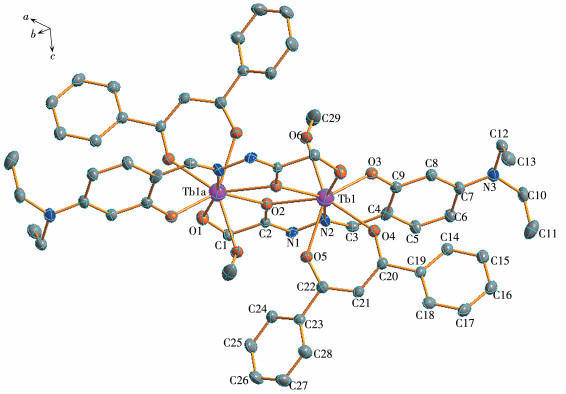
Hydrogen atoms have been omitted for clarity; Symmetry code: a: -x+1, -y+2, -z+1; Ellipsoids are drawn at a 30% probability level.
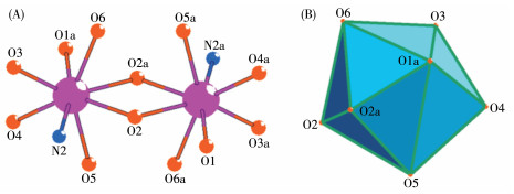
Symmetry code: a: -x+1, -y+2, -z+1; Color code: gray, C; blue, N; orange, O; fuchsia red, Tb.
To confirm the phase identity and purity of the bulk samples of complexes 1 and 2, the PXRD analysis of complexes 1 and 2 was carried out. As shown in Fig.S4, the PXRD patterns of the bulk samples were in line with the reality of the simulated ones, indicating the high phase identity and purity.
The thermal stabilities of complexes 1 and 2 were characterized in a temperature range of 30 - 800 ℃ under a nitrogen atmosphere. The analysis results are shown in Fig.S5. For 1 and 2, the TGA results were almost the same, and that's because they are isomorphic. Herein, complex 1 is an example of all the discussions and illustrations. It can be seen that the thermal decomposition of complex 1 mainly occurred in two stages. When the temperature was between 30 and 150 ℃, a weight loss of 1.88% was observed (Calcd.2.09%), which can be assigned to the decomposition of one coordinated CH3OH molecule. Then, as the temperature continued to increase and the weight loss rate continued to decrease, the framework of 1 began to break down and the skeleton began to collapse. The residue was considered Tb2O3 (Obsd. 31.48%, Calcd.24.67%).
The UV-Vis spectra for ligand H3L, complexes 1 and 2 were obtained in a methanol solution (Fig.S6).For H3L, two broad absorption bands were observed at ca. 226 and 356 nm, which are caused by the n-π* and π-π* transitions of the ligand itself. The UV-Vis spectra of complexes 1 and 2 displayed similar absorption profiles centered at ca. 226 and 392 nm. Compared with the absorption bands of H3L, the peaks at ca. 392 nm of 1 and 2 were red-shifted, which may be due to the coordination effects of dbm- and H2L- with the Ln(Ⅲ) ions[35].
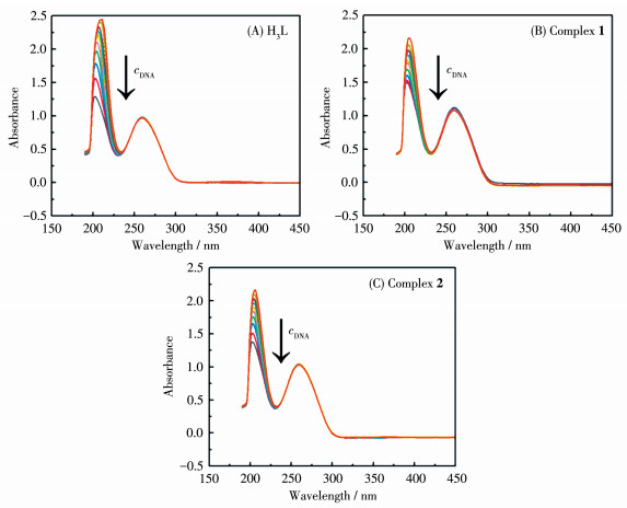
UV-Vis spectrometry is a direct and reliable approach to exploring the interaction between the complex and DNA[36-37]. Usually, the combination of the complex with DNA will lead to its absorption band widening, absorption peak redshift, and hypochromic effect. As exhibited in Fig. 4, the absorption spectra were recorded at room temperature between 190 and 450 nm by keeping the concentrations of the compounds (complexes 1 and 2 and ligand H3L) constant while varying the CT-DNA concentration. We can see that the absorption peaks of H3L, 1, and 2 at 205 nm had different levels of hypochromic effect. The reason for this phenomenon may be that π electron accumulation occurs between the compound and the base pair after the compound combines with DNA through insertion, and the π* empty orbital of the ligand and the π orbital of the DNA base pair will be coupled to cause the energy level to drop, and the π* orbital is partially filled with electrons after coupling, which reduces the probability of π-π* transition, resulting in hypochromic effect. The results indicated that the compound may bind to CT-DNA by embedding. With the increasing insertion, the hypochromic effect becomes more and more obvious.
EB molecule itself has very weak fluorescence because it is a conjugated plane molecule. It can be inserted specifically parallel between the base pairs inside the DNA double helix or triple helix, forming the EB-DNA complex, and its fluorescence intensity is significantly enhanced. If other compounds co-exist in the EB - DNA system, which can also have EB - like insertion with DNA, these compounds will compete for the binding site of DNA and EB, resulting in fluorescence quenching of the EB-DNA system. The degree of fluorescence quenching of the EB - DNA system by these compounds can indicate the strength of its interaction with DNA insertion, so EB is widely used as a DNA probe to analyze the interaction mode between compounds and DNA[38]. According to the Stern-Volmer equation: I0/I=1+Ksvr, where I0 is the fluorescence intensity of EB-DNA system without compound, I is the fluorescence intensity of EB-DNA system after adding different concentrations of compounds, r is the concentration ratio of compound to DNA, Ksv is a linear Stern -Volmer constant. If I0/I and r have a linear relationship, Ksv can be calculated from the linear slope, which can be used to evaluate the binding ability of the compound and DNA.
Based on the above theory, we performed the EB-DNA fluorescence experiments to explore the binding ability of H3L, complexes 1 and 2 with DNA, as shown in Fig. 5. After the addition of increasing amounts of H3L, 1, and 2, it was found that the fluorescence intensities of the EB-DNA composite all reduced in different levels (Fig. 5A-5C), which indicates that EB between DNA base pairs was extruded by the compound, and a new compound - CT - DNA system was formed, that is, the compound interposed with CT -DNA. From Fig. 5D-5F, the Ksv values for H3L, 1, and 2 were calculated to be 1.178, 4.160, and 7.341, respectively. The magnitude of binding strength was found to be 2 > 1 > H3L, revealing a slightly stronger binding affinity of 2 with CT - DNA as compared to those of 1 and H3L. It's telling that there's quite obviously a synergistic effect between the Ln3+ ion and ligand H3L.Compared with some Ln2 complexes reported by literature, the Ksv values of 1 and 2 were larger than those of some Ln2 complexes (Table S3).
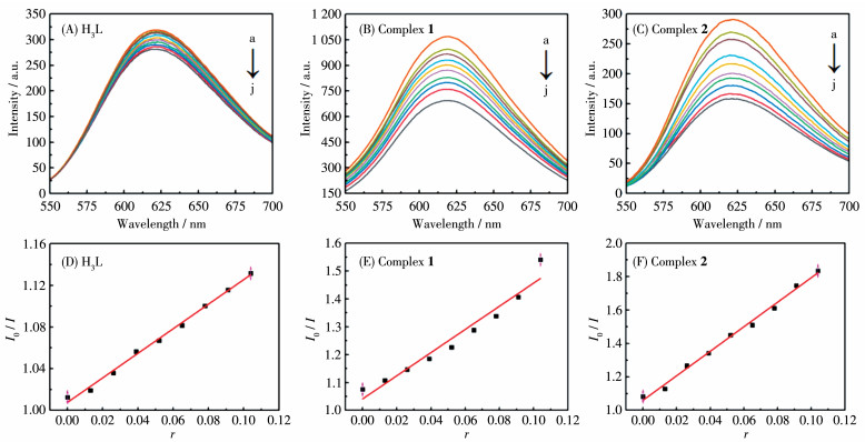
r=ccompound/cDNA; From a to j, Vcompound=0, 10, 20, 30, 40, 50, 60, 70, 80, 90 μL, respectively, and the initial concentration of the compound was 17 μmol·L-1.
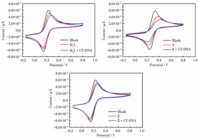
According to the changes in the electrochemical properties of the compound before and after adding DNA, the mode of interaction between the compound and DNA can be judged[39-40]. As shown in Fig. 6, the anodic peak potentials (Epa) and cathodic peak potentials (Epc) of H3L, 1, 2 were 0.149 8, 0.248 1, 0.379 8V, and 0.256 1, 0.140 3, 0.228 4 V, respectively, and the equation potentials were 0.203 0, 0.194 2, 0.304 1V, respectively. After the compound interacts with DNA, the anodic peak potentials (Epa) and cathodic peak potentials (Epc) of H3L, 1, 2 were 0.276 3, 0.260 3, 0.413 0 V, and 0.130 1, 0.133 8, 0.209 9 V, respectively, and the equation potentials were 0.203 2, 0.197 1, 0.311 5 V, respectively. The equation potentials of H3L, 1, and 2 were positive shifts, indicating that they were bound to the DNA molecule by insertion, and the interaction of 2 with DNA is greater than that of 1, consistent with the results obtained from spectroscopy.
Tris-HCl/NaCl buffer solution with pH=7.25 is an electrolyte solution..
In brief, we have synthesized two rare earth complexes [Ln2(dbm)2(HL)2(CH3OH)2]·4CH3OH where Ln=Tb (1) and Dy (2), Hdbm=dibenzoylmethane, H3L=hydroxy-acetic acid(4-diethylamino-2-hydroxy-benzyli-dene)-hydrazide. The X-ray structural analysis reveals that the two complexes are dinuclear and each Ln(Ⅲ) ion is eight-coordinated. The structures and DNA interaction of two complexes have been systematically studied. Subsequent studies strongly suggested that the two Ln2 complexes bound to calf thymus DNA (CT-DNA) through intercalative mode. This work has a certain reference value to study the interaction between complex and DNA.
Li W X, Situ Y Z, Ding L F, Chen Y L, Yang Q Y. MOF GRU: A MOFid aided deep learning model for predicting the gas separation performance of metal-organic frameworks[J]. ACS Appl. Mater. Interfaces, 2023, 15(51): 59887-59894. doi: 10.1021/acsami.3c11790
Zhang M X, Jiang J Y, Zhao H T, Wang Y, He X G, Chen M, Wang W, Wang S Y, Wang S, Wang M, Sun T M, Qin G P, Tang Y F, Cui H H. Flow channel with recognition corners in a stable La-MOF for onestep ethylene production[J]. Inorg. Chem, 2024, 63(3): 1507-1512. doi: 10.1021/acs.inorgchem.3c03852
El-Sewify I M, Ma S Q. Recent development of metal-organic frameworks for water purification[J]. Langmuir, 2024, 40(10): 5060-5076. doi: 10.1021/acs.langmuir.3c03818
Li W Y, Xu P, Wang Z W, He Y Z, Qin H, Zeng Y, Li Y C, Zhang Z Y, Gao J. MOFs meet membrane: Application in water treatment and separation[J]. Mater. Chem. Front, 2023, 7(21): 5140-5170. doi: 10.1039/D3QM00487B
司友琳, 孙树全, 杨俊松, 别子俊, 陈艳, 罗莉. 基于3, 3', 5, 5'-四咪唑基联苯配体的Zn(Ⅱ)金属有机骨架及其吸附性能[J]. 无机化学学报, 2024,40,(9): 1755-1762. doi: 10.11862/CJIC.20240061SI Y L, SUN S Q, YANG J S, BIE Z J, CHEN Y, LUO L. Synthesis and adsorption properties of Zn (Ⅱ) metal-organic framework based on 3, 3', 5, 5'-tetraimidazolyl biphenyl ligands[J]. Chinese J. Inorg. Chem., 2024, 40(9): 1755-1762. doi: 10.11862/CJIC.20240061
Seal N, Neogi S. Intrinsic-unsaturation-enriched biporous and chemorobust Cu (Ⅱ) framework for efficient catalytic CO2 fixation and pore-fitting actuated size-exclusive Hantzsch condensation with mechanistic validation[J]. ACS Appl. Mater. Interfaces, 2021, 13(46): 55123-55135. doi: 10.1021/acsami.1c16984
Kalhor S, Sepehrmansourie H, Zarei M, Zolfigol M A, Shi H. Application of functionalized Zn-based metal-organic frameworks (Zn-MOFs) with CuO in heterocycle synthesis via azide alkyne cycloaddition[J]. Inorg. Chem., 2024, 63(11): 4898-4914. doi: 10.1021/acs.inorgchem.3c03988
Syed Z H, Sha F, Zhang X, Kaphan D M, Delferro M, Farha O K. Metal-organic framework nodes as a supporting platform for tailoring the activity of metal catalysts[J]. ACS Catal., 2020, 10(19): 11556-11566. doi: 10.1021/acscatal.0c03056
Xiao C, Tian J D, Hong M C. Water-stable metal-organic frameworks (MOFs): Rational construction and carbon dioxide capture[J]. Chem. Sci., 2024, 15: 1570-1610. doi: 10.1039/D3SC06076D
梅震中, 王鸿宇, 亢秀琪, 邵永亮, 顾金忠. 三个包含四羧酸配体的配位聚合物的合成及其催化性质[J]. 无机化学学报, 2024,40,(9): 1795-1802. doi: 10.11862/CJIC.20240081MEI Z Z, WANG H Y, KANG X Q, SHAO Y L, GU J Z. Syntheses and catalytic performances of three coordination polymers with tetracarboxylate ligands[J]. Chinese J. Inorg. Chem, 2024, 40(9): 1795-1802. doi: 10.11862/CJIC.20240081
凌伟忠, 陈香云, 刘文静, 黄荧开, 黎彧. 三个含5-(4-羧基苯氧基)烟酸配体的锌(Ⅱ)、钴(Ⅱ)和镍(Ⅱ)配位聚合物的合成、晶体结构及催化性质[J]. 无机化学学报, 2024,2024,(9): 1803-1810. doi: 10.11862/CJIC20240068LING W Z, CHEN X Y, LIU W J, HUANG Y K, LI Y. Syntheses, crystal structures, and catalytic properties of three zinc (Ⅱ), cobalt (Ⅱ) and nickel (Ⅱ) coordination polymers constructed from 5-(4-carboxyphenoxy) nicotinic acid[J]. Chinese J. Inorg. Chem., 2024, 2024(9): 1803-1810. doi: 10.11862/CJIC20240068
Ariadni Z, George D, Geromichalos , Anna P, Antonios G. Hatzidimitriou, Evdoxia C A, Maria L K, Anastasia A P, George P. A palladium (Ⅱ) complex with the Schiff base 4-chloro-2-(N-ethyliminomethyl) phenol: Synthesis, structural characterization, and in vitro and in silico biological activity studies.[J]. J. Inorg. Biochem, 2019, 199: 110792. doi: 10.1016/j.jinorgbio.2019.110792
Xing F, Xu J W, Zhou Y X, Yu P Y, Zhe M, Xiang Z, Duan X, Ritz U. Recent advances in metal-organic frameworks for stimuli-responsive drug delivery[J]. Nanoscale, 2024, 16: 4434-4483. doi: 10.1039/D3NR05776C
Cedrún-Morales M, Ceballos M, Polo E, Del Pino P, Pelaz B. Nanosized metalorganic frameworks as unique platforms for bioapplications[J]. Chem. Commun, 2023, 59(20): 2869-2887. doi: 10.1039/D2CC05851K
Wang W M, Qiao N, Xin X Y, Wu Z L, Cui J Z. Octanuclear Ln(Ⅲ)based clusters assembled by a polydentate Schiff base ligand and a β-diketone co-ligand: Efficient conversion of CO2 to cyclic carbonates and large magnetocaloric effect[J]. Cryst. Growth Des, 2022, 23(1): 87-95.
Wang W M, Qiao N, Xin X Y, Yang C, Chen Y, Dong S S, Zhang C X. New wheel-shaped Ln6 clusters for conversion of CO2 and magnetic properties[J]. J. Rare Earths, 2023, 41(10): 1574-1582. doi: 10.1016/j.jre.2022.09.012
Xiong D H, Cheng J, Ai F X, Wang X Y, Xiao J X, Zhu F, Zeng K, Wang K, Zhang Z. Insight into the sensing behavior of DNA probes based on MOF-nucleic acid interaction for bioanalysis[J]. Anal. Chem, 2023, 95(12): 5470-5478. doi: 10.1021/acs.analchem.3c00832
Ghosh M K, Pathak S, Ghorai T K. Synthesis of two mononuclear Schiff base metal (M=Fe, Cu) complexes: MOF structure, dye degradation, H2O2 sensing, and DNA binding property[J]. ACS Omega, 2019, 4(14): 16068-16079. doi: 10.1021/acsomega.9b02268
Guan X F, Zhao C Y, Zhang Y X, Wang Y W, Wang Y Y, Shi X H, Shi Y, Wang W M. Crystal structure, fluorescence, magnetic properties and DNA interaction of four novel binuclear Ln2Ⅲ compounds with Schiff ligand[J]. J. Mol. Struct, 2023, 1282: 135207. doi: 10.1016/j.molstruc.2023.135207
Yu K H, Wei T S, Li Z J, Li J Y, Wang Z Y, Dai Z H. Construction of molecular sensing and logic systems based on site-occupying effectmodulated MOF-DNA interaction[J]. J. Am. Chem. Soc, 2020, 142(51): 21267-21271. doi: 10.1021/jacs.0c10442
Arola-Arnal A, Benet-Buchholz J, Neidle S, Vilar R. Effects of metal coordination geometry on stabilization of human telomeric quadruplex DNA by square-planar and square-pyramidal metal complexes[J]. Inorg. Chem., 2008, 47(24): 11910-11919. doi: 10.1021/ic8016547
熊欣婷, 熊志强, 肖攀蕾, 聂旭亮, 宋秀英, 易绣光. 两个含5-溴水杨酸钠(Ⅰ)/镉(Ⅱ)配合物的合成、晶体结构、Hirshfeld表面分析与抑菌活性[J]. 无机化学学报, 2024,40,(9): 1661-1670. doi: 10.11862/CJIC.20240145XIONG X T, XIONG Z Q, XIAO P L, NIE X L, SONG X Y, YI X G. Synthesis, crystal structures, Hirshfeld surface analysis, and antifungal activity of two complexes Na (Ⅰ)/Cd (Ⅱ) assembled by 5-bromo-2hydroxybenzoic acid ligands[J]. Chinese J. Inorg. Chem., 2024, 40(9): 1661-1670. doi: 10.11862/CJIC.20240145
Rosenberg B, Van Camp L, Krigas T. Inhibition of cell division in Escherichia coli by electrolysis products from a platinum electrode[J]. Nature, 1965, 205(4972): 698-699. doi: 10.1038/205698a0
Rosenberg B, Van Camp L, Grimley E B, Thomson A J. The inhibition of growth or cell division in Escherichia coli by different ionic species of platinum (Ⅳ) complexes[J]. J. Biol. Chem, 1967, 242(6): 1347-1352. doi: 10.1016/S0021-9258(18)96186-7
Das K, Nandi S, Mondal S, Askun T, Cantürk Z, Celikboyun P, Massera C, Garribba E, Datta A, Sinha C, Akitsu T. Triply phenoxo bridged Eu (Ⅲ) and Sm (Ⅲ) complexes with 2, 6-diformyl-4-methylphenol-di (benzoylhydrazone): Structure, spectra and biological study in human cell lines[J]. New J. Chem, 2015, 39(2): 1101-1114. doi: 10.1039/C4NJ01464B
Keshavarzian E, Asadi Z, Poupon M, Dusek M, Rastegari B. Heterodinuclear Cu-Gd (3d-4f) complex with dicompartmental Schiff base ligand in biological activity: Synthesis, crystal structure, catecholase activity and DNA & BSA-binding studies[J]. J. Mol. Liq, 2022, 345: 117785. doi: 10.1016/j.molliq.2021.117785
Pragti , Kundu B K, Upadhyay S N, Sinha N, Ganguly R, Grabchev I, Pakhira S, Mukhopadhyay S. Pyrene-based fluorescent Ru (Ⅱ)-arene complexes for significant biological applications: Catalytic potential, DNA/protein binding, two photon cell imaging and in vitro cytotoxicity[J]. Dalton Trans., 2022, 51(10): 3937-3953. doi: 10.1039/D1DT04093F
Côrte-Real L, Pósa V, Martins M, Colucas R, May N V, Fontrodona X, Romero I, Mendes F, Reis C P, Gaspar M M, Pessoa J C, Enyedy E A, Correia I. Cu (Ⅱ) and Zn (Ⅱ) complexes of new 8-hydroxyquinoline Schiff bases: Investigating their structure, solution speciation, and anticancer potential[J]. Chem., 2023, 62(29): 11466-11486.
闫金龙, 吴伟娜, 王元. 一例简单的席夫碱探针对次氯酸根的荧光开启识别及生物成像应用[J]. 无机化学学报, 2024,40,(9): 1653-1660. doi: 10.11862/CJIC.20240154YAN J L, WU W N, WANG Y. A simple Schiff base probe for the fluorescent turn-on detection of hypochlorite and its biological imaging application[J]. Chinese J. Inorg. Chem., 2024, 40(9): 1653-1660. doi: 10.11862/CJIC.20240154
Bhattacharya P, Bag R, Satpathi S, Pati S K, Butcher R J, Tang J K, Goswami S. Structure and magnetism of Ln2Ⅲ(Ln=Gd, Tb, Dy, and Ho) assemblies constructed from a bis (hydrazone) compartmental ligand: Slow magnetic relaxation in the Dy2Ⅲ analogue[J]. Cryst. Growth Des., 2023, 23(10): 7459-7471. doi: 10.1021/acs.cgd.3c00876
Mavragani N, Kitos A A, Mansikkamäki A, Murugesu M. New members of radical bridged Ln2 metallocene single molecule magnets based on the unsubstituted 1, 2, 4, 5 tetrazine ligand[J]. Chem. Front., 2023, 10(1): 259-266.
Shao D, Sahu P P, Tang W J, Zhang Y L, Zhou Y, Xu F X, Wei X Q, Tian Z F, Singh S K, Wang X Y. A single-ion magnet building block strategy toward Dy2 single-molecule magnets with enhanced magnetic performance[J]. Dalton Trans, 2022, 51(48): 18610-18621. doi: 10.1039/D2DT03046B
Guan X F, Shi P F, Xue M M, Fang Z X, Yang L R, Wang W M. Structures and magnetic properties of acyloxy O bridged Ln2 compounds: Gd2 compound displaying magnetic refrigeration property[J]. J. Mol. Struct., 2021, 1233: 129984. doi: 10.1016/j.molstruc.2021.129984
Guan X F, Zhao H J, Hao Y J, Guo X R, Yang Z P, Zhang F Y, Wang W M. Structures, luminescence properties and single molecule magnet behavior of four dinuclear lanthanide compounds[J]. J. Mol. Struct., 2021, 1245: 131010. doi: 10.1016/j.molstruc.2021.131010
Bols M L, Ma J, Rammal F, Plessers D, Wu X J, NavarroJaén S, Heyer A J, Sels B F, Solomon E I, Schoonheydt R A. In situ UV-VisNIR absorption spectroscopy and catalysis[J]. Chem. Rev., 2024, 124(5): 2352-2418. doi: 10.1021/acs.chemrev.3c00602
Liu Z C, Wang B D, Yang Z Y, Li Y, Qin D D, Li T R. Synthesis, crystal structure, DNA interaction and antioxidant activities of two novel water-soluble Cu2+ complexes derivated from 2-oxo-quinoline3-carbaldehyde Schiff-bases[J]. Eur. J. Med. Chem., 2009, 44(11): 4477-4484. doi: 10.1016/j.ejmech.2009.06.009
Liu Z C, Wang B D, Li B, Wang Q, Yang Z Y, Li T R, Li Y. Crystal structures, DNA binding and cytotoxic activities studies of Cu (Ⅱ) complexes with 2-oxo-quinoline-3-carbaldehyde Schiff-bases[J]. Eur. J. Med. Chem., 2010, 45(11): 5353-5361. doi: 10.1016/j.ejmech.2010.08.060
Taha Z A, Ajlouni A M, Al-Hassan K A, Hijazi A K, Faiq A B. Syntheses, characterization, biological activity and fluorescence properties of bis-(salicylaldehyde)-1, 3-propylenediimine Schiff base ligand and its lanthanide complexes[J]. Spectroc. Acta Pt. A-Molec. Biomolec., 2011, 81(1): 317-323. doi: 10.1016/j.saa.2011.06.018
Srinivasan S, Annaraj J, Athappan P R. Spectral and redox studies on mixed ligand complexes of cobalt (Ⅲ) phenanthroline/bipyridyl and benzoylhydrazones, their DNA binding and antimicrobial activity[J]. J. Inorg. Biochem, 2005, 99(3): 876-882. doi: 10.1016/j.jinorgbio.2005.01.002
Leone A M, Tibodeau J D, Bull S H, Feldberg S W, Thorp H H, Murray R W. Ion atmosphere relaxation and percolative electron transfer in Co bipyridine DNA molten salts[J]. J. Am. Chem. Soc, 2003, 125(22): 6784-6790. doi: 10.1021/ja0348795

Figure 1 Molecular structure for complex 1
Hydrogen atoms have been omitted for clarity; Symmetry code: a: -x+1, -y+2, -z+1; Ellipsoids are drawn at a 30% probability level.

Figure 2 (A) Coordination environment of Tb(Ⅲ) in complex 1; (B) Coordination polyhedrons for Tb1(Ⅲ) ions in 1
Symmetry code: a: -x+1, -y+2, -z+1; Color code: gray, C; blue, N; orange, O; fuchsia red, Tb.

Figure 3 (A) Coordination mode of ligand HL2- in complex 1; (B) Coordination modes of ligand dbm- in 1

Figure 4 UV-Vis spectra for the interaction between the compounds (17 μmol·L-1) and CT-DNA

Figure 5 Changes in fluorescence spectra of EB-DNA (cDNA=4.2 μmol·L-1) system upon the addition of H3L, complexes 1 and 2 (A-C) and corresponding Stern-Volmer plots (D-F)
r=ccompound/cDNA; From a to j, Vcompound=0, 10, 20, 30, 40, 50, 60, 70, 80, 90 μL, respectively, and the initial concentration of the compound was 17 μmol·L-1.

Figure 6 CV curves for the interaction between the compounds (17 μmol·L-1) and CT⁃DNA
Tris-HCl/NaCl buffer solution with pH=7.25 is an electrolyte solution..
Table 1. Crystallographic data and structure refinements for complexes 1 and 2
| Parameter | 1 | 2 |
| Formula | C62H80Tb2N6O16 | C62H80Dy2N6O16 |
| Formula weight | 1 483.16 | 1 490.32 |
| T/K | 150.0 | 150.0 |
| Crystal system | Monoclinic | Monoclinic |
| Space group | P21/c | P21/c |
| a/nm | 1.509 26(7) | 1.509 58(12) |
| b/nm | 1.210 40(6) | 1.211 74(8) |
| c/nm | 1.882 85(7) | 1.883 37(12) |
| β/(°) | 112.499 0(16) | 112.426(2) |
| V/nm3 | 3.177 8(2) | 3.184 6(4) |
| Z | 2 | 2 |
| Dc/(g·cm-3) | 1.550 | 1.554 |
| μ/mm-1 | 2.278 | 2.399 |
| θ range/(°) | 2.228-26.057 | 2.226-26.404 |
| F(000) | 1 504 | 1 508 |
| Reflection collected | 43 526 | 51 540 |
| Unique reflection | 6 253 | 6 514 |
| Rint | 0.067 5 | 0.061 1 |
| GOF(F2) | 1.096 | 1.094 |
| R1, wR2[I>2σ(I)] | 0.039 8, 0.087 1 | 0.033 2, 0.091 7 |
| R1, wR2 (all data) | 0.053 6, 0.094 1 | 0.043 3, 0.104 0 |
 下载: 导出CSV

 扫一扫看文章

扫一扫关注我们


 下载:
下载: