Scheme1 1.
Synthesis diagram of ECEI
(ⅰ) CH3CH2I, KOH, K2CO3, 2-methoxyethanol; (ⅱ) CH3MgCl, THF; (ⅲ) piperidine, EtOH.
A photothermal agent with high photothermal conversion efficiency and high stability for tumor therapy
Tao LIU , Yuting TIAN , Ke GAO , Xuwei HAN , Ru'nan MIN , Wenjing ZHAO , Xueyi SUN , Caixia YIN

Phototherapy has been documented for thousands of years. In ancient China, sunlight was often used to treat many diseases such as psoriasis and skin cancer[1]. Photothermal therapy (PTT) is a form of phototherapy that has demonstrated significant potential in cancer treatment[2-4]. The mechanism mainly depends on the photothermal agent. When these agents are irradiated by light of a specific wavelength, they change from the ground state to an excited singlet state, and part of the energy of the excited state is released through a non - radiative decay form. Since the excited photosensitizer collides with the surrounding molecules upon returning to the ground states, the increased kinetic energy leads to the heating of the surrounding micro-environment[5-7]. For cells, when the temperature rises to 41 ℃ the heat-shock proteins in the tissues are overexpressed to mitigate the effects of heat damage[8]. When the temperature rises to 42 ℃, tissue damage becomes irreversible. Cell death occurs almost instantaneously at temperatures above 60 ℃[9]. Therefore, a high photothermal conversion efficiency (PCE) is the first condition for an ideal photothermal agent.
In recent years, a mass of excellent photothermal agents have been successfully developed, focused on inorganic materials such as carbon - based nano - materials and transition metal nano-materials[10-13]. Although these materials have good photostability, the increased stability also brings the problem of poor biodegradability[14-16]. Unlike inorganic materials, organic small molecules can be modified easily, while considering bio-safety considerations[17-18]. In this context, many organic small-molecule photothermal agents have been developed[19-21]. However, photobleaching is a common problem for most organic molecules[22-23]. The photodegradation of dyes greatly limits their photothermal efficiency and seriously restricts multiple PTT upon one injection[24]. Therefore, an ideal photothermal agent's important element is to get good photostability.
Herein, we have developed a novel organic small molecule photothermal agent (ECEI). It displayed a significant photothermal effect under 660 nm laser irradiation. We summarized the photothermal agents reported in recent years. Bio - PPh3 - PT designed and synthesized by Tang's group had a PCE of 37.8%[25]. The PCE of PQs-TPA NPs synthesized by the research group Zhao reached (59.5±4)%[26]. In comparison, the PCE of ECEI was as high as 85.78%, and the temperature can be continuously increased to 71 ℃ within 8 min, representing the highest organic small molecule photothermal agent known to date[27-34]. Moreover, after 4-cycles of illumination, ECEI maintained a good photothermal effect. This shows that it has excellent photo-stability. Meanwhile, despite damage to the mitochondrial membrane potential, ECEI can effectively target it and induce cancer cell death under laser irradiation. In the tumor treatment experiments, it is found that ECEI inhibits tumor growth and achieves tumor ablation.
All chemicals and solvents were purchased from commercial suppliers and were not further purified. Phosphate buffered saline (PBS) was obtained by mixing 0.05 mol·L-1 Na2HPO4 water solution and 0.05 mol·L-1 NaH2PO4 water solution with the volume ratio 4∶1. All solution samples were made by dissolving each solid in water or dimethyl sulfoxide (DMSO). Thin layer chromatography (TLC) analysis was performed using precoated silica plates. Absorption and emission spectra were recorded using UV - 2600i and FS5, respectively. 1H and 13C nuclear magnetic resonance (NMR) experiments were performed with a Bruker Avance Ⅲ HD 400 and 600 MHz NMR spectrometer, respectively (Bruker, Billerica, MA). Coupling constants (J values) are reported in hertz. High - resolution mass spectra (HRMS) were carried out on AB Triple TOF 5600plus System (AB SCIEX, Framingham, U. S. A.). UNI-T UTi120S was employed to record temperature. CEL - NP2000 - 2(10)A (Beijing China Education Au - light Co., Ltd.) was used to detect the optical power density. Photothermal experiments were performed with an MDL-Ⅲ-FS660 (Changchun New Industries Optoelectronics Technology Co., Ltd.). A Zeiss LSM880 confocal laser scanning microscope measured the cell imaging.
Compound 1 : Compound 1 was synthesized according to the procedures reported in previous works[35].
Compound 2: To a round-bottomed flask containing ethylene glycol monomethyl ether was added compound 1 (1.06 g, 5 mmol), potassium hydroxide (0.56 g, 10 mmol), potassium carbonate (0.69 g, 5 mmol), and ethyl iodide (0.82 mL, 10 mmol), and the reaction was carried out at reflux under nitrogen at 120 ℃ for 12 h. After cooled to room temperature and poured into deionized water with ice, it was extracted with dichloro-methane (DCM), dewatered with anhydrous sodium sulfate and then spun dry, and purified by column chromatography using ethyl acetate and petroleum ether (1∶ 50, V/V) as the eluent to obtain an orange - red oily liquid, which was compound 2. 1H NMR (400 MHz, CDCl3): δ =8.24 (d, J=8.3 Hz, 1H), 8.04 (d, J=6.9 Hz, 1H), 7.67 (dd, J=8.3, 6.9 Hz, 1H), 6.95 (d, J=7.5 Hz, 1H), 6.82 (d, J=7.6 Hz, 1H), 3.96 (q, J=7.2 Hz, 2H), 3.25 (q, J=7.1 Hz, 4H), 1.37 (t, J=7.2 Hz, 3H), 1.09 (t, J=7.1 Hz, 6H); HR - MS [C17H20N2O + H] + : m/z Calcd. 269.164 8, Found 269.165 4.
Compound 3: compound 2 (1.07 g, 4 mmol) was placed in a two - necked flask containing anhydrous tetrahydrofuran (THF), protected by nitrogen, and methylmagnesium chloride solution (3 mol·L-1 in THF, 3.4 mL, 10 mmol) was added by syringe, and the reaction was refluxed at 60 ℃ for 4 h. At the end of the reaction, it was cooled to room temperature, poured into 300 mL of deionized water with ice, added 3 mL of perchloric acid, hydrolyzed for 30 min, and then filtered to get a large amount of solid. After washing and vacuum drying, compound 3 was obtained. The product can be used directly in the next step of the reaction without further purification. 1H NMR (400 MHz, DMSO-d6): δ=8.82 (d, J=7.5 Hz, 1H), 8.75 (d, J=8.2 Hz, 1H), 8.24 (d, J=8.9 Hz, 1H), 7.96 (t, J=7.8 Hz, 1H), 7.00 (d, J=9.0 Hz, 1H), 4.64 (q, J=7.3 Hz, 2H), 3.91 (q, J=7.0 Hz, 4H), 3.04 (s, 3H), 1.49 (t, J=7.3 Hz, 3H), 1.40 (t, J= 7.0 Hz, 6H); 13C NMR (101 MHz, DMSO-d6): δ 136.74, 133.30, 125.00, 110.94, 48.77, 41.51, 40.22, 40.01, 39.80, 16.17, 12.25, 12.06; HR - MS [C18H23N2] + : m/z Calcd. 267.185 5, Found 267.185 4.
Compound 4: Compound 4 was synthesized according to the method reported in the literature[36].
Compound ECEI: Compound 3 (0.8 g, 3 mmol) and compound 4 (0.49 g, 2 mmol) with piperidine (30 µL) were mixed into 20 mL of anhydrous ethanol (EtOH) and reflowed for 8 h at 80 ℃. After the completion of the reaction, it was cooled to room temperature, and the solvent was removed by vacuum distillation. The crude product was purified by silica gel column chromatography to obtain ECEI (0.41 g, 34.6%) as a black - green solid. The synthesis route of ECEI was shown in Scheme 1. 1H NMR (600 MHz, DMSO -d6): δ 9.03 (d, J=7.6 Hz, 1H), 8.67 (d, J=8.1 Hz, 1H), 8.49 (s, 1H), 8.14 (d, J=8.8 Hz, 1H), 8.09 (d, J=15.9 Hz, 1H), 8.03 7.98 (m, 2H), 7.50 (d, J=8.9 Hz, 1H), 7.02 (d, J= 8.7 Hz, 1H), 6.81 (d, J=8.9 Hz, 1H), 6.61 (s, 1H), 4.65 (d, J=6.9 Hz, 2H), 3.88 (d, J=6.8 Hz, 4H), 3.50 (d, J= 7.0 Hz, 4H), 1.49 (t, J=7.0 Hz, 3H), 1.39 (t, J=6.8 Hz, 6H), 1.16 (t, J=6.7 Hz, 6H). 13C NMR (151 MHz, DMSO-d6): δ 160.23, 156.83, 152.69, 152.66, 147.58, 145.96, 139.00, 135.07, 133.30, 131.32, 128.54, 127.02, 126.49, 124.36, 123.43, 121.12, 114.78, 114.40, 112.55, 110.85, 109.28, 96.84, 56.46, 48.80, 44.95, 19.01, 16.24, 12.91, 12.29. HR-MS: Calcd. for [C32H36N3O2]+, 494.280 2, Found 494.280 3.
(ⅰ) CH3CH2I, KOH, K2CO3, 2-methoxyethanol; (ⅱ) CH3MgCl, THF; (ⅲ) piperidine, EtOH.
The ECEI was configured as a 2 mmol·L-1 stock solution using DMSO. The stock solution was diluted to 10 µmol·L-1 with different solvents for ultraviolet-visible (UV - Vis) absorption and fluorescent emission tests. Subsequently, the stability of the probe under different bio - related substances was measured. In addition, the optical stability of ECEI was verified by UV-Vis absorption spectroscopy under the irradiation of a 660 nm (0.2 W·cm-2) laser.
The Eppendorf tubes were filled with 10 µmol·L-1 of aqueous ECEI solution and exposed to a 660 nm laser with an optical power density of 0.38 W·cm-2 at room temperature. The temperature fluctuations were monitored in real-time using an infrared thermography camera, and the PCE of ECEI was calculated. The photostability of ECEI was assessed by irradiating a 20 µmol·L-1 aqueous solution with a 660 nm laser (0.2 W·cm-2), timing and recording temperature changes, and repeating the heating-cooling cycle.
HeLa cells were inoculated in 96-well plates (2× 10³ cells per well) and cultured in Dulbecco's modified eagle medium (DMEM) medium supplemented with 10% fetal bovine serum (FBS) and 1% antibiotics for 24 h at 37 ℃ and 5% CO2 (volume fraction). After being treated with various concentrations (0, 1, 2.5, 5, 10 µmol·L-1) of ECEI for 0.5 h under dark conditions, the cells in the light group were irradiated with a 660 nm laser (0.2 W·cm-2) for 5 min. Subsequently, the cells were incubated for 4 h, washed with PBS, and treated with a 10% cell counting kit-8 (CCK-8) serum-free culture medium for 1 h. The absorbance at 450 nm was measured using an enzyme marker.
HeLa cells were co - cultured with 10 µmol·L-1 ECEI serum-free culture medium in 96-well plates for 0.5 h. Then, the cells were irradiated with 660 nm lasers of different optical power densities (0, 0.1, 0.2, 0.38 W·cm-2) in the light group. After irradiation and continued incubation for 4 h, the cells were washed with PBS and 100 µL of 10% CCK-8 serum-free culture medium was added to each well for 1 h. Finally, the absorbance at 450 nm was measured.
HeLa cells were cultivated in a culture medium (DMEM), which included a supplement of 10% FBS and 1% antibiotics at 37 ℃ in a humidified environment of 5% CO2. HeLa cells were incubated with ECEI (10 µmol·L-1) for 10 min and washed three times with PBS. Mitochondrial co - localization experiments were performed by staining with Mito - Tracker Green for 10 min before imaging. To assess the localization effect of ECEI in the presence of mitochondrial membrane potential damage, cells were further treated with carbonyl cyanide 3-chlorophenylhydrazone (CCCP) (10 µmol·L-1) for 10 min after staining with Mito - Tracker Green and ECEI, washed with PBS and imaged by laser confocal microscopy. The HeLa cells were incubated with ECEI, treated with Calcein - AM and pyridinium iodide (PI), and then imaged after being washed with PBS.
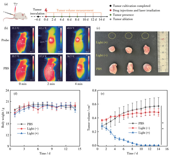
A mouse subcutaneous tumor model was established using 4T1 cells (the tumor volume of each mouse was approximately 200 mm3), and the tumor - bearing mice were divided into three groups of three mice each: (1) PBS, (2) ECEI (100 µL, 400 µmol·L-1), and (3) ECEI (100 µL, 400 µmol·L-1) with irradiation by a 660 nm laser (0.2 W·cm-2) for 10 min. The tumor volume and the body weight of the mice were monitored every two days, and the final tumors were harvested and measured at the end of PTT to assess the tumor inhibition effect of ECEI.
The experiments were performed in at least three parallel experiments and the results are expressed as mean±standard error. The data were evaluated by t-test and significant differences were considered statistically significant when *p < 0.05, **p < 0.01, and ***p < 0.001.
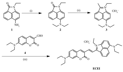
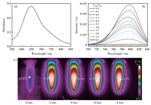
As a photothermal agent, it should be able to effectively absorb light and then transform it into a thermal effect. Therefore, we first studied its optical properties. The photophysical property parameters of ECEI are shown in Table 1. The absorption peak of ECEI in PBS was about 650 nm and trails to 850 nm (Fig. 1a). Moreover, ECEI exhibited fluorescence quenching in PBS (Fig. 1b), which indicates the potential of ECEI in PTT. The results of in vitro photothermal imaging showed that the temperature of ECEI increased to 71 ℃ within 8 min under the irradiation of 660 nm (0.38 W·cm-2) laser (Fig. 1c, 2a). Moreover, under the irradiation of a rated power laser, the temperature increased gradually with the increase of ECEI concentration (Fig. 2b). At the rated concentration, the temperature increased gradually as the laser power increased (Fig. 2c). Notably, in both cases, the ECEI temperature was stabilized above 60 ℃ within 8 min. These data indicate that ECEI has significant photothermal effects.
 下载:
导出CSV
下载:
导出CSV
| Probe | Solvent | λex / nm | λem / nm | ε / (L·mol-1·cm-1) | Φb | εφ / (L·mol-1·cm-1) |
| ECEI | PBS | 656 | — | 10 760 | 0.000 8 | 8.6 |
| EtOH | 710 | 800 | 18 756 | 0.030 6 | 573.9 | |
| a λex: excitation wavelength, λem: emission wavelength, ε: molar extinction coefficient, Φ: quantum yield of fluorescence, εφ: luminous brightness; b Rhodamine B (Ф=0.31/0.70 in water/EtOH) was chosen as the standard[37-38]. | ||||||
The concentration of ECEI was 10 µmol·L-1.
Next, we measured the PCE of ECEI by the method reported in the literature[39]. Encouragingly, ECEI demonstrates a very high PCE of about 85.78%. The temperature rose rapidly to about 72 ℃ within 8 min (Fig. 2a). This indicates that ECEI already possesses one of the characteristics of ideal photothermal agents: high PCE.
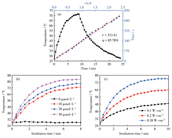
Excellent photostability is one of the essential elements of good photothermal agents. Therefore, we further measured the stability of ECEI. As shown in Fig. 3a, following irradiation with a 660 nm laser (0.2 W·cm-2), the UV - Vis absorption of ECEI reaches a steady state within two minutes. The maximum temperature of ECEI varied reversibly after four cycles with-out obvious loss (Fig. 3b). In addition, the complexity of the biological environment often causes the cracking of photothermic agents in vivo. Therefore, we evaluated the fluorescent intensity of ECEI when co-existing with different bio - related substances. As depicted in Fig. 3c, the fluorescent intensity of ECEI did not change when coexisting with different bio-related substances. These show that ECEI has another characteristic of ideal photothermal agents: good stability. These results indicate that ECEI with high PCE, excellent stability, and wide absorption band, would be expected to be a promising candidate for photothermal therapy.
(c)A: ECEI, B: Na+, C: Mg2+, D: K+, E: SO42-, F: HS-, G: Cl-, H: CO32-, I: SO32-, J: Glu, K: Cys, L: Gly, M: Hcy; I797: the fluorescent intensity at the wavelength of 797 nm.
The phototoxicity and dark toxicity of ECEI were performed on HeLa cells by CCK-8 assay. The results showed a concentration - dependent pattern (Fig. 4a), with the cell viability significantly lower in the light condition than in the non-light condition. Additionally, the cell viability decreased considerably with increased optical power density (Fig. 4b).

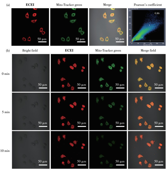
To demonstrate the localization ability of ECEI in cells, we co - stained HeLa cells with ECEI and Mito - Tracker Green. The images showed that the red fluorescence of ECEI overlapped well with the green fluorescence of the mitochondrial dye, and Pearson's correla-tion coefficient reached 0.86 (Fig. 5a). The co-localization results indicated that ECEI could accurately target mitochondria in living cells. On this basis, we further incubated the cells with CCCP (a reagent that causes loss of mitochondrial membrane potential) [40]. When the cells lost the mitochondrial membrane potential, the red fluorescence of ECEI in mitochondria did not decrease in intensity with time compared with the green fluorescence of Mito - Tracker (Fig. 5b). Thus, it can be inferred that ECEI can accurately locate mitochondria even under conditions of membrane potential damage.
Red channel: λex=633 nm, λem=(710±30) nm; Green channel: λex=488 nm, λem=(530±30) nm.
Based on the good photothermal conversion ability of ECEI, we investigated the photothermal ablation effect of ECEI on tumor cells using live/dead cell staining assay with Calcein - AM and PI. As shown in Fig. 6, fluorescent signals were seen in the green channel in both the probe group and the laser irradiation - only group. The fluorescent signal detected by the probe group in the red channel was due to the weak emission of ECEI. The probe+laser irradiation group showed obvious fluorescent signals in the red channel, while no fluorescent emission could be detected in the green channel. The experiments show that ECEI can effectively kill cancer cells by its photothermal effect under laser irradiation.
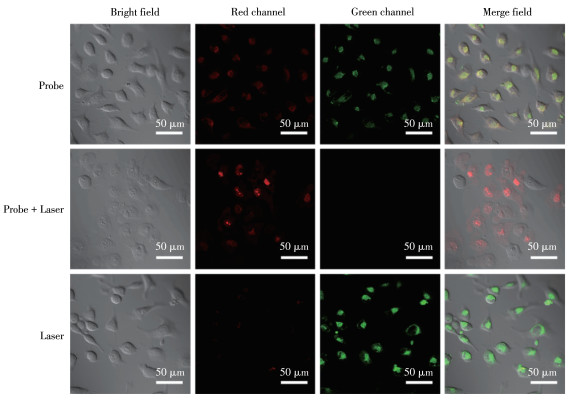
Red channel: λex=561 nm, λem=(620±30) nm; Green channel: λex=488 nm, λem=(530±30) nm.
The experimental process of photothermal therapy in vivo is depicted in Fig. 7a. To evaluate the tumor inhibition effect of photothermic agents on mouse models, we injected ECEI in situ into the mouse tumor, followed by irradiation (660 nm laser, 0.2 W·cm-2) at 10 min post - injection. Fig. 7b illustrates the results of tumor photothermal imaging, demonstrating that the temperature at the tumor site can reach nearly 50 ℃ after 4 min. Then, the body weights and tumor volumes were recorded during the subsequent 14 d (Fig. 7d, 7e). The weight of the three groups of mice did not change significantly, indicating negligible systemic cytotoxicity of ECEI during photothermal treatment. In the photothermal group, the tumor of mice disappeared on the tenth day after treatment and did not recur, indicating that ECEI effectively inhibited tumor growth. The tumor growth rates were similar in the without-irradiated and PBS - treated groups, suggesting that ECEI was non - toxic without light. Subsequently, the mice were treated on the 14th day and all the tumor tissues were stripped (Fig. 7c). All the above results indicated that ECEI had a superior inhibitory effect on tumor growth.
To sum up, we have successfully designed and synthesized photothermal agents (ECEI) based on small organic molecules. The photothermal agent shows excellent PCE (85.78%) and good photostability (after four cycles of heating and cooling without decay). Moreover, ECEI can be precisely located and stably present on mitochondria, inducing cell death upon laser irradiation. Remarkably, after a single photothermal treatment, the mouse tumors gradually disappeared within ten days and did not recur. This study provides a reference for improving the photothermal properties of organic small molecular dyes as photothermal agents and has positive significance for promoting photothermal treatment.
Ackroyd R, Kelty C, Brown N, Reed M. The history of photodetection and photodynamic therapy[J]. Photochem. Photobiol., 2001, 74(5): 656-669. doi: 10.1562/0031-8655(2001)074<0656:THOPAP>2.0.CO;2
Ferrari M. Cancer nanotechnology: Opportunities and challenges[J]. Nat. Rev. Cancer, 2005, 5: 161-171. doi: 10.1038/nrc1566
Zhang S, Xu J B, Chen H, Song Z F, Wu Y L, Dai X Y, Kong J. Acid-cleavable unimolecular micelles from amphiphilic star copolymers for triggered release of anticancer drugs[J]. Macromol. Biosci., 2017, 17(3): 1600258. doi: 10.1002/mabi.201600258
Duan X, Bai T, Du J J, Kong J. One - pot synthesis of glutathione-responsive amphiphilic drug self - delivery micelles of doxorubicin-disulfide-methoxy polyethylene glycol for tumor therapy[J]. J. Mater. Chem. B, 2018, 6(1): 39-43. doi: 10.1039/C7TB02817B
Li X S, Lovell J F, Yoon J Y, Chen X Y. Clinical development and potential of photothermal and photodynamic therapies for cancer[J]. Nat. Rev. Clin. Oncol., 2020, 17: 657-674. doi: 10.1038/s41571-020-0410-2
Liu Y J, Bhattarai P, Dai Z F, Chen X Y. Photothermal therapy and photoacoustic imaging via nanotheranostics in fighting cancer[J]. Chem. Soc. Rev., 2019, 48(7): 2053-2108. doi: 10.1039/C8CS00618K
Xie Z J, Fan T J, An J, Choi W, Duo Y H, Ge Y Q, Zhang B, Nie G H, Xie N, Zheng T T, Chen Y, Zhang H, Kim J S. Emerging combination strategies with phototherapy in cancer nanomedicine[J]. Chem. Soc. Rev., 2020, 49(22): 8065-8087. doi: 10.1039/D0CS00215A
Richter K, Haslbeck M, Buchner J. The heat shock response: Life on the verge of death[J]. Mol. Cell, 2010, 40(2): 253-266. doi: 10.1016/j.molcel.2010.10.006
Knavel E M, Brace C L. Tumor ablation: Common modalities and general practices[J]. Tech. Vasc. Interv. Radiol., 2013, 16(4): 192-200. doi: 10.1053/j.tvir.2013.08.002
Liu G X, Li B Q, Li J, Dong J X, Baulin V E, Feng Y J, Jia D C, Petrov Y V, Tsivadze A Y, Zhou Y. Photothermal carbon dots chelated hydroxyapatite filler: high photothermal conversion efficiency and enhancing adhesion of hydrogel[J]. ACS Appl. Mater. Interfaces, 2023, 15(48): 55335-55345. doi: 10.1021/acsami.3c11957
Irmania N, Dehvari K, Chang J Y. Multifunctional MnCuInSe/ZnS quantum dots for bioimaging and photodynamic therapy[J]. J. Biomater. Appl., 2022, 36(9): 1617-1628. doi: 10.1177/08853282211068959
Li B L, Zhao S J, Huang L, Wang Q, Xiao J F, Lan M H. Recent advances and prospects of carbon dots in phototherapy[J]. Chem. Eng. J., 2021, 408: 127245. doi: 10.1016/j.cej.2020.127245
Xu C, Pu K Y. Second near-infrared photothermal materials for com-binational nanotheranostics[J]. Chem. Soc. Rev., 2021, 50(2): 1111-1137. doi: 10.1039/D0CS00664E
Cheng L, Wang C, Feng L Z, Yang K, Liu Z. Functional nanomaterials for phototherapies of cancer[J]. Chem. Rev., 2014, 114(21): 10869-10939. doi: 10.1021/cr400532z
Zhou B J, Li Y Z, Niu G L, Lan M H, Jia Q Y, Liang Q L. Near-infrared organic dye-based nanoagent for the photothermal therapy of cancer[J]. ACS Appl. Mater. Interfaces, 2016, 8(44): 29899-29905. doi: 10.1021/acsami.6b07838
Peng F, Setyawati M I, Tee J K, Ding X G, Wang J P, Nga M E, Ho H K, Leong D T. Nanoparticles promote in vivo breast cancer cell intravasation and extravasation by inducing endothelial leakiness[J]. Nat. Nanotechnol., 2019, 14: 279-286. doi: 10.1038/s41565-018-0356-z
Xing M M, Han Y Y, Zhu Y L, Sun Y T, Shan Y Y, Wang K N, Liu Q X, Dong B L, Cao D X, Lin W Y. Two ratiometric fluorescent probes based on the hydroxyl coumarin chalcone unit with large fluorescent peak shift for the detection of hydrazine in living cells[J]. Anal. Chem., 2022, 94(37): 12836-12844. doi: 10.1021/acs.analchem.2c02798
Xin H T, Huang Y, Han Y Y, Tang L Y, Yang G Y, Zhang Y, Zhao S F, Wang K N, Li Y B, Cao D X. A two - photon iridium(Ⅲ) complex probe for sensitive detection of SO2 derivatives in living cell mitochondria[J]. Spectroc. Acta Pt. A - Molec. Biomolec. Spectr., 2023, 299(15): 122876.
Yoon H J, Lee H S, Lim J Y, Park J H. Liposomal indocyanine green for enhanced photothermal therapy[J]. ACS Appl. Mater. Interfaces, 2017, 9(7): 5683-5691. doi: 10.1021/acsami.6b16801
Chen Z, Zhao P F, Luo Z Y, Zheng M B, Tian H, Gong P, Gao G H, Pan H, Liu L L, Ma A Q, Cui H D, Ma Y F, Cai L T. Cancer cell membrane-biomimetic nanoparticles for homologous-targeting dual-modal imaging and photothermal therapy[J]. ACS Nano, 2016, 10(11): 10049-10057. doi: 10.1021/acsnano.6b04695
Jung H S, Verwilst P, Sharma A, Shin J, Sessler J L, Kim J S. Organic molecule - based photothermal agents: An expanding photothermal therapy universe[J]. Chem. Soc. Rev., 2018, 47(7): 2280-2297. doi: 10.1039/C7CS00522A
Van der Velde J H M, Oelerich J, Huang J Y, Smit J H, Jazi A A, Galiani S, Kolmakov K, Guoridis G, Eggeling C, Herrmann A, Roelfes G, Cordes T. A simple and versatile design concept for fluorophore derivatives with intramolecular photostabilization[J]. Nat. Commun., 2016, 7: 10144. doi: 10.1038/ncomms10144
Lei Z H, Zhang F. Molecular engineering of NIR-Ⅱ fluorophores for improved biomedical detection[J]. Angew. Chem. Int. Ed., 2021, 60(30): 16294-16308. doi: 10.1002/anie.202007040
Cheng P H, Pu K Y. Molecular imaging and disease theranostics with renal-clearable optical agents[J]. Nat. Rev. Mater., 2021, 6: 1095-1113. doi: 10.1038/s41578-021-00328-6
Wang H Y, Chang J J, Shi M W, Pan W, Li N, Tang B. A dual -targeted organic photothermal agent for enhanced photothermal therapy[J]. Angew. Chem. Int. Ed., 2019, 58(4): 1057-1061. doi: 10.1002/anie.201811273
Zhang L P, Kang L, Li X Q, Liu S Y, Liu T L, Zhao Y X. Pyrazino[2, 3-g] quinoxaline-based nanoparticles as near-infrared phototheranostic agents for efficient photoacoustic-imaging-guided photothermal ther-apy[J]. ACS Appl. Nano Mater., 2021, 4(2): 2019-2029. doi: 10.1021/acsanm.0c03346
Li C N, Lin W H, Liu S, Sun T T, Xie Z G. Structural optimization of organic fluorophores for highly efficient photothermal therapy[J]. Mater. Chem. Front., 2021, 5(1): 284-292. doi: 10.1039/D0QM00624F
Wang Z, Liu Y, He C X, Zhang X M, Li X, Li Y Y, Tang Y F, Lu X M, Fan Q L. Small-molecule phototheranostic agent with extended π-conjugation for efficient NIR-Ⅱ photoacoustic-imaging-guided photo-thermal therapy[J]. Small, 2024, 20(17): 2307829. doi: 10.1002/smll.202307829
Li S L, Deng Q Y, Zhang Y C, Li X Z, Wen G H, Cui X, Wan Y P, Huang Y W, Chen J X, Liu Z H, Wang L D, Lee C S. Rational design of conjugated small molecules for superior photothermal theranostics in the NIR -Ⅱ biowindow[J]. Adv. Mater., 2020, 32(33): 2001146. doi: 10.1002/adma.202001146
Jia W Y, Huang F F, Zhang Q, Zhao L L, Li C X, Lu Y. Novel conjugated small molecule-based nanoparticles for NIR - Ⅱ photothermal antibacterial therapy[J]. Chem. Commun., 2022, 58(43): 6340-6343. doi: 10.1039/D2CC00863G
Yang C Y, Guo L X, Zhang K X, Wang G, Yu Q S, Gan Z H, Gu X G. Diradical-featured organic small-molecule photothermal material based on 4, 6-di(2-thienyl)thieno[3, 4-c][1, 2, 5]thiadiazole for photothermal immunotherapy[J]. Adv. Funct. Mater., 2023, 33(52): 2306360. doi: 10.1002/adfm.202306360
Wu X J, Cai Y, Wang C, Fan L, Tang Q Y, Dong X C, Zhang Q. Tumor - targeting nanoparticles of small - molecule diketopyrrolopyr-role derivative for photothermal therapy[J]. J. Nanosci. Nanotechnol., 2018, 18(4): 2337-2344. doi: 10.1166/jnn.2018.14352
Cheng Z J, Zhang T, Wang W L, Shen Q, Hong Y, Shao J J, Xie X J, Fei Z H, Dong X C. D - A - D structured selenadiazolesbenzothiadia-zole - based near - infrared dye for enhanced photoacoustic imaging and photothermal cancer therapy[J]. Chin. Chem. Lett., 2021, 32(4): 1580-1585. doi: 10.1016/j.cclet.2021.02.017
Yue Y K, Xu Z, Ma K Q, Huo F J, Qin X M, Zhang K S, Yin C X. HSA shrinkage optimizes the photostability of embedded dyes fundamentally to amplify their efficiency as photothermal materials[J]. Chin. Chem. Lett., 2024, 35(8): 109223. doi: 10.1016/j.cclet.2023.109223
Jiang F, Hu Q H, Zhang Z M, Li H M, Li H L, Zhang D W, Li H W, Ma Y, Xu J J, Chen H F, Cui Y, Zhi Y L, Zhang Y M, Xu J Y, Zhu J P, Lu T, Chen Y D. Discovery of benzo[cd]indol-2(1H)-ones and pyrrolo[4, 3, 2-de]quinolin-2(1H) - ones as bromodomain and extra-terminal domain (BET) inhibitors with selectivity for the first bromodomain with potential high efficiency against acute gouty arthritis[J]. J. Med. Chem., 2019, 62(24): 11080-11107. doi: 10.1021/acs.jmedchem.9b01010
Yin G X, Niu T T, Gan Y B, Yu T, Yin P, Chen H M, Zhang Y Y, Li H T, Yao S Z. A multi -signal fluorescent probe with multiple binding sites for simultaneous sensing of cysteine, homocysteine, and glutathione[J]. Angew. Chem., Int. Ed., 2018, 57(18): 4991-4994. doi: 10.1002/anie.201800485
Beija M, Afonso C A M, Martinho J M G. Synthesis and applications of Rhodamine derivatives as fluorescent probes[J]. Chem. Soc. Rev., 2009, 38(8): 2410-2433. doi: 10.1039/b901612k
Jiang G W, Ren T B, D'Este E, Xiong M Y, Xiong B, Johnsson K, Zhang X B, Wang L, Yuan L. A synergistic strategy to develop photo-stable and bright dyes with long stokes shift for nanoscopy[J]. Nat. Commun., 2022, 13: 2264. doi: 10.1038/s41467-022-29547-3
Huang P, Lin J, Li W W, Rong P F, Wang Z, Wang S J, Wang X P, Sun X L, Aronova M, Niu G, Leapman R D, Nie Z H, Chen X Y. Biodegradable gold nanovesicles with an ultrastrong plasmonic coupling effect for photoacoustic imaging and photothermal therapy[J]. Angew. Chem., Int. Ed., 2013, 52(52): 13958-13964. doi: 10.1002/anie.201308986
Zhang X F, Sun Q, Huang Z L, Huang L R, Xiao Y. Immobilizable fluorescent probes for monitoring the mitochondria microenvironment: A next step from the classic[J]. J. Mater. Chem. B, 2019, 7(17): 2749-2758. doi: 10.1039/C9TB00043G

Scheme1 1 Synthesis diagram of ECEI
(ⅰ) CH3CH2I, KOH, K2CO3, 2-methoxyethanol; (ⅱ) CH3MgCl, THF; (ⅲ) piperidine, EtOH.

Figure 1 (a) UV absorption spectrum in PBS, (b) fluorescence emission spectra in different volume ratios of EtOH and PBS, (c) photothermal imaging by laser irradiation in vitro of ECEI
The concentration of ECEI was 10 µmol·L-1.

Figure 2 (a) Determination of PCE of ECEI (10 µmol·L-1) under laser irradiation; (b) Heating curves of different concentrations of ECEI in PBS at a power density of 0.38 W·cm-2; (c) Heating curves of ECEI (20 µmol·L-1) in PBS under irradiation with various laser power densities

Figure 3 (a) UV-Vis absorbance spectra of ECEI (10 µmol·L-1) under laser irradiation; (b) Four irradiation-cooling cycles of ECEI (20 µmol·L-1) under laser irradiation; (c) Fluorescent intensity of ECEI (10 µmol·L-1) at 660 nm after 30 min response with different substances (200 µmol·L-1)
(c)A: ECEI, B: Na+, C: Mg2+, D: K+, E: SO42-, F: HS-, G: Cl-, H: CO32-, I: SO32-, J: Glu, K: Cys, L: Gly, M: Hcy; I797: the fluorescent intensity at the wavelength of 797 nm.

Figure 4 (a) Cytotoxicity of different concentrations of ECEI after incubation for 30 min and laser irradiation for 5 min; (b) Cytotoxicity after incubation for 30 min and laser irradiation for 5 min under different light power

Figure 5 (a) Colocalization test of ECEI (10 µmol·L-1) in mitochondria; (b) Confocal fluorescence imaging of HeLa cells treated with CCCP (10 µmol·L-1) after incubation with ECEI (10 µmol·L-1) and Mito-Tracker Green (0.5 µmol·L-1)
Red channel: λex=633 nm, λem=(710±30) nm; Green channel: λex=488 nm, λem=(530±30) nm.

Figure 6 Fluorescence imaging of ECEI (10 µmol·L-1) pre-incubated HeLa cells after laser and no laser co-stained with Calcein-AM and PI
Red channel: λex=561 nm, λem=(620±30) nm; Green channel: λex=488 nm, λem=(530±30) nm.

Figure 7 (a) Experimental procedure for photothermal treatment of tumor; (b) Photothermal imaging of ECEI (400 µmol·L-1) by 0.2 W·cm-2 laser irradiation in vivo; (c) Tumor tissue of tumor mice under different conditions; (d) Body weights of the mice during the observation; (e) Tumor volume of the mice during the observation (n=3, **p < 0.01)
Table 1. Photophysical characterization of ECEIa
| Probe | Solvent | λex / nm | λem / nm | ε / (L·mol-1·cm-1) | Φb | εφ / (L·mol-1·cm-1) |
| ECEI | PBS | 656 | — | 10 760 | 0.000 8 | 8.6 |
| EtOH | 710 | 800 | 18 756 | 0.030 6 | 573.9 | |
| a λex: excitation wavelength, λem: emission wavelength, ε: molar extinction coefficient, Φ: quantum yield of fluorescence, εφ: luminous brightness; b Rhodamine B (Ф=0.31/0.70 in water/EtOH) was chosen as the standard[37-38]. | ||||||
 下载: 导出CSV

 扫一扫看文章

扫一扫关注我们
 下载:
下载: