
Scheme 1.
Synthesis of CPTH

It has been well-known to us that certain metal ions play an indispensable role in animals, plants and humans of nature. For example, copper is the third richest metal element in our body (after zinc and iron) and essential trace elements for life activities. Lack of copper in the human body can cause anemia, abnormal hair, arteries, and even brain disorders[1-2]. However, excess of copper ions will cause severe neurodegenerative diseases like Wilson disease[3], Alzheimer's disease[4], hypopigmentation, bone abnormalities. In addition, excessive copper as a pollutant causes a huge threat to the environment and ecosystem. The World Health Organization (WHO) determined copper ion as one of the most concerning chemical elements in drinking water. The United States Environmental Protection Agency (USEPA) has set the minimum detection limit of copper ion in potable water to 20 μmol·L-1 [5]. For this reason, sensitive, selective and biocompatible approaches for the detection of copper ions are important from an environmental or biological perspective.
There are many traditional analytical techniques for detecting copper ions. For example, atomic emission and absorption spectrometry[6-7], mass spectrometry[8] are widely used in recent years. However, the above mentioned methods have obvious shortcomings, such as high cost, tedious sample preparation procedures, and susceptible to interference from other ions. Alternatively, the fluorescent sensors with high selectivity, real time detection and low detection limits[9-14] for various heavy metal ions stand out and have received increasing attention recently.
A great deal of fluorescent sensors, derived from rhodamine, coumarin, and 1, 8 naphthalimide, etc., have been documented for detecting copper ions[15-16]. Among them, some rhodamine or 1, 8-naphthalimide-based probes are reported with some shortcomings. For example, rhodamine based probes have shown poor selectivity, interfering with other transition metals such as Co2+ [17] and Fe3+ [18]. 1, 8-Naphthalimide-based fluorescent probes may have high detection limits while their linear range is not clear enough[19]. In contrast, coumarin derivatives possess a series of excellent fluorescence properties, including photostability, low cytotoxicity, large Stokes displacement and high fluorescence quantum yield[20-25], and they have been inclusively utilized to prepare fluorescent sensors for the identification of various heavy metal ions. However, most of the known coumarin fluorescent probes face the problems of selectivity, sensitivity and biocompatibility[26-27]. It still remains a challenge to develop novel coumarin fluorescent probes for recognizing heavy metal ions with excellent selectivity, low detection limit and fast response time.
In this work, we try to combine the pyridine triazole (2-(5-(pyridine-3-yl) -4H-1, 2, 4 -triazol-3-yl)acetohydrazide) and coumarin moieties via a Schiff base bridge to build a CPTH molecule with rich coordination sites and extended π system (Scheme 1). And then, the photochemical properties and possible fluorescent sensors for certain metal ions have been explored for this coumarin derivative. The designed CPTH exhibited strong yellow fluorescence (excitation: 470 nm, emission: 520 nm), and the fluorescence was quenched quickly by adding copper ions. The detection limit of CPTH was found as low as 2 μmol·L-1, which is analogous to highly sensitive coumarin-based chemosensors[26]. Moreover, CPTH showed a high selectivity for Cu2+ without interfered by many other common metal ions.
Materials and measurement methods can be found in Support information.
Compound L3-Et (0.232 g, 1.0 mmol) and hydrazine hydrate (0.1 mL, 80% concentrated, 1.64 mmol) was added in absolute ethanol (20 mL). The solution was mixed together and refluxed well at 80 ℃ overnight until TLC demonstrated the finish of the reaction, then a portion of solvent was evaporated and concentrated under vacuumed condition by rotary evaporator. The obtained crude product was precipitated and filtered from the remaining solvent, then recrystallized from absolute ethanol to get white powder of 2 (0.175 g, Yield: 82%). m. p. 150~151 ℃. 1H NMR (400 MHz, DMSO-d6):δ 9.47 (s, 1H), 9.15 (dd, J=2.2, 0.8 Hz, 1H), 8.81 (dd, J=4.8, 1.6 Hz, 1H), 8.50~8.20 (m, 1H), 7.65 (m, 1H), 4.38 (s, 2H), 3.92 (s, 2H). 13C NMR (101 MHz, DMSO d6):δ 165.0, 163.2, 163.1, 153.0, 147.5, 134.6, 124.9, 120.4, 31.4. LC/MS: m/z=242.25 for [M+ Na] +. FT IR (KBr, cm-1): 3 382 (m), 3 276 (s), 3 177 (w), 1 661 (vs), 1 526 (m), 1 411 (s), 1 247 (w), 1 015 (m). Anal. Calcd. for C9H10N6O(%): C, 49.54; H, 4.62;N, 38.51. Found(%): C, 49.38; H, 4.88; N, 38.27.
Compound 1 (0.245 g, 1.0 mmol) and compound 2 (0.218 g, 1.0 mmol) in absolute ethanol (20 mL) was mixed and stirred at 80 ℃ for 24 h until the TLC showed the reaction was almost finished. The obtained yellow solution was cooled to room temperature and yellow solid was precipitated and filtered from the solution. The obtained crude product was recrystallized from ethanol to give orange powder of CPTH (0.356 g, Yield: 80%). m. p. 234~235 ℃. 1H NMR (400 MHz, DMSO d6):δ 11.83 (s, 1H), 9.19 (s, 1H), 8.82 (dd, J= 4.8, 1.7 Hz, 1H), 8.42~8.36 (m, 1H), 8.34 (s, 1H), 8.08 (s, 1H), 7.66 (m, 1H), 7.48 (d, J=9.0 Hz, 1H), 6.76 (dd, J=9.0, 2.3 Hz, 1H), 6.58 (d, J=2.2 Hz, 1H), 4.56 (s, 2H), 4.13 (s, 1H), 3.47 (q, J=6.8 Hz, 4H), 1.14 (t, J= 7.0 Hz, 6H). 13C NMR (101 MHz, DMSO -d6):δ 167.0, 163.6, 162.0, 161.6, 157.1, 152.4, 151.8, 147.9, 140.4, 139.3, 134.3, 130.5, 123.8, 120.4, 112.3, 109.8, 108.6, 97.2, 45.1, 31.4, 29.7, 12.5. LC/MS: m/z=447.20 for [M+H]+, m/z=469.20 for [M+Na]+. FT IR (KBr, cm-1): 2 974 (vw), 1 691 (vs), 1 627 (s), 1 602 (vs), 1 579 (m), 1 361 (w), 1 348 (w), 1 261 (m). Anal. Calcd. for C23H23N7O3(%): C, 62.01; H, 5.20; N, 22.01. Found(%): C, 61.90; H, 5.27; N, 21.87. Single crystals of CPTH suitable for X ray diffraction measurement were obtained by slow evaporation of a mixture of CHCl3 in air for one week.
CCDC: 1967788, CPTH.
The spectral analyses were done in a H2O-DMF PBS buffer (phosphate buffer saline) (10 mmol·L-1, pH=7.40, 1∶9, V/V) solution at 25 ℃. The concentration of CPTH was 10 μmol·L-1. Solutions of Ag+, Al3+, Ba2+, Cd2+, Ca2+, Co2+, Cu2+, Cr3+, Fe2+, Fe3+, K+, Hg2+, Mg2+, Mn2+, Na+, Zn2+ and Pb2+ ions were prepared with nitrate or acetate salts in water. The excitation wavelength was 470 nm and the fluorescence emission spectra were recorded within the scope of 480~650 nm.
The coumarin unit was first combined with the pyridine triazole unit to construct a new fluorescent sensor CPTH. As shown in Scheme 1, CPTH was prepared with a satisfactory yield through the imine condensation reaction between precursors 1 and 2 under the refluxing condition. The successful preparation of CPTH was confirmed by 1H/13C NMR, FTIR and ESIMS. It is worth noting that the single peak of the aldehyde proton in compound 1 at δ 10.13 disappeared after the Schiffbase condensation and the two amine protons of azine group at δ 3.92 were gone in the other starting compound 2 simultaneously. Instead, a new single peak at δ 7.66 was observed in CPTH, indicative of the formation of imine unit. At the same time, two methylene protons were low field shifted from δ 4.38 to 4.56 in 2 and CPTH. In addition, an LC/MS peak of CPTH was found at m /z=447.20 with the 100% abundance corresponding to the expected molecular ion peak. FT-IR spectral comparisons revealed the disappearance of 1 677 cm-1 in 1, 3 382 and 3 276 cm-1 in 2, and the emergence of 1 627 cm-1 in CPTH, corresponding to the functional radical transformation from aldehyde and amine to imine groups. Meanwhile, the absorption peak for carbonyl group of coumarin unit was shifted from 1 717 cm-1 in 1 to 1 691 cm-1 in CPTH after the Schiff base condensation, which was consistent with the alteration of force constant (k) for C=O and C=N bonds.
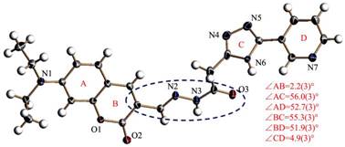
In order to further characterize our compound CPTH, its singlecrystal structure has been successfully acquired. The molecular structure of CPTH with atom-numbering schemes is shown in Fig. 1 and details of the data collection and refinement results are listed in Table 1. CPTH crystallizes in the orthorhombic Pbcn space group without the presence of any solvent molecule. The dihedral angles between the aromatic rings at both ends of CPTH are small, and the planarity of coumarin moiety can be extended to the carbonyl oxygen atom of the hydrazide unit. The central six nonhydrogen atoms (C3N2O) circled in Fig. 1 have the mean deviation of 0.002 4(4) nm from the least squares plane. However, the π-conjugated system is destroyed by the presence of an sp3 carbon atom linked to the triazole ring. This structural feature is consistent with the UV Vis spectrum of CPTH, as illustrated in Fig. SI1. In the crystal structure of CPTH, an offset dimeric packing mode is observed with a head-to-tail manner, as displayed in Fig. 2, where strong π-π stacking interactions are discovered between adjacent benzene and pyrone rings of coumarin moiety with the centroid centroid separation of 0.379(1) nm. In addition, every dimeric pair is linked with four close dimeric units via two types of weaker π-π stacking interactions. One comes from neighboring coumarin benzene rings with the longer centroid centroid distance of 0.397(1) nm, and the other stems from adjacent protruding triazole and pyrimidine rings with double complementary π-π stacking interactions (0.392(1) nm).
 下载:
导出CSV
下载:
导出CSV
| Empirical formula | C23H23N7O3 | Dc/(g·cm-3) | 1.389 |
| Formula weight | 445.48 | F(000) | 1 872 |
| Temperature/K | 190(2) | μ/mm-1 | 0.096 |
| Wavelength/nm | 0.071 07 | hmin, hmax | -9, 9 |
| Crystal size / mm | 0.10×0.10×0.11 | kmin, kmax | -15, 10 |
| Crystal system | Orthorhombic | lmin, lmax | -43, 47 |
| Space group | Pbcn | Data, parameter | 3 885, 304 |
| a/nm | 0.820 6(3) | Final R indices [I > 2σ(I)]* | R1=0.060 0, wR2=0.151 7 |
| b/nm | 1.312 8(4) | R indices (all data) | R1=0.115 9, wR2=0.185 0 |
| c/nm | 3.955 5(14) | S | 1.023 |
| V/nm3 | 4.261(3) | (Δρ)max, (Δρ)min/(e·nm-3) | 437, -544 |
| Z | 8 | ||
| *R1=Σ║Fo|-|Fc║/Σ|Fo|, wR2={Σ[w(Fo2-Fc2)2]/Σw(Fo2)2}1/2 | |||

As shown in Fig. SI1, the maximum absorption wavelength of compound 1 was located at 449 nm, indicative of the π-conjugated system of coumarin backbone. In contrast, no obvious absorption peak was found in 2 revealing the lack of extended π-system within the molecule. After the imine condensation, the maximum absorption wavelength of CPTH was the same as that of 1, indicating their analogous π-conjugated skeleton just by changing the heteroatoms from O to N.
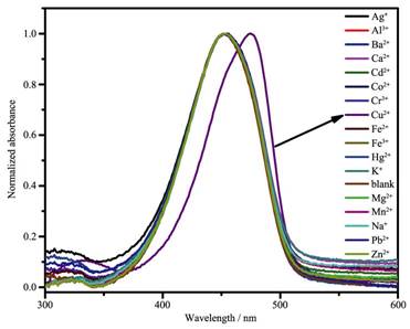
Since CPTH has multiple coordinating sites, we tried to explore its coordination with common metal ions. As shown in Fig. 3, when CPTH was mixed with Cu2+ ions, the maximum absorption wavelength was bathochromically shifted to 475 nm, whereas the addition of the other metal ions had no evident impact on the resultant absorption peak. Considering the presence of fluorescence active coumarin moiety, fluorescence spectrum was further studied for the interactions between this CPTH molecule and a variety of metal ions.
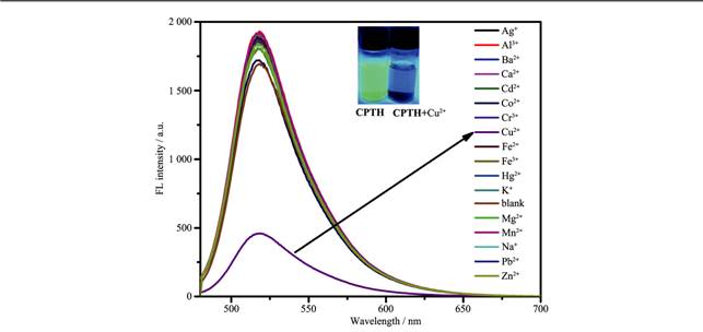
The rigid planar structure of the coumarin molecule gave it a large conjugated π bond, so most of the coumarin derivatives had fluorescence. As shown in Fig. 4, CPTH exhibited very strong yellow fluorescence (λex=480 nm, λem=520 nm) in the buffered DMF/PBS (10 mmol·L-1, pH=7.40, 1∶9, V/V) solution, which was similar to those reported in literature[28]. After the addition of 1.0 equiv. of Cu2+, the fluorescence of CPTH was instantly quenched. However, the addition of other common metal ions had no effect on the fluorescence spectrum of CPTH, suggesting excellent selectivity of CPTH to copper ions. The fluorescence quenching phenomenon may be ascribed to the selective combination of CPTH with Cu2+ ion[29-32].
The above experimental results showed that CPTH may have potential applications in the selective identification of copper ions, so the disturbance of other coexistent heavy metal cations was also carried out (Fig. 5a). CPTH (20 μmol·L-1) was mixed with 2.0 equiv. of Cu2+ in the presence of 2.0 equiv. of other common metal cations (Ag+, Al3+, Ba2+, Cd2+, Ca2+, Co2+, Cu2+, Cr3+, Fe2+, Fe3+, K+, Hg2+, Mg2+, Mn2+, Na+, Zn2+ and Pb2+ ions). The results displayed that the coexisting metal ions had negligible effects on the fluorescence quenching of CPTH, indicating excellent selective detection of Cu2+ under physiological conditions.
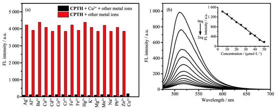
In order to assess the sensitivity limit of CPTH for Cu2+ detection, fluorescence spectral titrations were implemented. As depicted in Fig. 5b, the fluorescence intensity of the peak of CPTH (5 μmol·L-1) at 520 nm gradually decreased with the increase of Cu2+ concentration (from 0 to 2.0 equiv.), indicating concentration dependent fluorescence quenching. This observation can be explained by the formation of CPTH-Cu2+ complex. The detection limit of CPTH for Cu2+ was calculated to be 2 μmol·L-1, which was gotten by the linear fitting of Cu2+ concentrations from 10 to 50 μmol·L-1 (Inset of Fig. 5b). Specifically, the detection limit was calculated according to the formula of LOD=3σ/k, where k is the slope of the linear equation of y=kx+b between fluorescence intensity and concentration. The detection limit of CPTH for copper ions was far below the standard limit (20 μmol·L-1) for Cu2+ in potable water regulated by the USEPA[10].
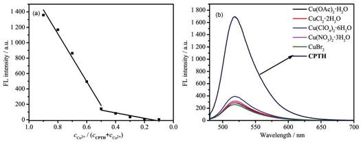
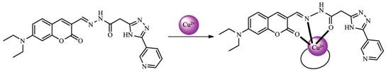
Job's plot was conducted to determine the stoichiometric ratio between Cu2+ and CPTH. The total concentration of Cu2+ and CPTH was 10 μmol·L-1. As can be seen in Fig. 6a, the inflection point was found approximately 0.5 molar fraction indicating a 1:1 stiochiometric ratio between Cu2+ and CPTH. It could be further confirmed by an ESI MS peak at m/z=509.42 (Fig.SI10) with the 100% abundance, corresponding to the molecular ion peak of [Cu2++CPTH] species. Based upon above-mentioned results and the molecular structure of CPTH, it is deduced that CPTH serves as a tridentate ligand to coordinate with Cu2+ in the 1:1 chelating fashion, where two fused five membered (CN2OCu) and six-membered (C3NOCu) chelating rings are formed, as displayed in Scheme 2. In addition, Cu(OAc)2·H2O, CuCl2·2H2O, Cu(ClO4)2 ·6H2O, CuBr2 and Cu(NO3)2·3H2O were used to explore the CPTH's antiinterference ability to anions. The results were given in Fig. 6b, in which the fluorescence quenching of CPTH was independent of the tested anions suggesting the strong anti-interference to anions for CPTH.
One of the main advantages of fluorescent probes for detecting metal ions compared to other traditional methods was their real time detection. Thus, the impact of the reaction time on the binding efficiency of Cu2+ ion to CPTH was investigated. As pictured in Fig. SI2. After the adding of 1.0 equiv. Cu2+ ion to CPTH solution (5 μmol·L-1), the fluorescence intensity of CPTH reached a stable value within 5 s and kept unchangeable from 5 to 120 s. Thus, a reaction time was determined as 5 s in this probe detection system. Moreover, the effect of pH stability on CPTH was tested when interacting with copper ions, where the fluorescent probe CPTH showed a relatively wide pH value range of 4~7.5 for the detection of copper ions (Fig. SI3).
In summary, the pyridine triazole unit was first used to construct a coumarin based fluorescent sensor via a simple synthetic approach. After the adding of Cu2+ ions, the maximum absorption wavelength of CPTH was red-shifted from 450 to 475 nm. And fluorescence quenching of CPTH was observed at 520 nm under the excitation of 470 nm, which did not take effect for other metal ions. Probe CPTH is highly selective for Cu2+ detection. This may be attributed to the fact that the Jahn-Teller deformation of Cu complexes provides excellent thermodynamic stability among all metal cations, so the d9 block metal Cu cation was preferentially used as the target ion during the metal transfer process. Job′s plot revealed that the binding ratio of copper ion to probe CPTH is 1:1, which could be further confirmed by the presence of a molecular ion peak for [Cu2++ CPTH] species in ESI-MS. Copper ion detection limit of probe CPTH was measured to be 2 μmol·L-1. Our contribution herein could provide certain new insights for exploring highly sensitive and selective coumarin based fluorescent sensors derived from aromatic heterocyclic Schiff base with multiple coordinating atoms.
Supporting information is available at http://www.wjhxxb.cn
Zhao H, Feng P, Cao J, et al. Org. Biomol. Chem., 2012, 10:3104-3109 doi: 10.1039/c2ob06980f
Halfdanarson T R, Kumar N, Li R L, et al. Eur. J. Haematol., 2008, 80:523-531 doi: 10.1111/j.1600-0609.2008.01050.x
Bull P C, Thomas G R, Rommens J M, et al. Nat. Genet., 1993, 5:323-337 doi: 10.1038/ng1293-323
Barnham K J, Masters C L, Bush A L, et al. Nat. Rev. Drug Discov., 2004, 3:205-214 doi: 10.1038/nrd1330
Chang H Q, Zhao X L, Wu W N, et al. J. Lumin., 2017, 182:268-273 doi: 10.1016/j.jlumin.2016.10.041
Otero-Romani J, Moreda-Pineiro A, Bermejo-Barrera A, et al. Anal. Chim. Acta, 2005, 536:213-218 doi: 10.1016/j.aca.2004.12.046
Gonzales A P S, Firmino M A, Nomura C S, et al. Anal. Chim. Acta, 2009, 636:198-204 doi: 10.1016/j.aca.2009.01.047
Becker J S, Matusch A, Depboylu C, et al. Anal. Chem., 2007, 79:6074-6080 doi: 10.1021/ac0700528
Xu Z, Zhang L, Guo R, et al. Sens. Actuators B, 2011, 156:546-552 doi: 10.1016/j.snb.2011.01.066
Xu Z H, Wang H W, Hou X F, et al. Sens. Actuators B, 2014, 201:469-474 doi: 10.1016/j.snb.2014.05.026
Qin J C, Yang Z Y, Wang G Q, et al. Tetrahedron Lett., 2015, 56:5024-5029 doi: 10.1016/j.tetlet.2015.07.023
Qin J C, Yang Z Y, Wang G Q, et al. J. Photochem. Photobiol. A, 2015, 310:122-127 doi: 10.1016/j.jphotochem.2015.05.010
徐帆, 吴芳辉, 程立春, 分析科学学报, 2016, 32(3):335339XU Fan, WU Fang-Hui, CHENG Li-Chun, et.al. J. Anal. Sci., 2016, 32(3):335339
齐琪, 李星.无机化学学报, 2019, 35(7):1301-1311 http://www.wjhxxb.cn/wjhxxbcn/ch/reader/view_abstract.aspx?file_no=20190723&flag=1QI Qi, LI Xing. Chinese J. Inorg Chem., 2019, 35(7):1301-1311 http://www.wjhxxb.cn/wjhxxbcn/ch/reader/view_abstract.aspx?file_no=20190723&flag=1
Xu J, Hou Y, Ma Q, et al. Spectrochim. Acta A, 2014, 124:416-422 doi: 10.1016/j.saa.2014.01.046
张长丽, 张虹, 何凤云, 等.无机化学学报, 2019, 35(10):1869-1876 doi: 10.11862/CJIC.2019.207ZHANG Chang-Li, ZHANG Hong, HE FengYun, et al. Chinese J. Inorg Chem., 2019, 35(10):1869-1876 doi: 10.11862/CJIC.2019.207
Li C R, Yang Z Y, Li S R, et al. J. Lumin., 2018, 198:327336
Gahlyan P, Bawa R, Jain H, et al. Chemistry Select, 2019, 4:7532-7540
Yuan L, Lin W, Chen B, et al. Org. Lett., 2012, 14:432-435 doi: 10.1021/ol202706k
Goswami S, Sen D, Das A K, et al. Sens. Actuators B, 2013, 183:518-525 doi: 10.1016/j.snb.2013.04.005
Wang M, Wen J, Qin Z, et al. Dyes Pigm., 2015, 120:208-212 doi: 10.1016/j.dyepig.2015.04.013
Shiraishi Y, Nakamura M, Hirai T, et al. Phys. Chem. Chem. Phys., 2015, 17:25027-25036 doi: 10.1039/C5CP03877D
Maiti S, Aydin Z, Zhang Y, et al. Dalton Trans., 2015, 44:8942-8949 doi: 10.1039/C4DT03792H
He L, Lin W, Xu Q, et al. Chem. Commun., 2015, 51:15101513
Kim K B, Kim H, Song E J, et al. Dalton Trans., 2013, 42:16569-16577 doi: 10.1039/c3dt51916c
Wang S, Wang Z, Yin Y, et al. J. Photochem. Photobiol. A, 2017, 333:213-219 doi: 10.1016/j.jphotochem.2016.10.030
Mergu N, Kim M, Son Y A, et al. Spectrochim. Acta A, 2018, 188:571-580 doi: 10.1016/j.saa.2017.07.047
Saini N, Prigyai N, Wannasiri C, et al. J. Photochem. Photobiol. A, 2018, 358:215-225 doi: 10.1016/j.jphotochem.2018.03.018
Borase P N, Thale P B, Shankarling G S, et al. Dyes Pigm., 2016, 134:276-284 doi: 10.1016/j.dyepig.2016.07.025
Huang L, Cheng J, Xie K, et al. Dalton Trans., 2011, 40:10815-10817 doi: 10.1039/c1dt11123j
Yang W, Chen X, Su H, et al. Chem. Commun., 2015, 51:9616-9619 doi: 10.1039/C5CC00787A
Su H, Chen X, Fang W, et al. Anal. Chem., 2014, 86:891-899 doi: 10.1021/ac4034592

图 1 ORTEP drawing of compound CPTH with the atom-numbering scheme
Displacement ellipsoids are drawn at the 30% probability level and the H atoms are shown as small spheres of arbitrary radii

图 2 Perspective view of the crystal packing of compound CPTH with the labeling of π-π stacking interactions

图 4 Fluorescence emission spectra of CPTH (10 μmol·L-1) after adding different metal ions
Excitation wavelength was 470 nm; Inset: digital photo showing the fluorescence color of CPTH and CPTH with Cu2+

图 5 (a) Fluorescence quenching response of CPTH (20 μmol·L-1) towards Cu2+ (2.0 equiv.) in the presence of other metal cations (2.0 equiv.); (b) Fluorescence spectra titration of CPTH (5 μmol·L-1) upon addition of Cu2+
Inset: fluorescence at 520 nm as a function of Cu2+ ion concentration

图 6 (a) Job's plot of the CPTH; (b) Fluorescence spectra of probe CPTH (10 μmol·L-1) upon addition of different copper salts
表 1 Crystal structural refinement details for compound CPTH
| Empirical formula | C23H23N7O3 | Dc/(g·cm-3) | 1.389 |
| Formula weight | 445.48 | F(000) | 1 872 |
| Temperature/K | 190(2) | μ/mm-1 | 0.096 |
| Wavelength/nm | 0.071 07 | hmin, hmax | -9, 9 |
| Crystal size / mm | 0.10×0.10×0.11 | kmin, kmax | -15, 10 |
| Crystal system | Orthorhombic | lmin, lmax | -43, 47 |
| Space group | Pbcn | Data, parameter | 3 885, 304 |
| a/nm | 0.820 6(3) | Final R indices [I > 2σ(I)]* | R1=0.060 0, wR2=0.151 7 |
| b/nm | 1.312 8(4) | R indices (all data) | R1=0.115 9, wR2=0.185 0 |
| c/nm | 3.955 5(14) | S | 1.023 |
| V/nm3 | 4.261(3) | (Δρ)max, (Δρ)min/(e·nm-3) | 437, -544 |
| Z | 8 | ||
| *R1=Σ║Fo|-|Fc║/Σ|Fo|, wR2={Σ[w(Fo2-Fc2)2]/Σw(Fo2)2}1/2 | |||
 下载: 导出CSV

 扫一扫看文章

扫一扫关注我们


 下载:
下载: