Table 1.
Crystal data and structure refinement for L, 1 and 2
Citation:
BU De-Yan, GUO Yan-Hong, ZHENG Tao, SUN Ai-Jing, WANG Zuo-Xiang. Syntheses, Crystal Structures and Spectral Properties of Cu(Ⅱ), Cd(Ⅱ) Complexes with 2-Methyl-5-(2-pyridyl)-1, 3, 4-oxadiazole[J]. Chinese Journal of Inorganic Chemistry,
2018, 34(10): 1875-1882.
doi:
10.11862/CJIC.2018.230

2-甲基-5-(2-吡啶基)-1, 3, 4-噁二唑Cu(Ⅱ), Cd(Ⅱ)配合物的合成, 晶体结构和表征
摘要:
以2-甲基-5-(2-吡啶基)-1,3,4-噁二唑(L)为配体合成了[Cu2L2(μ-Cl)2Cl2](1)和[CdL2(NO3)2](2),测定了X射线单晶结构,用红外光谱、紫外光谱、荧光及热重分析进行了表征。配体L和配合物2属于单斜晶系,配合物1属于三斜晶系。L,1和2的空间群分别为P21/c,P1和C2/c。配合物1是通过2个氯原子(Cl1,Cl1ⅰ)桥联形成的双桥双核Cu(Ⅱ)配合物,具有畸变四方锥构型[CuCl3N2]。配合物2具有畸变八面体构型[CdN4O2]。
-
关键词:
- 晶体结构
- / 1, 3, 4-噁二唑
- / 合成
- / Cu(Ⅱ)配合物
- / Cd(Ⅱ)配合物
English
Syntheses, Crystal Structures and Spectral Properties of Cu(Ⅱ), Cd(Ⅱ) Complexes with 2-Methyl-5-(2-pyridyl)-1, 3, 4-oxadiazole
Abstract:
2-Methyl-5-(2-pyridyl)-1, 3, 4-oxadiazole(L), [Cu2L2(μ-Cl)2Cl2] (1) and[CdL2(NO3)2] (2) were synthesized and characterized by single crystal X-ray diffraction, IR, UV-Vis, fluorescence and TGA. Ligand L and complex 2 crystallize in monoclinic system. Complex 1 crystallizes in triclinic system. The space groups of compound L, complexes 1 and 2 are P21/c, P1 and C2/c, respectively. Complex 1 is a binuclear Cu(Ⅱ) complex bridged by two Cl atoms (Cl1, Cl1ⅰ) and has a distorted tetragonal pyramidal geometry with[CuCl3N2]. Complex 2 has a distorted octahedral geometry with[CdN4O2].
-
Key words:
- crystal structure
- / 1, 3, 4-oxadiazole
- / synthesis
- / Cu(Ⅱ)
- / Cd(Ⅱ)complexes
-
0. Introduction
1, 3, 4-Oxadiazole and its derivatives are important compounds, which have gained considerable attentions in recent years. 1, 3, 4-Oxadiazole compounds play a significant role in medicine and pesticides. Some 1, 3, 4-oxadiazole derivatives have important physiolo-gical activities[1-5], such as analgesic[6], antibacterial[7], anticancer[8], anti-inflammatory[9] and insecticides[10]. The 1, 3, 4-oxadiazole ring can be introduced into organic compounds to synthesize special materials, such as electron transporting materials[11], corrosion inhibitors of metals[12-14] and stabilizers for polymer[15].
1, 3, 4-Oxadiazole rings have π-electron conju-gated planar structures. The nitrogen and oxygen atoms on the oxadiazole rings have strong electron donation capacities, which can coordinate to transition metal ions and form stable complexes[16-19]. Some metal complexes of 1, 3, 4-oxadiazole derivatives exhibit good biological activities[20-22]. Khler and Rentschler reported the first 1, 3, 4-oxadiazole based dinuclear iron(Ⅱ) complex showing spin crossover behavior in 2016[23]. Although some metal complexes with substituted l, 3, 4-oxadiazole have been reported, the metal complexes with 2-methyl-5-(2-pyridyl)-1, 3, 4-oxadiazole have not been reported so far. Here we reported two metal complexes [Cu2L2(μ-Cl)2Cl2] (1) and [CdL2(NO3)2] (2), where L=2-methyl-5-(2-pyridyl)-1, 3, 4-oxadiazole. The crystal structure, spectral properties and thermal stability are studied.
1. Experimental
1.1 Materials and measurements
All chemicals were analytical grade and used without further purification. The C, H, N elemental analyses were performed on a Perkin-Elmer240. Melting points were determined on an X4 digital microscopic melting point apparatus and uncorrected. The UV-Vis spectra were recorded on an UV-6000PC UV-Vis absorption spectrophotometer. The IR spectra were recorded from 400 to 4 000 cm-1 using KBr pellets on an IR prestige-21 Shimadzu infrared spectrophotometer. Fluorescence spectral data were obtained on a F-2700FL spectrophotometer. 1H NMR spectra were recorded on a Bruker Avance300 spectrometer at ambient temperature in CDCl3 using TMS as an internal standard. Single crystal data were collected on a Bruker SMART APEX Ⅱ CCD single crystal X-ray diffractometer. Thermogravimetric analysis (TGA) was performed on a NETZSCH STA49F3 thermal analyzer in nitrogen atmosphere at the heating rate of 10 K·min-1.
1.2 Synthesis of 2-methyl-5-(2-pyridyl)-1, 3, 4-oxadiazole (L)[24]
The 2, 5-disubstituted 1, 3, 4-oxadiazole are prep-ared from diacylhydrazines using p-toluenesulfonyl chloride or benzenesulfonyl chloride as dehydrating reagents. N-acetyl-N′-(2-picolinoyl)hydrazine (5.37 g, 30 mmol), p-toluenesulfonyl chloride (8.6 g, 45 mmol) and triethylamine (12.5 mL, 60 mmol) were added to acetonitrile (150 mL). The mixture was stirred for 5 h at room temperature, and then refluxed for 10 h at 90℃. Then the solvent was evaporated. The residue was dissolved in 10% NaOH solution (100 mL) and extracted with CHCl3 (3×80 mL). The organic phase was dried over MgSO4 and concentrated. Recrystalliza-tion from ethyl acetate gave a colorless crystals (4.13 g, Yield: 85.6%), m.p. 96~97 ℃. Anal. Calcd. For C8H7N3O(%): C 59.62, H 4.38, N 26.07; Found(%): C 59.45, H 4.27, N 25.93. 1H NMR (CDCl3): δ 2.704 (s, 3H), 7.470~8.792 (m, 4H). 13C NMR (CDCl3): δ 164.729, 164.023, 150.127, 143.462, 137.307, 125.754, 122.869, 11.168. IR (KBr, cm-1): 3 062, 3 000, 2 930, 1 578, 1 553, 1 454, 1 358, 1 254, 1 152, 1 103, 1 047, 1 034, 971, 955, 798, 749, 709. UV-Vis (λmax/nm): 240, 276.
1.3 Synthesis of complex 1
0.170 Grams of CuCl2·2H2O (1 mmol) was added to a warm solution of L (0.322 g, 2 mmol) in 30 mL acetonitrile. The solution turned green and a preci-pitation formed. The solution gradually became clear after 4 mL of distilled water was added, filtered, and the filtrate was left to stand at room temperature for slowly evaporating. Several days later green crystals were collected (0.236 g, Yield: 48.0%), and a single crystal suitable for X-ray was picked. Anal. Calcd. For C16H14Cl4Cu2N6O2(%): C 32.50, H 2.39, N 14.22; Found(%): C 32.38, H 2.33, N14.07. IR (KBr, cm-1): 3 074, 3 019, 2 965, 1 600, 1 575, 1 560, 1 488, 1 411, 1 248, 1 161, 1 097, 1 057, 1 046, 1 027, 940, 922, 788, 757, 740, 707. UV-Vis (λmax/nm): 240, 275.
1.4 Synthesis of complex 2
0.231 g of Cd(NO3)2·4H2O (0.75 mmol) was added to a warm solution of L (0.242 g, 1.5 mmol) in 20 mL acetonitrile. A colorless mixture was formed and filtered. The filtrate was left to stand at room tempera-ture for slowly evaporating. Several days later colorless crystals were obtained (0.199 g, Yield: 42.0%). A single crystal suitable for X-ray was picked. Anal. Calcd. for C16H14CdN8O8(%): C 34.39, H 2.53, N 20.05; Found(%): C 34.27, H 2.44, N 19.87. IR (KBr, cm-1): 3 072, 2 940, 1 583, 1 567, 1 555, 1 492, 1 433, 1 421, 1 398, 1 378, 1 289, 1 160, 1 135, 1 098, 1 047, 1 034, 1 011, 948, 920, 818, 803, 754, 735, 707. UV-Vis (λ/nm): 240, 276.
1.5 Crystal structure determination
Well-shaped single crystals of L, 1 and 2 were picked for X-ray diffraction studies. The data were collected at 296(2) K on a Bruker SMART APEX Ⅱ CCD X-ray diffractometer with a detector distance of 5 cm and frame exposure time of 10s using graphite-monochromated Mo Kα (λ=0.071 073 nm) radiation. The structures were solved by direct methods and refined on F2 by full-matrix least squares procedures using SHELXTL software[25]. All non-hydrogen atoms were anisotropically refined, and all hydrogen atoms on carbon atoms were generated geometrically and allowed to ride on the parent atoms, but not refined. Crystal data and structure refinement for L, 1 and 2 are listed in Table 1. Selected bond lengths and angles for L, 1 and 2 are listed in Table 2. The molecular graphics were prepared by using the DIAMOND 3.1 program.
Table 1

 下载:
导出CSV
下载:
导出CSV
Compound L 1 2 Formula C8H7N3O C8H7Cl2CuN3O C8H7Cd0.5O4 Formula weight 161.17 295.61 279.38 Crystal system Monoclinic Triclinic Monoclinic Space group P21/c P1 C2/c a/nm 0.786 3(8) 0.826 4(5) 1.090 0(3) b/nm 1.069 6(11) 0.851 7(5) 1.384 4(4) c/nm 0.938 9(10) 0.901 1(8) 1.362 3(4) α/(°) 109.627(8) β/(°) 99.344(13) 113.387(8) 99.090(4) γ/(°) 99.513(6) (V/nm3 0.779 1(14) 0.514 6(6) 2.029 9(11) Z 4 2 8 Dc/(g·cm3) 1.374 1.908 1.828 θ range/(°) 2.63-26.00 2.71-25.50 2.40-25.79 μ/mm-1 0.096 2.613 1.141 F(000) 336 294 1112 Rint 0.027 9 0.021 9 0.033 3 Reflection collected 5 584 3 752 7 223 Independent reflection 1 506 1 881 1 933 Observed reflection 1 188 1 771 1 682 Data, restrain, parameter 1 506, 0, 111 1 881, 0, 138 1 933, 2, 151 Goodness-of-fit on F2 1.050 1.091 1.108 R1, wR2 [I > 2σ(I)] 0.038 2, 0.100 4 0.023 0, 0.062 5 0.028 0, 0.060 8 R1, wR2 (all data) 0.049 3, 0.107 1 0.024 5, 0.063 1 0.034 6, 0.062 6 (Δρ)max, (Δρ)min/(e·nm-3) 183, -107 337, -313 361, -320 Table 2
Table 2. Selected bond lengths (nm) and bond angles (°) for L, 1 and 2下载:
导出CSV
L C2-N1 0.128 26(19) C3-O1 0.135 68(18) N1-N2 0.140 7(2) C2-O1 0.136 59(17) C3-N2 0.128 9(2) N1-C2-O1 112.36(14) C3-O1-C2 102.77(11) C3-N2-N1 106.30(10) N2-C3-O1 112.32(11) C2-N1-N2 106.25(12) 1 Cu1-Cl1 0.272 52(13) Cu1-Cl2 0.223 29(16) Cu1-N3 0.210 2(3) Cu1-C11ⅰ 0.226 86(10) Cu1-N2 0.202 5(2) N2-Cu1-N3 79.18(10) N3-Cu1-Cl2 166.14(6) Cl2-Cu1-Cl1 101.89(4) N2-Cu1-Cl2 93.43(9) N3-Cu1-C11ⅰ 91.19(8) Cl1-Cu1-Cl1ⅰ 90.89(5) N2-Cu1-C11ⅰ 168.72(6) N3-Cu1-Cl1 90.50(6) N2-Cu1-Cl1 95.00(7) Cl2-Cu1-Cl1ⅰ 94.76(7) 2 Cd1-N2 0.248 9(2) Cd1-02 0.245 4(2) Cd1-O3 0.247 9(2) Cd1-N3 0.238 9(2) N3ⅰ-Cd1-N3 113.98(12) N3-Cd1-N2 68.93(8) O2ⅰ-Cd1-N2ⅰ 154.16(7) N3-Cd1-O2ⅰ 104.18(8) N2ⅰ-Cd1-N2 130.77(11) O2ⅰ-Cd1-O2 80.72(10) N3-Cd1-O2 125.82(7) O2-Cd1-N2ⅰ 74.61(7) N3-Cd1-N2ⅰ 84.59(8) O2-Cd1-N2 154.16(8) Symmetry codes: ⅰ2-x, 1-y, 1-z for 1; ⅰ2-x, y, 3/2-z for 2. CCDC: 1851091, L; 1851090, 1; 1851089, 2.
2. Results and discussion
2.1 Crystal structural analyses
2.1.1 Crystal structure of L
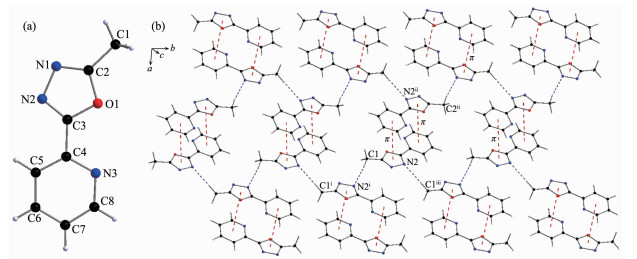
X-ray diffraction structure analysis reveals that L crystallizes in monoclinic system with space group P21/c. The oxadiazole ring and pyridyl ring of L are close to coplanar. The dihedral angle between them is 10.66°. In the crystals there is a non-classical hydrogen bond interactions of C1-H1A…N2ⅰ, and a weak face-to-face π…π interaction exists between the 1, 3, 4-oxadiazole ring and pyridyl ring (Table 3 and Fig. 1). All these interactions link the L molecules into a two-dimensional network structure.
Table 3
Table 3. Hydrogen bond and π…π interactions for align="left"下载:
导出CSV
D-H…A d(D-H)/nm d(H…A)/nm d(D…A)/nm ∠DHA/(°) C1-H1A …N2ⅰ 0.095 94 0.255 21 0.349 7(4) 168.40 d(Cg-Cg)/nm Dihedral angle/(°) Cg1…Cg2ⅱ 3.891(4) 10.66 Cg1: O1, C2, N1, N2, C3; Cg2: N3, C4, C5, C6, C7, C8; Symmetry codes: ⅰ2-x, -1/2+y, 1/2-z; ⅱ1-x, 2-y, -z. Figure 1
 Figure 1. (a) Structure of L with atomic labeling; (b) 2D structure of L linked by hydrogen bond and π…π interactions
Symmetry codes: ⅰ2-x, -1/2+y, 1/2-z; ⅱ1-x, 2-y, -z; ⅲ2-x, 1/2+y, 1/2-z
2.1.2 Crystal structure of 1
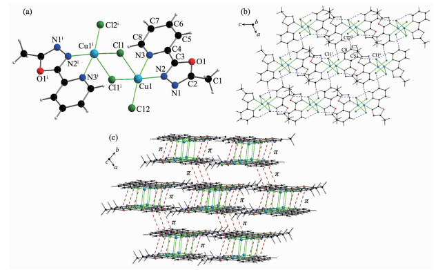
Complex 1 is a double-bridged binuclear Cu(Ⅱ) complex (Fig. 2) crystallizing in triclinic system with space group P1. Two copper(Ⅱ) ions are bridged by two Cl atoms (Cl1 and Cl1ⅰ). The N2, N3, Cl1ⅰ and Cl2 atoms are close to coplanar around the Cu1(Ⅱ) ion. The Cl1 atom sits at the side of the plane making the center Cu1(Ⅱ) ion form a distorted tetragonal geometry [CuCl3N2]. The bond lengths of Cu1-N2, Cu1-N3, Cu1-Cl1 and Cu1-Cl2 are 0.202 5(2), 0.210 2(3), 0.272 52(13) and 0.223 29(16) nm, respectively. The bond lengths of Cu-N are within the normal ranges observed for distorted tetragonal Cu(Ⅱ) complex or trigonal bipyramid Cu(Ⅱ) complexes[26].
Figure 2
 Figure 2. (a) Structure of 1 with atomic labeling; (b) 2D structure of 1 through the hydrogen bond interactions; (c) 3D structure of 1 linked by hydrogen bond and π…π interactions
Symmetry codes: ⅰ2-x, 1-y, 1-z; ⅱ2-x, 1-y, -z; ⅲ-1+x, -1+y, -1+z
In complex 1, there are three non-classical intermolecular hydrogen bonding interactions: C5-H5…Cl1ⅰ, C7-H7…Cl2ⅲ and C8-H8…Cl1ⅱ. All these intermolecular hydrogen bonding interactions link complex 1 molecules into a two-dimensional network structure (Table 4 and Fig. 2). A face-to-face π…π interaction exists between the 1, 3, 4-oxadiazole and pyridyl rings with a centroid-centroid distance of 0.387 4(4) nm (Table 4). Another face-to-face π…π interaction is found between the pyridyl rings with a centroid-centroid distance of 0.362 9(4) nm. Complex 1 molecules are linked into a layered three-dimensional networks through these π…π interactions (Fig. 2).
Table 4
Table 4. Hydrogen bond and π…π interactions for 1下载:
导出CSV
D-H…A d(D-H)/nm d(H…A)/nm d(D…A)/nm ∠DHA/(°) C8-H8…C11ⅰ 0.093 05 0.268 37 0.324 2(4) 119.30 C5-H5…C11ⅱ 0.092 97 0.270 21 0.352 8(4) 148.46 C7-H7…C12ⅲ 0.092 95 0.273 53 0.365 5(5) 170.21 d(Cg-Cg)/nm Dihedral angle/(°) Cg3…Cgⅱ 0.3874(4) 2.41 Cg4…Cg4ⅳ 0.3629(4) 0.00 Cg3: O1, C2, N1, N2, C3; Cg4: N3, C4, C5, C6, C7, C8; Symmetry codes: ⅰ2-x, 1-y, 1-z; ⅱ2-x, 1-y, -z; ⅲ-1+x, -1+y, -1+z; ⅳ1-x, 1-y, -z. 2.1.3 Crystal structure of 2
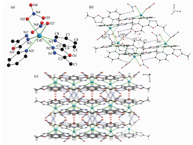
Complex 2 crystallizes in monoclinic system with space group C2/c. The center Cd1(Ⅱ) ion are coordinated by two molecules of L and two nitrate ions. Four coordinated nitrogen atoms (N2, N2ⅰ, N3, N3ⅰ) and two oxygen atoms (O2, O2ⅰ) make the center Cd1(Ⅱ) ion form a distorted octahedral geometry [CdN4O2] (Fig. 3). The bond lengths of Cd1-N2, Cd1-N3 and Cd1-O2 are 0.248 9(2), 0.238 9(2) and 0.245 4(2), respectively. The Cd-N bond lengths are within the normal ranges observed for the 1, 3, 4-oxadiazole based Cd(Ⅱ) complex[17].
Figure 3
 Figure 3. (a) Structure of 2 with atomic labeling; (b) Hydrogen bond and π…π interactions in 2; (c) 3D structure of 2
Symmetry codes: ⅰ2-x, y, 3/2-z; ⅱ3/2-x, 1/2+y, 3/2-z; ⅲ-1/2+x, 1/2-y, -1/2+z; ⅳ5/2-x, 1/2-y, 1-z
In complex 2, there are three non-classical intermolecular hydrogen bonding interactions: C1-H1C…O2ⅱ, C5-H5…O2ⅲ and C7-H7…O3ⅳ (Table 5 and Fig. 3). In addition, the pyridyl rings involve a face-to-face π…π interaction with a centroid-centroid distance of 0.391 01(19) nm. All these interactions link the complex 2 molecules into a three-dimensional network structure.
Table 5
Table 5. Hydrogen bond and π…π interactions for 2下载:
导出CSV
D-H…A d(D-H)/nm d(H…A)/nm d(D…A)/nm ∠DHA/(°) C1-H1C…O2ⅱ 0.096 05 0.255 58 0.342 2(5) 150.02 C5-H5…O2ⅲ 0.093 07 0.241 18 0.330 8(4) 161.60 C7-H7…O3ⅳ 0.092 97 0.250 81 0.339 8(4) 160.46 d(Cg-Cg)/nm Dihedral angle/(°) Cg2…Cg2ⅴ 0.391 1(2) 0.02 Cg2: N3, C4, C5, C6, C7, C8; Symmetry codes:ⅱ3/2-x, 1/2+y, 3/2-z; ⅲ-1/2+x, 1/2-y, -1/2+z; ⅳ5/2-x, 1/2-y, 1-z; ⅴ2-x, 1-y, 1-z. 2.2 Spectral characterization
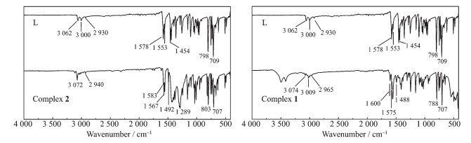
In the IR spectrum of L (Fig. 4), the bands of 1 578, 1 553 and 1 454 cm-1 are attributed to the aromatic rings stretching vibrations of pyridine and 1, 3, 4-oxadiazole. As the nitrogen atoms coordinate to the central Cu(Ⅱ) or Cd(Ⅱ) ions in complexes 1 and 2, the corresponding absorption bands are blue-shifted. In the spectra of 1 and 2, absorption bands at 1 600 cm-1 (or 1 583 cm-1), 1 575 cm-1 (or 1 567 cm-1) and 1 488 cm-1 (or 1 492 cm-1) are observed, which are assigned to the aromatic rings stretching vibrations. This indicates that the Cu1(Ⅱ) and Cd1(Ⅱ) ions are coordinated by nitrogen atoms from oxadiazole and pyridine rings. In complex 2, the bands of 1 289 cm-1 are attributed to NO3-.
Figure 4
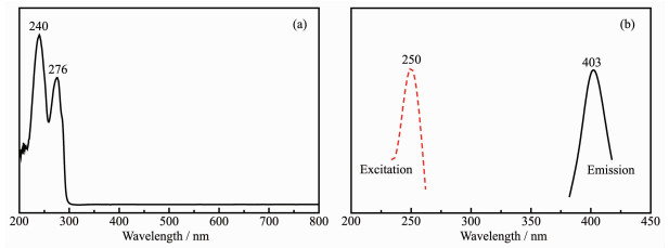
The UV-Vis spectra of L is given in Fig. 5(a). Maximum absorption peak appears at 240 and 276 nm, which are attributed to the π-π* and n-π* transitions, respectively. The fluorescence property of L has been examined in ethanol solution at room temperature. The fluorescence spectrum is shown in Fig. 5(b), which shows L is fluorescent. Maximum emission peak and excitation wavelength is 403 and 250 nm, respectively, which is attributed to π-π* electronic transition[27].
Figure 5
In the UV-Vis spectra of 1 and 2, the maximum absorption peak of 1 appears at 240.04 nm and 275.09 nm, and those for 2 appear at 240.13 nm and 276.05 nm. This is attributed to π-π* and n-π* transitions. The fluorescence properties of complexes 1 and 2 have been examined in methanol solution at room temperature, but no fluorescence properties were observed.
2.3 Thermal gravimetric analysis
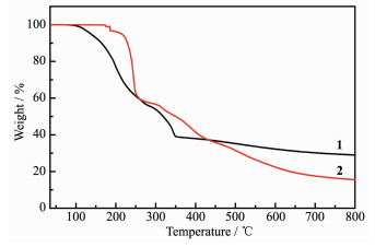
Thermal gravimetric analyses (TGA) of complexes 1 and 2 were accomplished in nitrogen atmosphere with the heating rate of 10 K·min-1. The TGA curves of complexes 1 and 2 are presented in Fig. 6. When the temperature is below 95 ℃, complex 1 is stable. Complex 1 decomposed gradually with the temperature raised between 95 and 800 ℃. Total weight loss was close to 71.00% at 800 ℃. The final residue is CuO (Calcd. 26.9%). Complex 2 is stable below 150 ℃. Complex 2 decomposed sharply with the temperature raised between 200 and 300 ℃. When the temperature reached to 650 ℃, total weight loss is close to 80.97% (Calcd. 77.02%).
Figure 6
3. Conclusions
Ligand L, complexes 1 and 2 were synthesized and characterized. The free ligand L exhibits fluorescence. Complex 1 is a double-bridged binuclear Cu(Ⅱ) complex. Two copper(Ⅱ) ions are bridged by two Cl atoms, and the central copper(Ⅱ) ions have distorted tetragonal geometries [CuCl3N2]. In complex 2, the central Cd(Ⅱ) ion have a distorted octahedral geometry [CdN4O2].
Supporting information is available at http://www.wjhxxb.cn
-
-
[1]
Holla B S, Gonsalves R, Shenoy S. Eur. J. Med. Chem., 2000, 35:267-271 doi: 10.1016/S0223-5234(00)00154-9
-
[2]
Boström J, Hogner A, Llinas A, et al. J. Med. Chem., 2012, 55:1817-1830 doi: 10.1021/jm2013248
-
[3]
Ishii M, Jorge S D, de Oliveira A A, et al. Bioorg. Med. Chem., 2011, 19:6292-6301 doi: 10.1016/j.bmc.2011.09.009
-
[4]
Bhandari S V, Parikh J K, Bothara K G, et al. J. Enzyme Inhib. Med. Chem., 2010, 25:520-530 doi: 10.3109/14756360903357585
-
[5]
刘建超, 王卫东, 贺红武.有机化学, 2014, 34(7):1447-1457 http://www.wanfangdata.com.cn/details/detail.do?_type=perio&id=yjhx201407023LIU Jian-Chao, WANG Wei-Dong, HE Hong-Wu. Chinese J. Org. Chem., 2014, 34(7):1447-1457 http://www.wanfangdata.com.cn/details/detail.do?_type=perio&id=yjhx201407023
-
[6]
Banerjee A G, Das N, Shengule S A, et al. Eur. J. Med. Chem., 2015, 101:81-95 doi: 10.1016/j.ejmech.2015.06.020
-
[7]
Ilango K, Valentina P, Kumar G, et al. Med. Chem., 2015, 11:753-763 doi: 10.2174/1573406411666150519112037
-
[8]
Verma G, Chashoo G, Ali A, et al. Bioorg. Chem., 2018, 77:106-124 doi: 10.1016/j.bioorg.2018.01.007
-
[9]
Dewangan, D, Verma V S, Nakhate K T, et al. Med. Chem. Res., 2016, 25:2143-2154 doi: 10.1007/s00044-016-1641-8
-
[10]
Wang B L, ZhuH W, Ma Y, et al. J. Agric. Food. Chem., 2013, 61:5483-5493 doi: 10.1021/jf4012467
-
[11]
Adachi C, Tokito S, Tsutsui T, et al. Jpn. J. Appl. Phys., 1988, 28:L269-L271
-
[12]
Bouanis M, Tourabi M, Nyassi A, et al. Appl. Surf. Sci., 2016, 389:952-966 doi: 10.1016/j.apsusc.2016.07.115
-
[13]
Gurudatt D M, Mohana K N. Ind. Eng. Chem. Res., 2014, 53:2092-2105 doi: 10.1021/ie402042d
-
[14]
Raj X J, Rajendran N. Prot. Met. Phys. Chem. Surf., 2013, 49:763-775 doi: 10.1134/S2070205113060257
-
[15]
Mohamed N A. Polym. Degrad. Stab., 2017, 146:42-45 doi: 10.1016/j.polymdegradstab.2017.09.017
-
[16]
Shavaleev N M, Scopelliti R, Gratzel M, et al. Inorg. Chim. Acta, 2013, 394:295-299 doi: 10.1016/j.ica.2012.07.026
-
[17]
Bharty M K, Bharti A, Dani R K, et al. J. Mol. Struct., 2012, 1011:34-31 doi: 10.1016/j.molstruc.2011.12.015
-
[18]
Slyvka Y, Goreshnik E, Veryasov G, et al. Polyhedron, 2017, 133:319-326 doi: 10.1016/j.poly.2017.05.052
-
[19]
Bharty M K, Dani R K, Nath P, et al. Polyhedron, 2015, 98:84-95 doi: 10.1016/j.poly.2015.05.045
-
[20]
Garcia A, Machado R C, Grazul R M, et al. J. Biol. Inorg. Chem., 2016, 21:275-292 doi: 10.1007/s00775-016-1338-y
-
[21]
Chaves J D S, Tunes L G, Franco C H D J, et al. Eur. J. Med. Chem., 2017, 127:727-739 doi: 10.1016/j.ejmech.2016.10.052
-
[22]
Gallego B, Kaluderovic M R, Kommera H, et al. Invest. New Drugs, 2011, 29:932-944 doi: 10.1007/s10637-010-9449-8
-
[23]
Köhler C, Rentschler E. Eur. J. Inorg. Chem., 2016, 13-14:1955-1960 doi: 10.1002/ejic.201501278/pdf
-
[24]
James C A, Poirier B, Grise C, et al. Tetrahedron Lett., 2006, 47:511-514 doi: 10.1016/j.tetlet.2005.11.057
-
[25]
Sheldrick G M. Acta Crystallogr. Sect. A, 2008, A64:112-122
-
[26]
Li Y, Zhang C G, Cai L Y. J. Coord. Chem., 2013, 66:3100-3112 doi: 10.1080/00958972.2013.826350
-
[27]
盛俊峰, 王宁, 蔡良英, 等.无机化学学报, 2015, 31(2):405-412 http://qikan.cqvip.com/article/detail.aspx?id=663728357SHENG Jun-Feng, WANG Ning, CAI Liang-Ying, et al. Chinese J. Inorg. Chem., 2015, 31(2):405-412 http://qikan.cqvip.com/article/detail.aspx?id=663728357
-
[1]
-
Figure 1 (a) Structure of L with atomic labeling; (b) 2D structure of L linked by hydrogen bond and π…π interactions
Symmetry codes: ⅰ2-x, -1/2+y, 1/2-z; ⅱ1-x, 2-y, -z; ⅲ2-x, 1/2+y, 1/2-z
Figure 2 (a) Structure of 1 with atomic labeling; (b) 2D structure of 1 through the hydrogen bond interactions; (c) 3D structure of 1 linked by hydrogen bond and π…π interactions
Symmetry codes: ⅰ2-x, 1-y, 1-z; ⅱ2-x, 1-y, -z; ⅲ-1+x, -1+y, -1+z
Figure 3 (a) Structure of 2 with atomic labeling; (b) Hydrogen bond and π…π interactions in 2; (c) 3D structure of 2
Symmetry codes: ⅰ2-x, y, 3/2-z; ⅱ3/2-x, 1/2+y, 3/2-z; ⅲ-1/2+x, 1/2-y, -1/2+z; ⅳ5/2-x, 1/2-y, 1-z
Table 1. Crystal data and structure refinement for L, 1 and 2
Compound L 1 2 Formula C8H7N3O C8H7Cl2CuN3O C8H7Cd0.5O4 Formula weight 161.17 295.61 279.38 Crystal system Monoclinic Triclinic Monoclinic Space group P21/c P1 C2/c a/nm 0.786 3(8) 0.826 4(5) 1.090 0(3) b/nm 1.069 6(11) 0.851 7(5) 1.384 4(4) c/nm 0.938 9(10) 0.901 1(8) 1.362 3(4) α/(°) 109.627(8) β/(°) 99.344(13) 113.387(8) 99.090(4) γ/(°) 99.513(6) (V/nm3 0.779 1(14) 0.514 6(6) 2.029 9(11) Z 4 2 8 Dc/(g·cm3) 1.374 1.908 1.828 θ range/(°) 2.63-26.00 2.71-25.50 2.40-25.79 μ/mm-1 0.096 2.613 1.141 F(000) 336 294 1112 Rint 0.027 9 0.021 9 0.033 3 Reflection collected 5 584 3 752 7 223 Independent reflection 1 506 1 881 1 933 Observed reflection 1 188 1 771 1 682 Data, restrain, parameter 1 506, 0, 111 1 881, 0, 138 1 933, 2, 151 Goodness-of-fit on F2 1.050 1.091 1.108 R1, wR2 [I > 2σ(I)] 0.038 2, 0.100 4 0.023 0, 0.062 5 0.028 0, 0.060 8 R1, wR2 (all data) 0.049 3, 0.107 1 0.024 5, 0.063 1 0.034 6, 0.062 6 (Δρ)max, (Δρ)min/(e·nm-3) 183, -107 337, -313 361, -320  下载: 导出CSV
Table 2. Selected bond lengths (nm) and bond angles (°) for L, 1 and 2
L C2-N1 0.128 26(19) C3-O1 0.135 68(18) N1-N2 0.140 7(2) C2-O1 0.136 59(17) C3-N2 0.128 9(2) N1-C2-O1 112.36(14) C3-O1-C2 102.77(11) C3-N2-N1 106.30(10) N2-C3-O1 112.32(11) C2-N1-N2 106.25(12) 1 Cu1-Cl1 0.272 52(13) Cu1-Cl2 0.223 29(16) Cu1-N3 0.210 2(3) Cu1-C11ⅰ 0.226 86(10) Cu1-N2 0.202 5(2) N2-Cu1-N3 79.18(10) N3-Cu1-Cl2 166.14(6) Cl2-Cu1-Cl1 101.89(4) N2-Cu1-Cl2 93.43(9) N3-Cu1-C11ⅰ 91.19(8) Cl1-Cu1-Cl1ⅰ 90.89(5) N2-Cu1-C11ⅰ 168.72(6) N3-Cu1-Cl1 90.50(6) N2-Cu1-Cl1 95.00(7) Cl2-Cu1-Cl1ⅰ 94.76(7) 2 Cd1-N2 0.248 9(2) Cd1-02 0.245 4(2) Cd1-O3 0.247 9(2) Cd1-N3 0.238 9(2) N3ⅰ-Cd1-N3 113.98(12) N3-Cd1-N2 68.93(8) O2ⅰ-Cd1-N2ⅰ 154.16(7) N3-Cd1-O2ⅰ 104.18(8) N2ⅰ-Cd1-N2 130.77(11) O2ⅰ-Cd1-O2 80.72(10) N3-Cd1-O2 125.82(7) O2-Cd1-N2ⅰ 74.61(7) N3-Cd1-N2ⅰ 84.59(8) O2-Cd1-N2 154.16(8) Symmetry codes: ⅰ2-x, 1-y, 1-z for 1; ⅰ2-x, y, 3/2-z for 2.
下载: 导出CSV
Table 3. Hydrogen bond and π…π interactions for align="left"
D-H…A d(D-H)/nm d(H…A)/nm d(D…A)/nm ∠DHA/(°) C1-H1A …N2ⅰ 0.095 94 0.255 21 0.349 7(4) 168.40 d(Cg-Cg)/nm Dihedral angle/(°) Cg1…Cg2ⅱ 3.891(4) 10.66 Cg1: O1, C2, N1, N2, C3; Cg2: N3, C4, C5, C6, C7, C8; Symmetry codes: ⅰ2-x, -1/2+y, 1/2-z; ⅱ1-x, 2-y, -z.
下载: 导出CSV
Table 4. Hydrogen bond and π…π interactions for 1
D-H…A d(D-H)/nm d(H…A)/nm d(D…A)/nm ∠DHA/(°) C8-H8…C11ⅰ 0.093 05 0.268 37 0.324 2(4) 119.30 C5-H5…C11ⅱ 0.092 97 0.270 21 0.352 8(4) 148.46 C7-H7…C12ⅲ 0.092 95 0.273 53 0.365 5(5) 170.21 d(Cg-Cg)/nm Dihedral angle/(°) Cg3…Cgⅱ 0.3874(4) 2.41 Cg4…Cg4ⅳ 0.3629(4) 0.00 Cg3: O1, C2, N1, N2, C3; Cg4: N3, C4, C5, C6, C7, C8; Symmetry codes: ⅰ2-x, 1-y, 1-z; ⅱ2-x, 1-y, -z; ⅲ-1+x, -1+y, -1+z; ⅳ1-x, 1-y, -z.
下载: 导出CSV
Table 5. Hydrogen bond and π…π interactions for 2
D-H…A d(D-H)/nm d(H…A)/nm d(D…A)/nm ∠DHA/(°) C1-H1C…O2ⅱ 0.096 05 0.255 58 0.342 2(5) 150.02 C5-H5…O2ⅲ 0.093 07 0.241 18 0.330 8(4) 161.60 C7-H7…O3ⅳ 0.092 97 0.250 81 0.339 8(4) 160.46 d(Cg-Cg)/nm Dihedral angle/(°) Cg2…Cg2ⅴ 0.391 1(2) 0.02 Cg2: N3, C4, C5, C6, C7, C8; Symmetry codes:ⅱ3/2-x, 1/2+y, 3/2-z; ⅲ-1/2+x, 1/2-y, -1/2+z; ⅳ5/2-x, 1/2-y, 1-z; ⅴ2-x, 1-y, 1-z.
下载: 导出CSV
-


 扫一扫看文章
计量
- PDF下载量: 2
- 文章访问数: 512
- HTML全文浏览量: 143
 下载:
下载:
