
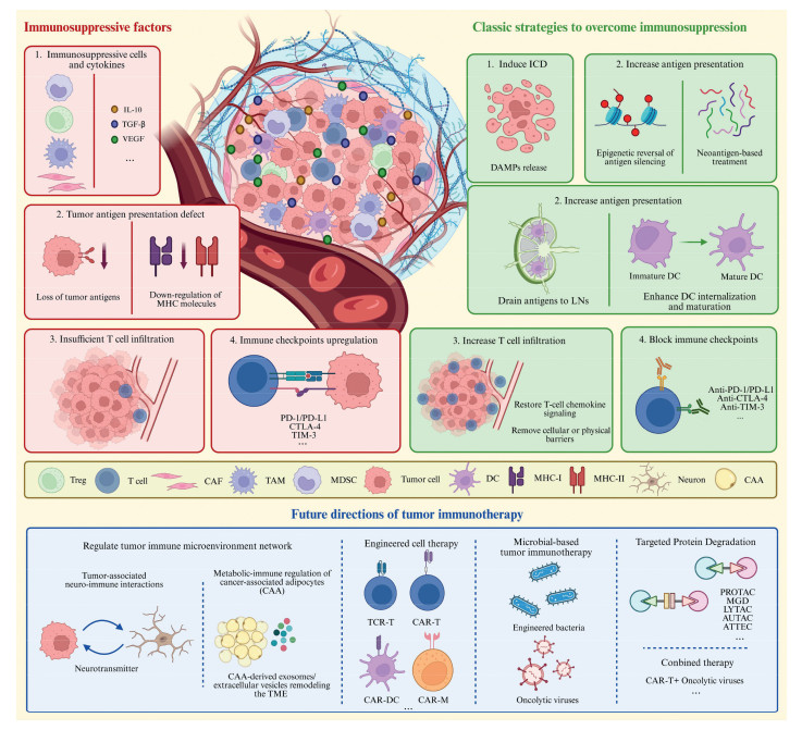
Scheme 1.
Schematic overview of immunosuppressive factors, classic therapeutic strategies, and future directions of tumor immunotherapy. Created by BioRender.


Tumor is a major public health challenge and is hard to treat completely. In 2024, an estimated 20.6 million new cancer cases and 9.8 million deaths occurred worldwide [1]. For patients with advanced or metastatic solid tumors, the 5-year survival rate remains below 30%, approximately 10% for pancreatic cancer [2]. Moreover, patients face a triple burden, physical decline, psychological distress, and financial toxicity that often depletes savings or interrupts care.
Immunotherapy, which can boost the body’s immune system to fight against tumor, is considered as the most promising strategy to overcome cancer. In the past decade, tens of immunotherapeutic drugs have been approved for clinical application, most of which are immune checkpoint inhibitors (ICIs).
However, the clinical efficacy of immunotherapy is often limited by the immunosuppressive tumor microenvironment (TME), non-specific inflammation, and drug resistance. For example, in pancreatic ductal adenocarcinoma and glioblastoma, the clinical benefits of current immunotherapies are extremely limited, and durable responses are primarily confined to patients with a high mutation burden [3,4]. Furthermore, immunotherapy drugs are associated with a range of toxic side effects, such as myocarditis, pneumonia, and colitis [5]. Therefore, researchers are striving to develop novel therapeutic platforms and innovative strategies to address the limitations of conventional immunotherapies [6]. In this review, we first analyze the major barriers that influence tumor immunotherapy. Next, we summarize current strategies to overcome these barriers. Finally, we discuss the emerging new strategies for tumor immunotherapy (Scheme 1).
TME refers to the internal and external environment in which tumor cells reside during tumor occurrence, proliferation and metastasis [7]. Due to disordered energy metabolism, insufficient blood perfusion and dysplastic cell proliferation, the structure and composition of TME differ from those of normal tissues, exhibiting special characteristics such as acidification and hypoxia [8]. The immune system plays a dual role in the TME: On the one hand, it can inhibit tumor initiation and progression by killing tumor cells; on the other hand, some components can promote tumor progression and metastasis by constructing a microenvironment suitable for tumor growth. TME suppresses antitumor immunity through multiple mechanisms, including immunosuppressive factors, deficiency in antigen presentation, inadequacy in T cell infiltration, and immune checkpoints expression, which collectively limit treatment efficacy.
Immunosuppressive cells and the cytokines they produce jointly maintain and regulate the immunosuppressive TME. Myeloid-derived suppressor cells (MDSCs), regulatory T cells (Tregs), tumor-associated macrophages (TAMs), and cancer-associated fibroblasts (CAFs) exert broad immunosuppressive functions through direct cell-cell contact and the secretion of immunosuppressive cytokines such as interleukin-10 (IL-10), transforming growth factor-β (TGF-β) and vascular endothelial growth factor (VEGF). Moreover, vascular abnormalities, acidity, and hypoxia caused by abnormal metabolism of tumor cells further enhance this effect.
MDSCs inhibit antigen-specific immune responses mainly by producing reactive oxygen species (ROS) and targeting T lymphocytes [9-11]. Tregs can promote the production of immunosuppressive cytokines (e.g., IL-10 and TGF-β), up-regulate the expression of negative inhibitory molecules such as cytotoxic T lymphocyte-associated antigen-4 (CTLA-4), programmed cell death 1 (PD-1) and programmed cell death ligand 1 (PD-L1), down-regulate the function of antigen-presenting cells (APCs) or compete for co-stimulatory molecules on them. Tregs can also inhibit the function of T lymphocytes, natural killer (NK) cells and dendritic cells (DCs) by consuming anti-tumor cytokines such as interferon-γ (IFN-γ), tumor necrosis factor-α (TNF-α) and IL-2 [12-15]. By expressing inhibitory ligands such as PD-L1 on the surface, M2 macrophages can directly bind to the corresponding receptors on CD8+ T lymphocytes (CTLs), thus preventing CTLs from attacking tumor cells [16]. M2 macrophages can also indirectly inhibit CTLs function by expressing C—C motif chemokine ligand 22 (CCL22) and inducing starvation of CTLs through exhaustion of essential amino acids. CAFs inhibit immune responses at the tumor site by secreting cytokines (e.g., IL-6, IL-4, IL-8, and IL-10) and chemokines (e.g., CCL2, CCL5, C-X-C motif chemokine ligand 9 (CXCL9), and CXCL10). These molecules limit DC maturation, inactive CTLs, induce MDSC differentiation, increase the number of M2 macrophages, and recruit and activate a large number of mast cells [17-19]. These immunosuppressive factors play important roles in the process of immunotherapy.
Major histocompatibility complex class Ⅰ (MHC-Ⅰ) and MHC-Ⅱ molecules present specific peptide sequences derived from tumor antigens to T cells, enabling recognition and activation of immune responses. Endogenous proteins are presented by MHC-Ⅰ molecules to CTLs, and exogenous proteins are presented by MHC-Ⅱ molecules to CD4+ T cells. Exogenous proteins can also be cross-presented to MHC-Ⅰ molecules by DCs and subsequently recognized by CTLs. Defective antigen presentation is a key mechanism by which tumors evade immune recognition and acquire resistance to T-cell-based immunotherapies. Tumors can reduce antigen presentation and achieve immune escape through various ways [20,21].
The expression of tumor antigens is heterogeneous and genetically unstable. Heterozygous mutations in antigen presentation related genes can lead to reduced antigen expression in tumors. Some tumor-associated antigens (TAAs) are also present on normal cells, causing weak immunogenicity and an inability to induce specific immune responses. Moreover, tumor cells often express more mucin and polysaccharide molecules than normal cells, resulting in antigens shielding by polysaccharides. Some tumor cells can secrete stimulating factors to activate the coagulation system, leading to the formation of fibrin cocoon around tumor cells, thereby isolating tumor antigens.
Studies have shown that in certain TME, the expression of MHC gene is decreased and both transcription and translation processes are blocked. The analysis of tumor samples from patients with bladder cancer (especially transitional cell carcinoma) revealed that 11 of the 72 patients (15.3%) exhibited complete deletion of class Ⅰ human leukocyte antigens (HLA-Ⅰ), and most of them showed the deletion of HLA-Ⅰ gene expression at the transcriptional level. In addition to down-regulate the transcription process, tumors also inhibit the surface expression of HLA-Ⅰ through post translational mechanisms involving autophagy [22,23].
T cells can only exert their killing effect after infiltrating the tumor site, but in practice, it is difficult for T cells to achieve a high degree of infiltration. According to the spatial distribution of cytotoxic immune cells in TME, tumors can be divided into three immunophenotypes: Immune-inflamed, immune-excluded and immune-desert. Based on quantitative scoring of CD3+ and CD8+ lymphocyte populations at the tumor center and invasive margin, tumors can also be classified as “hot” or “cold”. The immune-inflamed tumor is a “hot tumor” with high immune score, characterized by high T cell infiltration, high PD-L1 expression, high tumor mutation burden, and an activated IFN-γ signaling pathway. “Hot tumors” are more sensitive to immunotherapies such as immune checkpoint blockade (ICB) therapy. The other two immunophenotypes are “cold tumors” with low immune score. In immune-excluded tumors, CTLs only infiltrate the edge of the tumor, while in immune-desert tumors, CTLs are absent from the tumor and its surrounding areas. In addition to poor T cell infiltration, “cold tumors” also exhibit low neoantigen levels, low MHC-Ⅰ expression, immune deficiency (low PD-L1 expression) and high proliferation. Immunosuppressive cells such as TAMs, Tregs and MDSCs also exist in “cold tumors”. These characteristics indicate that “cold tumors” lack an active immune response and do not respond to certain immunotherapies, including ICB [24,25].
Under ideal circumstances, T cells are activated and transported to the tumor site through blood, where they contact with tumor cells and subsequently infiltrate the tumor bed. In practice, however, many factors may affect this process, ultimately leading to anti-tumor immune failure. These factors include defective T cell activation and impaired T cell homing to the tumor site. Carcinogenic pathway activation, β-catenin signaling pathway activation, chemokines deficiency (e.g., CXCL9, CXCL10), hormone secretion (e.g., prostaglandin E2), vascular abnormalities and hypoxia can all prevent T cells from homing to the tumor site [22,24,26-28]. Therefore, the efficacy of immunotherapy can be improved by improving the insufficient infiltration of T cells.
Although antitumor immunity can be activated by neoantigens or inflammatory signals, tumor cells have evolved countermeasures to escape immune surveillance and counteract immune responses by regulating immune checkpoint related pathways. Immune checkpoints are regulatory molecules that inhibit immune responses and prevent autoimmunity. CTLA-4, PD-1 and PD-L1 have been proved to play essential roles in tumor immune escape.
CTLA-4 is the first identified negative regulator of T cell activation, which has the same B7 ligands (CD80 and CD86) as CD28, the co-stimulatory molecule on T cells. However, the affinity of CTLA-4 to B7 ligands is about 100-fold higher than that of CD28, which means CTLA-4 is more likely to bind to CD80/CD86 in competition. This binding leads to decreased T cell proliferation [29]. What is more, CTLA-4 also inhibits CTL function by inducing the reverse signaling pathway of indoleamine 2,3-dioxygenase (IDO), phosphatidylinositol 3-kinase/protein kinase B (PI3K/Akt) signaling pathway, cell cycle dependent kinase and nuclear transcription factor (NF-κB) inhibition [30,31]. PD-1 is a transmembrane protein mainly expressed on the surface of activated T cells, B cells and macrophages. PD-L1 is the ligand of PD-1, which exhibits inhibitory activity on T cell effector function after binding to PD-1. The binding of PD-1 and PD-L1 can transmit survival signals to cancer cells and enhance their resistance to apoptosis induced by IFN-γ and CTLs. The PD-1/PD-L1 interaction forms a unique immune escape microenvironment and promotes tumor development and metastasis. It is also associated with chemotherapy resistance and poor prognosis. Therefore, PD-1/PD-L1 immune checkpoint pathway has become an ideal target for immunotherapy aimed at restoring antitumor effector function [32,33]. Checkpoint blockers targeting CTLA-4, PD-1 and PD-L1 have shown effective and lasting clinical effects in a variety of cancers, including melanoma, lung cancer, renal cell carcinoma, bladder cancer, ovarian cancer, triple negative breast cancer, gastric cancer, Hodgkin’s lymphoma and so on [34,35].
ICD is a type of regulatory cell death, which means that when cells undergo external stress-driven death, the dying cells are converted from non-immunogenic to immunogenic, thus stimulating anti-tumor immune response in the body. When tumor cells undergo ICD, they produce a series of signaling molecules called damage-associated molecular patterns (DAMPs). These signaling molecules mainly include calreticulin (CRT), high-mobility group box 1 (HMGB1), adenosine triphosphate (ATP) and heat shock proteins (HSPs, e.g., HSP70, HSP90) [36]. These signals are endogenous molecules that perform conventional intracellular functions, but become immunogenic when exposed to the extracellular environment. DAMPs can lead to the accumulation of DCs in tumor bed, promote DC phagocytosis of tumor antigens and their presentation to T cells, and stimulate specific anti-tumor immune response. ICD can be induced by a variety of stress sources, such as intracellular pathogens, traditional chemotherapeutic drugs (anthracycline drugs, DNA-damaging agents and protease inhibitors, etc.) and physical modalities (radiotherapy, photodynamic therapy (PDT), photoimmunotherapy (PIT), nano-pulse stimulation (NPS), etc.) [37-40].
Chemotherapeutic drugs such as anthracycline and oxaliplatin (OXA) can induce ICD by causing DNA damage, which leads to the release of DAMPs from dying tumors, promote the phagocytosis and maturation of DCs, and further stimulate antitumor immunity to eliminate tumors [37,41,42]. In the case of OXA, nanoparticle-based formulations have been shown to significantly enhance ICD induction and elicit stronger immune responses in DCs and T lymphocytes. Moreover, the combination of OXA with other therapies (such as PDT and immunotherapy) can further induce ICD of tumor cells and improve the antitumor efficiency [43,44]. For instance, bifunctional liposomes co-loading an OXA prodrug and the IDO inhibitor NLG919 have demonstrated synergistic antitumor effects, accompanied by increased intratumoral CTL infiltration, reduced Treg numbers, and markedly enhanced antitumor immunity (Fig. 1A) [45].
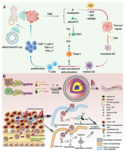
PDT can induce ICD by producing ROS and inducing CRT exposure [46]. Typical photosensitizers are hydrophobic, which can adversely affect their photophysical, chemical, and biological properties. Therefore, researchers have utilized nanoparticles as a delivery form, which can overcome these limitations and selectively transport photosensitizers to tumors to reduce side effects on normal tissues [47]. A hypoxia activatable nanodelivery system has been developed by co-loading the photosensitizer chlorin e6 (Ce6) and tirapazamine onto albumin, which enhances the therapeutic effect upon activation by PDT induced hypoxia [48]. However, nano-drugs based on enhanced permeability and retention (EPR) effects remain insufficient to meet the clinical needs, and the challenges that hinder nano-drug delivery should be overcome to achieve stronger therapeutic effects. To address this issue, shape changing and charge reversal nanoparticles have been designed, including a laser responsive shape changeable nanomedicine (I-P@NPs@M), in which ROS generated by laser irradiation triggers the transformation of spherical micelles into nanofibers, enabling sustained drug retention and release in the tumor areas (Fig. 1B) [49]. Similarly, a dual-responsive strategy integrating shape transformation and charge reversal has been established for chemo-photodynamic therapy. In this system, Ce6, berberrubine and a matrix metalloproteinase-2 (MMP-2) reactive peptide are self-assembled into nanoparticles. In the TME, this system achieves charge reversal, size reduction and MMP-2 responsive cleavage, thereby achieving enhanced tumor accumulation, prolonged retention time and enhanced anti-tumor effect [50].
PIT is a combination of PDT and immunotherapy for cancer. Compared with PDT, PIT only works when the targeting photosensitizer binds to the specific protein on cell membrane, thus selectively killing cancer cells with antibodies and helps generate more immune cells [51]. Researchers have identified a photoactivated chemical IRDye700DX (IR700), which initiates cell killing under the near infrared (NIR) at 690 nm. When coupled with antibody, it is highly lethal only to target cells irradiated by NIR, and has almost no off-target toxicity, phototoxicity or biotoxicity. This NIR-PIT can induce the activation of local DCs, initiate the proliferation of specific initial T cells, leading to cell-mediated cancer cell death. At present, NIR-PIT has been successfully used in tumors targeting epidermal growth factor receptor (EGFR), human epidermal growth factor receptor 2 (HER2), CD25, CD20, PD-L1, CD44 and CD133. Moreover, NIR-PIT can be used in any cancer that overexpresses a membrane protein and has suitable targeted monoclonal antibody. The combination of NIR-PIT and conventional anticancer drugs is expected to provide a more effective and reasonable treatment strategy [39,52,53].
NPS is a technology based on pulse power physics, which produces high-power and low-energy electric pulses by accumulating electrical energy and releasing it in the form of nanosecond pulse. NPS permeates all cells and organelles of tumor with high speed and large amplitude, which causes Ca2+ to escape from endoplasmic reticulum (ER) and leads to ER stress, promotes ROS release and then induces ICD. Studies have confirmed that NPS can eliminate a high percentage of primary 4T1 tumors (75%−100%) by inducing immunological responses and activating adaptive immune memory. Compared with drug-induced ICD, NPS is safer and less traumatic, so its clinical application is worthy of further study [39,54].
Enhancing tumor antigen presentation is critical to overcome immune evasion in refractory tumors. Generally speaking, tumor antigens that used as targets can be divided into two categories: Non-mutant proteins that are abnormally expressed or overexpressed in tumor cells and neoantigens formed by the somatic mutations.
Epigenetic therapy can enhance tumor immunogenicity by increasing the expression of tumor antigens. Some studies have shown that DNA methyltransferase inhibitors (DNMTis) and histone deacetylase inhibitors (HDACis) can enhance the expression of tumor antigens and other immune-related genes [24]. DNMTis, such as decitabine and azacitidine, can specifically inhibit the activity of DNA methyltransferases in tumor cells, reversing the methylation-induced silencing of key genes—including tumor antigens, MHC molecules, antigen-processing-related transporters (TAP), and proteasome subunits—and restoring the expression, processing, and presentation of endogenous antigens. In tumors such as breast cancer, low-dose DNMTis can significantly upregulate the expression of MHC-Ⅰ molecules and tumor antigens on the surface of tumor cells, reverse antigen silencing, and convert “cold tumor” into “hot tumor” [55-58]. HDACis, such as vorinostat and panobinostat, can increase histone acetylation levels, promote chromatin relaxation, and activate the transcription of silenced immune-related genes. This synergistically enhances antigen expression and presentation efficiency while upregulating the expression of co-stimulatory molecules, further strengthening immunogenicity [59,60]. A novel dual DNMT/HDAC inhibitor 15a has been developed to enhance tumor immunogenicity by promoting the expression of endogenous retroviral elements in breast cancer, thereby inducing a viral mimicry response and activating the RIG-I-MAVS pathway [61].
Non-mutant tumor autoantigens are not only expressed on the membrane of tumor cells, but also exist in healthy tissues. Therefore, they often lead to insurmountable central immune tolerance and potential off-target effects. Neoantigens are truly foreign proteins that are entirely absent from normal organs/tissues. As polypeptide sequences on the surface of tumor cells, they can be specifically recognized by neoantigen-specific T cell receptors (TCRs) in the background of MHC molecules. They have high immunogenicity and play a crucial role in tumor-specific T cell-mediated anti-tumor immune response and successful cancer immunotherapy [62-65]. A novel neoantigen vaccine platform based on high-purity linear hairpin DNA (hpDNA), delivered via electroporation, has been developed. In the MC38 tumor model, the hpDNA vaccine encoding 20 neoantigens (M20-hpDNA) successfully induced potent CD8+ and CD4+ neoantigen-specific T-cell responses, achieving dose-dependent protection in both preventive and therapeutic models, and effectively preventing lung metastasis [66]. Researchers have identified that after binding to the mutated protein, the KRASG12C inhibitor (sotorasib) generates a “synthetic neoantigen” (p*MHC) on the surface of cancer cells, composed of the drug and the mutated peptide, which is presented by the HLA-A3 superfamily. Based on this neoantigen, the team developed the engineered bispecific antibody AETX-R114, which binds to the synthetic neoantigen with high affinity and recruits T cells to kill tumors. This study marks the first time a drug-induced “synthetic neoantigen” has been transformed into a targetable immunotherapy target, opening new avenues for the use of neoantigens in immunotherapy [67].
LNs serve as critical hubs for the initiation of adaptive immune responses. LNs contain high concentrations of DC cells, T cells, and B cells. Tumor antigens can only effectively activate naive T cells and induce tumor-specific immune responses if they are efficiently delivered to the LNs and taken up by antigen-presenting cells for cross-presentation. Although the tumor microenvironment contains a large number of tumor antigens, most of these antigens remain confined to the tumor site and cannot come into contact with naive T cells in the LNs, making it difficult to initiate a systemic antitumor immune response. This is a major reason for the discrepancy between local antigen accumulation and the absence of an immune response [68,69].
Researchers have employed various strategies to drain tumor antigens to LNs. Passive targeting strategies rely on the physicochemical properties of nanoparticles (e.g., size, shape) to optimize lymphatic uptake and LN accumulation. An NIR-Ⅱ light-driven in situ nanovaccine (AuP/VNPR848) has been developed, which enables the in situ generation and LN delivery of antigens with optimized physical size (~28 nm) and good deformability. Under 1064-nm laser irradiation, AuP generates numerous tumor antigens, while VNPR848 captures the antigens and adjuvants and actively migrates to the LNs, significantly enhancing DC maturation and antitumor immune responses [70]. Active targeting strategies involve modifying the surface of drug delivery systems with specific ligands to enhance specific targeting of LNs or DCs. A mannose-modified in situ nanogel vaccine, CpG (5′-C-phosphate-G-3′) @Man-P/Tra/Gel, has been designed to utilize an MMP-2-sensitive nanogel for drug release at the tumor site, thereby creating an in situ antigen reservoir. The mannose modification enables active targeting of LNs, promoting DCs uptake and maturation, and inducing a potent antitumor immune response that suppresses ovarian cancer recurrence and metastasis [71]. In the context of biomimetic cell-based strategies, researchers have ingeniously used cells as “living carriers”. For example, the monocyte driven TRAP platform uses bioorthogonal chemistry to enable positively charged polymer mRNA complexes to be taken up by monocytes, and then exploits the natural chemotaxis of monocytes toward draining LNs to precisely deliver mRNA vaccines [72].
DCs are considered to be one of the professional APCs. Under normal and stable conditions, DCs both process and present antigens on MHC-Ⅰ and MHC-Ⅱ molecules [73]. Tumor microenvironmental factors (e.g., immunosuppressive cytokines, metabolic disorders, and hypoxia) suppress antigen internalization, processing, and maturation of DCs, rendering them tolerogenic or dysfunctional. This represents a critical final step in disrupting the antigen presentation pathway. Thus, restoring DC function to promote antigen internalization, efficient processing, and functional maturation is a core strategy to enhance tumor antigen presentation and induce robust antitumor immunity [74,75].
Based on this, researchers have developed various strategies to promote DC endocytosis and maturation, which can be classified into three categories based on their primary mechanisms of action. One category focuses on activating pattern recognition receptors (PRRs) on DCs. Toll-like receptor (TLR) agonists, such as CpG oligonucleotides and imidazoquinoline compounds, have been widely used to directly trigger DC activation by activating PRRs [76]. A solid self-emulsifying nanovaccine platform that co-delivers peptide antigens and a CpG-containing oligonucleotide adjuvant has been constructed, which enhances the internalization efficiency of DCs and increases the intensity of T-cell response by 40-fold (Fig. 2A) [77].
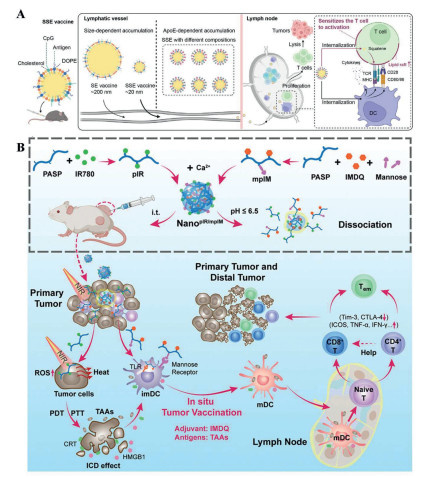
Another category targets intracellular signaling pathways that regulate DC maturation. The activation of PRRs triggers multiple downstream signaling cascades, including the NF-κB, mitogen-activated protein kinase pathway, and cyclic GMP-AMP synthase-stimulator of interferon genes (cGAS-STING) pathways, which collectively drive DC maturation and cytokines release. Directly regulating these core-signaling nodes can bypass or complement the involvement of PRRs, providing a more direct means of controlling DC function [78]. A nanoparticle based on a cyclic d/l-peptide (YhDh) that self-assembles through Mn2+ coordination and is coated with tumor cell membranes has been developed. This nanoparticle efficiently promotes DC internalization and maturation through a synergistic mechanism: The photothermal effect induces ICD to release antigens, while Mn2+ activates the cGAS-STING pathway. Together with the chiral structure of YhDh, these effects upregulate the expression of DC co-stimulatory molecules and promote DC maturation, ultimately enhancing the anti-tumor immune response [79]. A supramolecular hydrogel formed by Mn2+-coordinated self-assembly of a selenopeptide has been constructed, in which the hydrogel matrix itself directly provides therapeutic functions. This system enhances NK cell activity, activate cGAS-STING pathway, thus promoting DC maturation and enhancing antitumor immunity [80]. Furthermore, researchers used Ca2+ as the core building block to prepare a calcium–crosslinked nanocomplex. This complex encapsulates a TLR 7/8 agonist or a photosensitizer. Upon light stimulation, they efficiently promote DC maturation and inhibit the growth of both primary and distant tumors (Fig. 2B) [81].
In recent years, metabolic regulation strategies have attracted significant attention. Modulation of DC metabolism, particularly cholesterol homeostasis and glycolysis, has been shown to enhance antigen cross-presentation and T-cell activation. On one hand, cholesterol mobilization supports lipid raft formation and MHC-Ⅰ compartmentalization [82]; on the other hand, enhanced glycolytic metabolism provides the energy and biosynthetic precursors required for DC maturation and effector function [83]. Biomimetic calcium-chelating nanoparticles (RM-CDC@FX11&Ava) have been developed to co-deliver a lactate dehydrogenase A inhibitor (FX11) and avasimibe, synergistically regulating glycolysis and cholesterol metabolism. This nanosystem alleviates the acidic microenvironment by reducing lactate levels, while simultaneously increasing free cholesterol levels by activating sterol regulatory element-binding protein 2 and inhibiting cholesterol esterification, thereby promoting DC maturation and enhancing antitumor immunity [84]. Meanwhile, a lipid nanoparticle (LNP) platform capable of both mRNA delivery and DC metabolic reprogramming has been established, which stimulates glycolytic metabolism in DCs by activating the mechanistic target of rapamycin complex 2 (mTORC2) pathway, thereby promoting DC maturation and antigen presentation, which in turn enhances the antitumor immune response [85].
To address the problem that T cells failing to home to tumors, researchers have developed various strategies to promote T-cell infiltration by modulating the TME. These approaches primarily work through two mechanisms: Restoring T-cell chemokine signaling and removing physical or cellular barriers that hinder T-cell entry.
Several strategies can be used to restore the chemokine signaling that directs T cells to migrate toward tumor tissue, including carcinogenic pathway inhibitors, C-X-C chemokine receptor type 4 (CXCR4) antagonists and chemokine receptor inhibitors [24,28]. Researchers found that p21-activated kinase 4 (PAK4) is enriched in “cold tumors” and plays a vital role in the oncogenic WNT/β-catenin pathway, thereby limiting T-cell infiltration. Its overexpression is related to the development and progression of melanoma, pancreatic cancer and many other malignancies. Therefore, knocking out PAK4 or using PAK4 protein inhibitors can enhance T cell infiltration and thereby increase the therapeutic effect of PD-1 blockers [86]. In a Phase Ib study in patients with melanoma, the oral CXCR4 antagonist mavorixafor is shown to effectively promote T-cell infiltration into tumor tissue. Results showed a significant increase in the density of CD8+ T cells within the tumor, with the density of CD8+ T cells in the peripheral zone increasing 4-fold compared to pre-treatment levels [87].
Cellular barriers prevent T cells from penetrating the tumor stroma. The CXCL12/CXCR4 axis recruits immunosuppressive cells such as MDSCs and Tregs, forming a cellular barrier that excludes CTLs from tumor nests [88]. Targeting this axis, a CXCR4-targeted lipo-nanoenzyme platform (Pt-LNP@E5) has been developed, which exhibits higher affinity for CXCR4-overexpressing tumor cells and effectively antagonizes CXCR4/CXCL12-driven metastasis signals, thereby significantly inhibiting tumor cell migration and dissemination. In two hematological malignancy models, Pt-LNP@E5 demonstrated significant tumor growth inhibition, metastasis blockade, and immune microenvironment remodeling effects [89]. In addition, abnormal tumor vasculature and a dense extracellular matrix (ECM) constitute structural and physical barriers that hinder T-cell homing. Vascular normalization mediated by anti-angiogenic therapies (such as VEGF/VEGFR2 inhibitors) has been shown to restore the expression of endothelial adhesion molecules and promote T-cell extravasation [90,91]; meanwhile, ECM-degrading enzymes or matrix modulators can disrupt dense collagen barriers, opening pathways for T-cell infiltration [92]. As an example of combining anti-angiogenic and immunomodulatory functions, an aminated fullerene-based nanoplatform (TAPC@CNPs) has been designed to target VEGFR2 and block the downstream PI3K-AKT signaling pathway, while simultaneously reducing Treg levels and increasing T cell infiltration, thereby synergistically inhibiting tumor growth and remodeling the immune microenvironment [93].
ICB therapy achieves cancer immunotherapy by using specific antibodies or small-molecule drugs to block the signaling pathways of checkpoint molecules, thereby releasing the “braking” state of the immune system and enhancing the ability of CTLs. CTLA-4, PD-1 and PD-L1 have now become the most mature ICB targets [32,33,94]. Various ICIs including ipilimumab, tremelimumab, atezolizumab and pembrolizumab, have been approved by Food and Drug Administration (FDA) for the treatment of multiple types of malignant tumors [94-97].
Although the single drug of PD-1/PD-L1 and CTLA-4 has achieved great success in the treatment of multiple cancers, due to tumor immune escape, only a small number of patients are responsive to single-agent treatment. So, scientists have begun to explore how to combine ICB therapy with nanotechnology or bioengineering techniques to achieve better therapeutic results. A cell-cell combination drug delivery platform has been constructed based on conjugates of PD-L1 antibody-decorated platelets and hematopoietic stem cells (HSCs). This combination possesses the homing ability of HSCs to bone marrow and can effectively transmit anti-PD-1 in acute myeloid leukemia mouse model [98]. To enhance the ability of antibodies crossing the blood-brain barrier (BBB), researchers constructed a targeted nanoscale immunoconjugates on natural biopolymer scaffold, with covalently attached anti-CTLA-4 or anti-PD-1. The results showed that targeted polymer conjugated ICIs can effectively pass through BBB and can be used as an effective method for the treatment of glioblastoma [99].
The combination of ICB therapy with other treatments can also achieve better therapeutic effect than single drug. A combination approach integrating chemotherapy, autophagy inhibition, and PD-L1 blockade has been developed using functional gold nanoparticles. This strategy is shown to neutralize the immunosuppressive glioma microenvironment, enhance the anti-glioma effect, and effectively prevent glioma recurrence [100]. In another example, dimeric nanoparticles co-loading a chemotherapeutic agent and a hexokinase inhibitor have been combined with anti PD-L1 antibody, achieving superior tumor suppression and anti-metastatic effects compared with monotherapy [101]. The combination of PDT and ICB therapy also has great potential [102]. What is more, the combination of ICB therapy with DNA or mRNA vaccines can achieve better effects than monoclonal antibodies or vaccines alone [103-105].
At present, ICB therapy is rapidly expanding beyond CTLA-4 and PD-1/PD-L1. Drugs that block new immune checkpoints such as lymphocyte activating gene 3 (LAG-3), T cell immunoglobulin and mucin domain-containing protein 3 (TIM-3) or V-domain Ig suppressor of T cell activation are under investigation [106]. LAG-3 is expressed on CD4+ T cells, CTLs and Tregs. It binds to fibrin-like protein 1 (FGL1) overexpressed on tumor cells. LAG-3 and PD-1 on tumor infiltrating lymphocytes are often co-expressed and up-regulated, resulting in T cells exhaustion and promoting tumor growth [107,108]. Dual silencing of FGL1 and PD-L1 has been achieved using a ROS-sensitive nanoparticle loaded with FGL1 siRNA (siFGL1) and PD-L1 siRNA (siPD-L1). This system significantly reduced the protein levels of both targets on tumor cells, increased the infiltration of CD4+ T cells and CTLs, and effectively remodeled the TME (Fig. 3A) [109]. TIM-3 is a direct negative regulator of T cells and is expressed on NK cells and macrophages. TIM-3 indirectly promotes immunosuppression by inducing the expansion of MDSCs. Some studies have shown that TIM-3 plays a vital role in malignant tumors. Researchers combined dual PD-1 and TIM-3 blockade with PTT in an MC38 tumor model. Results showed that the combination successfully cleared the primary tumor, generated systemic immune response, suppressed the growth of distant tumors and prolonged the survival time. The indocyanine green-liposomes combined with PD-1/TIM-3 blockade strategy sheds light on the future clinical application of supramolecular PTT in cancer immunotherapy [106,110].
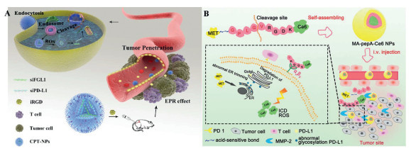
Although ICB therapy has made significant strides in cancer treatment, several challenges remain. For one, ICB therapy is usually associated with immune-related toxicity. This autoimmune response may result in organ-specific damage. For another, acquired drug resistance is also a serious problem faced by ICB therapy. The loss or down-regulation of tumor antigens and MHC molecules, the alterations in antigen presentation mechanism, the decrease of immune cell infiltration all contribute to tumor drug resistance. Therefore, a better understanding of the mechanism of drug resistance, the combination of ICB therapy with appropriate combination treatments, and the search for new biomarkers to achieve better efficacy have become the focus of current research. An MMP-2-cleavable self-delivery nanoparticle (MA-pepA-Ce6 NP) has been designed by conjugating a safe, acidic-sensitive small-molecule PD-L1 inhibitor (metformin) with a photosensitizer (Ce6). The TME dual-stimuli-sensitive system can achieve effective drug accumulation and potent antitumor immunotherapy, and its biodegradability and high safety profile suggest potential for controllable production (Fig. 3B) [111]. At present, a variety of classic strategies have been applied in clinical practice, bringing great benefits to patients (Table S1 in Supporting information).
Emerging evidence highlights that the nervous system is identified as a crucial regulator of cancer [112]. The nervous system can sense the immune status within the TME and transmit it to the brain, which then integrates the information and adjusts the immune system to support tumor growth and suppress anti-tumor immunity. At the same time, the immune system can also modulate neural activity and growth, exerting a significant impact on the nervous system [113,114]. Tumor-associated neuro-immune interactions are key factors driving tumor progression and metastasis [114,115].
As chemical messengers of the nervous system, neurotransmitters also serve as key modulators of the immune system. Major neurotransmitter classes include small molecule transmitters (acetylcholine, dopamine, epinephrine etc.) and neuropeptides, which directly target immune cells expressing neurotransmitter receptors, making them the most actively studied target category in neuro-immunology [114]. Norepinephrine, secreted by sympathetic nerves, binds to β2-adrenergic receptors (ADRB2) expressed on cancer cells and TAMs within the TME, promoting sympathetic neural crosstalk with these cells to accelerate tumor development and drug resistance. To block this adrenergic signaling, a biomimetic hybrid nanovesicle (Pro@hNV) has been constructed by fusing M1 macrophage-derived vesicles with pH-sensitive liposomes to encapsulate the ADRB2 blocker propranolol. This nano-system blocks ADRB2-mediated sympathetic-cancer cell and sympathetic-macrophage crosstalk, potently inhibits sympathetic signaling, reprograms M2-polarized TAMs to M1 phenotype, and markedly enhances anti-tumor immunity [116]. Calcitonin gene-related peptide, a key neuropeptide released by nociceptive sensory neurons, suppresses DC maturation and accelerates CTL exhaustion, thereby reinforcing tumor-neural crosstalk and facilitating immune evasion. Based on this, the LipoNCs@pGSDMB platform was developed to co-deliver a STING agonist and a gasdermin B (GSDMB) plasmid. This platform induces STING-driven immune priming and pyroptosis, while simultaneously restoring voltage-gated calcium channel expression in tumor cells, blocking calcium-dependent neural signaling and disrupting tumor-neural communication. In a breast cancer bone metastasis model, it achieved 94% tumor suppression and complete pain relief, demonstrating excellent neuro-immunotherapeutic efficacy (Fig. 4A) [117]. Overall, research in this field has evolved from simple drug delivery to “smart” nano-systems that actively engage and precisely modulate the neuro-immune network. These cutting-edge explorations open transformative avenues for developing more effective and less toxic cancer immunotherapies.
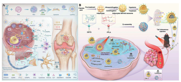
CAAs, predominantly located in adipose-rich tissues (e.g., breast, ovarian, colorectal and pancreatic tumors), undergo profound functional reprogramming upon exposure to cancer cells. Interactions between CAAs and immune cells play a critical role in reshaping the TME and are now recognized as key immune-metabolic regulators. CAAs directly modulate immune cell function via the secretion of adipokines, cytokines, and other soluble factors; additionally, they reprogram the metabolism of immune and tumor cells by supplying metabolic substrates such as fatty acids. Notably, CAA-derived extracellular vesicles (EVs) horizontally transfer bioactive molecules (e.g., lipids, RNA, proteins) between cells, further remodeling the TME [118-120]. Therefore, targeting CAA-related pathways has emerged as a promising therapeutic strategy in tumor immunotherapy.
A PLGA nanoparticle delivery system based on the natural compound hesperidin has been developed, which significantly reduces CCL2 secretion by CAAs while upregulating adiponectin production, thereby remodeling the TME and markedly enhancing the efficacy of anti-PD-1 immunotherapy [121]. To address lipid accumulation in tumor-associated dendritic cells (TADCs), a multi-level lipid reprogramming nanoparticle has been constructed that specifically targets the lipid transport receptor Msr1. By synergistically inhibiting exogenous lipid uptake, blocking de novo lipid synthesis, and suppressing adipogenic gene transcription in TADCs, this system systematically clears accumulated lipids, restores the function of dysfunctional TADCs, enhances their antigen presentation and T cell activation capacity, thereby reversing immunosuppression and significantly improving the efficacy of cancer immunotherapy [122]. Beyond lipid metabolic reprogramming, researchers have also applied CAA-derived exosomes/EVs for therapy. The biomimetic shell greatly enhances the natural affinity of nanoparticles for tumor cells, significantly improves cellular uptake and achieves efficient targeted combined chemo-photodynamic therapy (Fig. 4B) [123]. Looking ahead, in-depth exploration of the complex “molecular dialogue” among CAAs, tumor cells, and other immune cells will be key in this field. The combination of CAA-targeted nanotherapy with established immunotherapies such as ICB therapy is undoubtedly one of the most promising directions for the future.
ACT has become a major treatment modality for cancer, primarily comprising three technical routes: Tumor-infiltrating lymphocytes (TILs), TCR-T therapy, and chimeric antigen receptor (CAR) T-cell therapy. Among these, TILs utilize naturally occurring tumor-specific T cells, whereas TCR-T cell therapy and CAR-T cell therapy genetically engineer T cells to recognize and kill tumors. Both engineered routes share the core biological principle of T cell activation: Recognizing tumor antigens via receptors and initiating downstream signaling cascades to drive T cell proliferation and effector functions; but they differ in receptor design, antigen recognition mode, signaling mechanism, and clinical application scope.
TCR-T cell therapy. TCR-T cells are autologous T cells genetically engineered to express tumor-specific TCRs. They recognize antigenic peptides presented by MHC molecules on tumor cells, thereby activating T cells and triggering a series of immune responses that lead to tumor cell death. This MHC-dependent mechanism allows TCR-T cells to recognize intracellular tumor-associated or tumor-specific antigens presented via MHC molecules (e.g., NY-ESO-1 and MAGE-A4) as well as neoantigens derived from somatic mutations (e.g., KRAS G12D). This expands the range of targetable tumor antigens beyond cell-surface molecules, making TCR-T cell therapy a highly promising immunotherapy for solid tumors [124]. In 2024, the FDA approved the first TCR-T product, afamitresgene autoleucel (TecelraⓇ), for the treatment of certain adult patients with unresectable or metastatic synovial sarcoma (Table S2 in Supporting information).
Although clinical efficacy has been demonstrated in most treated patients, TCR-T cell therapy still faces numerous challenges, including immune toxicity from off-target effects, tumor immune escape through reduced or lost antigen expression, T cell exhaustion and dysfunction, and a lack of effective tumor-specific antigen targets [125]. To enhance TCR-T efficacy while mitigating off-target toxicity, histidine scanning has been employed to identify TCR hotspots capable of forming additional catch bonds. The introduction of histidine promotes new hydrogen bonds and salt bridges, thereby enabling the screening of low-affinity and high-activity TCR variants. This approach provides a new strategy for developing structure-independent universal low-affinity/high-potency TCR engineering [126]. A dual-target strategy combining an mp53-specific CD8+ T cell TCR with a CD4+ T cell-derived TCR that recognizes a mutant antigen presented on the cancer stroma has been shown to prevent tumor escape. This combination successfully prevented tumor recurrence and metastasis in preclinical models [127]. Furthermore, artificial intelligence (AI)-guided TCR specificity prediction enables rapid screening of candidate TCRs with high specificity and low cross-reactivity from massive TCR repertoires; base editing and precision genome engineering address chain mispairing while avoiding functional impairment from endogenous TCR knockout; and shifting targets from personalized to public neoantigens simplifies safety validation while improving platform universality [128-130]. Notably, nanotechnology has also been applied to empower TCR-T cell therapy. A dual-functional intelligent nanoplatform (AUTACE), composed of TCR-T cell-derived nanovesicles, utilizes TCRs for tumor targeting, anti-CD3 antibodies to activate CTLs, and low-intensity focused ultrasound to trigger degrader release and degrade mutant KRAS protein, effectively eliminating tumors in preclinical models [131].
CAR-T cell therapy. CAR-T cell therapy genetically modifies patient T cells to express CARs, which can recognize specific antigens on tumor cell surfaces, enabling precise tumor targeting and cytotoxicity. CAR design has undergone multiple generations. First-generation CARs contain only the CD3ζ signaling domain, which activates T cells but lacks persistence. Second-generation CARs incorporate one co-stimulatory domain (e.g., CD28 or 4–1BB), significantly enhancing T cell proliferation and in vivo persistence. Third-generation CARs further combine multiple co-stimulatory domains (e.g., CD3ζ-CD28-OX40 or CD3ζ-CD28–4–1BB), aiming to mimic a more complete T cell activation signal [124,132,133]. CAR-T cell therapy has achieved remarkable success in treating hematological malignancies. In 2017, the FDA approved the first CAR-T cell therapy (KymriahⓇ) for the treatment of relapsed or refractory B-cell acute lymphoblastic leukemia in children and young adults up to age 25. Since then, several additional CAR-T products have been approved (Table S2) [134]. However, CAR-T still faces challenges, such as limited efficacy in solid tumors, complex and costly ex vivo manufacturing, and toxicities such as cytokine release syndrome. Therefore, further optimization is needed to meet the higher treatment requirements [135].
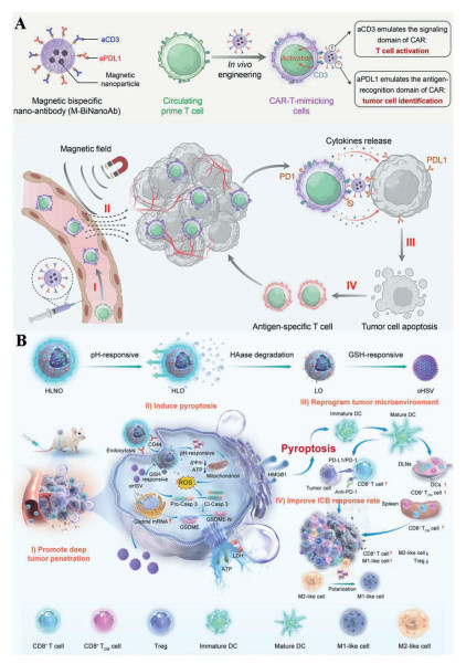
Various innovative strategies have been developed to enhance CAR-T function, including in vivo engineering, armored CAR design, and dual-antigen targeting. Targeted lipid nanoparticles (tLNPs) have been developed for the delivery of CAR mRNA, enabling the direct reprogramming of circulating CTLs into functional CAR-T cells in vivo, demonstrating robust tumor control. This tLNP platform may make CAR-T cell therapy more accessible and applicable to a broader range of clinical indications [136]. The toxicity previously associated with armored CAR-T due to non-specific peripheral cytokine expression has been addressed through a clustered regularly interspaced short palindromic repeats (CRISPR) knock-in strategy that employs endogenous promoters with tumor-restricted expression. This approach drives tumor-specific expression of cytokines such as IL-12 and IL-2, achieving both enhanced anti-tumor efficacy and improved safety [137]. To achieve dual-antigen targeting, researchers developed a magnetic bispecific nanobody (M-BiNanoAb) that couples anti-CD3 (CAR activation domain mimic) and anti-PD-L1 (CAR antigen recognition domain mimic) to magnetic nanoparticles. This system converts circulating T cells into CAR-T-like effector cells in vivo, and uses external magnetic fields for tumor-directed migration, significantly increasing intratumoral CTLs infiltration and activation and reducing exhaustion. This strategy can be adapted to diverse tumor antigens and extended to in vivo engineering of other immune cells such as NK cells and macrophages (Fig. 5A) [138].
Although CAR-T cell therapy and TCR-T cell therapy originate from different concepts, their future development shows profound convergent trends. The future of the ACT field lies in the complementary co-existence and synergistic evolution of multiple technology routes.
CAR-NK cell therapy has emerged as the most promising next-generation cellular immunotherapy after CAR-T. Unlike CAR-T cells, which rely on patient-derived autologous T cells and require a lengthy manufacturing process, CAR-NK cells originate from NK cells with inherent anti-tumor activity. After genetic engineering, CAR-NK cells not only retain MHC-unrestricted recognition and rapid tumor-killing capacity but also exhibit unique “off-the-shelf” potential and superior safety, opening a new chapter in tumor immunotherapy [139,140]. However, challenges such as insufficient efficacy, complex manufacturing process, high cost, and limited production capacity still hinder CAR-NK cells’ accessibility. Scientists are actively striving to develop more potent, persistent, and universal CAR-NK cells. SENTI-202 is an innovative CAR-NK cell therapy that uses two activating CARs (against CD33 and fms-like tyrosine kinase 3 (FLT3)) as an OR gate, destroying leukemia cells with either antigen to combat tumor heterogeneity. Endomucin, a marker on healthy stem cells, serves as a NOT gate: Upon recognition by inhibitory CARs, it suppresses NK cell activation and protects normal bone marrow [141]. To circumvent the safety and efficiency issues of viral transduction and gene editing, the CliCAR platform provides a non-genetic method: Recombinant CAR fusion proteins are “loaded” onto NK cells ex vivo, enabling them to exhibit CAR-like functions without genetic modification. This offers a safe, rapid, and customizable strategy for producing potent, off-the-shelf immunotherapies applicable to multiple tumor types [142]. CAR engineering has been further advanced to the hematopoietic stem and progenitor cell stage, which addresses the bottlenecks of complex and costly genetic modification of terminally differentiated NK cells, thereby providing a renewable, homogeneous, and batch-manufacturable source of effector cells [143]. In summary, CAR-NK is advancing steadily and dynamically, establishing a new therapeutic pillar in cellular immunotherapy based on its unique safety and universality (Table S3 in Supporting information).
CAR-Ms are genetically engineered to specifically recognize tumor-associated or tumor-specific antigens and achieve potent tumor clearance through phagocytosis. Their robust tumor infiltration capacity, high phagocytic efficiency, and unique potential to remodel the TME make CAR-Ms promising immunotherapeutic agents for T-cell-refractory solid tumors [144-147].
CAR-Ms exert anti-tumor effects through: (1) Direct elimination of tumor cells via phagocytosis and cytotoxicity [148]; (2) cross-presentation of tumor antigens to T cells after phagocytosis [149]; and (3) creating an immune-supportive niche by secreting pro-inflammatory cytokines (e.g., IL-12, TNF-β, IFN-γ), activating T and NK cells and reprogramming TAMs from a pro-tumor to an anti-tumor phenotype [147,150]. To date, CAR-M therapy has demonstrated promising anti-tumor efficacy in various cancer models including ovarian cancer, glioblastoma, melanoma, breast cancer, hepatocellular carcinoma, pancreatic cancer, and gastric cancer (Table S3) [144].
CAR-DC therapy genetically engineers DCs to express CARs that recognize tumor-specific antigens. Unlike the direct killing function of CAR-T, DCs are primarily responsible for antigen uptake, processing, and presentation, activating T cells to launch specific immune attacks against tumors. Therefore, CAR-DC therapy aims to combine the precise targeting of CAR technology with the superior immune-activating capacity of DCs, breaking immunosuppression, overcoming the “cold tumor” treatment dilemma, and addressing the limitations of CAR-T therapy [151]. Most researchers use CAR-DC therapy in combination with CAR-T cell therapy to achieve synergistic therapeutic effects [152]. Several clinical trials of CAR-DC therapy for multiple tumor types have been initiated (e.g., NCT06972576, NCT06946615) (Table S3). CAR-DC therapy represents a strategic evolution from “direct killing” to “intelligent regulation” in cellular immunotherapy, promising to break through current bottlenecks in solid tumor immunotherapy. Its unique therapeutic logic and clinical potential offer new hope for patients with refractory tumors.
Engineered cell therapies have developed diverse cellular platforms, exploiting the direct killing capacity of T cells, the natural killing and “off-the-shelf” potential of NK cells, the powerful phagocytosis and microenvironment-remodeling ability of macrophages, and the antigen presentation and immune activation advantages of DCs, all aimed at overcoming the core challenges of immunosuppression, heterogeneity, and poor infiltration in solid tumors. Currently, CAR-based therapies are evolving from single “direct killing” to multi-functional “systemic regulation”. The convergence of combination therapy, in vivo programming, universal manufacturing, and gene editing technologies will drive this field from personalized customization toward a new generation of large-scale, intelligent immunotherapeutic paradigms.
Multiple bacterial genera have been explored as potential cancer therapeutics, capable of naturally localizing to, penetrating, proliferating within, and shrinking solid tumors through various mechanisms [153]. Engineered bacterial cancer therapy harnesses the natural tumor tropism of bacteria, using synthetic biology and genetic engineering to transform bacteria into programmable “living drugs” that can precisely colonize tumors, produce anti-tumor agents in situ, and modulate the TME. This strategy transforms live bacteria from traditional pathogens into precise, intelligent anti-cancer platforms.
Engineered bacteria exert anti-tumor effects through multiple mechanisms including direct oncolysis, immune activation, targeted delivery of therapeutic payloads, and metabolic reprogramming of the TME [154-157]. In recent years, rapid advances in synthetic biology have enabled bacteria to evolve from “programmable cargo carriers” into intelligent biological platforms capable of sensing multiple input signals and executing complex logical operations. By integrating CRISPR technology, advanced inducible systems, and the integration of nanomaterials into hybrid platforms, current engineered bacteria possess the following capabilities: Sensing TME changes (e.g., hypoxia, low pH, or specific metabolite concentrations) and initiating context-dependent therapeutic responses [158-160] implementing self-limiting (e.g., population density control circuits with suicide switches) to avoid over-proliferation and off-target toxicity [161]; and synergizing with ICB therapy and CAR-T cell therapy to comprehensively remodel the TME [162,163]. Numerous tumor-related clinical trials of engineered bacteria have been initiated, marking the transition of this approach from proof-of-concept studies to early clinical translation in cancer immunotherapy (Table S4 in Supporting information).
Despite these advances, engineered bacteria still face challenges including safety concerns (e.g., unpredictable bacteremia and systemic inflammatory responses) and the complex regulatory landscape for live biotherapeutic products. To address these limitations, future research should focus on optimizing tumor-specific targeting elements to enhance selective bacterial accumulation, establishing standardized manufacturing processes and in vivo tracking methods, and combining them with existing immunotherapies. Through multidisciplinary collaboration, engineered bacterial therapy is expected to achieve broader clinical application in the era of precision cancer immunotherapy [164].
OVs are viral agents capable of selectively replicating within tumor cells, lysing them while simultaneously activating systemic antitumor immunity. OVs exert anti-tumor effects through multiple mechanisms, including direct oncolysis, activation of immunity and T cell priming, and remodeling of the immunosuppressive TME [165,166].
First-generation OV products (exemplified by OncorineⓇ) rely primarily on the virus’s inherent lytic effect without introducing exogenous transgenes, making it difficult to elicit robust systemic antitumor immune responses. Subsequent generations have progressively incorporated more precise immunomodulatory components and multiple transgenic payloads, significantly improving response rates and therapeutic outcomes in clinical studies [166,167]. Moreover, researchers have combined nanotechnology with OVs and integrated other immunotherapies (e.g., ICB, CAR-T) to overcome the efficacy limitations of monotherapy [168]. To reduce peripheral clearance and enhance tumor penetration, a triple-cascade responsive pneumatic nanomotor has been constructed for systemic delivery of oncolytic herpes simplex virus (oHSV). This system enables oHSV to evade immune clearance in peripheral circulation while specifically responding to TME stimuli, increasing CTL infiltration and M1 macrophage polarization, converting “cold” tumors to “hot” tumors (Fig. 5B) [169]. To overcome the limitations imposed by the immunosuppressive TME, a nanocapsule-coated OV platform (Epv@CMP) has been developed that delivers CD19 CAR target to gastrointestinal tumors, enabling a “double strike” in combination with CD19 CAR-T therapy. The combination of Epv@CMP with CD19 CAR-T achieves potent tumor clearance, validating the synergistic potential of integrating OV therapy with CAR-T immunotherapy for gastrointestinal solid tumors [170].
Overall, OV therapy is evolving from a traditional treatment concept focused solely on “direct oncolysis” into a multifunctional intelligent immunotherapy platform. With smart design of viral backbones, revolutions in delivery technology, precision and personalization of combination therapy, and deep integration with novel treatment modalities, OVs are poised to occupy an increasingly important position in tumor immunotherapy (Table S5 in Supporting information).
TPD represents the third paradigm in drug discovery after small molecule inhibitors and antibody drugs. Traditional small-molecule drugs operate in an “occupancy-driven” mode, requiring sustained binding to the functional site of the target protein to block its activity, which inherently restricts the range of druggable targets. What is more, traditional inhibitors often fail to maintain long-term efficacy against drug-resistant mutants. The key breakthrough of TPD lies in shifting the mode of action from “occupancy-driven” to “event-driven”. TPD molecules only need transient binding to trigger irreversible clearance of the target protein, without requiring continuous occupancy of the active site. This significantly reduces structural requirements for target proteins: Any protein that can be recognized by a TPD molecule is theoretically degradable. More importantly, due to their catalytic mechanism, TPD molecules can achieve complete target clearance at very low doses, effectively avoiding dose-dependent toxicity associated with traditional drugs. Depending on the endogenous degradation machinery utilized and the type of protein targeted, TPD can be classified into proteolysis-targeting chimeras (PROTACs), molecular glue degraders (MGDs), lysosome-targeting chimaeras (LYTACs), and autophagy-based methods (e.g., autophagy-targeting chimera (AUTACs) and autophagosome-tethering compound (ATTECs)) [171-173].
PROTACs are bifunctional molecules composed of a protein of interest (POI) ligand, a linker, and an E3 ubiquitin ligase ligand, which degrade POIs via the ubiquitin-proteasome system (UPS) inside cells [174,175]. Compared to traditional inhibitors that rely on occupation-driven mechanisms, this event-driven, catalytic degradation approach enables complete removal of target protein function, thereby fully blocking its activity. As such, it has emerged as a promising strategy to overcome limitations of existing immunotherapies. The application of PROTACs in tumor immunotherapy has formed a clear progressive strategy: First, degrading immune checkpoint proteins to directly break the physical “brake” on T cells and convert “cold” tumors to “hot”; second, modulating functional proteins within immune cells, optimizing the killing capacity of T and NK cells by degrading negative regulators or epigenetic modulators; third, systematically remodeling the TME by degrading key immunosuppressive factors (e.g., TGF-β, lactate metabolism enzymes), fundamentally reversing the immunosuppressive ecological niche [172,176]. On May 1, 2026, ARV-471 was officially approved by the FDA for the treatment of adult patients with advanced or metastatic ER+/HER2– breast cancer harboring ESR1 mutations (Table S6 in Supporting information).
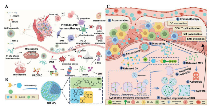
Despite accelerating clinical progress, PROTACs still face multiple challenges, including suboptimal pharmacokinetics, limited oral bioavailability and the “hook effect”, which are core bottlenecks limiting their clinical translation [177,178]. To address these issues, stimulus-responsive prodrugs and nano-delivery systems has been developed as cutting-edge solutions to improve PROTAC targeting selectivity and tissue accumulation. A multifunctional nanoplatform (dBET6@CFMPD) with MMP-2-responsive morphological change and mitochondrial targeting capability has been constructed. This platform enables the responsive release of the PROTAC drug (dBET6) and remodels the immunosuppressive TME. In multiple breast cancer models including brain metastases, it achieves effective suppression of both primary and metastatic tumors, overcoming challenges of poor distribution, limited accumulation, and inadequate monotherapy efficacy (Fig. 6A) [179]. Simultaneously, AI-assisted design provides a novel computational framework for ternary complex prediction, linker optimization, and de novo generation of new degraders [180,181]. Looking forward, PROTAC technology is expected to evolve into a programmable, customizable targeted degradation platform, shifting from broad “protein knockout” to precisely guided personalized medicine, continuously expanding the therapeutic scope of modern immunology.
MGDs share the same degradation mechanism as PROTACs, both recruiting E3 ubiquitin ligases to label POIs for proteasomal degradation. However, they differ fundamentally in chemical structure and mode of action. MGDs are typically monovalent, structurally concise small molecules that act as molecular bridges, inducing or stabilizing novel protein–protein interactions between the POI and an E3 ligase, thereby achieving target degradation without requiring a classical binding pocket. PROTACs, in contrast, are bifunctional molecules carrying both POI ligands and E3 ligase ligands, with more complex structures.
In tumor immunotherapy, MGDs, with their exquisite structures, restore immune system potency by degrading immune checkpoints, reprogramming T cells, and modulating MDSCs (Table S6) [182]. Traditionally, MGDs were discovered through phenotypic screening or serendipitous clinical observations (e.g., thalidomide), rather than rational design. Researchers are now attempting to systematically discover and optimize MGDs using AI and high-throughput screening (HTS) [183-185]. A recent study performed HTS on a library of 9600 HDAC-related compounds and identified a novel MGD, “Compound A”, which tripled immunoproteasome activity and enhanced the presentation of tumor-specific neoantigens by up to 100-fold, providing a novel approach for broadly applicable immunotherapy [186]. The future outlook of the integration of AI, structural biology, and proteomics is poised to revolutionize MGD discovery. Predictive glue libraries will become more systematic; E3 ligase “rewiring” will enable cell-type and tissue-specific degradation; and the integration of multi-omics data will facilitate personalized glue therapy based on the patient’s proteome [182].
Although PROTACs and MGDs are powerful, their utility is constrained by the substrate specificity of the UPS: They only degrade soluble cytoplasmic and nuclear proteins, failing to target extracellular proteins or membrane proteins. These proteins constitute about 40% of the human proteome and play critical roles in pathophysiology. First reported by Carolyn Bertozzi team in 2020, LYTAC technology fills this gap. LYTACs are bifunctional molecules consisting of a POI-binding ligand (antibody, peptide, or small molecule) and a ligand recognizing cell-surface lysosome-targeting receptors (LTRs), connected by a linker. The core mechanism is that LYTAC simultaneously binds the POI and an LTR (e.g., CI-M6PR/IGF2R) on the cell surface, triggering receptor-mediated endocytosis and delivering the POI to lysosomes for degradation via the endo-lysosomal pathway [172,187].
In tumor immunology, LYTACs exert therapeutic effects through multiple mechanisms: (1) Degrading key glycoproteins (e.g., MUC1) on tumor cell surfaces to disrupt physical barriers and expose tumor cells; (2) degrading critical immune checkpoints (e.g., PD-L1, CD47) and immunosuppressive cytokines (e.g., TGF-β, IL-6) to abrogate functional immune suppression by tumors; (3) degrading specific targets (e.g., tumor-surface galectin-1) to directly activate innate immune cells, promote antigen presentation, trigger T-cell responses, and bridge innate and adaptive immunity for synergistic effects [172,188-190]. To date, LYTAC technology still faces limitations: First, most LTRs show low abundance and restricted tissue distribution, affecting degradation efficiency; second, competition from endogenous ligands may impair LYTAC function; and finally, the large size and hydrophobicity of antibody-ligand conjugates pose pharmacokinetic challenges, including limited bioavailability and poor tumor penetration. Therefore, next-generation LYTACs are exploring novel ligand formats such as aptamers, nanoparticles, and peptides to expand the target spectrum, improve delivery efficiency, enhance degradation efficiency, and achieve better therapeutic outcomes [172].
A covalent dual-targeting Apt-cdLYTAC platform has been constructed by combining non-covalent recognition with photo-crosslinking covalent anchoring, employing a dual-ligand strategy to enhance local affinity. Compared to traditional LYTACs, Apt-cdLYTAC forms more stable degradation complexes with prolonged intracellular retention, significantly improving the degradation efficiency of target membrane proteins [191]. A linker-free nanovesicle LYTAC platform (NV-TACs) has been developed based on the simultaneous display of PD-1 and transferrin on the surface of endogenous exosomes, exploiting transferrin receptor-mediated specific lysosomal endocytosis to efficiently degrade PD-L1 on tumor cells and immune cells. When combined with a ferroptosis inducer, NV-TACs exhibits significant therapeutic effects in both anti-PD-1-responsive and non-responsive tumor models, with no obvious systemic toxicity. This study represents an important leap for LYTAC technology from traditional chemical conjugates to modular, biomimetic, programmable nano-delivery systems [192]. With deepening mechanistic research and breakthroughs in delivery technologies, LYTAC is expected to become a cornerstone of next-generation precision immunotherapy after PROTAC, fundamentally reversing the three barriers of physical shielding, metabolic suppression, and signaling escape, providing a novel catalytic engine for converting “cold” tumors to “hot”.
Autophagy is an important intracellular degradation pathway capable of degrading not only individual proteins but also protein aggregates, damaged organelles, and even invading pathogens. Autophagy-based degradation technologies further expand the substrate scope of TPD, enabling the targeting of pathological protein aggregates inaccessible to PROTACs or LYTACs.
AUTACs. AUTACs consist of a POI-ligand, a linker, and a guanine derivative (GD) degradation tag. During the degradation process, the POI ligand selectively binds to the POI, and the GD component induces S-guanosylation of the POI, resulting in K63-linked polyubiquitination and autophagosome recruitment for subsequent lysosomal degradation. This enables AUTAC not only to degrade individual proteins but also to target protein aggregates and subcellular structures such as damaged mitochondria [171,193,194].
To address the low bioavailability and non-specific distribution of AUTACs, a supramolecular nano-AUTAC system (GM NPs) has been developed by integrating AUTAC with nanotechnology. Targeting IDO, this system efficiently degrades this immune checkpoint via the autophagic-lysosomal pathway, suppressing tumor-associated immune tolerance and enhancing T cell function. The “IDO degradation + multi-modal immune activation” strategy improves tumor delivery of AUTACs, profoundly remodels the immunosuppressive TME and demonstrates broad adaptability and translational potential (Figs. 6B and C) [195]. Nevertheless, AUTACs remain in early-stage exploration; their degradation mechanism and structural optimization require further investigation. In-depth analysis of the structure-function relationship of AUTACs and comprehensive characterization of their autophagy-inducing molecular mechanisms are critical for guiding clinical translation.
ATTECs. ATTECs degrade proteins via the macro-autophagy pathway. ATTECs consist of three parts: A POI-ligand, a linker, and an LC3 (a key adaptor protein on autophagosome membranes) ligand. ATTECs adopt a bifunctional molecule strategy: One end binds LC3, the other binds the POI, tethering the target to autophagosomes for subsequent degradation via the autophagy-lysosome pathway [171].
Since the concept was proposed in 2019, the ATTEC platforms have continued to develop, achieving significant progress in protein degradation and providing new strategies for tumor immunotherapy. Researchers developed a ferritin-targeted ATTEC (16AA-C10-DPC@PPH) that selectively degrades ferritin, releasing endogenous Fe2+ and H2O2 to catalyze Fenton reactions and lipid peroxidation cascades, driving potent ferroptosis. This strategy does not rely on exogenous iron delivery; instead, it converts the compensatory defense mechanism of high ferritin expression in tumor cells into an “offensive” iron pool to induce cell death [196]. ATTEC platforms have also made progress in organelle degradation. Bio-ATTECs have been developed using bioorthogonal catalysis, which promote mitochondrial depletion and induce tumor cell death, highlighting their potential for organelle targeting [197]. Existing studies fully demonstrate the remarkable potential of the ATTEC platforms in achieving tissue-specific and autophagy-dependent degradation, offering new insights for future drug development and opening new possibilities for tumor immunotherapy.
As new strategies continue to emerge, a central focus in this field is the rational combination of different therapeutic approaches to maximize antitumor efficacy. A variety of classic combination therapies have been applied in clinical practice. Chemotherapy potentiates the efficacy of immunotherapy by inducing ICD and reshaping TME, while also facilitating more effective recognition and attack of tumor cells by the immune system. Accordingly, the combination of PD-1/PD-L1 inhibitors with chemotherapy has become a first line standard of care for many cancers. CTLA-4 and PD-1 act at different stages of T–cell activation, and their combined blockade produces synergistic effects. Therefore, PD-1/PD-L1 inhibitors (e.g., nivolumab and pembrolizumab) combined with CTLA-4 inhibitors (e.g., ipilimumab) have been approved for multiple cancer types, including melanoma and renal cell carcinoma.
There are also several combination regimens in preclinical or clinical development. Among these, the combination of OVs and CAR-T cell therapy has attracted particular attention. Insufficient infiltration of CAR T cells into solid tumors is a key barrier limiting their efficacy, and OVs can effectively overcome this bottleneck through various mechanisms. Genetically engineered OVs that express specific chemokines (e.g., CCL19) can actively recruit CAR-T cells to the tumor site via the chemokine-receptor axis, effectively overcoming the obstacle of poor CAR-T infiltration in solid tumors [198]. Studies have confirmed that recombinant Newcastle disease virus (rNDV19) expressing human CCL19 can significantly promotes CAR-T cell infiltration into tumor tissue via the CCL19-CCR7 chemotactic axis; the median survival of the combination therapy group was extended from 22 days to 36 days in an orthotopic lung cancer model [199]. Beyond recruitment and activation, there is also a bidirectional synergy between the two modalities. After CAR-T cells kill tumor cells, they can induce the release of intact viral particles, thereby enhancing the ability of OVs to infect surrounding tumor cells [200]. Concurrently, CAR-T cells can recognize OVs antigens via their own TCRs, giving rise to a TCR-activated CAR-T cell subset that exhibits stronger effector function and greater persistence upon subsequent exposure to tumor antigens [201]. This combined strategy has demonstrated synergistic antitumor effects in various solid tumor models, offering a promising approach to overcoming the limitations of CAR-T monotherapy in solid tumors.
The intersection of TPD and microbial-based immunotherapy also offers new opportunities for combination therapy. After OVs infect tumor cells, they induce ICD and upregulate the expression of immune checkpoints, such as PD-L1, on the surface of tumor cells, creating an ideal opportunity for LYTACs to target and degrade PD-L1 [202]. Engineered bacteria can also serve as delivery vehicles that release LYTACs in response to TME signals [203]. By leveraging their tumor colonizing ability, these bacteria enable sustained local production and release of LYTACs within the tumor, thereby avoiding the off-target risks associated with systemic administration.
The above cases demonstrate that the rational combination of emerging therapeutic approaches is expanding in multiple directions, driven by complementary mechanisms and functional synergy. It is foreseeable that cancer immunotherapy will shift from optimizing single modalities to integrating multiple strategies for multi target synergy, offering broader possibilities for conquering tumors.
Recent advances in tumor immunotherapy indicate that therapeutic efficacy is governed not only by the intrinsic activity of immunotherapeutic agents, but also by their capacity to operate and remodel the dynamic TME. The TME presents multifaceted barriers, including immunosuppressive networks, defective antigen presentation, insufficient T cell infiltration, and aberrant immune checkpoint signaling. Consequently, the TME has become a therapeutic target whose cellular constituents, molecular mediators, and spatial architecture must be actively reprogrammed to achieve durable antitumor immunity.
The emerging landscape of tumor immunotherapy is undergoing a conceptual transition from immune activation to immune-system engineering. Next generation strategies no longer simply boost endogenous antitumor responses; they are designed to rewire the biological circuits that govern tumor immune interactions. Engineered immune cells can be programmed to recognize, migrate toward, persist within, and function in tumor niches. Engineered bacteria and OVs act as tumor localizing biological devices that sense pathological cues, deliver therapeutic payloads, induce ICD, and remodel the TME. TPD offers a means to eliminate pathogenic or immunosuppressive proteins directly, rather than merely blocking their activity. Collectively, these developments signify a transition from “treating tumors” to “programming the tumor ecosystem”.
However, technological proliferation does not guarantee clinical success. A critical limitation of current approaches is that most platforms are optimized for single molecular targets or mechanisms, whereas tumors are heterogeneous, spatially organized, and continuously evolving ecosystems. Even with successful target engagement, resistance may arise through antigen loss, stromal barriers, metabolic competition, or adaptive immune suppression. For instance, engineered bacteria and OVs must concurrently address challenges in tumor penetration, replication, immune clearance, and safety; engineered cell therapies face issues of persistence, antigen escape, exhaustion, and a hostile TME. Therefore, future combination regimens should be rationally designed based on the immune status, spatial architecture, metabolic profile, and evolutionary trajectory of individual tumors.
Overall, the future of tumor immunotherapy will depend on our ability to interpret, predict, and therapeutically reprogram the evolving tumor immune ecosystem. Integrating spatial omics, single cell phenotyping, artificial intelligence, synthetic biology and nanotechnology may enable tumor classification based on actionable immune states, facilitating rational patient selection, target choice, regimen design, and combination partner identification. Thus, the field may be moving toward a dynamic, programmable, and patient specific immunotherapy paradigm, ushering in a predictive, engineering era for tumor treatment.
During the preparation of this work, the authors used Deepseek in order to have a more comprehensive understanding of the relevant fields. After using this tool, the authors reviewed and edited the content as needed and take full responsibility for the content of the published article.
Li Xia: Writing – review & editing, Data curation. Yujun Song: Writing – review & editing, Writing – original draft. Lan Xiao: Writing – review & editing, Writing – original draft. Fan Tong: Visualization. Miao Yu: Writing – review & editing. Huile Gao: Supervision, Conceptualization. Ran Ran: Supervision, Conceptualization.
The authors declare that they have no known competing financial interests or personal relationships that could have appeared to influence the work reported in this paper.
This work is supported by the 2023 Key Clinical Specialty Construction Project (General Surgery) (No. YB2023012).
Supplementary material associated with this article can be found, in the online version, at doi:
H. Sung, A.M. Filho, M. Laversanne, et al., CA Cancer J. Clin. 76 (2026) e70090.
R.L. Siegel, T.B. Kratzer, A.N. Giaquinto, et al., CA Cancer J. Clin. 75 (2025) 10–45. doi: 10.3322/caac.21871
A. Al-Khinji, N. Al-Korbi, S. Al-Kuwari, et al., Front. Oncol. 15 (2025) 1569884. doi: 10.3389/fonc.2025.1569884
P. Dasgupta, H. Lin, G. Kumar, et al., J. Neurooncol. 175 (2025) 453–461. doi: 10.1007/s11060-025-05131-9
S. Grabska, H. Grabski, T. Makunts, et al., Cancers 16 (2024) 2820. doi: 10.3390/cancers16162820
J. Hu, X. Yuan, F. Wang, et al., Chin. Chem. Lett. 32 (2021) 1341–1347. doi: 10.1016/j.cclet.2020.11.006
D.D. Shah, M.R. Chorawala, N.R. Raghani, et al., Med. Oncol. 42 (2025) 117. doi: 10.1007/s12032-025-02641-4
E. Boedtkjer, S.F. Pedersen, Annu. Rev. Physiol. 82 (2020) 103–126. doi: 10.1146/annurev-physiol-021119-034627
K. Li, H. Shi, B. Zhang, et al., Signal Transduct. Target. Ther. 6 (2021) 362. doi: 10.1038/s41392-021-00670-9
J. Lu, Y. Luo, D. Rao, et al., Exp. Hematol. Oncol. 13 (2024) 39. doi: 10.1186/s40164-024-00505-7
S. He, L. Zheng, C. Qi, Mol. Cancer 24 (2025) 5. doi: 10.1109/cisat66811.2025.11181725
N. Sobhani, D.R. Tardiel-Cyril, A. Davtyan, et al., Cancers 13 (2021) 1440. doi: 10.3390/cancers13061440
O. Goldmann, O.V. Nwofor, Q. Chen, et al., Front. Immunol. 15 (2024) 1328193. doi: 10.3389/fimmu.2024.1328193
Y. Shao, W.Y. Yang, G. Nanayakkara, et al., Int. J. Drug Discov. Pharm. 3 (2024) 100022.
J.T. Schnell, R.L. Briviesca, T. Kim, et al., Nat. Rev. Rheumatol. 21 (2025) 9–21. doi: 10.1038/s41584-024-01190-w
Q. Yang, N. Guo, Y. Zhou, et al., Acta Pharm. Sin. B 10 (2020) 2156–2170. doi: 10.1016/j.apsb.2020.04.004
Y. Li, L. Wang, W. Ma, et al., J. Transl. Med. 23 (2025) 697. doi: 10.1002/cjce.25399
J. Xiao, Z. Yang, S. Wang, et al., Sci. Rep. 14 (2024) 14343. doi: 10.1038/s41598-024-65435-0
J.S. Pawar, M.A. Salam, M.S.U. Dipto, et al., Cancers 17 (2025) 2484. doi: 10.3390/cancers17152484
M.Y. Lee, J.W. Jeon, C. Sievers, et al., J. Immunother. Cancer 8 (2020) e001111. doi: 10.1136/jitc-2020-001111
N. Pishesha, T.J. Harmand, H.L. Ploegh, Nat. Rev. Immunol. 22 (2022) 751–764. doi: 10.1038/s41577-022-00707-2
S. Jhunjhunwala, C. Hammer, L. Delamarre, Nat. Rev. Cancer 21 (2021) 298–312. doi: 10.1038/s41568-021-00339-z
J.M. Romero, P. Jiménez, T. Cabrera, et al., Int. J. Cancer 113 (2005) 605–610. doi: 10.1002/ijc.20499
Y.T. Liu, Z.J. Sun, Theranostics 11 (2021) 5365–5386. doi: 10.7150/thno.58390
J. Galon, D. Bruni, Nat. Rev. Drug Discov. 18 (2019) 197–218. doi: 10.1038/s41573-018-0007-y
K. Mortezaee, Clin. Exp. Med. 24 (2024) 15. doi: 10.1007/s10238-023-01274-z
F. Bayerl, P. Meiser, S. Donakonda, et al., Immunity 56 (2023) 1341–1358.e11 e11. doi: 10.1016/j.immuni.2023.05.011
P. Bonaventura, T. Shekarian, V. Alcazer, et al., Front. Immunol. 10 (2019) 168. doi: 10.3389/fimmu.2019.00168
K.P. Burke, A. Chaudhri, G.J. Freeman, et al., Immunity 57 (2024) 223–244. doi: 10.1016/j.immuni.2024.01.013
S. Singh, D. Dey, D. Barik, et al., Signal Transduct. Target. Ther. 10 (2025) 213. doi: 10.1007/978-981-96-5281-5_19
P. Azimnasab-Sorkhabi, M. Soltani-Asl, T.T. Yoshinaga, et al., Immunol. Res. 71 (2023) 679–686. doi: 10.1007/s12026-023-09378-0
J. Wang, T. Yang, J. Xu, Adv. Exp. Med. Biol. 1248 (2020) 619–649. doi: 10.1007/978-981-15-3266-5_23
F.K. Dermani, P. Samadi, G. Rahmani, et al., J. Cell. Physiol. 234 (2019) 1313–1325. doi: 10.1002/jcp.27172
L. Huang, H. Zhu, Y. Shi, J. Hematol. Oncol. 18 (2025) 107.
J. Wang, Q. Chen, T. Liang, et al., Cancer Lett. 617 (2025) 217616. doi: 10.1016/j.canlet.2025.217616
A.O. Elzoghby, O. Samir, H.E. Emam, et al., Acta Pharm. Sin. B 14 (2024) 2475–2504. doi: 10.1016/j.apsb.2024.03.022
G. Kroemer, L. Galluzzi, O. Kepp, et al., Annu. Rev. Immunol. 31 (2013) 51–72. doi: 10.1146/annurev-immunol-032712-100008
M.L. Lotsberg, K. Wnuk-Lipinska, S. Terry, et al., J. Thorac. Oncol. 15 (2020) 973–999. doi: 10.1016/j.jtho.2020.01.015
J. Zhou, G. Wang, Y. Chen, et al., J. Cell. Mol. Med. 23 (2019) 4854–4865. doi: 10.1111/jcmm.14356
A. Serrano-del Valle, A. Anel, J. Naval, et al., Front. Cell Dev. Biol. 7 (2019) 50. doi: 10.3389/fcell.2019.00050
Q. Wang, X. Ju, J. Wang, et al., Cancer Lett. 438 (2018) 17–23. doi: 10.1016/j.canlet.2018.08.028
H. Zhao, S. Dai, Front. Pharmacol. 17 (2026) 1732407. doi: 10.3389/fphar.2026.1732407
X. Zhao, K. Yang, R. Zhao, et al., Biomaterials 102 (2016) 187–197. doi: 10.1016/j.biomaterials.2016.06.032
Z. Huang, Y. Chen, J. Zhang, et al., ACS Appl. Mater. Interfaces 13 (2021) 39934–39948. doi: 10.1021/acsami.1c11327
F. Shen, L. Feng, Y. Zhu, et al., Biomaterials 255 (2020) 120190. doi: 10.1016/j.biomaterials.2020.120190
L. Jia, Y. Hong, X. He, et al., Chin. Chem. Lett. 36 (2025) 109957. doi: 10.1016/j.cclet.2024.109957
X. Duan, C. Chan, W. Lin, Angew. Chem. Int. Ed. 58 (2019) 670–680. doi: 10.1002/anie.201804882
X. Sun, J. Sun, J. Lv, et al., J. Mater. Chem. B 7 (2019) 5797–5807. doi: 10.1039/c9tb01346f
R. Liu, Y. An, W. Jia, et al., J. Control. Release 321 (2020) 589–601. doi: 10.1016/j.jconrel.2020.02.043
W. Jia, R. Liu, Y. Wang, et al., Acta Pharm. Sin. B 12 (2022) 3354–3366. doi: 10.1016/j.apsb.2022.03.010
D. Allen, M.J. Szoo, T.D. van Bergen, et al., Antib. Ther. 8 (2025) 68–85. doi: 10.1093/abt/tbaf001
H. Kobayashi, P.L. Choyke, Acc. Chem. Res. 52 (2019) 2332–2339. doi: 10.1021/acs.accounts.9b00273
K. Nakajima, H. Takakura, Y. Shimizu, et al., Cancer Sci. 109 (2018) 2889–2896. doi: 10.1111/cas.13713
A. Nanajian, M. Scott, N.I. Burcus, et al., Vaccines 12 (2024) 633. doi: 10.3390/vaccines12060633
F.R. Riyas Mohamed, S. Aldubaisi, A. Akbar, et al., Cancers 17 (2025) 2953. doi: 10.3390/cancers17182953
T.J. Lai, L. Sun, K. Li, et al., Cancer Res. Commun. 4 (2024) 1834–1849. doi: 10.1158/2767-9764.crc-23-0566
F.R. Riyas Mohamed, A. Yaqinuddin, Med. Oncol. 42 (2025) 213. doi: 10.1007/s12032-025-02760-y
Y. Zhang, P. Naderi Yeganeh, H. Zhang, et al., Nat. Immunol. 25 (2024) 1858–1870. doi: 10.1038/s41590-024-01932-8
B. Moran, M. Davern, J.V. Reynolds, et al., Cancer Lett. 559 (2023) 216121. doi: 10.1016/j.canlet.2023.216121
W. Dong, B. He, Y. Cao, et al., Cell. Oncol. 48 (2025) 249–264. doi: 10.1007/s13402-024-00989-9
W. Huang, Q. Zhu, Z. Shi, et al., Cell Death Discov. 10 (2024) 143. doi: 10.1002/9781119719830.ch6
A. Apavaloaei, M.P. Hardy, P. Thibault, et al., Cancers 12 (2020) 2607. doi: 10.3390/cancers12092607
T. Jiang, T. Shi, H. Zhang, et al., J. Hematol. Oncol. 12 (2019) 93. doi: 10.1186/s13045-019-0787-5
P. Xu, H. Luo, Y. Kong, et al., Biomed. Pharmacother. 131 (2020) 110640. doi: 10.1016/j.biopha.2020.110640
S.N. Khattak, K.P. Adzavon, W. Zhao, et al., Chin. Chem. Lett. 37 (2026) 112244. doi: 10.1016/j.cclet.2025.112244
C. Tonini, A. Bianchi, M. Esposito, et al., J. Immunother. Cancer 14 (2026) e013812. doi: 10.1136/jitc-2025-013812
L. Maso, S.A. Mosure, S.A. Rodriguez-Aponte, et al., Nat. Commun. 16 (2025) 11264. doi: 10.1038/s41467-025-66132-w
X. Li, R. Cheng, D. Junyi, et al., Biomaterials 327 (2026) 123735. doi: 10.1016/j.biomaterials.2025.123735
S. You, S. Li, L. Zeng, et al., Cancer Cell 42 (2024) 1415–1433.e12. doi: 10.1016/j.ccell.2024.06.014
Y. Huang, M. Su, W. Lin, et al., Theranostics 15 (2025) 7677–7692. doi: 10.7150/thno.114347
Y. Li, F. Tong, Y. Wang, et al., Acta Pharm. Sin. B 14 (2024) 4102–4117. doi: 10.1016/j.apsb.2024.06.003
Q. Ren, X. Zhao, L. Zhou, et al., Nat. Biomed. Eng. (2026), doi: 10.1038/s41551-026-01672-0.
M. Sadeghzadeh, S. Bornehdeli, H. Mohahammadrezakhani, et al., Life Sci. 254 (2020) 117580. doi: 10.1016/j.lfs.2020.117580
B. Zhang, L. Zhao, H. Li, et al., Front. Immunol. 17 (2026) 1744671. doi: 10.3389/fimmu.2026.1744671
Y. Yao, Y. Luo, Z. Wang, et al., J. Immunother. Cancer 13 (2025) e012710. doi: 10.1136/jitc-2025-012710
H. Wang, W. Zhang, C. Huang, et al., Chin. Chem. Lett. 36 (2025) 111092. doi: 10.1016/j.cclet.2025.111092
X. Shen, S. Fan, J. He, et al., Adv. Sci. 13 (2026) e12139. doi: 10.1002/advs.202512139
W. Qian, J. Ye, S. Xia, Front. Mol. Biosci. 11 (2024) 1391046. doi: 10.3389/fmolb.2024.1391046
W. Hu, J. Xu, M. Lv, et al., Nano Today 66 (2026) 102931. doi: 10.1016/j.nantod.2025.102931
L. Peng, Y. Kong, J. Yan, et al., Adv. Funct. Mater. 36 (2026) e77396. doi: 10.1002/adfm.77396
Y. Liu, H. Li, H. Zhao, et al., Adv. Healthc. Mater. 11 (2022) 2102781. doi: 10.1002/adhm.202102781
M. Belabed, M.D. Park, C.M. Blouin, et al., Nat. Immunol. 26 (2025) 188–199. doi: 10.1038/s41590-024-02065-8
M.A. Nour, M. Rajabivahid, M.S.S. Mehdi, et al., Cancer Cell Int. 25 (2025) 149. doi: 10.1186/s12935-025-03781-3
Z. Wang, M. Wu, Y. Jiang, et al., J. Control. Release 380 (2025) 362–374. doi: 10.3390/electronics14020362
D. Kim, N. Gong, M.G. Alameh, et al., Nat. Mater. (2026), doi: 10.1038/S41563-026-02512-X.
G. Abril-Rodriguez, D.Y. Torrejon, W. Liu, et al., Nat. Cancer 1 (2019) 46–58. doi: 10.1038/s43018-019-0003-0
R.H. Andtbacka, Y. Wang, R.H. Pierce, et al., Cancer Res. Commun. 2 (2022) 904–913. doi: 10.1158/2767-9764.crc-22-0090
K. Mortezaee, Life Sci. 249 (2020) 117534. doi: 10.1016/j.lfs.2020.117534
M. Wang, Y. Zhao, M. Lu, et al., J. Control. Release 395 (2026) 114982. doi: 10.1016/j.jconrel.2026.114982
Y. Xie, I. Ho, Z. Liu, et al., Front. Immunol. 17 (2026) 1760044. doi: 10.3389/fimmu.2026.1760044
J. Wang, Q. Zhang, Y. Li, et al., Chin. Chem. Lett. 35 (2024) 108746. doi: 10.1016/j.cclet.2023.108746
J. Hu, H. Singh, Y. Jin, et al., Proc. Natl. Acad. Sci. U. S. A. 122 (2025) e2507542122. doi: 10.1073/pnas.2507542122
J. Huo, Y. Wang, L. Liu, et al., Bioact. Mater. 62 (2026) 248–262.
A. Ribas, J.D. Wolchok, Science 359 (2018) 1350–1355. doi: 10.1126/science.aar4060
B. Comin-Anduix, H. Escuin-Ordinas, F.J. Ibarrondo, OncoTargets Ther. 9 (2016) 1767–1776.
R. Jotte, F. Cappuzzo, I. Vynnychenko, et al., J. Thorac. Oncol. 15 (2020) 1351–1360. doi: 10.1016/j.jtho.2020.03.028
P. Kawczak, I.J. Feszak, T. Bączek, Cancers 17 (2025) 2805. doi: 10.3390/cancers17172805
X. Han, H. Li, D. Zhou, et al., Acc. Chem. Res. 53 (2020) 2521–2533. doi: 10.1021/acs.accounts.0c00339
A. Galstyan, J.L. Markman, E.S. Shatalova, et al., Nat. Commun. 10 (2019) 3850. doi: 10.1038/s41467-019-11719-3
S. Ruan, R. Xie, L. Qin, et al., Nano Lett. 19 (2019) 8318–8332. doi: 10.1021/acs.nanolett.9b03968
X. Yang, C. Hu, F. Tong, et al., Adv. Funct. Mater. 29 (2019) 1901896. doi: 10.1002/adfm.201901896
N. Zhang, J. Song, Y. Liu, et al., J. Control. Release 306 (2019) 15–28. doi: 10.1016/j.jconrel.2019.05.036
C.G. Buss, S.N. Bhatia, Proc. Natl. Acad. Sci. U. S. A. 117 (2020) 13428–13436. doi: 10.1073/pnas.2001569117
A. Monfaredan, S. Şen, N.K. Fathi, et al., Int. J. Mol. Sci. 26 (2025) 8448. doi: 10.3390/ijms26178448
Y. Yin, J. Guo, S. Zhang, et al., Chin. Chem. Lett. 36 (2025) 110771. doi: 10.1016/j.cclet.2024.110771
S. Dadfar, Y. Eivazzadeh, H. Molavi, et al., Inflamm. Res. 75 (2026) 58. doi: 10.1007/s00011-026-02211-y
J. Wang, C. Klein, J.R. Cochran, et al., Trends Pharmacol. Sci. 46 (2025) 638–652. doi: 10.1016/j.tips.2025.05.008
Q. Lecocq, M. Keyaerts, N. Devoogdt, et al., Int. J. Mol. Sci. 22 (2020) 75. doi: 10.3390/ijms22010075
W.J. Wan, G. Huang, Y. Wang, et al., Acta Biomater. 136 (2021) 473–484. doi: 10.1016/j.actbio.2021.09.040
T.Y. Huang, G.L. Huang, C.Y. Zhang, et al., Front. Chem. 8 (2020) 1. doi: 10.3389/fchem.2020.00001
C. Hu, X. He, Y. Chen, et al., Adv. Funct. Mater. 31 (2021) 2007149. doi: 10.1002/adfm.202007149
R. Mancusi, M. Monje, Nature 618 (2023) 467–479. doi: 10.1038/s41586-023-05968-y
T. Pu, J. Sun, G. Ren, et al., Signal Transduct. Target. Ther. 10 (2025) 176. doi: 10.1038/s41392-025-02241-8
F.V. Abeele, M. Salzet, Cancer Lett. 634 (2025) 218070. doi: 10.1016/j.canlet.2025.218070
M. Amit, T. Eichwald, A. Roger, et al., Nat. Rev. Cancer 25 (2025) 573–589. doi: 10.1038/s41568-025-00831-w
J. Liu, J. Qin, W. Zheng, et al., Nat. Commun. 17 (2026) 6230. doi: 10.1038/s41467-026-72847-1
Z. Zhang, P. Chen, Y. Zheng, et al., Sci. Adv. 12 (2026) eady1292. doi: 10.1126/sciadv.ady1292
W. Wang, H. Chen, S. Yin, et al., J. Cancer Res. Clin. Oncol. 149 (2023) 7969–7979. doi: 10.1007/s00432-023-04685-3
T.H. Kim, S.H. Lim, H. Lee, et al., Exp. Mol. Med. 58 (2026) 366–381. doi: 10.1038/s12276-026-01650-1
J. Park, J. Cho, Biomol. Ther. 34 (2026) 491–505. doi: 10.4062/biomolther.2026.036
N. Luo, L. Ma, N. Ma, et al., Int. Immunopharmacol. 140 (2024) 112759. doi: 10.1016/j.intimp.2024.112759
C. Xu, X. Ji, Y. Zhou, et al., Adv. Mater. 35 (2023) 2211415. doi: 10.1002/adma.202211415
X. Liu, J. Zhang, S. Zheng, et al., J. Control. Release 370 (2024) 168–181. doi: 10.1016/j.jconrel.2024.04.031
T. Sanomachi, Y. Katsuya, T. Nakatsura, et al., Cancer 17 (2025) 1945. doi: 10.3390/cancers17121945
R. Xu, S. Du, J. Zhu, et al., Cancer Lett. 546 (2022) 215840. doi: 10.1016/j.canlet.2022.215840
Y. Wang, Y. Wang, W. Yuan, et al., Cell 189 (2026) 1680–1700.e48. e48. doi: 10.1016/j.cell.2025.12.050
V. Anastasopoulou, H. Schreiber, C.E. Lee, et al., Oncoimmunology 14 (2025) 2514041. doi: 10.1080/2162402X.2025.2514041
Y. Gao, Y. Gao, Y. Fan, et al., Nat. Mach. Intell. 5 (2023) 236–249. doi: 10.1038/s42256-023-00619-3
D. Su, F. Zhu, H. Shi, et al., Cell. Mol. Immunol. 23 (2026) 686–700. doi: 10.1038/s41423-026-01418-7
Y. Zhang, Y. Liu, Q. Wang, et al., Front. Immunol. 17 (2026) 1754735. doi: 10.3389/fimmu.2026.1754735
L. Li, B. Li, Y. Hao, et al., Adv. Sci. 13 (2026) e18455. doi: 10.1002/advs.202518455
L. Yu, Y. Liu, X. Lin, Cell. Mol. Immunol. 22 (2025) 712–729. doi: 10.1038/s41423-025-01304-8
M. Hiltensperger, A.M. Krackhardt, Front. Immunol. 14 (2023) 1121030. doi: 10.3389/fimmu.2023.1121030
H.Q. Wang, R. Fu, Q.W. Man, et al., J. Clin. Med. 12 (2023) 2173. doi: 10.3390/jcm12062173
L. Wang, L. Zhang, L.C. Dunmall, et al., Cancer Lett. 591 (2024) 216871. doi: 10.1016/j.canlet.2024.216871
T.L. Hunter, Y. Bao, Y. Zhang, et al., Science 388 (2025) 1311–1317. doi: 10.1126/science.ads8473
A.X. Chen, K.M. Yap, J.S. Kim, et al., Nature 644 (2025) 241–251. doi: 10.1038/s41586-025-09212-7
Y. Zhu, C. Chen, J. Chen, et al., Adv. Mater. 38 (2026) e20493. doi: 10.1002/adma.202520493
P. Yao, Y.G. Liu, G. Huang, et al., Exp. Hematol. Oncol. 13 (2024) 118. doi: 10.1186/s40164-024-00583-7
F.D. Dos Reis, Y. Saidani, P. Martín-Rubio, et al., Front. Immunol. 16 (2025) 1603757. doi: 10.3389/fimmu.2025.1603757
B.S. Garrison, H. Deng, G. Yucel, et al., Blood 138 (2021) 2799. doi: 10.1182/blood-2021-154201
S.Y. Lim, Y. Kim, S. Han, et al., Biomed. Pharmacother. 193 (2025) 118691. doi: 10.1016/j.biopha.2025.118691
F. Hu, J. Li, Y. Wang, et al., Nat. Biomed. Eng. 10 (2026) 765–784.
M. Wang, Z. Qin, X.W. Bian, et al., Cancer Commun. 45 (2025) 1344–1366. doi: 10.1002/cac2.70053
J. Lu, Y. Ma, Q. Li, et al., Biomark. Res. 12 (2024) 86. doi: 10.1186/s40364-024-00637-2
K.M. Maalej, M. Merhi, V.P. Inchakalody, et al., Mol. Cancer 22 (2023) 20. doi: 10.1186/s12943-023-01723-z
K.A. Reiss, M.G. Angelos, E.C. Dees, et al., Nat. Med. 31 (2025) 1171–1182. doi: 10.1038/s41591-025-03495-z
M. Klichinsky, M. Ruella, O. Shestova, et al., Nat. Biotechnol. 38 (2020) 947–953. doi: 10.1038/s41587-020-0462-y
S. Chen, Y. Wang, J. Dang, et al., Nat. Commun. 16 (2025) 4069. doi: 10.1039/d5ob00425j
S. Pierini, R. Gabbasov, M.C. Oliveira-Nunes, et al., Nat. Commun. 16 (2025) 706. doi: 10.1038/s41467-024-55770-1
Y. Zhang, R. Hu, X. Xie, et al., Ann. Hematol. 104 (2025) 4305–4317. doi: 10.1007/s00277-025-06538-0
R. Zhang, J. Zhang, H. Zhang, et al., Clin. Transl. Med. 15 (2025) e70536. doi: 10.1002/ctm2.70536
L. Guo, J. Ding, W. Zhou, Chin. Chem. Lett. 35 (2024) 108557. doi: 10.1016/j.cclet.2023.108557
M. Jayaprakash, D.V. Kumar, G. Chakraborty, et al., Biomed. Pharmacother. 192 (2025) 118559. doi: 10.1016/j.biopha.2025.118559
M. Sun, J. Yang, L. Li, et al., Chin. Chem. Lett. 36 (2025) 111093. doi: 10.1016/j.cclet.2025.111093
L. Niu, Z. Deng, Y. Jin, et al., Protein Cell 17 (2026) 279–303. doi: 10.1093/procel/pwaf085
H. Wang, F. Xu, C. Wang, Semin. Cancer Biol. 112 (2025) 58–70. doi: 10.1016/j.semcancer.2025.03.003
Z.P. Zou, X.G. Wang, X.R. Shi, et al., Adv. Sci. 12 (2025) e06486. doi: 10.1002/advs.202406486
V. Staedtke, N. Sun, R. Bai, Semin. Cancer Biol. 100 (2024) 39–48. doi: 10.1016/j.semcancer.2024.03.003
D.H. Nguyen, A. Chong, Y. Hong, et al., Nat. Commun. 14 (2023) 3553. doi: 10.1038/s41467-023-39224-8
M.O. Din, T. Danino, A. Prindle, et al., Nature 536 (2016) 81–85. doi: 10.1038/nature18930
T. Shao, M. Xu, J. Li, et al., Acta Pharm. Sin. B 15 (2025) 1703–1705. doi: 10.1016/j.apsb.2025.01.020
C.J. Thomas, K. Delgado, K. Sawant, et al., Cancers 16 (2024) 3810. doi: 10.3390/cancers16223810
S. Liu, Z. Zhong, Q. Tu, et al., Front. Immunol. 16 (2025) 1698158. doi: 10.3389/fimmu.2025.1698158
Y. Li, X. Qin, C. Liang, et al., Front. Pharmacol. 17 (2026) 1751206. doi: 10.3389/fphar.2026.1751206
Z. Zhao, Y. Shen, T. Liang, Clin. Transl. Med. 15 (2025) e70503. doi: 10.1002/ctm2.70503
J. Smestad, J. Rieth, D. Laux, et al., Cells 14 (2025) 1620. doi: 10.3390/cells14201620
J. Wen, S.J. Li, R. Xiao, X. Shi, Z.J. Sun, et al., Adv. Mater. 38 (2026) e19222. doi: 10.1002/adma.202519222
W.D. Wang, C.H. Yang, W.T. Mo, et al., ACS Nano 20 (2026) 9784–9801. doi: 10.1021/acsnano.5c19047
H. Zhang, K. Wen, T. Liu, et al., Biomaterials 332 (2026) 124143. doi: 10.1016/j.biomaterials.2026.124143
K. Ma, T. Liu, W.B. Huan, et al., Nano Today 67 (2026) 102945. doi: 10.1016/j.nantod.2025.102945
S. Li, Y. Jin, H. Wu, et al., J. Hematol. Oncol. 19 (2025) 5. doi: 10.1186/s13045-025-01765-4
X. Huang, F. Wu, J. Ye, et al., Acta Pharm. Sin. B 14 (2024) 2402–2427. doi: 10.1016/j.apsb.2024.01.010
F. Liu, Y. Zhang, Y. Xu, et al., Bioorg. Chem. 179 (2026) 110045. doi: 10.1016/j.bioorg.2026.110045
Z. Feng, S. Zhou, X. Li, et al., Chin. Chem. Lett. 37 (2026) 112338. doi: 10.1016/j.cclet.2025.112338
P. Liu, Q. Chen, J. Li, Bioorg. Chem. 180 (2026) 110042. doi: 10.1016/j.bioorg.2026.110042
N. Wu, Z. Xu, S. Dai, et al., Front. Pharmacol. 17 (2026) 1815569. doi: 10.3389/fphar.2026.1815569
K. Li, G. Dong, S. Wu, et al., Chin. Chem. Lett. 36 (2025) 110280. doi: 10.1016/j.cclet.2024.110280
F. Tong, Y. Wang, Y. Xu, et al., Nat. Commun. 15 (2024) 10382. doi: 10.1038/s41467-024-54854-2
F. Xue, M. Zhang, S. Li, et al., Nat. Commun. 16 (2025) 5514. doi: 10.1038/s41467-025-61272-5
Q. Hu, Y. Cao, P. Ren, et al., Proc. Natl. Acad. Sci. U. S. A. 123 (2026) e2518248123. doi: 10.1073/pnas.2518248123
A. Pandey, A.K. Chettupalli, S.P.N. Bukke, et al., Int. Immunopharmacol. 167 (2025) 115694. doi: 10.1016/j.intimp.2025.115694
O. Eladl, Biochem. Pharmacol. 242 (2025) 117297. doi: 10.1016/j.bcp.2025.117297
V. Oleinikovas, P. Gainza, T. Ryckmans, et al., Annu. Rev. Pharmacol. Toxicol. 64 (2024) 291–312. doi: 10.1146/annurev-pharmtox-022123-104147
K. Baek, R. Metivier, S. Roy Burman, et al., Nat. Commun. 16 (2025) 6831. doi: 10.1038/s41467-025-62099-w
J.J. Driscoll, J.J. Ignatz-Hoover, J. Immunother. Cancer 13 (2025) 1162.
S.M. Banik, K. Pedram, S. Wisnovsky, et al., Nature 584 (2020) 291–297. doi: 10.1038/s41586-020-2545-9
B. Li, Y. Li, J. Zhang, et al., Cell Chem. Biol. 33 (2026) 433–460. doi: 10.1016/j.chembiol.2026.01.008
W. Zhang, J. Jiang, Y. Sun, et al., J. Transl. Med. 24 (2026) 660. doi: 10.1145/3801228.3801330
Y. Jiang, G. Ahn, M. Rahman, et al., Cancer Res. 84 (2024) 3880. doi: 10.1158/1538-7445.am2024-3880
T. Peng, M. Su, Z. Zhang, et al., JACS Au 6 (2026) 144–153. doi: 10.1021/jacsau.5c00978
Y. Xiao, X. Zhou, X. Niu, et al., J. Chem. Soc. 148 (2026) 7008–7023. doi: 10.1021/jacs.5c17801
D. Takahashi, H. Arimoto, Cell Chem. Biol. 28 (2021) 1061–1071. doi: 10.1016/j.chembiol.2021.05.006
Y. Zhang, X. Liu, D.J. Klionsky, et al., Life Med. 1 (2022) 120–148. doi: 10.1093/lifemedi/lnac043
Y. Wang, L. Yang, C. Yan, et al., Sci. Adv. 10 (2024) eadn8079. doi: 10.1126/sciadv.adn8079
Z. Song, Y. Gao, X. Zou, et al., J. Control. Release 389 (2026) 114450. doi: 10.1016/j.jconrel.2025.114450
M. Liu, Z. Liu, G. Qin, et al., Nano Lett. 23 (2023) 4965–4973. doi: 10.1021/acs.nanolett.3c00798
K. Watanabe, H. Nishikawa, Pathol. Int. 76 (2026) e70117. doi: 10.1111/pin.70117
M. Liu, C. Li, L.T. Feng, et al., J. Immunother. Cancer 13 (2025) e011783. doi: 10.1136/jitc-2025-011783
T. Zhou, J. Yang, Y. Tan, et al., Chaos Soliton. Fract. 197 (2025) 116418. doi: 10.1016/j.chaos.2025.116418
O. Liseth, E. Appleton, B. Kendall, et al., Sci. Adv. 12 (2026) eaef5331. doi: 10.1126/sciadv.aef5331
S.N. Mirbahari, M. Bakhtiyaridovvombaygi, H. Mahdizadeh, et al., J. Clin. Pharm. Ther. 2025 (2025) 6036890. doi: 10.1155/jcpt/6036890
L.Y. Su, Y. Tian, Q. Zheng, et al., Cell Chem. Biol. 31 (2024) 1219–1230.e5. doi: 10.1016/j.chembiol.2024.01.002

Scheme 1 Schematic overview of immunosuppressive factors, classic therapeutic strategies, and future directions of tumor immunotherapy. Created by BioRender.

Figure 1 Examples of strategies inducing ICD. (A) Scheme illustrating the synergistic therapeutic efficacy of bifunctional aNLG/Oxa(Ⅳ)-Lip. Copied with permission [45]. Copyright 2020, Elsevier Ltd. (B) Schematic illustration of shape changeable I-P@NPs@M fulfilling its function in tumor region. Copied with permission [49]. Copyright 2020, Elsevier B.V.

Figure 2 Examples of strategies promoting the internalization and maturation of DCs. (A) Schematic illustration of the SSE vaccine for cancer immunotherapy. Copied with permission [77]. Copyright 2025, Wiley-VCH GmbH. (B) Synthesis of NanopIR/mpIM and schematic depiction of induced anti-tumor immunotherapy in vivo. Copied with permission [81]. Copyright 2022, Wiley-VCH GmbH.

Figure 3 Examples of strategies blocking immune checkpoints. (A) Schematic of co-delivering siFGL1 and siPD-L1 using ROS-sensitive nanoparticles facilitated by tumor-penetrating peptide iRGD for efficient cancer therapy. Copied with permission [109]. Copyright 2021, Elsevier Ltd. (B) Diagram depicting the design of self-delivery nanoparticles for combination breast cancer immunotherapy. Copied with permission [111]. Copyright 2021, Wiley-VCH GmbH.

Figure 4 Examples of new strategies regulating tumor immune microenvironment network. (A) Schematic illustration of immune and nerve modulation mediated by LipoNCs@pGSDMB for enhanced bone metastasis therapy. Copied with permission [117]. Copyright 2026, The American Association for the Advancement of Science. (B) Schematic illustration of hybrid adipocyte-derived exosome nano platform for cancer immunotherapy. Copied with permission [123]. Copyright 2024, Elsevier B.V.

Figure 5 Examples of engineering therapy. (A) Schematic illustrating the CAR-T-mimicking cells generated by M-BiNanoAb for anti-tumor immunity. Copied with permission [138]. Copyright 2026, Wiley-VCH GmbH. (B) Schematic illustration of triple-cascade responsive pneumatic nanomotor for triggering GSDME-mediated pyroptosis and enhancing antitumor immune responses. Copied with permission [169]. Copyright 2026, American Chemical Society.

Figure 6 Examples of new strategies using TPD. (A) Schematic illustration of the composition of dBET6@CFMPD and its therapeutic effect on primary breast cancer and brain metastases. Copied with permission [179]. Copyright 2024, The authors. (B) Assembly process of supramolecular artificial nano-AUTACs for synergistic tumor therapy. (C) Supramolecular artificial nano-AUTACs enable tumor-specific metabolism protein degradation for synergistic immunotherapy. Copied with permission [195]. Copyright 2024, The American Association for the Advancement of Science.

 扫一扫看文章


扫一扫关注我们
 DownLoad:
 下载:
 下载: