Department of Emergency, Department of Gynecology, The First Affiliated Hospital of Guangxi Medical University, Nanning 530021, China
b.
Department of Clinical Nutrition, The First Affiliated Hospital of Guangxi Medical University, Nanning 530021, China
c.
Department of Prosthodontics, College & Hospital of Somatology, Guangxi Medical University, Guangxi Key Laboratory of Oral and Maxillofacial Restoration and Reconstruction, Nanning 530021, China
d.
Department of Orthopedics, Guangxi Maternal and Child Health Hospital, Nanning 530005, China
e.
Life Sciences Institute, Guangxi Medical University, Nanning 530021, China
f.
School of Chemistry, South China Normal University, Guangzhou 510006, China
kathaineliu@163.com (Y. Liu). 1 These authors contributed equally to this work.
Received Date:
03 June 2026 Accepted Date:
10 July 2026 Revised Date:
09 July 2026 Available Online:
15 December 2026
Abstract:
Cartilage defect (CD) refers to the local loss of cartilage covering the joint surface, often accompanied by the excessive reactive oxygen species (ROS) and inflammation response. Reprogramming cartilage cavity microenvironment is the most effective strategy to treat CD. Herein, the nanosized cerium dioxide (CeO2) was incorporated into 4% methacrylic modified carboxymethyl cellulose to establish nanocomposited hydrogel scaffold (4%CC), applied as a CD therapeutic strategy with the functions of antioxidant, anti-inflammation and tissue repair. The incorporation of CeO2 in 4%CC, ensuring a stable release of Ce ions, had confirmed its excellent abilities of lowering intracellular ROS levels, downregulating inflammatory factors expression, promoting chondrogenic factors expression, inhibiting nuclear factor kappaB (NF-κB) pathway, and inducing macrophage M2 polarization both in lipopolysaccharide induced cells, and CD rat models. It may provide a novel perspective on the design of hydrogel scaffold with multiple functions for future tissue engineering application.
Cartilage defect (CD), a common joint disease, refers to the local loss of cartilage covering on the surface of joint due to injury or degeneration. It commonly happens in weight-bearing areas such as knee and hip joints, which can cause pain and limited mobility, and seriously affect the life quality in patients [1]. CD is usually caused by the combination of mechanical pressure, metabolic changes, and genetic factors. Inside the articular cavity, the cell necrosis occurs while the damaged cells release the inflammatory factors [2,3]. In specific, the stimulated immune cells in synovial tissue can also release a large number of inflammatory factors, causing the persistent inflammation [4]. This inflammation impairs cartilage repair ability, and delays the healing of CD patients. Thus, combining cartilage repair with antioxidant and anti-inflammation abilities to reprogram cartilage cavity microenvironment is the most effective strategy to treat CD [5].
Generally, the regeneration of damaged cartilage tissue is a complex cascade reaction process, requiring the initiation of the immune system to reprogram the microenvironment. At the initial stage of CD repair, it can trigger local inflammation and immune microenvironment, recruiting multiple immune cells to participate in inflammation regulation [6]. Among these, macrophages, as the most common immune cells, participate in all phases of cartilage healing. Specifically, macrophages, in response to external stimuli, can form two types: Pro-inflammatory (M1) and anti-inflammatory macrophages (M2). M1 type macrophages induce excessive fibrosis, and inhibit healing by generating reactive oxygen species (ROS), and secreting proinflammatory cytokines, while M2 macrophages tend to promote angiogenesis and osteogenic through the secretion of multiple anti-inflammatory cytokines [7,8]. Therefore, it is likely that inducing macrophage M2 directional polarization can create a suitable immune microenvironment, helpful to cartilage repair.
Currently, the cartilage repair is compromised due to the deficiency of blood vessels, and the limitation of innate self-proliferation ability [9]. To develop a proper strategy for CD repair, various functional biomaterials have been developed [10–12]. In particular, hydrogels derived from natural polymers, such as collagen, cellulose, alginate and chitosan, show great potentials in bone tissue engineering due to their 3D aqueous network architectures, excellent biocompatibility and biodegradability [13,14]. In recent studies, researchers had used silk as a natural hydrogel scaffold. Combined with 3D printing, it ensured the good cell viability, proliferation, and differentiation in vitro. The animal experiments showed that the new formed cartilage tissue was observed around the scaffold [15]. In addition, Wang et al. prepared a chondroitin sulfate hydrogel scaffold for CD repair in Bama pigs, displaying the optimum therapeutic effect in repairing cartilage and subchondral bone tissue [16]. However, the effectiveness of hydrogel scaffolds was limited by entirely relying on its own functional characteristics [17]. It is still a major challenge to construct novel hydrogel scaffolds with multiple functions such as antioxidant and anti-inflammation to meet the clinic requirements.
Nanozymes are a novel class of mimic enzymes with nanomaterial properties and catalytic functions, widely applied in biomedical fields due to their high catalytic efficiency, strong stability, and large-scale preparation [18–20]. Among them, cerium dioxide (CeO2) has the reversible Ce3+/Ce4+ transition and oxygen vacancies, exhibiting various catalytic functions, and is considered as a promising nanozyme with great potentials in diseases' treatment [21–23]. Besides, under normal physiological conditions, CeO2 is relatively stable. However, in ROS or acidic condition, Ce ions, a degradation product of CeO2, also exhibit multiple biological functions, mainly due to their unique redox ability and biocompatibility [24,25]. Apart from scavenging ROS to protect cells, they can also regulate stem cell differentiation, thereby inhibiting adipogenic differentiation, promoting osteogenic differentiation, enhancing collagen deposition and calcification [26,27]. Significantly, they can protect osteoblasts from oxidative stress damage, and enhance cartilage regeneration efficiency [28]. Guo's team developed a core-shell structured nanozyme called CeO2@ZIF-8, which automatically released the CeO2 in acidic environments, and exhibited strong antioxidant capacity. It had confirmed the ability of preventing the destructive behaviors of osteoclasts, inducing protective transcription programs in chondrocytes, enhancing the energy metabolism of cells, and repairing chondrocytes [29]. Therefore, the application of CeO2 in CD therapy may reduce ROS levels, provide oxygen supply, and boost Ce ions metabolism to promote cartilage formation. However, nanomaterials, including CeO2, are unstable and easy to be cleared by synovium in joint cavity [30]. Most importantly, a certain of ROS levels in the joint cavity could promote stem cell proliferation, and further induce chondrocytes differentiation [31,32]. Therefore, developing a CeO2 nanocomposite hydrogel to achieve the sustained release of CeO2, and improving the formation of cartilage is highly desired.
In this work, we proposed two hypotheses and verified them. One was that Ce ions could be helpful to regulate cartilage formation. The other was that reprogramming articular cavity microenvironment could be related to CD therapy. The findings revealed that CeO2 incorporated hydrogels promoted the growth and proliferation of chondrocytes, decreased the intracellular ROS levels, downregulated inflammatory factors expression, induced macrophage M2 polarization, and promoted cartilage tissue repair in vitro and in vivo (Fig. 1). To effectively deliver an appropriate concentration of Ce ions in a sustained release manner to the joint cavity, nanosized CeO2 was applied as a donor for Ce ions, and methacrylic (MA) modified carboxymethyl cellulose (CM) as a carrier. The CeO2 nanocomposite hydrogel scaffold showed a stable release of Ce ions in the joint cavity, further effectively scavenging ROS levels, relieving the inflammation progression, and accelerating the cartilage repair. This nanocomposite hydrogel scaffold was envisioned as a novel delivery system that integrated material properties with biomedicine, making a valuable contribution to the development of future drug delivery strategies.
Figure 1
Figure 1.
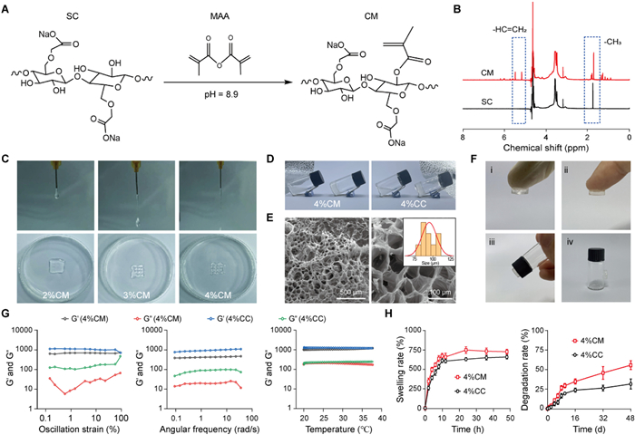
Preparation and physicochemical characterization of hydrogel scaffold. (A) The synthesis procedure of CM. (B) The NMR spectra of SC and CM. (C) The injectability of 2%CM, 3%CM and 4%CM. (D) The gelation procedure of 4%CM and 4%CC. (E) The SEM image of 4%CC, and the average porous size. Scale bar: 500 or 100 µm. (F) The adhesion ability of 4%CM: rubber (ⅰ), skin (ⅱ), plastic (ⅲ) and glass (ⅳ). (G) The rheological ability of 4%CM and 4%CC in three modes: Oscillation strain, angular frequency and temperature. (H) The swelling rate and degradation rate of 4%CM and 4%CC. Data are presented as mean ± standard deviation (SD) (n = 3).
To prepare nanosized CeO2, cerium acetate (Ce(OAc)3) was used as the precursor, followed by the addition of ammonia (NH3·H2O). The reaction was carried out at 200 ℃ in a high pressure reactor. After reaction and purification, the CeO2 was collected for further characterization. From the transmission electron microscope (TEM) images, it was in bulk shape with the diameter of 30.36 ± 0.13 nm, and the obvious Ce and O elements existed in CeO2 (Fig. S1 in Supporting information). And the obvious crystallization peaks were observed at 28.56°, 47.48° and 56.34° in CeO2 (Fig. S2 in Supporting information). By X-ray photoelectron spectrometry (XPS) results, Ce and O elements were observed, and the multivalent Ce ions existed in in CeO2 (Fig. S3 in Supporting information). Meanwhile, the zeta potential was −26.5 ± 1.7 mV in CeO2 by zeta sizer (Fig. S4 in Supporting information). Herein, it confirmed the successful preparation of CeO2, which was in nanoscale. The existence of Ce3+ and Ce4+ contributed to the ROS scavenging ability of CeO2 [33].
Sodium carboxymethyl cellulose (SC) was applied as the starting material for hydrogel preparation due to its plenty of hydroxyl groups in the main chain, where could be modified with various functional groups [34]. To prepare CM, the hydroxyl groups of SC were activated and modified with MA groups in the weak alkaline condition. And then, the MA groups of CM were crosslinked by ultraviolet (UV) exposure to form CM hydrogels. Besides, to form CC hydrogels, a certain amount of CeO2 was also incorporated in CM solutions before UV exposure (Fig. 1A). And the existence of CeO2 also offered an extra physical interaction inside CC hydrogels [35]. As illustrated in Fig. S5 (Supporting information), compared to SC with the obvious peak in 1550 cm−1 (-CH2COO-), the extra peaks were observed in 3400 and 1550 cm−1, corresponding to -OH and -C=O groups, in the Fourier transform infrared spectroscopy (FTIR) results of CM. In addition, by nuclear magnetic resonance (NMR) spectra, the obvious two protons ware displayed at 5.0 and 5.5 ppm, corresponding to the double bonds, in CM compared to that of SC. And the chemical shift appeared at around 1.7 ppm in CM, attributed by the -OCH3 group of SC, and -CH3 group of MA (Fig. 1B). After calculation, the degree of substitution (DS) of MA groups was 6.84% in CM by calculating the integral area of -CH=CH2 and -CH3 groups (Table S1 in Supporting information). Both of them gave the proof of the successful fabrication of CM.
For three dimentional (3D) printing, it was expected that the pregel solution (ink) was uniform and fine with a certain viscosity during printing. After printing, it could form a regular structure [36]. To valid the 3D printability, different weight concentrations (2%, 3% and 4%) of CM hydrogels, defined as 2%CM, 3%CM and 4%CM were considered. As shown in Fig. 1C, the uniform and fine fluid was observed in 4%CM, which also displayed a neat and intact mesh structure via 3D printing after UV exposure. Thus, 4%CM was applied for further experiments. Next, the gelation transition was shown in Fig. 1D and Fig. S6 (Supporting information). For SC, CM and CC with the weight concentration of 4%, all of them were in pregel solution state. After UV exposure, it became hydrogels in 4%CM and 4%CC, while it was still in solution state in 4%SC. Similarly, by scanning electron microscope (SEM), the obvious interconnected network was observed in 4%CC, with the pore size of 95.6 ± 10.1 µm (Fig. 1E). Significantly, to various kinds of substrate, 4%CC possessed the good adhesion ability (Fig. 1F).
The rheological behaviors of hydrogels were tested in three modes: Angular frequency, oscillation strain and temperature changes. Generally, the elasticity of hydrogels was characterized by the fact that their storage modulus (G′) was higher than the loss modulus (G′′). As shown in Fig. 1G, the G′ and G′′ of 4%CC was higher than those of 4%CM. CeO2 incorporation was helpful to the enhanced viscoelasticity of hydrogels [37]. And both of 4%CM and 4%CC maintained the relative stable with the increase of oscillation strain from 0.1% to 100%. Only in high angular frequency (100 rad/s), their G′ started to decrease, while the G′′ was slightly ascended. Similarly, during the range of angular frequency from 0.1 rad/s to 100 rad/s, the G′ was always higher than G′′ for both 4%CM and 4%CC. And the G′ and G′′ of 4%CC kept higher than those of 4%CM. In particular, the G′ and G′′ maintained almost the same levels between 4%CM and 4%CC during this temperature range. All of these indicated that 4%CM and 4%CC possessed the stable hydrogel network structure. CeO2 incorporation provided the extra physical interaction in 4%CC, displaying the higher viscoelasticity than that of 4%CM.
Subsequently, as displayed in Fig. 1H, both hydrogels slowed swollen in phosphate buffered saline (PBS) versus time. And it almost reached the swelling equilibrium after 12 and 24 h in 4%CM and 4%CC, with the swelling rate (SR) of 607.3% ± 19.3% and 661.0% ± 38.0% respectively. In the meantime, it slowly degraded in both of 4%CM and 4%CC. The degradation rate (DR) of 4%CM was higher than that of 4%CC. After 48 days, the DR was 31.6% ± 5.3% in 4%CM, decreased to 55.7% ± 4.7% in 4%CC. From the above, it indicated that the good swelling ability of 4%CM was due to the chemical crosslinking formed by MA groups. And the existence of CeO2 contributed the extra physical crosslinking, leading to the high crosslink degree, and further low SR and DR [38].
As known to all, a certain of ROS was helpful to tissue repair. Nevertheless, the excessive ROS could induce inflammation, and inhibit the regeneration of cartilage tissue [39]. By ROS testing kits, the ROS scavenging capacities of hydrogels were tested. As illustrated in Fig. S7 (Supporting information), the ·OH scavenging ratio was 27.7% ± 1.9% in 4%CM, while it became 64.5% ± 1.6% in 4%CC. Meanwhile, the similar tendency was observed in ·O2− scavenging capacities, which was 29.0% ± 0.4% in 4%CM, and 67.9% ± 0.5% in 4%CC (Table S2 in Supporting information). A little of ROS scavenging ability happened to 4%CM due to its hydroxyl groups. However, the incorporation of CeO2 obviously increased the ROS scavenging ability of 4%CC.
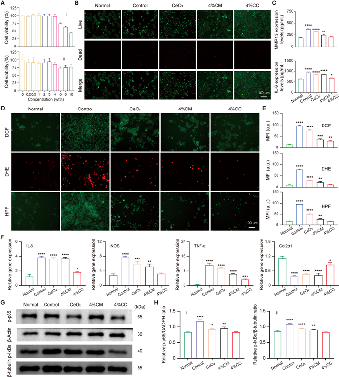
To assess the cell viability, cell counting kit-8 (CCK-8) assay was applied. For CC hydrogels with different weight concentrations of CeO2, it maintained the good biocompatibility with the cell viability above 96%. When its concentration was above 4%, the cell viability was significantly declined in both of chondrocytes and mouse monocytic macrophage cells (RAW264.7) (Fig. 2A). Thus, 4%CC was utilized as the final hydrogel scaffold for investigation. Lipopolysaccharide (LPS), also known as endotoxins, are widely presented in the cell walls of Gram negative bacteria. It has strong pro-inflammatory properties, and is widely applied in the construction of in vitro inflammatory cell models [40,41]. From the live/dead staining, compared to normal group with few dead cells (red fluorescence), a few of live cells (green fluorescence) were observed, and many dead cells existed in control group. The treatments could obviously decrease the number of dead cells (Fig. 2B). After statistical analysis, the live cell ratio was 98.9% ± 0.2% in normal group, sharply declined to 54.7% ± 2.6% in control group. The treatments could recover the live cell ratio to 86.1% ± 1.0% in CeO2, 96.6% ± 0.5% in 4%CM, and 99.3% ± 0.2% in 4%CC (Fig. S8 in Supporting information).
Figure 2
Figure 2.
Antioxidant and anti-inflammation abilities in cellular levels. (A) The cell viability of 4%CM with CeO2 of different concentrations ranging from 0 to 100 wt%: Chondrocytes (ⅰ) and RAW264.7 (ⅱ). (B) The live/dead staining images of treated chondrocytes. (C) The inflammatory factors expression levels of treated chondrocytes by ELISA: MMP13 and IL‐6. (D) The intracellular ROS levels of treated cells by fluorescent microscopy: DCF, DHE and HPF, and (E) the corresponding quantified results. (F) The relative genes expression levels of treated chondrocytes by RT‐qPCR: IL‐6, iNOS, TNF-α and Col2α1. (G) The pathway related proteins expression levels of treated cells by WB, and (H) the corresponding quantified results: p-p65 (ⅰ) and p-IκBα (ⅱ). The corresponding groups were: cells without treatments (normal group), and cells pre‐treated with LPS followed by incubating with PBS (control group), CeO2 (CeO2), 4%CM (4%CM), and 4%CC (4%CC). Scale bar: 100 µm. *P < 0.05, **P < 0.01, ***P < 0.001, ****P < 0.0001 vs. normal group. Data are presented as mean ± SD (n = 3).
In addition, by enzyme-linked immunosorbent assay (ELISA), compared to normal group (193.5 ± 5.7 and 627.1 ± 21.8), both of matrix metalloproteinase 13 (MMP13) and interleukin-6 (IL-6) expression was in the relatively high levels in control group (376.5 ± 17.1 and 939.3 ± 27.7) (P < 0.0001). The treatments could decrease their expression levels. In particular, in 4%CC, the MMP13 and IL-6 expression levels were 210.8 ± 1.8 and 690 ± 6.6 (P < 0.05) (Fig. 2C). Subsequently, the intracellular ROS levels were analyzed by immunofluorescent staining. As displayed in Fig. 2D, the 2′,7′-dichlorodihydrofluorescein diacetate (DCF), dihydroethidium (DHE) and hydroxyphenyl fluorescein (HPF) levels were the highest in control group, while they were the lowest in normal group. Compared to control group, the treatments could decrease the intracellular ROS levels. After statistical analysis, the mean fluorescent intensity (MFI) of DCF, DHE and HPF was 77.6 ± 2.2, 94.9 ± 2.9 and 93.9 ± 2.2 in control group (P < 0.0001). After treatments, the MFI of DCF, DHE and HPF was 29.8 ± 0.9, 73.6 ± 4.6 and 50.3 ± 3.7 in CeO2, 21.0 ± 3.6, 35.8 ± 3.0 and 28.0 ± 2.2 in 4%CM, and 11.5 ± 0.5, 28.2 ± 3.8 and 15.6 ± 1.5 in 4%CC respectively (Fig. 2E).
Subsequently, by reverse transcription-quantitative polymerase chain reaction (RT-qPCR), the IL-6 (3.8 ± 0.1), inducible nitric oxide synthase (iNOS) (7.7 ± 0.2) and tumor necrosis factor-alpha (TNF-α) (11.3 ± 0.9) genes expression levels were relatively high in control group (P < 0.0001). Compared to control group, their expression levels were declined after treatments, which was 3.7 ± 0.1, 6.0 ± 0.6 and 9.6 ± 0.2 in CeO2, 3.7 ± 0.1, 5.3 ± 0.6 and 6.4 ± 0.1 in 4%CM, and 1.9 ± 0.1, 3.3 ± 0.1 and 3.4 ± 0.3 in 4%CC. Conversely, for collagen type Ⅱ alpha 1 chain (Col2α1) gene expression, it was in the highest levels in normal group (1.1 ± 0.1), which was significantly reduced in control group (0.4 ± 0.1) (P < 0.0001). After treatments, it became 0.5 ± 0.1, 0.4 ± 0.1 and 0.9 ± 0.1 in CeO2, 4%CM and 4%CC (P < 0.0001, P < 0.0001 and P < 0.05, respectively) (Fig. 2F). Similarly, the SRY-box transcription factor 9 (SOX9) and aggrecan (ACAN) genes expression was the highest in normal group (0.9 ± 0.1 and 1.2 ± 0.1), significantly declined in control group (0.4 ± 0.1 and 0.5 ± 0.1) (P < 0.0001). After treatments, their expression had been improved. Especially in 4%CC, the SOX9 and ACAN genes expression became 0.7 ± 0.1 and 0.9 ± 0.1 (P < 0.01 and P < 0.05) (Fig. S9 in Supporting information).
The nuclear factor kappaB (NF-κB) signaling pathway plays a central role in the inflammatory response. It can be activated through various pathways, promoting the release of inflammatory cytokines, thereby exacerbating inflammation related diseases [42]. In CD therapy, inhibiting NF-κB pathway is beneficial for achieving anti-inflammatory effects, and promoting cartilage tissue repair [43]. To explore the therapeutic mechanism, Western blot (WB) was applied. As displayed in Fig. 2G and Fig. S10 (Supporting information), the phosphorylated p65 subunit (p-p65) and phosphorylated inhibitor of kappa B alpha (p-IκBα) expression was in the high levels in control group. The treatments could lower their expression levels, especially in 4%CC. By statistical analysis, the relative p-p65/GAPDH and p-IκBα/β-tubulin ratio was 0.83 ± 0.02 and 0.85 ± 0.02 in normal group, significantly ascended to 1.18 ± 0.04 and 1.09 ± 0.02 in control group. However, they became 0.93 ± 0.02 and 0.95 ± 0.01 in CeO2, 0.96 ± 0.04 and 0.91 ± 0.01 in 4%CM, and 0.83 ± 0.02 and 0.83 ± 0.01 in 4%CC respectively (Fig. 2H).
In cellular levels, it confirmed that 4%CC possessed the good biocompatibility. For LPS induced cells, 4%CC could protect them avoiding from the attack of oxidative stress, lower the intracellular ROS levels, downregulate the inflammatory factors expression levels, promote the chondrogenic genes expression levels, and inhibit NF-κB pathway. For CeO2 alone, it presented the poor antioxidant and anti-inflammatory abilities due to the fact that the nanosized CeO2 easily formed aggregates, and failed to be uptake by the cells [44]. And the better antioxidant and anti-inflammation behaviors happened to 4%CC due to the incorporation of CeO2, not only offering the slow release of CeO2, and also avoiding the formation of aggregates [45,46].
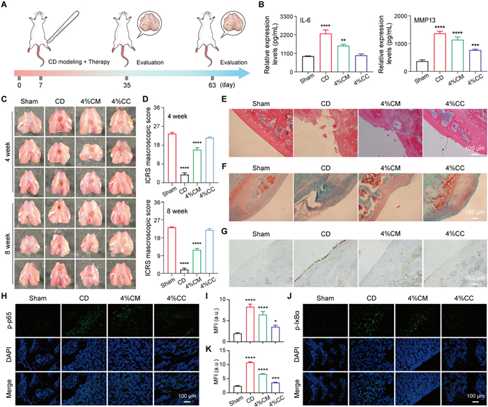
The CD rats models were established by punching a hole in the joint of Sprague-Dawley rats. All animal experiments involved had been approved by the Animal Experiment Ethics Committee of Guangxi Medical University (No. 202105010). On one hand, the defect could not be repaired through self-healing. On the other hand, it ensured the possible healing through treatments [47,48]. As shown in Fig. 3A, after adaptive feeding for 7 days, the CD rats were established together with the treatments. After 4 and 8 weeks, the rats were sacrificed for therapeutic evaluation respectively. Generally, it could present a certain of therapeutic effects after 4 weeks' treatment. However, CD are always accompanied by joint inflammation, which gradually worsens with the progression [49,50]. Therefore, 8 weeks' treatment were also evaluated. At 4 weeks, by ELISA, the IL-6 expression levels were 1220.3 ± 90.3 in the synovial fluid of sham group, significantly jumped to 2135.8 ± 126.7 in that of CD (P < 0.0001). After treatments, it became 1799.7 ± 43.6 in 4%CM (P < 0.001), and 1512.6 ± 67.5 in 4%CC (P < 0.05). Similarly, for MMP13 expression, it was the same tendency with the order of CD (930.4 ± 56.3) > 4%CM (650.9 ± 1.9) > 4%CC (527.1 ± 5) > sham group (238.2 ± 16.3) (Fig. S11 in Supporting information). Similarly, at 8 weeks, the IL-6 and MMP13 expression levels of synovial fluid were 928.1 ± 12.8 and 363.1 ± 43.5, 2274.4 ± 174.2 and 1367.7 ± 63.1, 1550.5 ± 66.7 and 1139.1 ± 80.7, and 984.4 ± 63.0 and 766.3 ± 24.9 in sham group, CD, 4%CM and 4%CC (Fig. 3B).
Figure 3
Figure 3.In vivo CD therapy evaluation. (A) The time schedule of in vivo CD therapy. (B) The inflammatory factors expression levels in the synovial fluid of treated rats at week 8 by ELISA: IL-6 and MMP13. (C) The macroscopic observation in the joint of treated rats at week 4 and 8, and (D) the corresponding ICRS score. (E) The H&E staining images in the joint tissue of treated rats. (F) The SO/FG staining images in the joint tissue of treated rats. (G) The IL-6 expression levels in the joint tissue of treated rats. (H) The p-p65 expression levels in the joint tissue of treated rats, and (I) the corresponding quantified results. (J) The p-IκBα expression levels in the joint tissue of treated rats, and (K) the corresponding quantified results. The corresponding groups were: Rats without treatments (sham group), and rats with joint defect followed by treatment with PBS (CD), 4%CM (4%CM), and 4%CC (4%CC). Scale bar: 100 µm. *P < 0.05, **P < 0.01, ***P < 0.001, ****P < 0.0001 vs. sham group. Data are presented as mean ± SD (n = 3).
From the macroscopic observation, there were no damages in the femur of sham group, with the smooth cartilage surface. However, the significant defects and inflammation were observed in the cartilage of CD, and its inflammation gradually became worsened over time, indicating the poor self-healing ability. At 4 weeks, the defects decreased after treatments, where the defect of 4%CC was significantly reduced. At the same time, some inflammation was observed in the cartilage of CD at 8 weeks. Specifically, in 4%CC, there was no obvious inflammation area in the cartilage. And the new cartilage tissue completely filled the defect site, and also formed a good fusion with the surrounding normal cartilage (Fig. 3C). By international cartilage repair society (ICRS) scoring [51], it was 23.7 ± 0.5 in sham group, sharply decreased to 4.0 ± 0.8 in CD. By treatments, it became 16.0 ± 0.8 in 4%CM, and 21.7 ± 0.5 in 4%CC at 4 weeks. Similarly, at 8 weeks, the ICRS score was 23.3 ± 0.5 in sham group, 2.0 ± 0.8 in CD, 12.0 ± 0.8 in 4%CM, and 22.0 ± 0.8 in 4%CC (Fig. 3D).
The pathological features of cartilage tissue was also analyzed. From hematoxylin and eosin (H&E) staining images, compared to sham group with smooth joint surface and regular cartilage structure, the obvious defect was observed in the cartilage of CD. The treatments could recover the above conditions, where 4%CC was the most effective one (Fig. 3E). After calculation, the modified O' Driscoll score (MODS) was 26.0 ± 0.1, 7.0 ± 0.8, 14.3 ± 0.5 and 22.3 ± 0.5 in sham group, CD, 4%CM and 4%CC (Fig. S12 in Supporting information). Besides, the ROS levels in the cartilage tissue of treated rats were investigated. In CD, the ROS levels of cartilage tissue were relatively high compared to that of sham group. The treatments could reduce the ROS levels of cartilage tissue (Fig. S13A in Supporting information). After statistical analysis, the MFI of ROS levels was 1.1 ± 0.1 in sham group, increased to 9.4 ± 0.3 in CD (P < 0.0001). The treatments reduced the MFI to 5.1 ± 0.1 in 4%CM (P < 0.0001) and 2.8 ± 0.1 in 4%CC (P < 0.0001) (Fig. S13B in Supporting information).
Meanwhile, there was almost no tissue repair or filling in the defect area of CD by Safranin O and fast green (SO/FG) staining. And in 4%CM, the repair was not complete, and the SO staining was negative, indicating that the repair of cartilage defect mainly relied on the proliferation of fibrous tissue, and a little of transparent cartilage tissue was formed. In contrast, the repair surface appeared smooth in 4%CC. And the extracellular matrix of the newly formed tissue showed higher similarity to the surrounding normal cartilage tissue (Fig. 3F). After osteoarthritis cartilage histopathology assessment system (OARSI) scoring, it was 0.3 ± 0.5 in sham group, increased to 13.3 ± 0.9 in CD (P < 0.0001), 3.7 ± 0.5 in 4%CM (P < 0.01), and 1.2 ± 0.2 in 4%CC (Fig. S14 in Supporting information). For IL-6 expression, it was in the highest levels in the cartilage tissue of CD. The treatments could decrease its expression levels (Fig. 3G). By statistical analysis, the average optical density (AOD) was 5.4 ± 1.0 in sham group, 17.1 ± 1.6 in CD (P < 0.001), 14.5 ± 2.3 in 4%CM (P < 0.01), and 10.4 ± 1.0 in 4%CC (Fig. S15 in Supporting information).
Mechanistically, the p-p65 expression was relatively high in the joint of CD with high green fluorescence existed. The treatments could lower its expression levels (Fig. 3H). After quantification, the MFI of p-p65 expression was 2.0 ± 0.1 in the joint of sham group, increased to 8.3 ± 0.5 in the joint of CD. The treatments lowered its MFI to 6.4 ± 0.6 and 3.6 ± 0.3 in the joint of 4%CM and 4%CC (Fig. 3I). Similarly, the p-IκBα expression was relatively high in the joint of CD with the MFI of 10.8 ± 0.2. The treatments could decrease its expression in the joint of 4%CM and 4%CC with the MFI of 6.7 ± 0.1 and 3.6 ± 0.1 (Figs. 3J and K). The above demonstrated that 4%CC more efficiently achieved CD therapy via NF-κB pathway inhibition both in vitro and in vivo. Finally, from the H&E staining images of major organs, there were no significant differences observed (Fig. S16 in Supporting information), indicating the good in vivo biosafety.
All together, it indicated that the cartilage defect could not repair, and the obvious high ROS levels and inflammation existed in the cartilage tissue of CD. Compared to 4%CM, 4%CC most efficiently decreased the ROS levels, downregulated the inflammatory factors expression, and accelerated the tissue repair in the cartilage. The existence of CeO2 in 4%CC not only could effectively scavenge ROS and achieve anti-inflammation, and also promote cartilage tissue repair [52]. Besides, it also displayed the good biosafety by this therapeutic strategy.
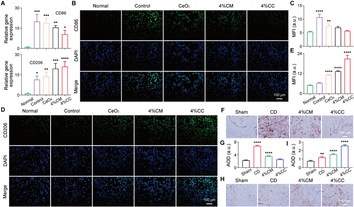
Generally, macrophages M2 polarization plays a crucial role in the protection and repair of cartilage tissue, mainly through antioxidant, anti-inflammatory, and promoted tissue regeneration [53]. It can inhibit inflammation and cartilage degradation, regulate oxidative stress and mitochondrial function, and protect the integrity of cartilage tissue [54]. Thus, the 4%CC mediated macrophage M2 polarization was verified both in vitro and in vivo. As illustrated in Fig. 4A, the cluster of differentiation 86 (CD86) gene expression levels were relatively low in normal group (1.0 ± 0.1), sharply increased to 19.9 ± 4.3 in control group (P < 0.001). The treatments could decrease the CD86 gene expression levels to 18.7 ± 2.8, 15.5 ± 1.2 and 10.5 ± 2.3 in CeO2, 4%CM and 4%CC (P < 0.001, P < 0.01 and P < 0.05, respectively). Conversely, the CD206 gene expression was relatively low in normal group (1.0 ± 0.3) and control group (11.4 ± 2.2). Significantly, its gene expression was enhanced after treatments, which was 13.7 ± 2.5, 19.5 ± 3.1 and 21.0 ± 3.0 in CeO2 (P < 0.01), 4%CM (P < 0.001) and 4%CC (P < 0.0001).
Figure 4
Figure 4.In vitro and in vivo CD immunotherapy. (A) The CD86 and CD206 genes expression levels of treated RAW264.7 by RT‐qPCR. (B) The CD86 expression levels of treated RAW264.7 by immunofluorescent microscope, and (C) the corresponding quantified results. (D) The CD206 expression levels of treated RAW264.7 by immunofluorescent microscope, and the corresponding quantified results (E). The corresponding groups were: Cells without treatments (normal group), and cells pre‐treated with LPS followed by incubating with PBS (control group), CeO2 (CeO2), 4%CM (4%CM), and 4%CC (4%CC). (F) The CD86 expression levels in the synovial tissue of treated rats, and (G) the corresponding quantified results. (H) The CD206 expression levels in the synovial tissue of treated rats, and (I) the corresponding quantified results. The corresponding groups were: rats without treatments (sham group), and rats with joint defect followed by treatment with PBS (CD), 4%CM (4%CM), and 4%CC (4%CC). Scale bar: 100 µm. *P < 0.05, **P < 0.01, ***P < 0.001, ****P < 0.0001 vs. normal group in cellular levels, or sham group in animal levels. Data are presented as mean ± SD (n = 3).
Similarly, by immunofluorescent staining, in control group, a lot of green fluorescence was observed, corresponding to high CD86 expression levels. Compared to control group, the green fluorescence was reduced. Among them, the weakest green fluorescence happened to 4%CC (Fig. 4B). By statistical analysis, the MFI was 5.4 ± 0.1 in normal group, increased to 10.8 ± 0.9 in control group (P < 0.0001), 7.6 ± 0.5 in CeO2 (P < 0.01), 7.0 ± 0.3 in 4%CM, and 5.7 ± 0.1 in 4%CC (Fig. 4C). On the contrary, the CD206 expression (green fluorescence) was relatively low in normal group and control group with the MFI of 5.2 ± 0.1 and 6.5 ± 0.1. After treatments, CD206 expression was enhanced, sequentially with the MFI of 11.2 ± 0.2, 13.4 ± 0.2 and 20.8 ± 1.6 in CeO2 (P < 0.0001), 4%CM (P < 0.0001) and 4%CC (P < 0.0001) (Figs. 4D and E).
Significantly, in the synovial tissue of treated rats, the CD86 expression was the highest in CD. The treatments obviously reduced its expression levels (Fig. 4F). After statistical calculation, the AOD was 6.7 ± 0.2 in CD (P < 0.0001), 3.5 ± 0.1 in 4%CM (P < 0.0001), and 2.5 ± 0.2 in 4%CC (Fig. 4G). However, the CD206 expression was relatively low in sham group and CD with the AOD of 0.7 ± 0.1 and 1.2 ± 0.1. The treatment could promote the CD206 expression with the AOD of 1.5 ± 0.1 and 2.5 ± 0.1 in 4%CM and 4%CC (Figs. 4H and I). In this part, it both confirmed that 4%CC could effectively induce macrophages M2 polarization, further to achieve anti-inflammation, and promote cartilage tissue repair.
In summary, 4%CC hydrogel scaffold was prepared by incorporating the nanosized CeO2 in CM followed by UV exposure. The intrinsic physicochemical properties of hydrogels including morphology, rheological behaviors, swelling properties and degradability were systematically investigated. The incorporation of CeO2 effectively contributed to the excellent ROS scavenging ability in 4%CC, due to the slow release of Ce element, and avoiding the aggregation of nanosized CeO2. Such a slow release of CeO2 in 4%CC further lead to the decrease of intracellular ROS levels, downregulation of inflammatory factors, NF-κB pathway inhibition, macrophages M2 polarization as well as promotion of cartilage tissue repair, which were verified both in vitro and in vivo. Thus, as an effective hydrogel scaffold with the functionalities of antioxidant, anti-inflammation and promoted tissue repair, it finally demonstrated its excellent CD therapeutic effects with high biosafety. The novel concept of combining antioxidant and anti-inflammation effects with tissue repair provided a viable strategy for tissue damage-inflammation treatment.
The authors declare that they have no known competing financial interests or personal relationships that could have appeared to influence the work reported in this paper.
Acknowledgments
This work was financially supported by the project of the National Natural Science Foundation of China (No. 82160430), the Joint Project on Regional High-Incidence Diseases Research of Guangxi Natural Science Foundation (No. 2025GXNSFBA069275), the Guangdong Basic and Applied Basic Research Foundation (No. 2024A1515030080), the Middle/Young aged Teachers' Research Ability Improvement Project of Guangxi Higher Education (No. 2024KY0098), and the Major Talent Project of Guangxi Autonomous Region.
Supplementary materials
Supplementary material associated with this article can be found, in the online version, at doi:10.1016/j.cclet.2026.113206.
Figure 1
Preparation and physicochemical characterization of hydrogel scaffold. (A) The synthesis procedure of CM. (B) The NMR spectra of SC and CM. (C) The injectability of 2%CM, 3%CM and 4%CM. (D) The gelation procedure of 4%CM and 4%CC. (E) The SEM image of 4%CC, and the average porous size. Scale bar: 500 or 100 µm. (F) The adhesion ability of 4%CM: rubber (ⅰ), skin (ⅱ), plastic (ⅲ) and glass (ⅳ). (G) The rheological ability of 4%CM and 4%CC in three modes: Oscillation strain, angular frequency and temperature. (H) The swelling rate and degradation rate of 4%CM and 4%CC. Data are presented as mean ± standard deviation (SD) (n = 3).
Figure 2
Antioxidant and anti-inflammation abilities in cellular levels. (A) The cell viability of 4%CM with CeO2 of different concentrations ranging from 0 to 100 wt%: Chondrocytes (ⅰ) and RAW264.7 (ⅱ). (B) The live/dead staining images of treated chondrocytes. (C) The inflammatory factors expression levels of treated chondrocytes by ELISA: MMP13 and IL‐6. (D) The intracellular ROS levels of treated cells by fluorescent microscopy: DCF, DHE and HPF, and (E) the corresponding quantified results. (F) The relative genes expression levels of treated chondrocytes by RT‐qPCR: IL‐6, iNOS, TNF-α and Col2α1. (G) The pathway related proteins expression levels of treated cells by WB, and (H) the corresponding quantified results: p-p65 (ⅰ) and p-IκBα (ⅱ). The corresponding groups were: cells without treatments (normal group), and cells pre‐treated with LPS followed by incubating with PBS (control group), CeO2 (CeO2), 4%CM (4%CM), and 4%CC (4%CC). Scale bar: 100 µm. *P < 0.05, **P < 0.01, ***P < 0.001, ****P < 0.0001 vs. normal group. Data are presented as mean ± SD (n = 3).
Figure 3In vivo CD therapy evaluation. (A) The time schedule of in vivo CD therapy. (B) The inflammatory factors expression levels in the synovial fluid of treated rats at week 8 by ELISA: IL-6 and MMP13. (C) The macroscopic observation in the joint of treated rats at week 4 and 8, and (D) the corresponding ICRS score. (E) The H&E staining images in the joint tissue of treated rats. (F) The SO/FG staining images in the joint tissue of treated rats. (G) The IL-6 expression levels in the joint tissue of treated rats. (H) The p-p65 expression levels in the joint tissue of treated rats, and (I) the corresponding quantified results. (J) The p-IκBα expression levels in the joint tissue of treated rats, and (K) the corresponding quantified results. The corresponding groups were: Rats without treatments (sham group), and rats with joint defect followed by treatment with PBS (CD), 4%CM (4%CM), and 4%CC (4%CC). Scale bar: 100 µm. *P < 0.05, **P < 0.01, ***P < 0.001, ****P < 0.0001 vs. sham group. Data are presented as mean ± SD (n = 3).
Figure 4In vitro and in vivo CD immunotherapy. (A) The CD86 and CD206 genes expression levels of treated RAW264.7 by RT‐qPCR. (B) The CD86 expression levels of treated RAW264.7 by immunofluorescent microscope, and (C) the corresponding quantified results. (D) The CD206 expression levels of treated RAW264.7 by immunofluorescent microscope, and the corresponding quantified results (E). The corresponding groups were: Cells without treatments (normal group), and cells pre‐treated with LPS followed by incubating with PBS (control group), CeO2 (CeO2), 4%CM (4%CM), and 4%CC (4%CC). (F) The CD86 expression levels in the synovial tissue of treated rats, and (G) the corresponding quantified results. (H) The CD206 expression levels in the synovial tissue of treated rats, and (I) the corresponding quantified results. The corresponding groups were: rats without treatments (sham group), and rats with joint defect followed by treatment with PBS (CD), 4%CM (4%CM), and 4%CC (4%CC). Scale bar: 100 µm. *P < 0.05, **P < 0.01, ***P < 0.001, ****P < 0.0001 vs. normal group in cellular levels, or sham group in animal levels. Data are presented as mean ± SD (n = 3).


 DownLoad:

 下载:


 下载:

