Citation:
Tuo Wang, Yueping Ding, Yuanfang Wang, Qingbai Tian, Xing Xu. Interfaces and synergy in heterogeneous material-microorganism hybrid systems for enhanced water remediation: Mechanisms and application strategies[J]. Chinese Chemical Letters,
2026, 37(12): 113188.
doi:
10.1016/j.cclet.2026.113188
Interfaces and synergy in heterogeneous material-microorganism hybrid systems for enhanced water remediation: Mechanisms and application strategies
English
Interfaces and synergy in heterogeneous material-microorganism hybrid systems for enhanced water remediation: Mechanisms and application strategies
School of Architecture and Engineering, Yan’an University, Yan’an 716000, China
b.
Department of Chemistry and Chemical & Environmental Engineering, Weifang University, Weifang 261061, China
c.
Shandong Key Laboratory of Water Pollution Control and Resource Reuse, School of Environmental Science and Engineering, Shandong University, Qingdao 266237, China
Received Date:
25 April 2026 Accepted Date:
07 July 2026 Revised Date:
05 July 2026 Available Online:
15 December 2026
Abstract:
This review critically examines the rapidly developing field of heterogeneous material-microbe hybrid systems for advanced wastewater remediation. To address the inherent limitations of conventional physico-chemical and biological methods, such as low efficiency and poor stability, these systems combine conductive or photoactive materials (e.g., zero-valent iron, magnetite, graphitic carbon nitride) with functional microorganisms to construct synergistic interfaces. These interfaces facilitate crucial direct or indirect electron transfer, significantly enhance microbial metabolic activity, and provide protective microenvironments. Recent advances underscore their remarkable effectiveness in the degradation of refractory organic pollutants, enhanced nitrogen removal and bioreduction of heavy metal, even under challenging low-substrate conditions. Despite promising progress, significant challenges persist, including a limited mechanistic understanding of interfacial electron transfer at the molecular level and a notable gap in the design of efficient reactor configurations. This work provides a systematic analysis of interfacial electron transfer mechanisms in heterogeneous material-microorganism hybrid systems, categorizes the photocatalytic biohybrid systems and non-photocatalytic biohybrid systems for enhanced water remediation, and evaluates reactor design strategies. Finally, it outlines future research priorities, emphasizing the development of stable and cost-effective heterogeneous materials, advanced microbial protection strategies, integration with reactor engineering and real-world scenarios, as well as long-term economic and environmental assessment.
The escalating global crisis of water pollution, characterized by the persistence of refractory organic contaminants, excess nutrients and heavy metals, has starkly exposed the limitations of conventional physico-chemical and biological treatment technologies [1-4]. These traditional methods often struggle with low treatment performance, high sensitivity to environmental fluctuations, and insufficient electron donor availability, particularly under low-substrate conditions [2,5-8]. To surmount these bottlenecks, the strategic integration of heterogeneous materials with microbial processes has emerged as a transformative frontier in environmental remediation [9,10]. This heterogeneous material-microorganism hybrid approach leverages the synergistic coupling between conductive or photoactive materials (e.g., iron minerals, zero-valent iron (Fe0), semiconductors) and functional microorganisms [2,11-15]. By constructing efficient “material–microbe” interfaces, these systems can facilitate direct interspecies electron transfer, optimize microbial metabolic energy acquisition, and provide protective microenvironments for biomass retention [16-18]. Notably, recent studies have demonstrated that these interfaces can be engineered through surface functionalization to enhance biocompatibility and electron transfer efficiency [17,19,20]. Consequently, such coupled systems offer distinct advantages over standalone biological or chemical methods, including superior degradation efficiency, deep mineralization of pollutants, enhanced operational stability against shock loads, and a significantly reduced carbon footprint [16,19,21].
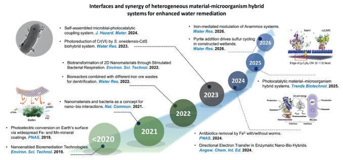
Recent years have witnessed substantial progress in elucidating the mechanisms and expanding the applications of these material–microbe hybrid systems (Fig. 1). Extensive research has demonstrated that natural conductive minerals (e.g., magnetite, pyrite) and synthetic materials (e.g., Fe0, graphitic carbon nitride (g-C3N4)) act as critical electron shuttles or nucleation cores that stimulate microbial activity through diverse pathways [4,20-24]. For instance, iron-based materials have been shown to enrich anaerobic ammonium oxidation (anammox) bacteria by serving as alternative electron donors or acceptors, thereby accelerating nitrogen removal even under substrate-limited conditions [11,25,26]. Similarly, photocatalytic biohybrids effectively harness solar energy to drive enzymatic reactions, enabling the simultaneous degradation of antibiotics and reduction of heavy metals [27-29]. Studies further reveal that these materials can upregulate key functional genes, promote the secretion of extracellular polymeric substances (EPS), and induce sludge granulation, collectively enhancing the system's resilience and overall treatment capacity [25,30-33]. Particularly promising are recent developments in nanomaterial-microbe complexes, where precisely engineered nanoparticles create optimal microenvironments for specialized microbial communities [4,21,25,32,34].
Figure 1
Figure 1.
Timeline of the interfaces and synergy of heterogeneous material-microorganism hybrid systems for enhanced water remediation. Reproduced with permission [42]. Copyright 2021, Nature Publishing Group. Reproduced with permission [74]. Copyright 2023, Elsevier B.V. Reproduced with permission [32]. Copyright 2024, John Wiley and Sons.
Despite these promising advancements, critical knowledge gaps and challenges remain that hinder widespread implementation. At the microscopic level, the dynamic mechanisms governing interfacial electron transfer, particularly the molecular interactions between outer-membrane of microbes and material surfaces under varying redox or light illumination, are not yet fully understood [28,35,36]. Although batch experiments have validated the concept at the bench scale, translating this success into scalable reactors remains a critical bottleneck. Key engineering challenges, such as optimizing light distribution in photocatalytic setups, managing hydraulic retention, and ensuring the long-term stability of material–biofilm composites in real wastewater, are still unresolved. Additionally, the economic viability and environmental safety of these hybrid systems demand thorough life-cycle assessment and risk evaluation before industrial applications [37].
In this review, we aim to provide a comprehensive synthesis of the current state of heterogeneous material–microbe coupled systems for enhanced water remediation. Review flow of heterogeneous material-microorganism hybrid systems for enhanced water remediation is presented in Fig. S1 (Supporting information). We will critically examine the fundamental mechanisms of interfacial electron transfer, categorize the synergistic roles of various heterogeneous materials (i.e., minerals, metal oxides, Fe0, g-C3N4) on the performance of emerging non-photocatalytic or photocatalytic biohybrid systems. Special attention will be devoted to analyzing reactor designs to bridge the gap between mechanistic understanding and engineering applications. Finally, we will outline future research priorities, focusing on the development of robust, self-healing materials. Crucially, we need practical guidelines to bridge the gap between current innovations and resilient, large-scale wastewater solutions.
2.
Concept of heterogeneous material-microorganism hybrid systems
2.1
Core concept for heterogeneous material-microorganism hybrid systems
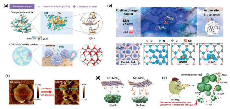
It is well known that nature synthesizes various metal enzymes by selectively binding specific metal ions (such as iron, nickel and manganese) into protein frameworks as active sites [32,37-40]. These complex biological catalysis systems can simultaneously promote various oxidation and reduction reactions within organelles without affecting each other's catalytic efficiency or selectivity. In fact, many catalysts (especially nano-based catalysts, and single-atom catalysts (SACs)) exhibited characteristics similar to the activities of peroxidase, catalase, superoxide dismutase and oxidase [32,38-40]. Cobalt myoglobin, featuring a central cobalt ion as its active site, serves as a quintessential biomimetic prototype. Zhang et al. developed a hierarchical hydrophilic cobalt hydroxide structure that mimicked the cobalt myoglobin [41], which could efficiently activate peroxymonosulfate (PMS) to achieve ultrafast antibiotic degradation. The catalyst shared significant structural similarities with natural cobalt myoglobin, particularly electronic microenvironment modulated by adjacent amino acid residues critically influenced the catalytic behavior of the cobalt center (Fig. 2a). Drawing inspiration from the cobalt-nitrogen (Co–N) coordination found in dehalogenase active centers, Min et al. designed a cobalt single-atom catalyst supported on a carbon-doped boron nitride substrate [38]. This design replicated both the enzymatic active site and the electric field microenvironment derived from surrounding peptide chains (Fig. 2b), achieving nearly 100% dechlorination efficiency of chloramphenicol. Shi et al. synthesized two-dimensional molybdenum disulfide nanosheets (NR-MoS2) rich in nanopores [42], which could significantly enhance the electron transfer between the nanomaterial and the biofilm (Figs. 2c and d). Such enhanced transfer effectively disrupted key biofilm components, including proteins, extracellular polysaccharides, and extracellular DNA (Fig. 2e).
Figure 2
Figure 2.
(a) Biomimetic LDH@Co(OH)2 catalyst for antibiotics degradation via PMS activation. Reproduced with permission [41]. Copyright 2026, John Wiley and Sons. (b) Engineering Co-SACs on carbon-modified boron nitride to replicate enzyme active centers and microenvironments. Reproduced with permission [38]. Copyright 2021, Nature Publishing Group. (c) Atomic force microscopy (AFM) images of MoS2 samples. (d) Illustration of biofilm suppression via nanohole-enhanced electron transport. (e) Diagram depicting the NR-MoS2-mediated suppression of biofilm formation in physiological environments. Reproduced with permission [42]. Copyright 2021, Nature Publishing Group.
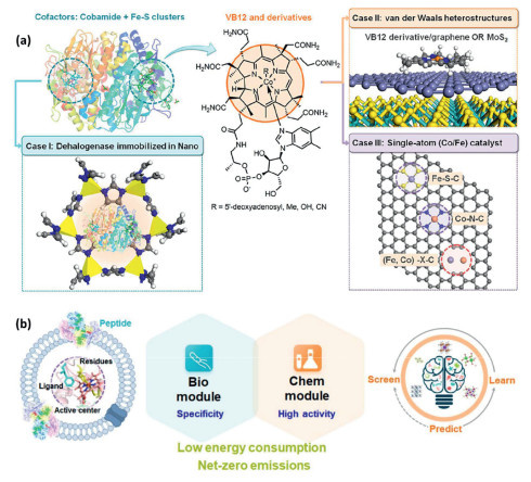
In fact, both biological and chemical transformations of pollutants predominantly involve redox reactions in the heterogeneous material-microorganism hybrid systems, wherein interfacial electron transfer plays a pivotal role. This process is complex and intrinsic to chemical and biological redox systems, commencing with either direct electron transfer or the generation of electron carriers [43,44]. These carriers subsequently migrate across the micro-interface, facilitating the transfer of electrons to specific pollutant molecules, thereby catalyzing redox reactions that lead to the transformation or degradation of these pollutants. Consequently, there exists a connection between the pathways of artificial enzymes in various catalysts (i.e., carbonocatalysts, metal oxides, SACs) and natural enzymes within microbial systems during interfacial electron transfer processes [44,45]. Leveraging this cross-interface electron transfer synergy can enhance wastewater treatment efficiency and broaden application scenarios. Chen conducted a comparative study on three kinds of biomimetic catalyst designs (Fig. 3a), aiming to improve the degradation efficiency of halogenated pollutants [44]. Specifically, these design frameworks include: (ⅰ) Bio-enzyme/inorganic component coupled catalysts (Case Ⅰ); (ⅱ) organic molecule/inorganic component coupled catalysts (Case Ⅱ); (ⅲ) cobalt-amide metal center/inorganic material catalyst (Case Ⅲ). In contrast to the other two systems, case Ⅲ was capable of selectively concentrating the halogenated pollutants through dipole-dipole interactions of carbon-halogen bonds, thus substantially improving the dehalogenation efficiency of pollutants. As for the microorganisms, particularly extracellular respiratory bacteria (ERBs), they are capable of transferring electrons to external solid-phase electron acceptors via outer membrane (OM) proteins or redox mediators [46-48]. This extracellular electron transport process is central to microbial metabolism, driving the transformation of pollutants (e.g., organic compounds and heavy metals) into less toxic forms or complete mineralization [46-48]. Therefore, the key to achieve efficient removal of pollutants is to improve the electron transfer efficiency of the interface. Yu et al. analyzed the dynamic structural changes during the OmcA/hematite interface interaction at the molecular level [40], and systematically proved that the outer membrane protein OmcA could inhibit the production of hematite photocurrent, while the process of iron cycling was driven by the release of biological electrons in the dark and anoxic environment, thus revealing the mechanism of iron dissolution on the mineral surface mediated by microbial proteins.
Figure 3
Figure 3.
(a) Three possible schemes based on the design of dehalogenase catalytic systems. (b) Integrated biological and chemical modules to achieve effective removal of pollutants. Reproduced with permission [44]. Copyright 2023, American Chemical Society.
Basically, the essence of heterogeneous material-microorganism hybrid systems lies in the universal nature of interfacial electron transfer, where these heterogeneous materials emulate natural metalloenzymes to forge efficient electrical connections with microbes. By harnessing this shared electron flow, these heterogeneous material-microorganism hybrids can synergistically achieve diverse pollutants removal, merging the catalytic activity of natural or synthetic materials with the metabolic adaptability of microorganisms (Fig. 3b).
2.2
Simulation methods for understanding the interfacial electron transfer in biohybrid systems
In the material-microbe synergistic systems for pollutant removal, interfacial electron transfer constitutes the rate-limiting step that governs overall removal efficiency in biohybrid systems [44]. However, this process occurs at complex material–microbe interfaces and involves the instantaneous coupling of multiple interdependent factors, including protein conformational dynamics, interfacial electronic transitions, and energetic alignment, rendering it inaccessible to comprehensive mechanistic analysis by conventional experimental approaches [49-54]. To address this challenge, the research paradigm is shifting from single-modality characterization toward an integrated strategy that synergistically couples multi-scale computational modeling with operando high-resolution techniques [44,45,55].
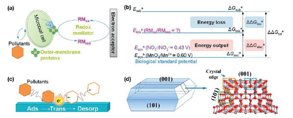
Researchers can leverage advanced computational techniques such as quantum mechanics/molecular mechanics (QM/MM) and density functional theory (DFT) combined with single-molecule fluorescence imaging. These methods allow them to not only visualize the specific locations where reactions occur but also calculate the energetic pathways of electron transfer, thereby unraveling complex bio-electrochemical processes at the atomic level [44,56]. The QM/MM multiscale simulation approach designated the porphyrin–iron center within the outer membrane (OM) proteins and associated mediator molecules as the quantum region for high-accuracy electronic structure calculations, while treating the surrounding bulk amino acid environment with molecular mechanics [49,56]. This partitioning enabled robust quantification of the reaction’s Gibbs free energy change and reduction potential (Figs. 4a and b). Combined with in-situ spectroscopic data, this strategy could reveal the microscopic mechanism of proton-coupled electron transfer and identified interfacial hydrogen bonding as the key accelerators for enhancing efficiency [57]. Acting like a bridge, these hydrogen bonding would shorten the electron transfer distance and lower the energy barrier, thereby offering a mechanistic rationale for the experimentally observed acceleration of interfacial electron transfer and the enhancement of pollutant degradation induced by hydroxyl micro-interface modifications [44,57].
Figure 4
Figure 4.
(a) Mediator-bridged electron transport between microbes and materials. (b) Redox potential rules dictating reaction predictability. (c) Catalyst-driven redox cascades for pollutant removal. (d) Dependence of catalytic efficiency on crystal facets and atomic configurations in anatase titanium dioxide (TiO2). Reproduced with permission [44]. Copyright 2023, American Chemical Society.
Furthermore, to precisely map active sites within the complex material–microbe architecture and guide the rational design of biohybrid systems, single-molecule fluorescence lifetime imaging offers a transformative perspective [44,58]. This technique enabled the direct visualization of electron transfer hotspots and revealed that catalytic reactions at these biohybrid interfaces occur predominantly at specific under-coordinated regions of the catalyst surface, such as edges and corners (Figs. 4c and d), where microbial outer membrane cytochromes preferred to attach. Theoretical simulations using single-particle models could corroborate these findings by demonstrating that metal atoms at these edge sites exhibited outwardly directed orbitals. These orbitals not only facilitated pollutant adsorption but also optimized energetic alignment with microbial electron carriers, thereby maximizing interfacial coupling efficiency [44,50,59]. This closed-loop methodology bridged direct imaging of the bio-interface with theoretical modeling to elucidate how the crystal phase structure and atomic arrangement of the material would govern the kinetics of microbial extracellular electron transfer. These insights provided a robust foundation for the precision customization of catalysts with tailored atomic configurations, advancing pollution control strategies from empirical trial-and-error to mechanism-driven engineering of high-performance material–microbe synergies.
3.
Coupled heterogeneous materials with microbes for pollutant removal
The integration of heterogeneous materials with microbial processes represents a promising frontier for treating refractory organic pollutants, nitrogen and heavy metals, distinguished by its low carbon footprint, high removal efficiency, and capacity for deep mineralization [60-63]. This synergistic strategy generally encompasses two primary paradigms: Photocatalytic biohybrid systems and non-photocatalytic biohybrid systems, each defined by distinct material properties and microbial functionalities [64-66]. In photocatalytic biohybrid systems, semiconductors (e.g., TiO2, cadmium sulfide (CdS), g-C3N4) serve as light-harvesting antennas that generate electron-hole pairs upon irradiation [64-66]. These photo-excited electrons are subsequently transferred to specific microbes, often facultative anaerobes or engineered phototrophs, which utilize them as reducing power to drive reductive degradation of refractory organic pollutants or denitrification. On the other hand, non-photocatalytic systems rely on conductive materials (e.g., magnetite, graphene, carbon nanotubes, or Fe0) that function primarily as electrical bridges rather than light harvesters [16-19,67]. These non-photocatalytic materials facilitate direct or indirect electron transfer or serve as solid-state electron acceptors/donors, bridging the metabolic gap between electroactive bacteria (such as Geobacter or Shewanella species) and the target contaminants. In essence, photocatalytic biohybrid systems harness solar energy to fuel microbial metabolism via photon input, whereas non-photocatalytic systems enhance microbial respiration by lowering the energy barriers associated with extracellular electron exchange [17,68-70]. Despite these different mechanisms, both approaches rely on a precisely engineered conductive material–microbe interface to ensure efficient and directional electron flow.
3.1
Photocatalytic biohybrid systems
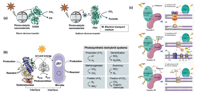
The integration of microorganisms with photocatalytic nanomaterials focuses on enhancing the efficiency of photogenerated electron conversion, contingent on the material's capacity to transfer high-energy photogenerated electrons to enzymatic active sites [68,69,71,72]. Occurring via direct or indirect pathways [68,69,71], this electron transfer leverages the unique metal and non-metal cofactors embedded in enzymes to achieve high selectivity and catalytic activity across diverse applications (Figs. 5a and b). As for the environmental remediation (especially water purification), this synergistic relationship is essential [37,68,69]: Under aerobic conditions, photogenerated electrons and holes initiate redox reactions that generate reactive oxygen species (ROS), collaborating with microbial activity to break down refractory organic compounds. Under anaerobic conditions, charge transfer follows three primary mechanisms (Fig. 5c) [37]: Photogenerated electrons directly degrade pollutants, drive microbial enzymatic pathways (e.g., denitrification, anaerobic ammonium oxidation), or serve as electron donors for microbes to reduce specific contaminants like heavy metals [36,73], and refractory organic contaminants [37,69].
Figure 5
Figure 5.
(a) Two intrinsic electron transfer modes in nanomaterial-enzyme hybrid systems. Reproduced with permission [71]. Copyright 2024, Elsevier B.V. (b) Photosynthetic biohybrid systems: A promising approach for energy and environmental applications. Reproduced with permission [68]. Copyright 2025, American Chemical Society. (c) Charge transfers in photocatalytic biohybrid systems for water decontamination and its application examples. Reproduced with permission [37]. Copyright 2025, Elsevier B.V.
3.1.1
Metal-based photocatalytic biohybrid systems for enhanced water remediation
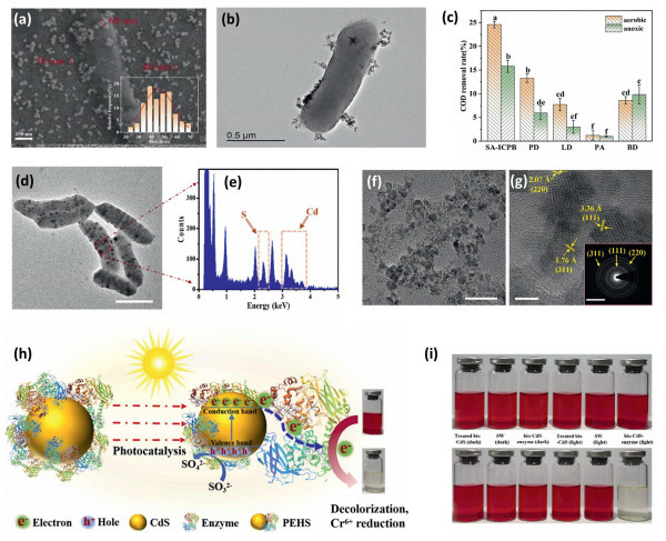
In recent years, the direct coupling of photocatalysis and biodegradation has been widely adopted for the removal of dyes and antibiotics, as well as for denitrification and dechlorination, proving its efficacy in practical wastewater treatment [5,74-76]. Photocatalytic materials control the conversion of solar energy to chemical energy, and their properties have a significant impact on photocatalytic biohybrid systems [37,77]. Ideal materials should have good biocompatibility, low cytotoxicity, and high energy conversion capacity. Currently, commonly used photocatalytic materials include metal sulfides, metal oxides, minerals. Most of these materials are chemically synthesized, and some, such as cadmium sulfide (CdS), can be obtained through biosynthesis [74-76,78]. Qiu and co-workers harnessed the metabolic activity of the electroactive bacterium Shewanella to enable the in-situ biosynthesis of nanoscale CdS photocatalysts from aqueous Cd2+ [74], thereby establishing a spatially integrated photocatalytic–biodegradation system on bacterial cells. The resulting bio-CdS exhibited uniform crystallinity and a narrow size distribution centered at 40–50 nm (Figs. 6a and b), with homogeneous dispersion across the bacterial surface. Antibiotic removal proceeded via concurrent biosorption and enzymatic biodegradation with enzymatic biodegradation accounting for the majority of total antibiotic elimination (Fig. 6c). Wang et al. [75] developed a facile biomineralization approach to construct this hybrid system and demonstrated that cytochrome enzymes spontaneously self-assemble onto biosynthesized CdS during the biomineralization process mediated by Shewanella oneidensis (S. oneidensis) MR–1 (Figs. 6d-g). Importantly, the immobilized cytochrome enzymes could efficiently accept the photogenerated electrons from CdS and function as photocatalytic reduction centers, thereby forming a well-defined photocatalytic-enzyme coupled architecture with high photocatalytic decolorization rate of 69.4 mg g–1 min–1 (Figs. 6h and i).
Figure 6
Figure 6.
(a) Scanning electron microscope (SEM) image of polyurethane sponge carrier as well as the particle size distribution of bio-CdS. (b) Scanning transmission electron microscope (STEM) image of bio-CdS loaded microorganism. (c) chemical oxygen demand (COD) removal by the bio-CdS loaded microorganism. Reproduced with permission [74]. Copyright 2023, Elsevier B.V. (d, e) Biological CdS synthesized by S. oneidensis MR–1, and energy spectrum of the selected area. (f) High-resolution transmission electron microscope (HRTEM) image of isolated biological CdS nanoparticles. (g) HRTEM image of isolated biological CdS nanoparticles. (h) Proposed mechanism of photocatalysis by bio-CdS/enzyme system. (i) Photos of photocatalytic decolorization by different systems. Reproduced with permission [75]. Copyright 2024, Elsevier B.V.
In addition, photosensitive minerals could absorb photons with energies surpassing their bandgap, which excited the electrons from the valence band to the conduction band and creates electron–hole pairs [72,79]. These photogenerated charge carriers could act as vital drivers of microbial physiology by stimulating growth, sustaining cellular metabolism, shaping community structures, and facilitating the biogeochemical transformations required for environmental remediation [32,72]. For example, dissimilatory metal-reducing bacteria (DMRB) could exemplify this process, as they employed outer-membrane cytochromes to reduce hematite (α-Fe2O3) and played a pivotal role in the iron biogeochemical cycle [40]. It was also important to understand the molecular dynamics governing the binding between cytochromes and hematite, along with how this interaction regulated the injection, storage, and utilization of photogenerated electrons during iron redox cycling. Yu and colleagues focused on the photoelectrochemical performance of α-Fe2O3 hybridized with OmcA [40]. This protein, a decaheme outer-membrane cytochrome isolated from S. oneidensis MR–1, consisted of four structural domains, where domained Ⅱ and Ⅳ each contain five histidine-ligated heme groups (Figs. S2a and b in Supporting information). The OmcA was immobilized onto α-Fe2O3 at specific sites via carboxyl, amide Ⅱ, and amide I moieties, where electrostatic attraction and hydrogen bonding acted synergistically to govern the binding process (Figs. S2c-e in Supporting information). This interaction induced conformational rearrangements in OmcA that accelerated interfacial electron transfer kinetics and promoted Fe(Ⅲ) reduction. Notably, the OmcA layer functioned not merely as a passive conduit but as a dynamic biological capacitor; it accumulated photogenerated electrons under illumination and sustained Fe(Ⅲ) reduction during subsequent dark, anoxic periods, thereby effectively decoupling redox activity from light availability (Figs. S2f-h in Supporting information). In addition, the greenockite (chemical composition CdS), a typical photosensitive sulfide mineral, could significantly increase the expression level of outer-membrane c-type cytochromes (OM c-Cyts) in Thiobacillus denitrificans by about 3.35 times under light conditions, thereby mediating the transfer of photogenerated electrons to denitrification enzymes [80]. However, CdS exhibited inherent photoinstability, as prolonged irradiation triggers oxidative decomposition that released toxic Cd2+ ions and precipitated elemental sulfur (S0). This dual degradation pathway undermined catalytic longevity and introduced risks of secondary heavy-metal contamination [72,80]. Therefore, the rational selection of photosensitive minerals for environmental applications required balancing three key criteria: Spectral responsiveness that matched ambient light conditions, interfacial compatibility with target microbial electron-transfer machinery, and long-term chemical and photochemical stability under operational constraints. In summary, the mineral–microbe interface behaved as a dynamic, electrochemically active region instead of a fixed border, functioning as the site for converting light energy into biological redox work. Developing robust solar-driven bioremediation methods depended on clarifying how structure influenced function in photoinduced electron transfer. This process involved factors ranging from atomic binding patterns and protein movements to the overall speed of electron storage and release.
3.1.2
Nonmetal-based photocatalytic biohybrid systems for enhanced water remediation
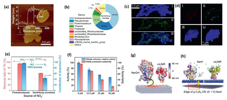
Beyond conventional metal-based systems (including the artificial metallic materials, and photosensitive minerals), the g-C3N4 has gained significant interest owing to its biocompatibility, capacity to absorb visible light, and tunable electronic structure [72,81,82]. Bian et al. demonstrated that photo-excited electrons generated at the edge -C≡N sites of cyano-rich g-C3N4 (g-C3N4—CN) could be directly injected into nitrate reductase (NarGH) [32]. The g-C3N4—CN nanosheets featuring coexisting nitrogen vacancies and terminal -C≡N groups showed an average thickness of ~7.2 nm (Fig. 7a). These nanosheets exhibited spatial compatibility with the NarGH dimer (a bacterial respiratory enzyme, 8 nm × 10 nm), enabling the efficient formation of the biohybrid (Figs. 7b-d). In addition, the -C≡N group provided a unique pathway for the transfer of photoelectrons from g-C3N4—CN nanosheets to the NarGH crude enzyme. They also reported that the NO3— reduction performance of g-C3N4—CN nanosheets was significantly enhanced after coupling with enzymes (Figs. 7e and f). Electron transfer between the g-C3N4—CN nanosheets and the NarGH dimer occurred through a direct, mediator-free interfacial mechanism. This unidirectional electron flow, which was driven by a favorable energy-level alignment between the materials, enabling selective catalytic reduction by specifically delivering electrons to the active site of NarGH (Figs. 7g and h). The photo-enzyme cooperative reduction to generate NO2— could also be coupled with other microbial nitrogen transformation pathways, such as Anammox, significantly enhancing the efficiency of biological nitrogen removal and ammonia synthesis [32]. Dong and colleagues first revealed how interspecies cooperation could promote the photogenerated electron transfer, leading to efficient nitrogen removal in a g-C3N4-Anammox consortia biohybrid system [81]. Liu et al. also reported an intimately coupled photocatalysis (Fe3+/g-C3N4) and biofilm system with superior antibiotic removal performance compared with sole photocatalysis and sole biodegradation system [82]. These findings confirmed that g-C3N4 could effectively facilitate the transfer of photogenerated electrons and substantially improve the pollutant removal efficiency across diverse microbial platforms. As a result, g-C3N4 emerged as a highly promising photocatalyst for constructing light-driven biohybrid systems aimed at achieving the enhanced pollutant removal.
Figure 7
Figure 7.
(a) AFM of g-C3N4—CN. (b) Taxonomic composition of the microbial community, displaying the relative abundance of the top ten genera. (c, d) Confocal laser scanning microscopy (CLSM) visualizations of the g-C3N4—CN/enzyme complex and the broader g-C3N4—CN hybrid material. (e) Efficiency of total nitrogen removal via a partial-denitrification/anammox pathway. (f) Impact of exogenous cytochrome c concentrations on nitrate conversion rates. (g) Electrostatic potential distributions mapped onto g-C3N4—CN, NarGH, and cytochrome cd1 nitrite reductase (cd1NiR). (h) Molecular docking models illustrating the final adsorption configurations of the NarH subunit and cd1NiR on the g-C3N4—CN edge ((0-1-0) facet). Reproduced with permission [32]. Copyright 2024, John Wiley and Sons.
3.1.3
Carriers in photocatalytic biohybrid systems for enhanced water remediation
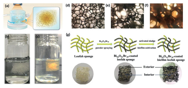
In addition to the photocatalysts, microporous carriers are also the core components of the photocatalytic biohybrid systems, and their role goes far beyond mere physical support [83-88]. An ideal carrier should have a density close to that of water for good mixing, high porosity to load photocatalytic materials and microorganisms, and excellent biocompatibility [12,84,86-91]. Beyond its structural role, the carrier acts as a protective barrier, shielding the biofilm from ultraviolet exposure and free radical damage while absorbing fluctuations in water flow. This micro-environmental stability is key to maintaining high microbial activity and achieving consistent, long-term system performance [12,92]. Currently, the widely used carriers in the photocatalytic biohybrid systems mainly include polyurethane sponges and biomass materials [12,84]. Polyurethane sponges are widely adopted due to their high porosity, strong water absorption, and good biocompatibility [12,93]. Studies have demonstrated that they could facilitate rapid microbial attachment, support high-density biofilm formation, and enable stable immobilization of photocatalysts [44,84,94,95]. Hu et al. engineered a novel, tightly integrated photocatalysis-biodegradation system by immobilizing denitrifying sludge and an Ag3PO4/Bi4Ti3O12 photocatalyst within a polyurethane sponge matrix (Figs. 8a-c) [95]. Xu et al. immobilized TiO2 nanocrystals with exposed reactive crystal facets onto polyurethane sponges for enhanced photocatalytic degradation of cephalexin [12]. Microscopic observations revealed that microorganisms could densely wrap around the sponge fibers and form biofilms (Figs. 8d-f). Xiong and co-workers further demonstrated that the polyurethane sponge-based biohybrid system could achieve efficient degradation of 2,4,6-trichlorophenol, with cyclic operation enabling in-situ catalyst regeneration and sustained high photocatalytic efficiency [94]. Polyurethane sponge-based biohybrid systems enabled the stepwise transformation of contaminants, first into bioavailable low-molecular-weight intermediates for microbial assimilation, and then ultimately into CO2, H2O, and inorganic ions through complete mineralization. However, the synthesis of polyurethane sponges often leaves behind toxic residues like 2,4-dinitrotoluene. These compounds can suppress specific microbial communities, prompting a shift toward more eco-friendly biomass-based carriers [84,96]. For instance, luffa sponge, a natural three-dimensional porous biocarrier, demonstrates significantly reduced inhibition of microbial activity and provides superior support for bacterial colonization relative to polyurethane sponge [84,96,97]. Li et al. developed a low-cost reusable bio-carrier by coating Bi24O31Br10 photocatalyst onto natural loofah sponge [97], which was subsequently incubated with the unacclimated sludge to cultivate biofilms (Fig. 8g). The loofah carrier allowed for greater bacterial colonization and activity, selectively enriching the microbial populations that efficiently degrade photocatalytic intermediates. This architecture could facilitate a robust synergy between photocatalysis and biodegradation via an optimized material-biofilm interface. Complementing these findings, the successful integration of bagasse cellulose and nonwoven biomass matrices further underscores the versatility of agricultural waste-derived supports, paving the way for scalable, cost-effective, and sustainable hybrid treatment systems [13,92,98].
Figure 8
Figure 8.
(a) Schematic diagram of the reactor with visible light. (b, c) Formation of biofilm on carriers. Reproduced with permission [95]. Copyright 2024, Elsevier B.V. Morphological evolution of the sponge carrier in the photocatalytic biohybrid systems: (d) bare carrier, (e) after photocatalyt coating, and (f) after biofilm attachment. Reproduced with permission [12]. Copyright 2023, Elsevier B.V. (g) Images of the loofah sponge carrier. Reproduced with permission [97]. Copyright 2021, Elsevier B.V.
In summary, the direct coupling of photocatalysis and biodegradation, particularly systems utilizing various photocatalysts (e.g., CdS, g-C3N4, natural photoactive materials), demonstrated significant promise for advanced wastewater treatment. Despite this considerable potential, the large-scale advancement of this technology is currently constrained by several challenges, including material cost, long-term stability, and complexities in analyzing reactions within real wastewater matrices. To bridge the gap between laboratory research and practical application, future efforts should focus on several key priorities: (ⅰ) The development of novel, robust photocatalysts with high specific surface areas, self-healing properties, and corrosion resistance, coupled with optimized light-source integration for sustained performance; (ⅱ) a shift from single-pollutant models to elucidating the complex interactions and precise mechanisms in multi-pollutant systems; and (ⅲ) the engineering of sustainable porous carriers and the use of genetic tools to construct multifunctional biofilms with enhanced microbial tolerance and metabolic diversity. Addressing these challenges is pivotal for enabling the scalable deployment and industrialization of photocatalysis-bio hybrid systems.
3.2
Non-photocatalytic biohybrid systems
Beyond photocatalytic materials, researchers also explored heterogeneous systems based on natural conductive minerals (such as magnetite and pyrite) and synthetic non-photoactive conductors like zero-valent iron (Fe0) or supported transition metal catalysts [60,61,63,72,99-102]. These materials proved effective in non-photocatalytic biohybrid setups. Mechanistic studies showed that electroactive microorganisms formed direct electron transfer channels with these surfaces using outer membrane cytochromes or conductive nanowires [60,61,72]. In this process, microbes sent metabolic electrons to the materials, while the materials acted as electron donors to support anaerobic respiration [60,61]. This two-way electron exchange, which worked without soluble mediators, not only improved denitrification and pollutant breakdown but also helped microbes gain energy more efficiently [103-105].
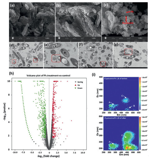
Natural conductive minerals, ubiquitous in soil and aquatic environments, historically served as critical bridges linking microbial metabolism to geochemical cycles. Building on this foundation, Bai et al. demonstrated that Rhodopseudomonas palustris (R. palustris) could reduce ferrihydrite (Fh) extracellularly through direct physical contact [60]. This interaction triggered the in-situ formation of goethite, creating a conductive mineral-biological interface that facilitated electron flow into the cells (Figs. 9a-g). Consequently, this electrochemical coupling process markedly enhanced the degradation efficiency of antibiotics in water, and the primary degradation intermediates exhibited low developmental toxicity. The extracellular reduction of Fh upregulated the expression of key functional genes related to photosynthetic metabolism, extracellular electron transfer, and organic matter decomposition in R. palustris (Fig. 9h). It also stimulated the bacteria to secrete photosensitive compounds, which worked together with the minerals to drive both direct biodegradation and indirect photosensitive degradation of antibiotics (Fig. 9i), revealing the dynamic electrochemical interaction between anaerobic photosynthetic bacteria and conductive iron minerals. Su and colleagues immobilized the denitrifying strain Aquabacterium sp. XL4 onto various iron-rich ore wastes [61], enabling concurrent nitrate reduction and simultaneous removal of copper and lead ions from aqueous solution. Zhang et al. reported that the electron transfer efficiency in sulfide-based constructed wetlands could be enhanced by amending with pyrite (CW-FeS2/S), achieving simultaneous removal of sulfamethoxazole (SMX) and total inorganic nitrogen [63]. Fundamentally, these minerals functioned as electron donors or acceptors, accelerating pollutant removal primarily by modulating microbial metabolism rather than degrading contaminants directly [72,106]. By exchanging electrons with microbes, these minerals boosted metabolic activity and drove contaminant transformation. Redox-active minerals specifically served as terminal electron acceptors, streamlining biodegradation pathways [106-108]. Moreover, once reduced by microorganisms, mineral surfaces displayed heightened reductive capacity, directly facilitating heavy metal reduction and organic pollutant degradation. For example, Tang et al. reported that adding Fe(Ⅲ)-citrate and ferrihydrite accelerated phenol degradation [108], reducing the treatment times by 10–20 days. This role of minerals as electron sinks also promoted the selective enrichment of dissimilatory metal-reducing bacteria (DMRB), further enhancing overall biotransformation efficiency.
Figure 9
Figure 9.
SEM images of (a) Fh, (b) R. palustris, and (c) R. palustris-Fh. transmission electron microscope (TEM) of (d) bioreduced Fh by R. palustris. (e) Partial mineral dissolution of Fh. (f, g) TEM of dissolved Fh particles in the interior of R. palustris cells or embedded in the cell membrane. (h) Volcanic analysis revealing significant differential gene expression patterns among experimental groups. (i) Spectral evolution of EPS components before and after co-cultivation with R. palustris and Fh. Reproduced with permission [60]. Copyright 2023, Elsevier B.V.
In addition, these natural iron minerals (such as magnetite Fe3O4 and pyrite FeS2) and artificial iron-based materials (such as iron filings and scrap iron wires) could also serve as multifunctional interface carriers in the anammox systems [25,34]. They could significantly promote extracellular electron transfer by providing electron donor/acceptor sites, thereby enhancing the metabolic coupling among microorganisms [34,109]. Moreover, solid iron materials could act as nucleation cores for microbial attachment and growth, promoting sludge granulation and enhancing the mechanical stability of granular structure, which would help to improve the system's shock resistance and long-term operational stability in the anammox systems [11,109]. Furthermore, some iron-sulfur minerals (such as FeS2) could stimulate the Fe-N-S coupling cycle [25,110]. In this process, the sulfur cycle fueled autotrophic denitrification and worked together with the Anammox pathway [103,104,111]. This collaboration created a multi-pathway network that significantly improved the overall efficiency of nitrogen removal.
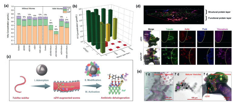
In addition to the natural conductive minerals, the coupled iron oxides (Fe2O3, Fe(OH)3), and Fe0 with the microorganisms have also been intensively used for various pollutants removal [11,25,31,33,112,113]. Recent researches indicated that anammox bacteria were capable of using insoluble iron oxides, such as Fe2O3 and Fe(OH)3, as extracellular electron acceptors through extracellular electron transfer, eliminating the requirement for nitrite in the electron-accepting role [26,31,113,114]. This metabolic capability was significantly enhanced by flavin-enriched extracellular polymeric substances (EPS) that formed conductive coatings on iron oxide surfaces [31,113]. These bridging structures could promote the intimate electrical connections between anammox cells and iron oxides, yielding a considerable electron flux and superior ammonium removal efficiencies even in the absence of nitrite [114]. In addition, the Fe0 noted for its high specific surface area and strong reducing capacity (redox potential E0 = −0.44 V), has been shown to improve nitrogen removal efficiency in anammox processes [11]. These iron-based materials would serve as nucleation sites for microbial aggregation, thereby promoting sludge granulation and enhancing settleability [33]. In addition to anammox processes, the synergistic interaction between Fe0 and microorganisms offers a promising strategy for accelerating antibiotic degradation in aquatic environments [17,115,116]. Li et al. demonstrated that S. oneidensis MR–1, an anaerobic iron-reducing bacterium, could facilitate electron transfer within the Fe0-coupled system, significantly enhancing the removal efficiency of sulfamethoxazole [115]. Ri et al. demonstrated that ball-milled Fe0/FeS2 nanocomposites promoted close physical and electrochemical integration between S. oneidensis MR- and the surrounding anaerobic microbial consortium [17], thereby enabling efficient reductive degradation of 2,4-dichloronitrobenzene. Hou et al. further explored the synergistic effect of pollutant-tolerant tubeworm (tubifex) and Fe0 for the efficient degradation of multiple antibiotics (Figs. 10a and b) [117]. They observed that Fe0 nanoparticles were rapidly adsorbed by the worms to form the “nZVI-augmented worms”, and then these particles underwent progressive depassivation concomitant with iron oxide formation (Fig. 10c). Exposure to Fe0 nanoparticles triggered the secretion of structural proteins (tubulin and actin) on the tubeworm surface, forming a biomolecular corona (Fig. 10d). Additionally, functional proteins (Pccb and thioredoxin) were released via tubulin-coated vesicles under co-exposure conditions, interacting with nanoscale zero-valent iron (nZVI) aggregates on the epidermis and facilitating chemical modification of the iron particles (Fig. 10e).
Figure 10
Figure 10.
(a) Antibiotics removal by Fe0 with/without wormst. (b) Florfenicol removal by Fe0-augmented worms. (c) Design concept and potential antibiotic removal applications of Fe0-augmented worms. (d) Protein localization and proteomic analysis of the biomolecular corona from CLSM data. (e) CLSM visualization of biomolecular corona assembly dynamics. Reproduced with permission [117]. Copyright 2024, National Academy of Sciences.
Basically, coupling the microorganisms (including certain aquatic organisms) on the surface of these non-photocatalytic heterogeneous materials could enhance the transmembrane electron transfer efficiency. This improvement stemmed from strengthened interfacial interactions that bridged the materials with microbial intra- and extracellular electron transfer chains [32,38-40,117]. Beyond facilitating electron flow, these materials would function as electron donors, acceptors, or mediators, thereby regulating microbial metabolism and accelerating pollutant biodegradation. Moreover, the remediation efficacy of these material–microorganism composites depended critically on the synthesis and secretion of specific functional proteins and metabolites [117-119]. These biomolecules would not only contribute directly to degradation but also mediate surface modifications of the materials, synergistically boosting overall system performance.
4.
Reactor setup for heterogeneous material-microorganism hybrid systems
Although heterogeneous materials provided the chemical foundation for efficient pollutant removal in biohybrid systems, their practical success hinged on robust reactor engineering. The reactor acted as the critical interface where materials met microbial communities, governing mass transfer, light distribution, and biomass retention. However, a universally applicable design proved ineffective, as photocatalytic systems require precise control of illumination, while anaerobic mineral–microbe interactions necessitate strict light exclusion and stable microenvironmental conditions. Consequently, research has increasingly branched into specialized directions tailored to these distinct physiological requirements.
4.1
Reactor setup for photocatalytic biohybrid systems
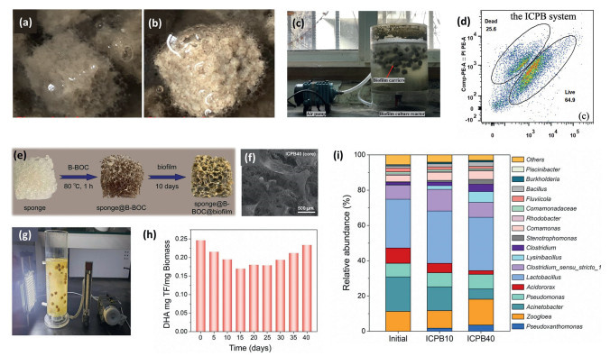
As for the photocatalytic biohybrid systems, the reactor configuration is a core engineering element that determines the performance of the integrated photocatalytic-biodegradation process in removing antibiotics and its engineering scalability from laboratory principles to practical wastewater treatment engineering applications [14,15,82]. Liu et al. developed a continuous-flow reactor specifically for photocatalytic-biological coupling systems to treat antibiotic wastewater [82]. The reactor had an effective volume of 600 mL and was equipped with g-C3N4-loaded polyurethane sponge as a composite functional carrier (Figs. 11a and b). A dedicated groove at the bottom was designed to fix the microporous aeration device, ensuring uniform oxygen supply and good mixing. The outer wall of the reactor was circumferentially integrated with light-emitting diode (LED) light sources (λ = 420–430 nm) to provide directional and stable irradiation to the photocatalytic unit (Fig. 11c). They observed that bacterial viability remained above 60% in the biohybrid system, a level comparable to that in the controlled biofilm system, demonstrating that the integrated photocatalytic process did not impose additional cytotoxic stress on the microbial community (Fig. 11d). Dong et al. engineered a biofilm-supporting porous sponge functionalized with B/Bi3O4Cl (Figs. 11e–g), and integrated it into a visible-light-driven photocatalytic biohybrid reactor system [14]. The biohybrid reactor could achieve the superior degradation capacity (~95%) towards the ciprofloxacin (CIP) within 12 h of hydrodynamic residence time. They also reported that the microorganisms (such as Bacillus, Lysinibacillus, Pseudomonas, and Burkholderia) could progressively acclimate to the coupled photocatalytic-biodegradation environment during the mid-phase process (Figs. 11h and i).
Figure 11
Figure 11.
Photograph of biofilm carrier in the intimately coupled photocatalysis and biodegradation (ICPB) system before (a) and after (b) the experiment. Reproduced with permission [82]. Copyright 2022, Elsevier B.V. (c) Actual photo of the biofilm culture reactor. (d) Fluorescent images of live/dead bacteria in the ICPB system after the antibiotic removal. Reproduced with permission [15]. Copyright 2022, Elsevier B.V. (e) Schematic illustration of the sponge@B-BOC@biofilm architecture. (f) SEM of core structure of sponge@B-BOC@biofilm following a 40-day operational. (g) Photo of the sponge@B-BOC@biofilm apparatus. (h) Temporal evolution of microbial dehydrogenase activity throughout the CIP degradation process in the ICPB system. (i) Comparative analysis of biofilm genus-level relative abundances in different degradation trial. Reproduced with permission [14]. Copyright 2022, Elsevier B.V.
Collectively, these representative studies reflected the advancing state of photocatalytic-biological hybrid reactor systems for antibiotic wastewater treatment, from initial proof-of-concept batch setups to engineered continuous-flow configurations featuring spatially optimized illumination, controlled aeration, and rationally designed biofilm-supporting carriers. Key performance milestones include sustained microbial viability under operational photocatalytic conditions, high-efficiency degradation of recalcitrant antibiotics, and documented microbial community acclimation to the coupled physicochemical–biological stress environment. Nevertheless, critical barriers persist in translating these systems to full-scale application, particularly concerning long-term operational stability, energy input per unit pollutant removed, and capital/operational cost competitiveness relative to conventional biological processes. Strategic research priorities should therefore emphasize improving photon utilization efficiency through spectral matching and light distribution engineering, advancing scalable, mechanically stable, and catalytically active biofilm carriers, and rigorously evaluating system resilience and treatment efficacy under realistic wastewater matrices, including variable organic load, ionic strength, and background scavengers. Ultimately, synergistic integration of tailored microbial consortia design with next-generation photocatalytic materials offers a robust pathway toward sustainable, resilient, and implementable advanced wastewater treatment technologies.
4.2
Reactor setup for non-photocatalytic biohybrid systems
The design of reactor configurations for coupled heterogeneous material–microbe systems is pivotal for their scalable application in pollutant removal [19,120]. Notably, the most extensive research into these hybrid systems has focused on the anaerobic ammonium oxidation process [19,121]. This predominance stems from the unique challenges of anammox bacteria, which exhibit extremely slow growth rates and high sensitivity to environmental fluctuations [19,121,122]. Consequently, heterogeneous materials are indispensable in anammox reactors not only for biomass retention via biofilm formation but also for creating protective microenvironments that ensure process stability and accelerate startup.
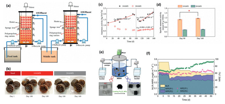
Liu et al. developed the two-stage fixed bed biofilm reactor (each bed volume of 7 L, Fig. 12a) to incubate the anammox biofilm under low nitrogen conditions [121]. The polypropylene carriers (filling rate: 26.9%) were attached with anaerobic ammonium oxidation biofilms (Fig. 12b), and were fixed in two beds through acrylic glass supports and nylon lines. Each bed was sealed and shaded with black sponge to avoid the adverse effects of oxygen and light. They demonstrated that supplementing the system with sponge iron (42.8 g/L) significantly enhanced both the apparent activity of anammox bacteria and the production of extracellular polymeric substances (EPS) in biofilms cultivated under low nitrogen loading conditions (Figs. 12c and d). Abdulatti et al. investigated the enhancement of nitrate removal from real municipal wastewater by integrating an electric-magnetic field (EMF) with Fe0 (3 g/L) in an up-flow microbial sludge reactor [67]. Their study demonstrated that this synergistic EMF-Fe0 system could significantly enrich the denitrifying bacteria, achieving a 67% nitrate removal efficiency. Chang et al. engineered a novel carrier by incorporating magnetite nanoparticles (Fe3O4 NPs) into a polypropylene matrix for use in moving bed biofilm reactors (MBBRs) for stimulating anammox bacterial growth (Fig. 12e) [30]. Comparative analysis demonstrated that the biofilms formed on these iron-amended carriers harbored an anammox bacterial population roughly four times denser than those on pristine carriers. Such significant proliferation of functional microbes was identified as the primary driver for the superior nitrogen removal observed in the modified reactors (Fig. 12f).
Figure 12
Figure 12.
(a) Schematic diagram of experimental setup and (b) morphology of observed anammox biofilm. (c) Assessment of anammox performance in a continuous fixed-bed reactor and (d) comparative analysis of specific anammox activity before and after the introduction of sponge iron. Reproduced with permission [121]. Copyright 2024, Elsevier B.V. (e) Scheme of the MBBRs laden with iron-modified carriers. (f) Nitrogen loading and nitrogen removal in the reactor. Reproduced with permission [30]. Copyright 2024, Elsevier B.V.
Despite promising laboratory results, the transition from small-scale prototypes to full-scale treatment plants remains a significant challenge. Key priorities for the future reactor setup for heterogeneous material-microorganism hybrid systems include: (ⅰ) Developing cost-effective and durable carrier materials, (ⅱ) designing energy-efficient illumination systems, and (ⅲ) validating long-term stability in pilot-scale studies using real wastewater. As a result, to fully realize their potential as practical wastewater treatment solutions, these heterogeneous material-microorganism systems will benefit from continued focus on improving cost-effectiveness, durability, and operational adaptability beyond the laboratory stage.
5.
Conclusions and perspectives
This review has critically synthesized the current state of heterogeneous material–microbe coupled systems, elucidating the fundamental mechanisms of interfacial electron transfer that drive enhanced water remediation. We have categorized the synergistic roles of diverse heterogeneous materials, including minerals, metal oxides, Fe0, and g-C3N4, in both photocatalytic and non-photocatalytic biohybrid systems. Furthermore, by analyzing existing reactor designs, we have highlighted the critical pathways required to bridge the gap between mechanistic understanding and engineering application. Although significant progress has been made in laboratory-scale proofs-of-concept, the transition to resilient, large-scale wastewater solutions demands a paradigm shift towards robustness, economic viability, and environmental sustainability. Based on this analysis, we propose the following future research priorities:
(ⅰ) Development of stable and cost-effective heterogeneous materials. Future efforts must prioritize the design of heterogeneous materials that balance high catalytic activity with long-term stability and low cost. For non-photocatalytic systems, research should focus on earth-abundant minerals and modified industrial by-products that resist corrosion and passivation in complex wastewater matrices. In photocatalytic applications, the development of visible-light-responsive materials with enhanced charge separation efficiency is crucial, yet these must be synthesized via scalable, green methods to reduce production costs. Special attention should be given to creating "self-healing" materials capable of regenerating active sites in situ, thereby extending the operational lifespan of the hybrid system without frequent material replacement.
(ⅱ) Advanced microbial protection strategies. The vulnerability of microbial communities to material toxicity, oxidative stress, and environmental fluctuations remains a primary bottleneck. Future studies should explore innovative encapsulation techniques and biomimetic interfaces that provide a protective microenvironment for microbes while facilitating efficient electron transfer. This includes the design of hierarchical structures where conductive materials act as both electron shuttles and physical shields against harsh conditions. Additionally, the selection and engineering of electroactive microorganisms with enhanced tolerance to toxic pollutants and material-induced stress will be essential for maintaining system performance under real-world operating conditions.
(ⅲ) Integration with reactor engineering and real-world scenarios. To bridge the lab-to-field gap, future work must move beyond batch experiments to continuous-flow reactor designs optimized for hydrodynamics, mass transfer, and light distribution (for photocatalytic systems). Research should validate these systems using real wastewater matrices containing complex organic matter, inorganic ions, and fluctuating pH levels, rather than synthetic surrogates. Pilot-scale demonstrations are urgently needed to assess the scalability of material–microbe coupling in diverse scenarios, such as municipal wastewater treatment plants, industrial effluent polishing, and decentralized rural sanitation systems.
(ⅳ) Long-term economic and environmental assessment. Finally, the viability of heterogeneous material–microbe coupled systems must be evaluated through comprehensive life cycle assessment (LCA) and techno-economic analysis (TEA). Future research should quantify the long-term economic benefits relative to conventional treatment technologies, accounting for material synthesis costs, energy consumption, maintenance requirements, and sludge disposal. Concurrently, the environmental footprint, including the potential leaching of nanomaterials and the formation of toxic byproducts, must be rigorously monitored. Establishing practical guidelines based on these assessments will be pivotal for policymakers and engineers to adopt these hybrid technologies as sustainable solutions for global water security.
(ⅴ) Emergence of AI-driven, self-adaptive biohybrid ecosystems. Future research should also integrate machine learning (ML) with in situ sensing technologies to predict optimal electron transfer pathways and automatically adjust operational parameters (e.g., light intensity, hydraulic retention time, or redox potential). Furthermore, leveraging synthetic biology, we can engineer smart microbial consortia equipped with genetic circuits that sense specific pollutants and trigger the on-demand expression of extracellular electron transfer proteins or protective biofilms only when needed. This convergence of digital intelligence and biological programming will transform passive treatment units into autonomous, self-optimizing bio-reactors that maximize remediation efficiency while minimizing energy input and material wear.
In summary, realizing the full potential of heterogeneous material–microbe coupled systems requires a multidisciplinary approach that integrates materials science, microbiology, and process engineering. By addressing these priorities, the next generation of biohybrid systems can deliver robust and economically feasible solutions for advanced water remediation.
The authors declare that they have no known competing financial interests or personal relationships that could have appeared to influence the work reported in this paper.
Acknowledgments
The work was supported by Shandong Provincial Excellent Youth (No. ZR2022YQ47). The authors also want to thank Conghua Qi from Scientific Compass (www.shiyanjia.com).
Supplementary materials
Supplementary material associated with this article can be found, in the online version, at doi:10.1016/j.cclet.2026.113188.
[1]
L. Wu, Z. Sun, Y. Zhen, et al., Environ. Sci. Technol. 55 (2021) 15400–15411. doi: 10.1021/acs.est.1c04600
Figure 1
Timeline of the interfaces and synergy of heterogeneous material-microorganism hybrid systems for enhanced water remediation. Reproduced with permission [42]. Copyright 2021, Nature Publishing Group. Reproduced with permission [74]. Copyright 2023, Elsevier B.V. Reproduced with permission [32]. Copyright 2024, John Wiley and Sons.
Figure 2
(a) Biomimetic LDH@Co(OH)2 catalyst for antibiotics degradation via PMS activation. Reproduced with permission [41]. Copyright 2026, John Wiley and Sons. (b) Engineering Co-SACs on carbon-modified boron nitride to replicate enzyme active centers and microenvironments. Reproduced with permission [38]. Copyright 2021, Nature Publishing Group. (c) Atomic force microscopy (AFM) images of MoS2 samples. (d) Illustration of biofilm suppression via nanohole-enhanced electron transport. (e) Diagram depicting the NR-MoS2-mediated suppression of biofilm formation in physiological environments. Reproduced with permission [42]. Copyright 2021, Nature Publishing Group.
Figure 3
(a) Three possible schemes based on the design of dehalogenase catalytic systems. (b) Integrated biological and chemical modules to achieve effective removal of pollutants. Reproduced with permission [44]. Copyright 2023, American Chemical Society.
Figure 4
(a) Mediator-bridged electron transport between microbes and materials. (b) Redox potential rules dictating reaction predictability. (c) Catalyst-driven redox cascades for pollutant removal. (d) Dependence of catalytic efficiency on crystal facets and atomic configurations in anatase titanium dioxide (TiO2). Reproduced with permission [44]. Copyright 2023, American Chemical Society.
Figure 5
(a) Two intrinsic electron transfer modes in nanomaterial-enzyme hybrid systems. Reproduced with permission [71]. Copyright 2024, Elsevier B.V. (b) Photosynthetic biohybrid systems: A promising approach for energy and environmental applications. Reproduced with permission [68]. Copyright 2025, American Chemical Society. (c) Charge transfers in photocatalytic biohybrid systems for water decontamination and its application examples. Reproduced with permission [37]. Copyright 2025, Elsevier B.V.
Figure 6
(a) Scanning electron microscope (SEM) image of polyurethane sponge carrier as well as the particle size distribution of bio-CdS. (b) Scanning transmission electron microscope (STEM) image of bio-CdS loaded microorganism. (c) chemical oxygen demand (COD) removal by the bio-CdS loaded microorganism. Reproduced with permission [74]. Copyright 2023, Elsevier B.V. (d, e) Biological CdS synthesized by S. oneidensis MR–1, and energy spectrum of the selected area. (f) High-resolution transmission electron microscope (HRTEM) image of isolated biological CdS nanoparticles. (g) HRTEM image of isolated biological CdS nanoparticles. (h) Proposed mechanism of photocatalysis by bio-CdS/enzyme system. (i) Photos of photocatalytic decolorization by different systems. Reproduced with permission [75]. Copyright 2024, Elsevier B.V.
Figure 7
(a) AFM of g-C3N4—CN. (b) Taxonomic composition of the microbial community, displaying the relative abundance of the top ten genera. (c, d) Confocal laser scanning microscopy (CLSM) visualizations of the g-C3N4—CN/enzyme complex and the broader g-C3N4—CN hybrid material. (e) Efficiency of total nitrogen removal via a partial-denitrification/anammox pathway. (f) Impact of exogenous cytochrome c concentrations on nitrate conversion rates. (g) Electrostatic potential distributions mapped onto g-C3N4—CN, NarGH, and cytochrome cd1 nitrite reductase (cd1NiR). (h) Molecular docking models illustrating the final adsorption configurations of the NarH subunit and cd1NiR on the g-C3N4—CN edge ((0-1-0) facet). Reproduced with permission [32]. Copyright 2024, John Wiley and Sons.
Figure 8
(a) Schematic diagram of the reactor with visible light. (b, c) Formation of biofilm on carriers. Reproduced with permission [95]. Copyright 2024, Elsevier B.V. Morphological evolution of the sponge carrier in the photocatalytic biohybrid systems: (d) bare carrier, (e) after photocatalyt coating, and (f) after biofilm attachment. Reproduced with permission [12]. Copyright 2023, Elsevier B.V. (g) Images of the loofah sponge carrier. Reproduced with permission [97]. Copyright 2021, Elsevier B.V.
Figure 9
SEM images of (a) Fh, (b) R. palustris, and (c) R. palustris-Fh. transmission electron microscope (TEM) of (d) bioreduced Fh by R. palustris. (e) Partial mineral dissolution of Fh. (f, g) TEM of dissolved Fh particles in the interior of R. palustris cells or embedded in the cell membrane. (h) Volcanic analysis revealing significant differential gene expression patterns among experimental groups. (i) Spectral evolution of EPS components before and after co-cultivation with R. palustris and Fh. Reproduced with permission [60]. Copyright 2023, Elsevier B.V.
Figure 10
(a) Antibiotics removal by Fe0 with/without wormst. (b) Florfenicol removal by Fe0-augmented worms. (c) Design concept and potential antibiotic removal applications of Fe0-augmented worms. (d) Protein localization and proteomic analysis of the biomolecular corona from CLSM data. (e) CLSM visualization of biomolecular corona assembly dynamics. Reproduced with permission [117]. Copyright 2024, National Academy of Sciences.
Figure 11
Photograph of biofilm carrier in the intimately coupled photocatalysis and biodegradation (ICPB) system before (a) and after (b) the experiment. Reproduced with permission [82]. Copyright 2022, Elsevier B.V. (c) Actual photo of the biofilm culture reactor. (d) Fluorescent images of live/dead bacteria in the ICPB system after the antibiotic removal. Reproduced with permission [15]. Copyright 2022, Elsevier B.V. (e) Schematic illustration of the sponge@B-BOC@biofilm architecture. (f) SEM of core structure of sponge@B-BOC@biofilm following a 40-day operational. (g) Photo of the sponge@B-BOC@biofilm apparatus. (h) Temporal evolution of microbial dehydrogenase activity throughout the CIP degradation process in the ICPB system. (i) Comparative analysis of biofilm genus-level relative abundances in different degradation trial. Reproduced with permission [14]. Copyright 2022, Elsevier B.V.
Figure 12
(a) Schematic diagram of experimental setup and (b) morphology of observed anammox biofilm. (c) Assessment of anammox performance in a continuous fixed-bed reactor and (d) comparative analysis of specific anammox activity before and after the introduction of sponge iron. Reproduced with permission [121]. Copyright 2024, Elsevier B.V. (e) Scheme of the MBBRs laden with iron-modified carriers. (f) Nitrogen loading and nitrogen removal in the reactor. Reproduced with permission [30]. Copyright 2024, Elsevier B.V.


 DownLoad:

 下载:


 下载:

