
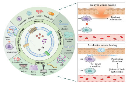
Figure 1.
Schematic illustration of engineered exosome delivery systems for diabetic wound healing via macrophage polarization modulation.


Affecting millions worldwide, diabetes is a chronic metabolic disease marked by insulin deficiency or resistance and subsequent dysregulation of glucose and lipid metabolism [1]. Alarmingly, this number is projected to reach 1.3 billion by 2050 [2]. Diabetic impaired wound healing (DIWH) is one of the common and severe complications of diabetes, with diabetic foot ulcer (DFU) being its most typical manifestation. Approximately 15%–25% of diabetic patients develop a foot ulcer in their lifetime, with about 20% of these cases leading to amputation, severely impacting quality of life and increasing healthcare burden [3]. Unlike acute wounds, diabetic wounds are marked by chronic inflammation, impaired angiogenesis, abnormal extracellular matrix remodeling, and recurrent infections, resulting in significantly delayed or stalled healing [4].
Conventional treatments for diabetic wounds remain inadequate for many patients, largely due to a persistent imbalance in macrophage polarization, a key driver of impaired healing. In normal healing, macrophages transition from a pro-inflammatory (M1) to an anti-inflammatory/reparative (M2) phenotype, orchestrating inflammation resolution and tissue repair [5]. However, under hyperglycemic conditions, macrophages are trapped in the M1 state, leading to sustained chronic inflammation and hindered repair [6]. Therefore, promoting M2 macrophage polarization has emerged as a promising therapeutic strategy for diabetic wound management.
Exosomes are cell-secreted extracellular vesicles, typically 30–150 nm in diameter, ubiquitously present in various body fluids including blood, saliva, urine, and breast milk [7,8]. Due to their low immunogenicity and excellent biocompatibility, some exosomes exhibit unique advantages in diabetic wound therapy [9]. They carry bioactive molecules, including proteins, lipids, mRNA, and non-coding RNAs (such as microRNA (miRNA), circular RNA (circRNA), long non-coding RNA (lncRNA)), which can be taken up by recipient cells to regulate their biological functions [10]. Extensive research has demonstrated that exosomes derived from mesenchymal stem cells (MSCs) [11-13], macrophages themselves [14], and other cell types [15,16] can effectively reprogram pro-inflammatory M1 macrophages into reparative M2 phenotypes by delivering specific functional cargo and modulating signaling pathways. This process significantly ameliorates the inflammatory microenvironment in diabetic wounds and accelerates the healing process.
Despite their therapeutic potential, topical application of naïve exosomes faces key challenges: Suboptimal active cargo, rapid clearance by tissue fluids or blood flow, enzymatic degradation in the protease/nuclease-rich wound microenvironment, and poor intrinsic targeting. Consequently, engineering exosomes and enabling their transdermal delivery are indispensable for unlocking their full therapeutic potential. This review systematically overviews engineered exosomes (e.g., efficient cargo loading) and their local delivery (e.g., via hydrogels, microneedles, sponges) designed to regulate macrophage polarization, thereby promoting diabetic wound healing. This review addresses the core bottleneck of "delivery", encompassing both the vehicle and cargo, to provide novel insights and a theoretical foundation for developing next-generation, efficient immunomodulatory therapies for diabetic wounds (Fig. 1).
Exosomes originate from the endocytic pathway, where inward budding of endosomal membranes generates intraluminal vesicles (ILVs) [17]. Released upon multivesicular body fusion with the plasma membrane, these exosomes are enriched in tetraspanins (CD9, CD63, CD81) and cholesterol/sphingomyelin microdomains, ensuring high stability and prolonged circulation [18]. Their ability to travel long distances in bodily fluids and be internalized by target cells underpins their role in intercellular communication [19]. Within the diabetic wound microenvironment, exosomes function as "intelligent messengers", delivering bioactive cargo (proteins, lipids, nucleic acids) to reprogram macrophage polarization and restore functional balance [7].
Multiple cell types have been exploited as sources of therapeutic exosomes for diabetic wound healing, each offering distinct mechanistic advantages in driving M2 macrophage polarization. Table 1 [15,16,20-29] summarizes the key bioactive molecules, signaling pathways, and primary mechanisms of exosomes derived from bone marrow MSCs (BMSC-Exos), adipose-derived stem cells (ADSC-Exos), human umbilical cord MSCs (hUCMSC-Exos), platelet-rich plasma (PRP-Exos), M2 macrophages (M2-Exos), and keratinocytes (KC-Exos).
 DownLoad:
CSV
| Exosome types | Cellular origin | Key bioactive molecules/signaling pathways | Primary mechanism | Ref. |
| BMSC-Exos | Bone marrow MSCs | miR-23a-3p, CD73-adenosine axis | Inhibiting the interferon regulatory factor 1 (IRF1)/NF-κB pathway to promote M2 macrophage polarization, function impaired under hyperglycemia due to CD73 down regulation | [20,21] |
| ADSC-Exos | Adipose-derived stem cells | circRps5/miR–124–3p axis | Promoting M2 macrophage polarization and enhancing collagen synthesis | [22] |
| hUCMSC-Exos | Human umbilical cord MSCs | miR-146a-5p, insulin (INS), superoxide dismutase 1 (SOD1) | Suppressing tumor necrosis factor receptor-associated factor 6 (TRAF6)/signal transducer and activator of transcription 1 (STAT1) signaling to drive M2 macrophage polarization; inhibiting pro-inflammatory activation | [23-25] |
| PRP-Exos | Platelet-rich plasma | miR-26b-5p, sphingosine-1-phosphate (S1P)/Akt/fibronectin 1 (FN1) pathway | Reducing neutrophil infiltration, promotes angiogenesis, and enhances M2 macrophage polarization | [26-28] |
| M2-Exos | M2 macrophages | CCL22, IL-4, milk fat globule-epidermal growth factor 8 (MFG-E8), etc. | Directly suppresses inducible nitric oxide synthase (iNOS) while upregulating Arg-1 and CD206, thereby inducing M1-to-M2 conversion | [29] |
| KC-Exos | Keratinocytes | Metastasis associated lung adenocarcinoma transcript 1 (MALAT1) /miR–1914–3p/MFG-E8 axis | Modulates macrophage function via miRNA regulation, promoting diabetic wound healing | [15,16] |
Natural exosomes exhibit limited therapeutic efficacy in regulating macrophage polarization because of poor targeting, insufficient active component loading, and rapid in vivo clearance. This approach, which utilizes exosomes as engineered drug delivery vehicles, can circumvent their natural limitations. By functioning as "precision packages", these modified exosomes present a far more promising therapeutic proposition.
A wide array of therapeutic agents has been developed to induce M2 macrophage polarization, showing great promise for diabetic wound healing. These agents span functional proteins and inhibitors (MBD2 [30], MLN4924 [31]), cytokines (interleukin-25 (IL-25 [32] and IL-27 [33]), nucleic acids (miR-146a [34], miR-20a-5p [35], miR-132–3p [36]); siMMP9 [37], siAkt2 [38], and small molecules (MCC950 [39]). Natural products (e.g. resveratrol [40], C-30 [41], breviscapine [42], puerarin [43], quercetin [44], paeoniflorin [45], and other phytochemicals) have also demonstrated efficacy in driving M2 macrophage polarization. Notably, antibiotic-loaded exosomes (vancomycin/lysostaphin) have been engineered to couple bacterial killing with macrophage phenotype switching [46]. Various exogenous and endogenous techniques have been developed to overcome these loading challenges, each with distinct pros and cons tailored to specific cargo and applications (Fig. 2).
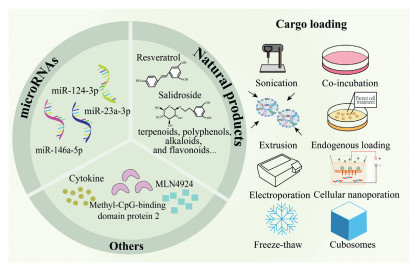
Common exogenous loading techniques include sonication, electroporation, co-incubation, freeze–thaw cycles and extrusion. Sonication offers high loading efficiency. However, the high-intensity mechanical shear forces generated during this process can significantly compromise the integrity of the exosome membrane, leading to irreversible structural changes [47,48]. Electroporation uses electrical pulses to create nanopores in the lipid bilayer, allowing nucleic acids (e.g., siRNA, mRNA) to enter via electrophoresis. While effective for macromolecules, suboptimal parameters can cause exosome aggregation [49], and the siRNA precipitation induced by electroporation will also mask the efficiency of siRNA loading onto exosomes [50]. Co-incubation relies on passive diffusion of hydrophobic drugs; it is gentle and preserves exosome integrity but suffers from low encapsulation efficiency and drug aggregation [51,52]. Freeze–thaw cycles transiently disrupt membranes via ice crystal formation, facilitating drug entry. However, this method may induce exosome fusion and size heterogeneity, reducing yield [53,54]. Extrusion mechanically shears the exosome–drug mixture through nanoporous membranes to disrupt and reconstitute the lipid bilayer. It achieves high loading efficiency for certain small molecules, but may alter the zeta potential and membrane protein properties of the exosomes [55].
Endogenous loading strategies primarily involve genetic modification of parental cells or co-culture approaches. Genetic modification uses plasmid or viral vectors to transfect parental cells with sequences encoding therapeutic proteins or nucleic acids, leveraging cellular sorting machinery to package these cargoes into nascent exosomes. This method preserves protein conformation and post-translational modifications, ensuring bioactivity [56]. The co-culture method incubates parental cells with high concentrations of small-molecule drugs, which enter cells via diffusion or uptake and are partially sorted into exosomes [57]. While technically simple and avoiding genetic manipulation, this approach often suffers from low and inconsistent loading efficiency due to factors like drug cytotoxicity and lysosomal degradation [58].
Parent cell pretreatment offers a versatile strategy to enrich exosomes with therapeutic cargo. Mechanistically, exosomes from LPS-preconditioned MSCs are enriched with let-7b, which targets Toll-like receptor (TLR)/nuclear factor kappa-light-chain-enhancer of activated B cells (NF-κB) signaling to induce M2 macrophage polarization [59]. Hypoxia preconditioning yields exosomes enriched with different effectors: miR-4645–5p in BMSC-Exos inhibits the MAPK activated protein kinase 2 (MAPKAPK2)/tumor necrosis factor-alpha (TNF-α)/sterol regulatory element binding protein 2 (SREBP2) axis [60]. While circ-Snhg11 in ADSC-exos modulates the miR-144–3p/hypoxia-inducible factor 1-alpha (HIF-1α) pathway, both promoting M2 macrophage polarization and diabetic wound healing [61].
To overcome the limitations of conventional loading methods, especially for large nucleic acids, recent advances have introduced membrane fusion–based and nanoscale stimulation–based strategies. Cubosome-mediated fusion utilizes lipid nanoparticles with a bicontinuous cubic phase, offering exceptional membrane fusion capability. Upon simple mixing with exosomes at room temperature, spontaneous fusion occurs within 10 min, yielding hybrid exosomes with efficient cargo transfer. This method achieves >85% loading efficiency for doxorubicin and IgG, and nearly 100% for mRNA transcripts up to 1 kb, while fully preserving exosome functionality, including blood-brain barrier penetration and homing capabilities [62]. In addition, the apolipoprotein H-enriched protein corona on cubosomes significantly enhances macrophage uptake efficiency, thereby promoting M2 macrophage polarization [63]. Nevertheless, several limitations remain. The impact of this method on exosomal membrane protein functions has not been comprehensively evaluated. The fusion process may lead to the dilution, altered orientation, or partial loss of these proteins, potentially compromising the targeting precision of exosomes. This concern is supported by previous studies showing that shear stress generated during membrane fusion-related procedures can damage exosome protein integrity [64]. Furthermore, to achieve efficient fusion, specific lipids or lipid combinations must be selected to form the desired cubic phase structure [65]. This requirement not only limits formulation flexibility but also increases the difficulty of individualized application.
More significantly, CNP has emerged as a paradigm–shifting approach for loading therapeutic nucleic acids into exosomes. Unlike conventional electroporation, which acts directly on isolated exosomes and often causes aggregation or RNA precipitation, CNP employs an array of nanochannels to deliver electrical pulses to adherent parental cells [66,67]. This transient and localized stimulation triggers cellular influx of plasmid DNA encoding therapeutic RNA, followed by the natural biogenesis of exosomes with substantially increased loading efficiency and maintained integrity [68]. Yang et al. demonstrated that CNP increases exosome production by approximately 50-fold and improves functional mRNA loading efficiency by >1000-fold compared with conventional methods. CNP-generated exosomes loaded with phosphatase and tensin homolog (PTEN) mRNA successfully restored protein function, inhibited tumour growth, and prolonged survival in a glioblastoma mouse model [69]. Although this study focused on cancer, the platform directly applies to diabetic wound healing, where poor loading of immunomodulatory agents (e.g., IL-4 mRNA, miR-146a-5p, or siRNAs targeting NF-κB) limits natural exosome efficacy. CNP enables parental-cell loading of M2-polarizing cargos into exosomes at unprecedented scale and efficiency, overcoming the suboptimal active cargo bottleneck.
The key to active macrophage targeting lies in modifying exosomes with high-affinity ligands to enhance recognition and binding.
In diabetic wounds, persistently high C—C motif chemokine ligand 2 (CCL2) levels recruit monocytes/macrophages via C—C motif chemokine receptor 2 (CCR2) [70,71]. Under hyperglycemia, CCR2+ macrophages skew toward M1 phenotype and secrete more CCL2, creating a positive feedback loop that impairs healing [72]. Leveraging this, exosomes displaying CCR2 can bind wound CCL2, enabling homing to inflammatory macrophages. Studies show CCR2-modified exosomes (ExoCCR2) accumulate in lesion areas and promote M2 macrophage polarization [73]. Additionally, MSC–derived ExoCCR2 inhibits CCL2–induced macrophage migration and M1 activation while upregulating M2 marker Arg–1 [74]. This strategy offers high specificity, though receptor saturation and the need to maintain CCR2 functionality remain concerns.
AGEs and their receptor RAGE accumulate in diabetic wounds, sustaining chronic inflammation. RAGE ligand S100A8/A9 is upregulated in M1 macrophages [75,76]. Thus, exosomes displaying RAGE ligands (e.g., S100A8/A9-derived peptides or anti-RAGE scFv) enable precise M1 macrophage targeting via RAGE-mediated endocytosis. Similarly, phosphatidylserine (PS), an "eat me" signal on apoptotic cells, can be recognized by macrophages. PS-modified nanocarriers effectively target M1 macrophages, alleviating inflammation and promoting repair [77]. This approach selectively targets M1 macrophages, but RAGE is widely expressed on various cell types, which may lead to off–target uptake. Moreover, the stability of peptide ligands in the protease–rich wound environment remains a concern.
Mannose receptors are highly expressed on macrophages. Existing research indicates that N–glycan molecules can enhance the uptake of keratinocyte–derived exosomes by macrophages [15]. Studies have demonstrated that exosomes loaded with miR–23b–3p and modified with mannose effectively target alveolar macrophages and alleviate acute lung injury, suggesting the potential applicability of this strategy in diabetic wounds [78]. Additionally, MMP-2/9 overexpression in wounds enables targeted activation. Coating exosomes with MMP-cleavable peptides and polyethylene glycol (PEG) layers exposes targeting ligands specifically in MMP-rich regions, improving precision. While no exosome-specific studies are yet available, similar strategies have been validated in synthetic nanoparticle systems [79]. Sugar–based modification is simple and biocompatible, though its binding affinity is modest and may be competitively inhibited by endogenous glycans. Enzyme–responsive strategies offer high spatiotemporal control, but their efficacy depends on sufficient and consistent matrix metalloproteinase (MMP) activity at the wound site, which can vary with the healing stage.
Efficiently anchoring targeting ligands onto exosome membranes is key for active delivery. Current strategies include click chemistry, membrane fusion/genetic engineering, hybrid systems, and parental cell engineering.
Click chemistry enables mild, efficient, and site-specific conjugation. For instance, Smyth et al. modified exosomes with alkynes via carbodiimide chemistry, followed by copper-catalyzed reaction with azide-fluorescein for targeted labeling [80,81]. The major advantage lies in its high specificity and minimal disruption to exosome integrity; however, the requirement for copper catalysts raises biocompatibility concerns, and residual organic solvents may affect cargo activity.
Membrane fusion/genetic engineering involves engineering parental cells to overexpress transmembrane ligand-fusion proteins (e.g., Lamp2b fused with Rabies virus glycoprotein (RVG) peptide [66]), yielding exosomes with naturally displayed targeting ligands for neuron-specific delivery [82]. This approach holds promise for macrophage-targeted diabetic wound therapy. While this approach ensures stable and uniform ligand presentation, it requires complex genetic manipulation and may potentially interfere with exosome biogenesis or sorting efficiency.
Exosome-liposome hybrid systems create hybrid vesicles via freeze-thaw [83] or DNA-zipper-mediated fusion [84]. Co-incubating exosomes with targeting ligand-modified liposomes enables co-loading of M2-polarizing agents (e.g., miR-21–5p, miR-146a) for synergistic therapy. This method is technically straightforward and highly versatile, but the fusion efficiency can be variable, and the resulting hybrids may exhibit altered membrane protein distribution and pharmacokinetic properties.
Parental cell engineering uses lentiviral transduction or CRISPR/Cas9 to stably engineer cells that secrete exosomes displaying targeting ligands. Li et al. employed this strategy to generate targeted exosomes from placental MSCs, which were combined with silk fibroin patches for diabetic wound treatment, demonstrating promising therapeutic potential [85]. Stable engineering allows scalable production of uniformly targeted exosomes, representing a promising route for clinical translation. Nevertheless, the genetic modification of parental cells introduces regulatory and safety hurdles that require careful evaluation.
While exosome therapy shows promise for wound repair, its therapeutic efficacy is often limited by delivery efficiency [86]. The integration of exosomes with advanced delivery systems is crucial for effective diabetic wound treatment, enabling targeted, sustained, and multifunctional therapeutic interventions.
Exosomes, particularly those derived from MSCs, target and inhibit key pro-inflammatory signaling pathways such as TLR4/NF-κB by delivering anti-inflammatory molecules (such as miR-146a and miR-145–5p) to macrophages. This reduces the production of pro-inflammatory factors such as TNF-α and IL-6, and synergizes with anti-inflammatory factors such as IL-10, effectively driving the polarization of macrophages from the pro-inflammatory M1 phenotype to the anti-inflammatory and repairing M2 phenotype. This is the core molecular mechanism underlying their anti-inflammatory and tissue repair functions [87-89].
Hydrogels are three-dimension (3D) hydrophilic polymer networks (~90% water) that maintain a moist wound environment, promote debridement, and enable sustained exosome release through tunable physicochemical properties [90].
Hydrogel mechanics influence immune cell behavior. Macrophages on soft (~11 kPa) to moderately stiff (~88 kPa) substrates preferentially polarize to the M2 phenotype and secrete IL-10 [91,92]. Certain hydrogels possess inherent immunomodulatory effects. For instance, starch-based hydrogels reduce pro-inflammatory cytokines (IL-6, TNF-α) [93]. Chitosan and hyaluronic acid promote M2 macrophage polarization via receptor interactions. Gelatin containing RGD sequences binds integrins to guide phenotype switching [94]. Additionally, protocatechuic aldehyde-hybridized collagen hydrogels [95] and glycyrrhizic acid-based hierarchical hydrogels (HGAS) can directly convert M1 to M2 macrophages without external intervention [96].
The porous network of hydrogels mimics the extracellular matrix, shielding exosomes from proteases, aberrant pH, and reactive oxygen species (ROS) in the diabetic wound environment [97]. For example, a sprayable exosome-loaded hydrogel (Exo@AMCN) enables stage-specific release, promoting cell migration, angiogenesis, and M2 macrophage polarization [98].
Effective macrophage polarization requires sustained therapeutic signals [99]. By tuning cross-linking density, pore size, and degradable bonds, hydrogels achieve controlled release based on degradation kinetics or microenvironmental stimuli, preventing burst release and maintaining local therapeutic concentrations [100-103]. Release typically follows Fickian diffusion, correlating with pore size [104]; adjusting cross-linker ratios can achieve near-zero-order kinetics [105]. Poly(lactic-co-glycolic acid) (PLGA)-based hydrogels allow tunable release from days to months by modulating lactic/glycolic acid ratios [106].
Recent advances enable on-demand exosome release through environmental responsiveness. Table 2 [102,107-114] summarizes key stimuli-responsive hydrogels (ROS–, pH–, enzyme–, glucose–, and thermosensitive) developed for engineered exosome delivery to promote M2 macrophage polarization, including their representative materials, triggering conditions, and functional outcomes. Building on these single–responsive systems, current research focuses on multi–responsive smart hydrogels that integrate two or more environmental response mechanisms. Such systems aim to achieve spatiotemporally coordinated exosome release and systemic regulation of the wound microenvironment, thereby guiding M2 macrophage polarization more efficiently and precisely to promote orderly healing of diabetic wounds. However, several challenges remain to be addressed for clinical translation, including potential foreign body responses, difficulty in achieving precise in vivo release kinetics, and scale-up reproducibility. Overall, hydrogels offer key advantages for exosome delivery, including protection from enzymatic degradation, tunable and sustained release, and inherent immunomodulatory properties that can synergize with exosomes to promote M2 macrophage polarization.
DownLoad:
CSV
| Hydrogel type | Representative materials | Stimulus/triggering condition | Impact on macrophage polarization | Ref. |
| ROS-responsive hydrogels | Hydrogels containing thioketal/boronate ester bonds | High ROS microenvironment | Smart release of exosomes to enhance M2 macrophage polarization | [102,107,108] |
| pH-responsive hydrogels | Hydrogels containing carboxyl/amino/Schiff base groups | Slightly acidic microenvironment (pH 5.5–6.5) | Preferential release at inflammatory sites, promoting phenotype switching | [109,110] |
| Enzyme-responsive hydrogels | MMP-9-sensitive peptide-cross-linked hydrogels | High MMP-9 expression | Precise release to promote M2 macrophage polarization | [111] |
| Glucose-responsive hydrogels | Phenylboronic acid group-modified hydrogels | Hyperglycemic microenvironment | Cooperatively modulate macrophage phenotype and reduce inflammation | [112] |
| Thermosensitive hydrogels | Pluronic F-127 | Body temperature (~37 ℃) | Sustained and controlled release to enhance M2 marker expression | [113,114] |
Conventional topical exosome suspensions have limited penetration through thickened stratum corneum and biofilm barriers in diabetic wounds [115]. Microneedles (MNs), a minimally invasive transdermal technology, create micron-sized channels in the skin to enable precise exosome delivery to macrophage-rich dermal regions, enhancing bioavailability while avoiding systemic exposure [116,117].
Based on material science and loading mechanisms, exosome-delivering MNs are classified into three main types (Fig. 3). Dissolving MNs are fabricated by mixing exosomes with biodegradable materials such as hyaluronic acid, CMC, PVA, or PVP [118,119]. Needle tips dissolve in interstitial fluid for sustained release [120]. This approach is simple to operate and avoids the discomfort associated with conventional injections. For example, Cao et al. developed PLT-Exos@ADMMA-MN for sustained transdermal delivery of platelet–derived exosomes (PLT-Exos) to induce M2 macrophage polarization [121]. Hollow MNs feature a central channel that allows direct infusion of exosomes in solution into the dermis, suitable for rapid or controlled drug release. Jiang et al. combined MXene-1M2 exosome nanohybrid hydrogel (FM-Exo) with hollow MNs for precise delivery, promoting M2 macrophage polarization via phosphoinositide 3-kinase/protein kinase B (PI3K/Akt) activation [122]. Coated MNs achieve rapid release during insertion by adsorbing or covalently linking exosomes onto the needle surface [123]. A study reported A double-layer coated MN (MEs@PMN) increased CD206+ M2 macrophages by 2.8-fold and upregulated Arg-1 by 3.2-fold in RAW264.7 cells [124]. Responsive MNs fabricated from smart materials can sense wound microenvironment signals (e.g., pH [125], ROS [126]) for on–demand exosome release tailored to the healing stage. Integrated "poke-and-stay" system innovatively merges microneedle arrays with exosome-loaded hydrogel backing, enabling both efficient skin penetration and sustained wound-site retention in a single platform [127]. For example, a GelMA/PEGDA composite MN system with drug-free needles and a GelMA backing loaded with human umbilical vein endothelial cells (HUVEC)-derived exosomes demonstrated slow degradation (fluorescence detectable at day 8), highlighting excellent wound retention and sustained release [128].
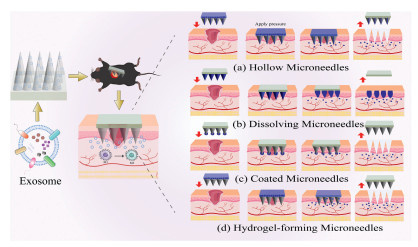
In general, microneedle-based delivery offers the advantages of improved transdermal efficiency, reduced systemic side effects, and the potential for sustained or on-demand release. However, practical limitations include relatively low loading capacity (especially for coated and dissolving MNs), the risk of needle clogging or tissue damage (hollow MNs), and increased fabrication complexity and cost for responsive or integrated systems. Moreover, the rapid dissolution or diffusion of exosomes in moist wound environments may lead to suboptimal sustained release kinetics. Despite these challenges, MNs remain a promising platform for delivering engineered exosomes to modulate macrophage polarization in diabetic wounds.
Beyond traditional carriers, novel scaffolds, including porous sponges, electrospun membranes, and 3D-printed frameworks, offer tunable architectures that create a pro-regenerative microenvironment, actively promoting M2 macrophage polarization and synergistic tissue repair.
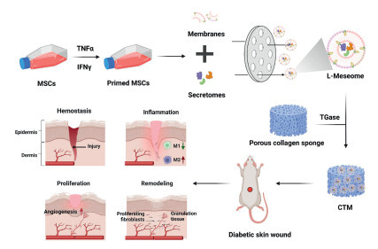
Porous sponge scaffolds feature high porosity, interconnected channels, and strong fluid absorption, mimicking the native extracellular matrix (ECM) to facilitate macrophage infiltration and phenotypic switching. Their pore architecture dictates macrophage behavior: Pores of 100–300 µm most effectively promote infiltration, while larger pores favor angiogenesis [129]. Furthermore, the hydrophilic surface of collagen- or gelatin-based sponges promotes the binding of integrins associated with the M2 macrophage phenotype, consequently reducing the secretion of pro-inflammatory cytokines [130]. When loaded with exosomes capable of inducing M2 macrophage polarization, such scaffolds can form a dynamic “immunological niche”, continuously releasing reparative signals, reversing chronic inflammation in diabetic wounds, and promoting M2 macrophage polarization. For instance, collagen sponge scaffolds loaded with either efficacy–potentiated exosome–mimicking nanovesicles (Fig. 4) [131] or trichostatin A pretreated BMSC-Exos [132] have been shown to drive M2 macrophage polarization, promote angiogenesis, and accelerate diabetic wound healing. Despite these advantages, the relatively large pore size may lead to rapid exosome diffusion out of the scaffold, and the mechanical strength of porous sponges is often insufficient for load-bearing applications.
Electrospinning generates ECM-mimicking nanofibers whose physical features, including fiber alignment, diameter, and porosity, actively modulate macrophage behavior. Aligned fibers and diameters in the 300–700 nm range preferentially induce M2 macrophage polarization, while interconnected pores facilitate macrophage infiltration [133-135].
For exosome loading, coaxial electrospinning offers key advantages over physical adsorption or blend electrospinning by encapsulating exosomes within a hydrophilic core-shell structure, protecting cargo bioactivity and enabling sustained release. Core–shell fibers via coaxial electrospinning encapsulate exosomes for sustained release, while ECM-based shells provide cell adhesion signals. Wen et al. developed an M2 macrophage-derived exosome-loaded biomimetic periosteum using coaxial electrospinning; this scaffold recruited endogenous stem cells via aptamer modification and promoted M2 macrophage polarization and bone regeneration (Fig. S1 in supporting information) [136]. Similarly, Tao's team developed a Janus–structured patch (G–Avs) by electrospinning an exosome–encapsulated GelMA layer onto an anti–adhesive PLGA/PCL backing. The upper functional layer was constructed via coaxial electrospinning, forming core–shell nanofibers. This design ensured sustained exosome release directly into the wound bed, effectively shifted macrophage polarization from the pro–inflammatory M1 phenotype toward the pro–healing M2 phenotype, resolved chronic inflammation, and accelerated diabetic wound healing [137]. The main challenges include the high voltage used during electrospinning, which may compromise exosome integrity, and the difficulty in achieving uniform fiber diameter and pore distribution at scale.
3D bioprinting enables precise layer-by-layer deposition of exosome-containing "bio-inks", allowing spatiotemporal control over exosome distribution and construction of biomimetic tissues with complex architectures. As reviewed by Zhang et al., advanced 3D printing techniques such as extrusion–based direct ink writing and light–curing methods have greatly expanded the design freedom of hydrogel scaffolds, enabling precise tuning of pore size, stiffness, and topography to modulate cellular responses [138]. This technology enables precise control of scaffold geometry, pore architecture, and exosome distribution, guiding macrophage infiltration and polarization with high accuracy. Scaffolds featuring 250–400 µm pores optimally promote M1-to-M2 transition and neovascularization [139]. Zhang et al. designed a 3D–printed scaffold for sustained release of hUCMSC–derived exosomes, which effectively alleviated neuroinflammation and promoted tissue repair (Fig. S2 in Supporting information) [140]. Additionally, immunomodulatory hydrogel bio–inks loaded with M2 macrophage–derived exosomes enable in situ delivery and real–time macrophage phenotype modulation during wound healing, as demonstrated by Dutta and colleagues using a 3D–bioprinted construct that actively induced angiogenesis and M2 macrophage polarization [141]. Although 3D bioprinting offers unparalleled design flexibility, the printing process often involves shear stress and UV exposure that may damage exosome bioactivity, and the high cost and low throughput currently limit its widespread adoption [142,143].
In addition to biological scaffolds, MOFs have recently emerged as a versatile platform for exosome delivery and immune remodeling in diabetic wounds. MOFs are crystalline porous materials consisting of metal ions (e.g., Zn2+, Zr4+, Fe3+, Mg2+) coordinated with organic ligands. Their ultrahigh surface area, tunable porosity, and facile surface functionalization make them ideal carriers for encapsulating exosomes and achieving controlled release [144,145]. Notably, the natural phospholipid structure of exosomes facilitates their association with MOFs, enabling the assembly of functional nanocomposites [146].
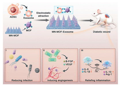
Importantly, MOFs serve not merely as passive delivery vehicles; their metal ion components also confer unique bioactivities, such as antibacterial, anti-inflammatory, and pro-angiogenic effects, thereby accelerating wound healing [147,148]. Certain MOFs can actively participate in modulating macrophage polarization toward the pro–healing M2 phenotype, either through their intrinsic physicochemical properties or through synergistic effects with the loaded exosomes [149]. For instance, Xiang et al. developed a MOF–based microneedle patch that electrostatically adsorbed exosomes, allowing precise delivery of the nanocomplex to the dermis of diabetic wounds. Upon degradation, the MOF simultaneously released Zn2+ and exosomes: Zn2+ exerted antibacterial and anti–inflammatory actions, while the exosomes induced M2–type macrophage polarization via activation of the extracellular signal-regulated kinase 1/2 (ERK1/2) and PI3K–Akt pathways (Fig. 5) [150]. Nevertheless, challenges such as potential metal ion toxicity, inconsistent degradation behavior, and insufficient large–scale production reproducibility remain to be addressed before MOF–based exosome systems can be clinically translated for diabetic wound therapy.
In conclusion, transdermal delivery of engineered exosomes holds significant promise for diabetic wound healing by promoting macrophage M2 polarization and resolving chronic inflammation. However, translating this strategy from bench to bedside requires addressing distinct challenges in fundamental research and clinical application.
For fundamental research, future investigations should focus on four areas. First, the temporal and metabolic dynamics of M2 polarization, specifically how exosome release kinetics correlate with the M1-to-M2 switch and the associated shift from glycolysis to oxidative phosphorylation, remain largely unexplored. Second, the comparative efficacy of different engineering modalities (e.g., cargo loading vs. surface modification) and their potential synergies warrant systematic evaluation. Third, strategies to enhance the yield of exosomes with potent M2-polarizing capacity warrant dedicated investigation, including cytokine preconditioning (IL-4/IL-13), hypoxia, metabolic modulators, and three-dimensional bioreactor cultures. Fourth, the impact of the transdermal delivery process on exosome integrity and bioactivity requires rigorous assessment to identify mechano-protective design principles.
For clinical translation, several hurdles must be overcome. The scalable guanosine monophosphate (GMP)-compliant production of engineered exosomes with validated quality control metrics is a prerequisite for clinical testing. The safety profile, including potential off-target effects and risks associated with chronic M2 polarization, must be established in large animal models and early-phase trials. Additionally, patient stratification strategies based on wound characteristics or inflammatory biomarkers may be necessary given the heterogeneity of diabetic wounds. Finally, regulatory frameworks specifically tailored to exosome-based therapeutics need to evolve in parallel with technological advances. Addressing these fundamental and translational challenges in parallel will accelerate the eventual clinical adoption of this promising therapeutic strategy.
Lv Geng: Writing – original draft. Xiaoya Yu: Writing – original draft. Xinyue Lei: Writing – original draft. Xiaoju Zhou: Conceptualization, Supervision, Writing – review & editing.
The authors declare that they have no known competing financial interests or personal relationships that could have appeared to influence the work reported in this paper.
This work was supported by the National Natural Science Foundation of China (No. 82273875), the National Key Research and Development Program of China (No. 2022YFA0912500).
Supplementary material associated with this article can be found, in the online version, at doi:
B. Arneth, R. Arneth, M. Shams, Int. J. Mol. Sci. 20 (2019) 2467. doi: 10.3390/ijms20102467
GBD 2021 Diabetes Collaborators, Lancet 402 (2023) 203–234. doi: 10.1016/S0140-6736(23)01301-6
D.G. Armstrong, A.J.M. Boulton, S.A. Bus, N. Engl. J. Med. 376 (2017) 2367–2375. doi: 10.1056/NEJMra1615439
V. Falanga, Lancet 366 (2005) 1736–1743. doi: 10.1016/S0140-6736(05)67700-8
T.A. Wynn, K.M. Vannella, Immunity 44 (2016) 450–462. doi: 10.1016/j.immuni.2016.02.015
R. Mirza, T.J. Koh, Cytokine 56 (2011) 256–264. doi: 10.1016/j.cyto.2011.06.016
Y. Li, Z. Zhu, S. Li, et al., J. Nanobiotechnol. 22 (2024) 398. doi: 10.1186/s12951-024-02684-1
M.D. Hade, C.N. Suire, Z. Suo, Cells 10 (2021) 1959. doi: 10.3390/cells10081959
D. Ha, N. Yang, V. Nadithe, Acta Pharm. Sin. B 6 (2016) 287–296. doi: 10.1016/j.apsb.2016.02.001
H. Yu, H. Feng, H. Zeng, et al., Int. J. Biol. Sci. 20 (2024) 1778–1795. doi: 10.7150/ijbs.92897
M. Arabpour, A. Saghazadeh, N. Rezaei, Int. Immunopharmacol. 97 (2021) 107823. doi: 10.1016/j.intimp.2021.107823
W. Liu, M. Yu, D. Xie, et al., Stem Cell Res. Ther. 11 (2020) 259. doi: 10.1186/s13287-020-01756-x
J. Zhao, X. Li, J. Hu, et al., Cardiovasc. Res. 115 (2019) 1205–1216. doi: 10.1093/cvr/cvz040
Y. Qian, G. Chu, L. Zhang, et al., J. Nanobiotechnol. 22 (2024) 72. doi: 10.1186/s12951-024-02336-4
X. Zhou, B.A. Brown, A.P. Siegel, et al., ACS Nano, 14 (2020) 12732–12748. doi: 10.1021/acsnano.0c03064
L. Kuang, C. Zhang, B. Li, et al., Int. J. Nanomed. Volume 18 (2023) 949–970. doi: 10.2147/ijn.s399785
M. Colombo, G. Raposo, C. Théry, Annu. Rev. Cell Dev. Biol. 30 (2014) 255–289. doi: 10.1146/annurev-cellbio-101512-122326
S. Ghadami, K. Dellinger, Front. Mol. Biosci. 10 (2023) 1198044. doi: 10.3389/fmolb.2023.1198044
M. Yáñez-Mó, P.R. Siljander, Z. Andreu, et al., J. Extracell. Vesicles. 4 (2015) 27066.
Z. Li, Q. Li, K. Tong, et al., Stem Cell Res. Ther. 13 (2022) 295. doi: 10.1186/s13287-022-02975-0
Y. Dou, S. Yu, S. Cao, et al., Exp. Cell Res. 450 (2025) 114653. doi: 10.1016/j.yexcr.2025.114653
D. Yin, G. Shen, Immun. Inflamm. Dis. 12 (2024) e1274. doi: 10.1002/iid3.1274
X. Chen, H. An, Y. Du, et al., Stem Cell Res. Ther. 16 (2025) 384. doi: 10.1021/jacs.4c11028
X. Li, M. Chen, J. Cao, et al., Heliyon, 10 (2024) e37002. doi: 10.1016/j.heliyon.2024.e37002
Y. Zhang, X. Le, S. Zheng, et al., Stem Cell Res. Ther. 13 (2022) 171. doi: 10.1186/s13287-022-02855-7
T. Chen, P. Song, M. He, et al., Burns Trauma. 11 (2023) tkad003. doi: 10.1093/burnst/tkad003
S. Rui, L. Dai, X. Zhang, et al., J. Control. Release 372 (2024) 221–233. doi: 10.1016/j.jconrel.2024.06.050
L. He, N. Zhao, X. Chen, et al., Clin. Exp. Med. 25 (2025) 163. doi: 10.1007/s10238-025-01651-w
H. Kim, S.Y. Wang, G. Kwak, et al., Adv. Sci. 6 (2019) 1900513. doi: 10.1002/advs.201900513
Y. Wang, L. Zhang, G.R. Wu, et al., Sci. Adv. 7(2021) eabb6075. doi: 10.1126/sciadv.abb6075
R. Zeng, B. Lv, Z. Lin, et al., Bioact. Mater. 34 (2024) 366–380.
S. Li, X. Ding, H. Zhang, et al., Int. Immunopharmacol. 106 (2022) 108605. doi: 10.1016/j.intimp.2022.108605
L. Tao, S. Wu, Q. Wang, et al., Int. Immunopharmacol. 155 (2025) 114575. doi: 10.1016/j.intimp.2025.114575
X. Peng, F. He, Y. Mao, et al., J. Mol. Endocrinol. 69 (2022) 315–327. doi: 10.1530/jme-21-0019
J. Mao, S. Qian, Q. Zhao, et al., Med 5 (2024) 148–168.e8.e148. doi: 10.1016/j.medj.2024.01.006
Y. Huang, L. Zhu, J. Wang, et al., Burns Trauma. 13 (2025) tkaf060. doi: 10.1093/burnst/tkaf060
Y. Li, K. Du, D. Peng, et al., Biomaterials 322 (2025) 123411. doi: 10.1016/j.biomaterials.2025.123411
S. Cui, X. Wu, X. Yu, et al., Adv. Mater. 38 (2026) e10507. doi: 10.1002/adma.202410507
W. Yan, T. Ni, Q. Zhang, et al., Int. Immunopharmacol. 142 (2024) 112983. doi: 10.1016/j.intimp.2024.112983
Y. Ding, P. Yang, S. Li, et al., Pharm. Biol. 60 (2022) 2328–2337. doi: 10.1080/13880209.2022.2149821
J. Wang, L. Zhang, W. Duan, et al., Int. Immunopharmacol. 167 (2025) 115708. doi: 10.1016/j.intimp.2025.115708
A. Shu, J. Chen, S. Shao, et al., Phytomedicine 151 (2026) 157794. doi: 10.1016/j.phymed.2026.157794
S. Li, P. Yang, X. Ding, et al., Burns Trauma. 10 (2022) tkac046. doi: 10.1093/burnst/tkac046
J. Fu, J. Huang, M. Lin, et al., J. Surg. Res. 246 (2020) 213–223. doi: 10.1016/j.jss.2019.09.011
H. Yang, L. Song, B. Sun, et al., Mater. Today Bio 12 (2021) 100139. doi: 10.1016/j.mtbio.2021.100139
J. Zou, W. Cui, N. Deng, et al., J. Control. Release. 382 (2025) 113730. doi: 10.1016/j.jconrel.2025.113730
M.S. Kim, M.J. Haney, Y. Zhao, et al., Nanomedicine 12 (2016) 655–664. doi: 10.5021/ad.2016.28.5.655
P. Wang, H. Wang, Q. Huang, et al., Theranostics, 9 (2019) 1714–1727. doi: 10.7150/thno.30716
J.L. Hood, M.J. Scott, S.A. Wickline, Anal. Biochem. 448 (2014) 41–49. doi: 10.1016/j.ab.2013.12.001
S.A.A. Kooijmans, S. Stremersch, K. Braeckmans, et al., J. Control. Release 172 (2013) 229–238. doi: 10.1016/j.jconrel.2013.08.014
M. Deng, S. Wu, P. Huang, et al., Asian J. Pharm. Sci. 18 (2023) 100870.
S. Guo, N. Perets, O. Betzer, et al., ACS Nano 13 (2019) 10015–10028. doi: 10.1021/acsnano.9b01892
A. Akbari, F. Nazari-Khanamiri, M. Ahmadi, et al., Pharmaceutics 15 (2022) 66. doi: 10.3390/pharmaceutics15010066
M.J. Haney, N.L. Klyachko, Y. Zhao, et al., J. Control. Release 207 (2015) 18–30. doi: 10.1016/j.jconrel.2015.03.033
M. Khongkow, T. Yata, S. Boonrungsiman, et al., Sci. Rep. 9 (2019) 8278. doi: 10.1038/s41598-019-44569-6
Z. Erana-Perez, M. Igartua, E. Santos-Vizcaino, et al., Trends Pharmacol. Sci. 45 (2024) 350–365. doi: 10.1016/j.tips.2024.02.006
Y. Yang, Y. Chen, F. Zhang, et al., Int. J. Hyperthermia 31 (2015) 498–506. doi: 10.3109/02656736.2015.1036384
L. Pascucci, V. Coccè, A. Bonomi, et al., J. Control. Release 192 (2014) 262–270. doi: 10.1016/j.jconrel.2014.07.042
D. Ti, H. Hao, C. Tong, et al., J. Transl. Med. 13 (2015) 308. doi: 10.1186/s12967-015-0642-6
Y. Shi, S. Wang, K. Wang, et al., Small 20 (2024) e2309276. doi: 10.1002/smll.202309276
N. Hu, Z. Cai, X. Jiang, et al., Acta Biomater. 157 (2023) 175–186. doi: 10.1016/j.actbio.2022.11.057
G. Son, J. Song, J.C. Park, et al., Nat. Commun. 16 (2025) 4799. doi: 10.1038/s41467-025-59489-5
S. Subramaniam, P. Joyce, L. Donnellan, et al., J. Colloid Interface Sci. 641 (2023) 36–47. doi: 10.1016/j.jcis.2023.03.048
A. Liu, G. Yang, Y. Liu, et al., Front. Bioeng. Biotechnol. 10 (2022) 939441. doi: 10.3389/fbioe.2022.939441
B.P. Dyett, H. Yu, J. Strachan, et al., Nat. Commun. 10 (2019) 4492. doi: 10.1038/s41467-019-12508-8
L. Alvarez-Erviti, Y. Seow, H. Yin, et al., Nat. Biotechnol. 29 (2011) 341–345. doi: 10.1038/nbt.1807
R. Hao, Z. Yu, J. Du, et al., Small 17 (2021) e2102150. doi: 10.1002/smll.202102150
Y. Sheng, Z. Huang, T. Zhang, et al., J. Am. Chem. Soc. 144 (2022) 9443–9450. doi: 10.1021/jacs.2c02268
Z. Yang, J. Shi, J. Xie, et al., Nat. Biomed. Eng. 4 (2020) 69–83.
S. Kuang, F. He, G. Liu, et al., Biomaterials 275 (2021) 120963. doi: 10.1016/j.biomaterials.2021.120963
A.E. Boniakowski, A.S. Kimball, A. Joshi, et al., Eur. J. Immunol. 48 (2018) 1445–1455. doi: 10.1002/eji.201747400
H. Al Sadoun, Cells 11 (2022) 2430. doi: 10.3390/cells11152430
H.C. Yang, M. Zhang, R. Wu, et al., World J. Stem. Cells 12 (2020) 152–167.
B. Shen, J. Liu, F. Zhang, et al., Stem. Cells Int. 2016 (2016) 1240301. doi: 10.1155/2016/1240301
D.M. Mossel, K. Moganti, V. Riabov, et al., Front. Immunol. 11 (2020) 1071. doi: 10.3389/fimmu.2020.01071
S. Ostrand-Rosenberg, T. Huecksteadt, K. Sanders, Cancers 15 (2023) 1026. doi: 10.3390/cancers15041026
D. Sun, G. Zhang, M. Xie, et al., J. Nanobiotechnol. 21 (2023) 172. doi: 10.1186/s12951-023-01930-2
J. Lin, L. Yang, T. Liu, et al., J. Control. Release 379 (2025) 832–847. doi: 10.1016/j.jconrel.2025.01.073
W. Qi, L. Jin, S. Huang, et al., Acta Biomater. 181 (2024) 425–439. doi: 10.3390/aerospace11060425
Y. Takayama, K. Kusamori, M. Nishikawa, Molecules 24 (2019) 172. doi: 10.3390/molecules24010172
T. Smyth, K. Petrova, N.M. Payton, et al., Bioconjugate Chem. 25 (2014) 1777–1784. doi: 10.1021/bc500291r
M. Bahadorani, M. Nasiri, K. Dellinger, et al., Int. J. Nanomed. 19 (2024) 7137–7164. doi: 10.2147/ijn.s464249
Y.T. Sato, K. Umezaki, S. Sawada, et al., Sci. Rep. 6 (2016) 21933. doi: 10.1038/srep21933
M. Xie, Y. Wu, Y. Zhang, et al., Adv. Mater. 36 (2024) e2403935. doi: 10.1002/adma.202403935
Q. Li, W. Hu, Q. Huang, et al., Signal Transduct. Target. Ther. 8 (2023) 62. doi: 10.1038/s41392-022-01263-w
D. Sivaraj, K. Chen, A. Chattopadhyay, et al., Front. Bioeng. Biotechnol. 9 (2021) 660145. doi: 10.3389/fbioe.2021.660145
R. Madhyastha, H. Madhyastha, Q.I. Nurrahmah, et al., Inflammation 44 (2021) 1274–1287. doi: 10.1007/s10753-021-01415-0
Z. Jiang, J. Zhang, Cell Cycle 20 (2021) 993–1009. doi: 10.1080/15384101.2021.1919825
H. Wu, H. Fan, Z. Shou, et al., Int. Immunopharmacol. 68 (2019) 204–212. doi: 10.1016/j.intimp.2018.12.043
S. Nasra, M. Patel, H. Shukla, et al., Life Sci. 334 (2023) 122232. doi: 10.1016/j.lfs.2023.122232
R. Sridharan, B. Cavanagh, A.R. Cameron, et al., Acta Biomater. 89 (2019) 47–59. doi: 10.1016/j.actbio.2019.02.048
S. Butenko, R.R. Nagalla, C.F. Guerrero-Juarez, et al., Nat. Commun. 15 (2024) 6820. doi: 10.1038/s41467-024-50072-y
L. Ye, Y. Zhang, Q. Wang, et al., ACS Appl. Mater. Interfaces 8 (2016) 15710–15723. doi: 10.1021/acsami.6b03098
R. Nie, Q.Y. Zhang, Z.Y. Feng, et al., Int. J. Biol. Macromol. 268 (2024) 131643. doi: 10.1016/j.ijbiomac.2024.131643
Y.J. Fu, Y.F. Shi, L.Y. Wang, et al., Adv. Sci. 10 (2023) e2206771. doi: 10.1002/advs.202206771
X.C. Li, L. Guan, X.L. Li, et al., Chin. Chem. Lett. 36 (2025) 110661. doi: 10.1016/j.cclet.2024.110661
Y. Zhao, Y. Zhao, B. Xu, et al., J. Tissue Eng. 15 (2024) 20417314241253290. doi: 10.1177/20417314241253290
B. Liu, L. Chen, C. Huang, et al., Mater. Today Bio 34 (2025) 102159. doi: 10.1016/j.mtbio.2025.102159
M. Jung, Y. Ma, R.P. Iyer, et al., Basic Res. Cardiol. 112 (2017) 33. doi: 10.1007/s00395-017-0622-5
N.A. Peppas, Y. Huang, M. Torres-Lugo, et al., Annu. Rev. Biomed. Eng. 2 (2000) 9–29. doi: 10.1146/annurev.bioeng.2.1.9
S.S. Yerneni, S. Lathwal, J. Cuthbert, et al., Biomacromolecules 23 (2022) 1713–1722. doi: 10.1021/acs.biomac.1c01636
Y. Chen, X. Wang, S. Tao, et al., Mil. Med. Res. 10 (2023) 37.
Y. Zhang, W. Yan, L. Wu, et al., Front. Bioeng. Biotechnol. 13 (2025) 1545636. doi: 10.3389/fbioe.2025.1545636
E. Axpe, D. Chan, G.S. Offeddu, et al., Macromolecules, 52 (2019) 6889–6897. doi: 10.1021/acs.macromol.9b00753
Y. Li, Y. Liu, R. Ma, et al., ACS Appl. Mater. Interfaces 9 (2017) 13056–13067. doi: 10.1021/acsami.7b00957
A.I. Visan, I. Negut, Gels 10 (2024) 497. doi: 10.3390/gels10080497
X. Wang, J. Dong, J. Kang, et al., J. Am. Chem. Soc. 147 (2025) 16362–16378. doi: 10.1021/jacs.5c02184
Y. Ye, Y. Liu, S. Ma, et al., Bioact. Mater. 52 (2025) 422–437.
P. Yang, Y. Ju, N. Shen, et al., Adv. Healthc. Mater. 14 (2025) e2403304. doi: 10.1002/adhm.202403304
M. Deng, Y. Wu, Y. Ren, et al., J. Control. Release. 350 (2022) 613–629. doi: 10.1016/j.jconrel.2022.08.053
H. Meng, J. Su, Q. Shen, et al., Adv. Healthc. Mater. 14 (2025) e2404966. doi: 10.1002/adhm.202404966
W. Yuan, C. Huang, W. Deng, et al., Int. J. Biol. Macromol. 280 (2024) 135562. doi: 10.1016/j.ijbiomac.2024.135562
J. Yang, Z. Chen, D. Pan, et al., Int. J. Nanomed. 15 (2020) 5911–5926. doi: 10.2147/ijn.s249129
S. Wang, J. Wu, K. Ren, et al., ACS Appl. Mater. Interfaces 17 (2025) 27923–27936. doi: 10.1021/acsami.5c02705
W. Jin, Y. Li, M. Yu, et al., Burns Trauma. 13 (2025) tkae078. doi: 10.1093/burnst/tkae078
T.M. Tuan-Mahmood, M.T. McCrudden, B.M. Torrisi, et al., Eur. J. Pharm. Sci. 50 (2013) 623–637. doi: 10.1016/j.ejps.2013.05.005
M. Liu, J. Jiang, Y. Wang, et al., Mater. Today Bio 29 (2024) 101321. doi: 10.1016/j.mtbio.2024.101321
K. Ahmed Saeed Al-Japairai, S. Mahmood, S. Hamed Almurisi, et al., Int. J. Pharm. 587 (2020) 119673. doi: 10.1016/j.ijpharm.2020.119673
N. Qiang, Z. Liu, M. Lu, et al., Materials 16 (2023) 3417. doi: 10.3390/ma16093417
S. Mu, H. Chang, F. Qu, J. Vis. Exp. 209 (2024) e67109.
Y. Cao, B. Chen, Q. Liu, et al., Bioact. Mater. 42 (2024) 32–51. doi: 10.1117/12.3031964
X. Jiang, J. Ma, K. Xue, et al., ACS Nano 18 (2024) 4269–4286. doi: 10.1021/acsnano.3c09721
T. Waghule, G. Singhvi, S.K. Dubey, et al., Biomed. Pharmacother. 109 (2019) 1249–1258. doi: 10.1016/j.biopha.2018.10.078
J. Zeng, Z. Sun, F. Zeng, et al., Mater. Today Bio 20 (2023) 100649. doi: 10.1016/j.mtbio.2023.100649
A. Ullah, H. Khan, H.J. Choi, et al., Polymers 11 (2019) 1834. doi: 10.3390/polym11111834
H. Ding, Y. Cui, J. Yang, et al., J. Control. Release. 360 (2023) 365–375. doi: 10.1016/j.jconrel.2023.03.060
H. Lee, C. Song, Y.S. Hong, et al., Sci. Adv. 3 (2017) e1601314. doi: 10.1126/sciadv.1601314
M. Yuan, K. Liu, T. Jiang, et al., J. Nanobiotechnol. 20 (2022) 147. doi: 10.1214/21-aos2099
F. Mukasheva, L. Adilova, A. Dyussenbinov, et al., Front. Bioeng. Biotechnol. 12 (2024) 1444986. doi: 10.3389/fbioe.2024.1444986
B.H. Cha, S.R. Shin, J. Leijten, et al., Adv. Healthc. Mater. 6 (2017) 1700289. doi: 10.1002/adhm.201700289
X. Liu, Y. Liu, J. Zhou, et al., ACS Biomater. Sci. Eng. 10 (2024) 975–986. doi: 10.1021/acsbiomaterials.3c01282
T. Wang, Y. Xue, W. Zhang, et al., Int. J. Biol. Macromol. 269 (2024) 131948. doi: 10.1016/j.ijbiomac.2024.131948
L. Tian, M.P. Prabhakaran, S. Ramakrishna, Regen. Biomater. 2 (2015) 31–45. doi: 10.1093/rb/rbu017
Y. Jia, W. Yang, K. Zhang, et al., Acta Biomater. 83 (2019) 291–301. doi: 10.1016/j.actbio.2018.10.040
K. Garg, N.A. Pullen, C.A. Oskeritzian, et al., Biomaterials 34 (2013) 4439–4451. doi: 10.1016/j.biomaterials.2013.02.065
Z. Wen, S. Li, Y. Liu, et al., Bioact. Mater. 50 (2025) 95–115.
Y. Tao, T. Liu, F. Jing, et al., ACS Nano 19 (2025) 21271–21289. doi: 10.1021/acsnano.4c16058
A.K. Zhang, F. Wang, L. Chen, et al., Chin. Chem. Lett. 32 (2021) 2923–2932. doi: 10.1016/j.cclet.2021.03.073
X. Yang, J. Gao, S. Yang, et al., Int. J. Bioprint. 9 (2023) 755. doi: 10.18063/ijb.755
A. Zhang, B. Sun, C. Nan, et al., Stem Cell Res. Ther. 16 (2025) 196. doi: 10.70251/hyjr2348.33196200
S.D. Dutta, J.M. An, J. Hexiu, et al., Bioact. Mater. 45 (2025) 345–362.
S. Manzoli, E. Merotto, M. Piccoli, et al., J. Funct. Biomater. 16 (2025) 436. doi: 10.3390/jfb16120436
B. Li, Z. Wang, C. Huang, et al., Med. Eng. Phys. 145 (2025) 104448. doi: 10.1016/j.medengphy.2025.104448
D. Ma, G. Wang, J. Lu, et al., Eur. J. Med. Chem. 261 (2023) 115884. doi: 10.1016/j.ejmech.2023.115884
X. Zhao, Z. Chen, S. Zhang, et al., J. Nanobiotechnol. 22 (2024) 387. doi: 10.4067/s0717-95022024000200387
X. Qin, B. Wei, Y. Xiang, et al., Biosens. Bioelectron. 245 (2024) 115828. doi: 10.1016/j.bios.2023.115828
S. Yao, J. Chi, Y. Wang, et al., Adv. Healthc. Mater. 10 (2021) e2100056. doi: 10.1002/adhm.202100056
X. Xia, X. Song, Y. Li, et al., Front. Bioeng. Biotechnol 10 (2022) 1026743. doi: 10.3389/fbioe.2022.1026743
F.F. Wang, H. Yao, X.Y. Wu, et al., Chin. Chem. Lett. 35 (2024) 108925. doi: 10.1016/j.cclet.2023.108925
J. Xiang, Z. Li, S. Tseng, et al., Mater. Today Bio 33 (2025) 102076. doi: 10.1016/j.mtbio.2025.102076

Figure 1 Schematic illustration of engineered exosome delivery systems for diabetic wound healing via macrophage polarization modulation.

Figure 4 Porous collagen sponge loaded with large efficacy-potentiated exosome-mimicking nanovesicles for diabetic skin wound healing. Copied with permission [121]. Copyright 2024, ACS Publication.

Figure 5 MOF and exosome loaded MN patch for promoting diabetic wound healing. Exosomes autonomously assemble on the surface of MOF loaded microneedles through electrostatic attraction. Copied with permission [150]. Copyright 2025, Elsevier.
Table 1. Polarization-regulating functions of exosomes from different cellular origins.
| Exosome types | Cellular origin | Key bioactive molecules/signaling pathways | Primary mechanism | Ref. |
| BMSC-Exos | Bone marrow MSCs | miR-23a-3p, CD73-adenosine axis | Inhibiting the interferon regulatory factor 1 (IRF1)/NF-κB pathway to promote M2 macrophage polarization, function impaired under hyperglycemia due to CD73 down regulation | [20,21] |
| ADSC-Exos | Adipose-derived stem cells | circRps5/miR–124–3p axis | Promoting M2 macrophage polarization and enhancing collagen synthesis | [22] |
| hUCMSC-Exos | Human umbilical cord MSCs | miR-146a-5p, insulin (INS), superoxide dismutase 1 (SOD1) | Suppressing tumor necrosis factor receptor-associated factor 6 (TRAF6)/signal transducer and activator of transcription 1 (STAT1) signaling to drive M2 macrophage polarization; inhibiting pro-inflammatory activation | [23-25] |
| PRP-Exos | Platelet-rich plasma | miR-26b-5p, sphingosine-1-phosphate (S1P)/Akt/fibronectin 1 (FN1) pathway | Reducing neutrophil infiltration, promotes angiogenesis, and enhances M2 macrophage polarization | [26-28] |
| M2-Exos | M2 macrophages | CCL22, IL-4, milk fat globule-epidermal growth factor 8 (MFG-E8), etc. | Directly suppresses inducible nitric oxide synthase (iNOS) while upregulating Arg-1 and CD206, thereby inducing M1-to-M2 conversion | [29] |
| KC-Exos | Keratinocytes | Metastasis associated lung adenocarcinoma transcript 1 (MALAT1) /miR–1914–3p/MFG-E8 axis | Modulates macrophage function via miRNA regulation, promoting diabetic wound healing | [15,16] |
 下载: 导出CSV
Table 2. Stimuli-responsive hydrogel systems for engineered exosome delivery in diabetic wound healing.
| Hydrogel type | Representative materials | Stimulus/triggering condition | Impact on macrophage polarization | Ref. |
| ROS-responsive hydrogels | Hydrogels containing thioketal/boronate ester bonds | High ROS microenvironment | Smart release of exosomes to enhance M2 macrophage polarization | [102,107,108] |
| pH-responsive hydrogels | Hydrogels containing carboxyl/amino/Schiff base groups | Slightly acidic microenvironment (pH 5.5–6.5) | Preferential release at inflammatory sites, promoting phenotype switching | [109,110] |
| Enzyme-responsive hydrogels | MMP-9-sensitive peptide-cross-linked hydrogels | High MMP-9 expression | Precise release to promote M2 macrophage polarization | [111] |
| Glucose-responsive hydrogels | Phenylboronic acid group-modified hydrogels | Hyperglycemic microenvironment | Cooperatively modulate macrophage phenotype and reduce inflammation | [112] |
| Thermosensitive hydrogels | Pluronic F-127 | Body temperature (~37 ℃) | Sustained and controlled release to enhance M2 marker expression | [113,114] |
下载: 导出CSV

 扫一扫看文章


扫一扫关注我们


 下载: