The Affiliated Hospital of Guizhou Medical University, State Key Laboratory of Discovery and Utilization of Functional Components in Traditional Chinese Medicine, Guizhou Medical University, Guiyang 561113, China
b.
Department of Chemistry, Bengbu Medical University, Bengbu 233030, China
c.
Department of Chemistry, University of Namur, Rue de Bruxelles 61, Namur B-5000, Belgium
tlei1974@163.com (L. Tang). 1 These authors contributed equally to this work.
Received Date:
28 January 2026 Accepted Date:
13 May 2026 Revised Date:
12 May 2026 Available Online:
15 December 2026
Abstract:
Herein, we report an efficient fragment co-cyclization strategy for the synthesis of an orthogonally functionalized copillar[3 + 2]arene bearing propargyl and bromoethyl groups, achieving a yield of up to 50%. Its programmable architecture was unambiguously confirmed by single-crystal X-ray diffraction. This versatile scaffold enabled the precise construction of the water-soluble glycopeptide mimic GM10via sequential orthogonal click reactions. GM10, featuring rationally integrated lectin-targeting and cationic modules, exhibits a dual inhibitory mechanism: It competitively binds to the Pseudomonas aeruginosa (P. aeruginosa) lectin LecB to block lectin-mediated adhesion, while simultaneously sequestering the key quorum-sensing (QS) signal molecule 3-oxo-C12-HSL through host–guest complexation, thereby suppressing QS and attenuating virulence factor expression. This mimic demonstrates potent, non-bactericidal anti-biofilm activity in vitro and accelerates wound healing in a murine infection model. Our work not only presents a novel synthetic route to functionalized pillar[5]arenes but also underscores the potential of programmable copillar[3 + 2]arene scaffolds for developing next-generation anti-virulence agents against biofilm-associated resistance.
The emergence of antibiotic resistance, especially in Gram-negative bacteria, has become a major global health challenge [1]. Pseudomonas aeruginosa (P. aeruginosa) is a multidrug-resistant opportunistic pathogen and a major cause of healthcare-associated infections, particularly in patients with cystic fibrosis, burns, or cancer [2]. Its strong ability to form biofilms greatly reduces antibiotic efficacy and facilitates the spread of resistance, making anti-biofilm therapy an attractive alternative to conventional bactericidal strategies [3].
Biofilm formation in P. aeruginosa is regulated by multiple pathways. Among them, the extracellular lectin LecB plays an important role in bacterial adhesion and early biofilm development [4-7], while quorum sensing (QS) controls biofilm maturation and virulence through self-produced autoinducers such as 3-oxo-C12-HSL [8,9]. Mutants deficient in lectins or QS show impaired biofilm formation without obvious growth defects [7,10-12], indicating that both pathways are promising anti-biofilm targets. However, inhibition of either pathway alone often shows limited efficacy because of the complexity of biofilm regulation, making dual-target intervention an attractive strategy [13-19]. Notably, Zhu et al. recently showed that conjugating a QS inhibitory motif with a lectin ligand can synergistically suppress biofilm formation [20,21]. Nevertheless, conventional conjugation-based dual inhibitors targeting both lectins and QS still face major limitations, because lectins are extracellular, whereas QS receptors are intracellular, and their ligand-binding environments are also fundamentally different [7,22-24]. These issues make it difficult to develop a single molecule that effectively addresses both targets through direct receptor binding. Accordingly, macrocycle-engineered systems have emerged as promising platforms for combating biofilm-associated infections [25].
Pillar[5]arenes are a class of macrocycles with symmetrical cylindrical structures and electron-rich cavities. Since their discovery, they have attracted considerable attention because of their host-guest properties, facile synthesis, and versatile functionalization [26-31]. Thanks to the tunability of the type and number of substituents, they have been used in molecular separation, sensing, catalysis, and functional materials [32-37]. Recently, cationic pillar[5]arenes have emerged as anti-biofilm agents, but the reported systems are mainly based on homopillar[5]arenes bearing identical groups on both rims and generally show limited efficacy against Gram-negative biofilms, especially those of P. aeruginosa [38-41]. It has been reported that a cationic rotaxane based on a galactosylated homopillar[5]arene scaffold and bearing fucose and arginine tetrapeptide modules can achieve lectin-dependent, synergistic, and non-bactericidal inhibition of P. aeruginosa biofilms [42]. However, this strategy suffers from tedious synthesis and cumbersome purification. Therefore, readily functionalizable pillar[5]arene derivatives that enable the regioselective incorporation of diverse functional groups are highly desirable. In this context, propargyl- and bromoethyl-functionalized copillar[4 + 1]arenes have been developed as heteromultivalent scaffolds for orthogonal click functionalization [43], and one such scaffold was further used to construct highly efficient artificial water channels by tuning the hydrophilic-lipophilic balance and channel geometry [44]. Inspired by this versatility, we sought to develop a programmable copillar[3 + 2]arene scaffold for the precise integration of lectin-targeting and cationic motifs.
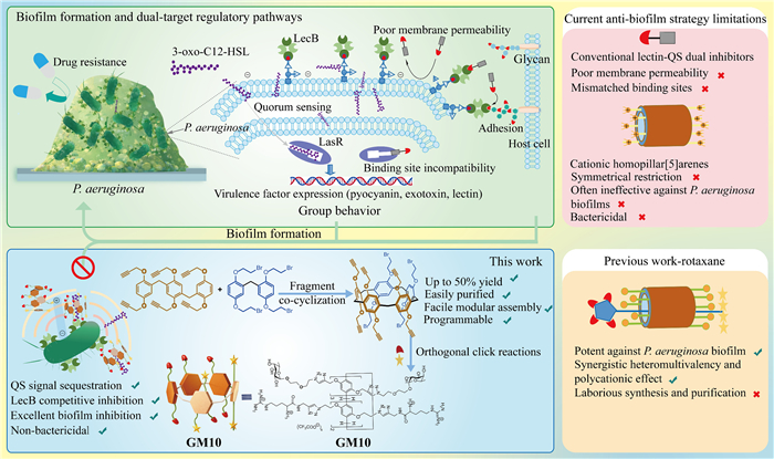
However, preparing multifunctionalized copillar[5]arenes remains challenging because constitutional isomers are readily formed, often resulting in low yields (<8%) and tedious purification. Although Ma's group improved the yields of tetrafunctionalized copillar[3 + 2]arenes to 15%−17% through step-growth cyclooligomerization [45], the reported examples are mainly limited to alkyl-substituted derivatives, thus offering limited scope for further functionalization. Considering the reversibility of the Friedel-Crafts alkylation cyclization step, we hypothesized that increasing the proportion of fragments with less electron-withdrawing substituents would favor pentameric cyclization and improve the yield of the target scaffold. Herein, we developed an efficient method for synthesizing multifunctionalized copillar[3 + 2]arenes bearing propargyl and bromoethyl groups as programmable scaffolds. Using this platform, we constructed GM10, a novel anti-biofilm agent integrating lectin ligands and cationic motifs. As shown in Fig. 1, GM10 was designed to combine extracellular LecB targeting by peripheral L-fucoside units with sequestration of the QS signal molecule 3-oxo-C12-HSL by the electron-rich cavity.
Figure 1
Figure 1.
Schematic illustration of the design and proposed dual-mode action of the programmable copillar[3 + 2]arene-based anti-biofilm agent GM10. Core functional modules and extracellular mechanisms are labeled with text annotations.
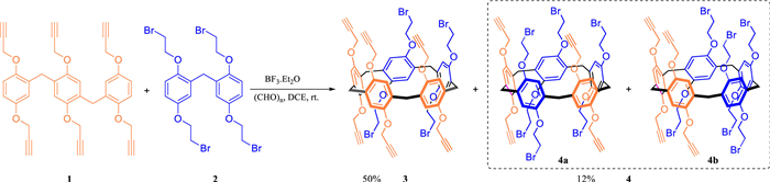
Guided by the electronic-density rationale, we developed an efficient fragment co-cyclization strategy for the synthesis of functionalized copillar[3 + 2]arene 3 bearing orthogonal propargyl and bromoethyl groups for modular modification (Scheme 1). After optimization of trimer 1 and the co-cyclization step with dimer 2, the target scaffold 3 was obtained in 50% yield under the optimized conditions (1,2-dichloroethane, 1:2 fragment ratio), outperforming conventional methods for unsymmetrical copillararenes. More broadly, the stoichiometry-tuning principle may also be extended to other heteromultivalent systems, such as copillar[4 + 1]arenes. Full synthetic details, condition optimization, and byproduct analysis are provided in the Supporting Information (Section S2, Table S1, and Schemes S1–S3 in Supporting information).
Scheme 1
Scheme 1.
Synthesis of copillar[3 + 2]arene 3via co-cyclization of fragments 1 and 2.
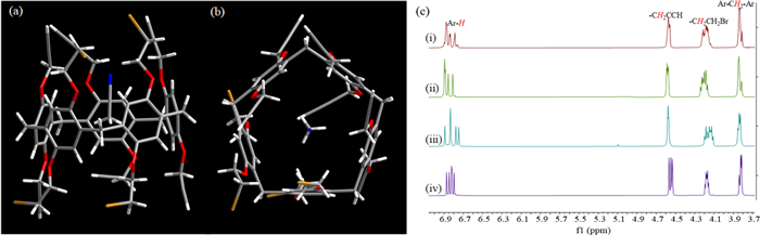
Single-crystal X-ray diffraction unambiguously established the regioselective [3 + 2] structure of the major product 3, with three propargyl-substituted units and two bromoethyl-substituted units (Figs. 2a and b). An acetonitrile molecule was observed in the pentagonal cavity, indicating the host-guest binding capability of the scaffold. In addition to 3, another copillar[3 + 2]arene fraction was isolated in 12% total yield and identified by high-performance liquid chromatography (HPLC) as a 1:1 mixture of constitutional isomers 4a and 4b (Fig. S66 in Supporting information). After separation by preparative HPLC, comparison of their aromatic proton splitting patterns in the partial proton nuclear magnetic resonance (1H NMR) spectra with that of 3 allowed tentative assignment of their regiochemistries (Fig. 2c (ⅰ–ⅳ) and Fig. S67 in Supporting information).
Figure 2
Figure 2.
Structural characterization of copillar[3 + 2]arene 3 and its regioisomers. (a) Single-crystal X-ray structure of 3 showing the side view. (b) Top view. (c) Partial 1H NMR spectra: mixture 4 (ⅰ), isolated isomers 4a (ⅱ) and 4b (ⅲ), and major product 3 (ⅳ).
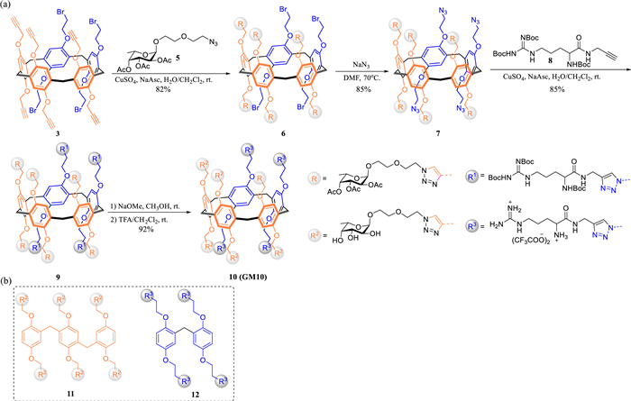
With the precisely defined scaffold 3 in hand, we explored its potential as a programmable platform for constructing anti-biofilm agents. As a representative example, GM10 was synthesized through sequential orthogonal CuAAC click reactions. L-Fucoside and arginine modules were conjugated to the distinct sites of the [3 + 2] scaffold, followed by global deprotection to afford the water-soluble product in 55% overall yield (Fig. 3a). To evaluate the advantages of this asymmetric architecture, we synthesized the L-fucoside-functionalized fragment 11 and the arginine-functionalized dimer fragment 12 for comparison (Fig. 3b, Schemes S8 and S9 in Supporting information). PP5 is a well-known potent anti-biofilm agent against P. aeruginosa, acting through a direct bactericidal mechanism [46], and was used as a control.
Figure 3
Figure 3.
Synthesis of GM10 and related reference fragments. (a) Sequential orthogonal CuAAC construction of GM10, followed by deprotection. (b) Structures of the reference fragments 11 and 12.
The antibacterial activities of all compounds were first assessed in standard LB medium (Table S3 and Fig. S68 in Supporting information). The cationic controls 12 and PP5 exhibited potent activity, with minimum inhibitory concentrations (MICs) of 100 and 50 µmol/L, respectively; the value for PP5 is consistent with the reported data [46]. In contrast, GM10 and the neutral fragment 11 showed no growth inhibition at concentrations up to 200 µmol/L. Notably, a 1:1 physical mixture of 11 and 12 displayed the same MIC as 12 alone, suggesting that covalent integration onto the rigid three-dimensional scaffold plays an important role in minimizing direct killing. To address the solubility issues of compounds 11 and PP5 at high concentrations in LB medium, we also determined MICs in the defined biofilm-supporting medium ABTGC. In this medium, GM10 remained non-bactericidal (MIC > 200 µmol/L), whereas the activities of the two cationic controls changed in a medium-dependent manner (Table S3 and Fig. S69 in Supporting information). These results establish GM10 as a consistently non-bactericidal compound.
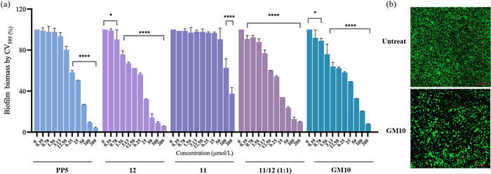
Given this non-bactericidal profile and the presence of LecB-targeting L-fucoside groups, we reasoned that GM10 might inhibit biofilm formation by interfering with early-stage bacterial adhesion. Its anti-biofilm potency was evaluated by the crystal violet assay in ABTGC (Fig. S70 in Supporting information). Surprisingly, GM10 showed a minimum biofilm inhibitory concentration required for 50% inhibition (MBIC50) of 14.6 µmol/L, which was superior to that of PP5 (20.6 µmol/L) and fragment 11 (145 µmol/L), and comparable to that of fragment 12 (10.2 µmol/L). In contrast, the 1:1 physical mixture of 11 and 12 gave an MBIC50 of 12.5 µmol/L, nearly identical to that of 12 alone and distinct from GM10 (Fig. 4a). Together with the MIC results, these data indicate that the anti-biofilm activity of GM10 is independent of direct bacterial killing and relies on the integrated copillar[3 + 2]arene architecture. Confocal laser scanning microscopy (CLSM) further confirmed that treatment with 50 µmol/L GM10 markedly reduced the P. aeruginosa PAO1 biofilm biomass (Fig. 4b). The discrete green fluorescence observed in the treated samples suggests bacterial aggregation, consistent with a lectin-mediated anti-adhesion mechanism [47].
Figure 4
Figure 4.In vitro anti-biofilm activity of GM10. (a) Dose-dependent inhibition of P. aeruginosa PAO1 biofilm formation by GM10, fragments 11 and 12, their 1:1 mixture (11/12), and PP5. Data are presented as mean ± SD (n = 3). *P < 0.05, ****P < 0.0001. (b) Representative CLSM images of untreated and GM10-treated biofilms. Scale bar: 20 µm.
To quantify the interaction between GM10 and its target LecB, isothermal titration calorimetry (ITC) was performed, showing that GM10 binds LecB with a dissociation constant (Kd) of 527 nmol/L (Table S4 in Supporting information). To further elucidate the structural advantages of the copillar[3 + 2]arene scaffold, we compared GM10 with its arginine-free counterpart 6a (Scheme S11 in Supporting information). Remarkably, the co-presentation of arginine motifs led to an almost 10-fold increase in binding affinity (Kd = 5.26 µmol/L, Fig. S73 in Supporting information), suggesting that the cationic modules promote lectin engagement through favorable electrostatic interactions with the acidic patches on the LecB surface. However, compared with the monovalent reference α-L-FucOMe (Kd = 430 nmol/L), GM10 did not show a pronounced classical multivalent enhancement (β = 0.8). The stoichiometry (N = 0.18) indicates a sub-stoichiometric binding mode, which may be related to the steric shielding effect of the rigid copillar[3 + 2]arene core and the ball-shaped, distantly spaced binding sites of the LecB tetramer, limiting simultaneous multivalent binding [42,48]. In addition, the dual-PEG linker length in GM10 may be insufficient to bridge these sites efficiently [49]. Nevertheless, the scaffold enables precise preorganization of the L-fucoside and arginine motifs, and the arginine motifs clearly enhance LecB binding.
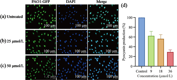
Having established the binding affinity of GM10 for LecB, we further evaluated its ability to inhibit bacterial adhesion, a key step in infection and biofilm formation mediated by this lectin [7,23]. In a pathogen-host infection model using green fluorescent protein (GFP)-expressing P. aeruginosa PAO1 and A549 lung epithelial cells, CLSM analysis showed that the association of bacterial cells with host cell membranes was significantly and dose-dependently attenuated by GM10 treatment (Figs. 5a-c), supporting its anti-adhesive activity. We also evaluated its ability to disperse established P. aeruginosa biofilms. Treatment with 25 µmol/L GM10 for 24 h produced a mild 12% dispersion of the biofilm biomass (Fig. S71 in Supporting information), indicating that GM10 can also affect mature biofilms.
Figure 5
Figure 5.
Effects of GM10 on bacterial adhesion and pyocyanin production. (a-c) CLSM images of A549 cells infected with P. aeruginosa PAO1-GFP (green, MOI = 10) and treated with GM10 at 0 (a), 25 µmol/L (b) or 50 µmol/L (c). Cell nuclei were stained with DAPI (blue). Scale bar: 100 µm. (d) Dose-dependent inhibition of pyocyanin production by GM10 (up to 100 µmol/L) in P. aeruginosa PAO1. Data are presented as mean ± SD (n = 3).
Given that the potent anti-biofilm activity of GM10 arises from its integrated three-dimensional copillar[3 + 2]arene architecture rather than a simple additive effect of its functional units, we further investigated its effect on quorum sensing (QS). Remarkably, GM10 significantly decreased the production of pyocyanin, a QS-regulated virulence factor, in a concentration-dependent manner, with 38% inhibition observed at 9 µmol/L (Fig. 5d and Fig. S74 in Supporting information). Concentrations up to 100 µmol/L (∼7-fold the MBIC50) were selected to capture a full dose-response profile under non-bactericidal conditions. GM10 also dose-dependently suppressed the fluorescence outputs of the lasB-, pqsA-, and rhlA-GFP reporter strains, while the constitutive GFP control and the corresponding growth curves remained largely unchanged (Fig. S75 in Supporting information). Together with the cell-free pyocyanin assay, these results effectively rule out indirect interference from bacterial aggregation or growth alterations. Given the pronounced stereochemical and polarity differences between GM10 and the QS signal molecule 3-oxo-C12-HSL, direct competitive binding to the LasR receptor is highly improbable. We therefore speculated that the pre-organized cavity of the copillar[3 + 2]arene core may sequester the hydrophobic QS signal molecule 3-oxo-C12-HSL through host-guest complexation.
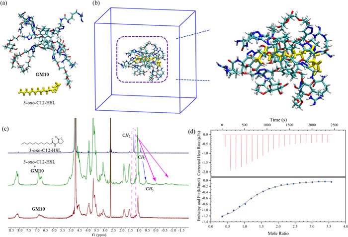
To verify the cavity-sequestration hypothesis, computational and spectroscopic approaches were employed. Molecular dynamics (MD) simulations suggested that 3-oxo-C12-HSL can be accommodated within the cavity of GM10, with its hydrophobic alkyl chain inserted into the aromatic cavity and the lactone headgroup exposed at the portal (Figs. 6a and b, Fig. S76 in Supporting information). Although the 100-ps trajectory was used only to assess the initial recognition pose and geometrical feasibility of this binding mode, density functional theory (DFT) calculations and subsequent experiments provided further support. Using a simplified core model, DFT calculations gave a substantial binding energy of −23.09 kcal/mol (Fig. S77 and Table S5 in Supporting information). In 1H NMR studies, the guest alkyl-chain protons showed pronounced upfield shifts (up to ∼1.5 ppm), and nuclear Overhauser effect spectroscopy (NOESY) correlations were observed between the guest alkyl protons and the aromatic protons of the pillar[5]arene core, indicating close spatial proximity in solution (typically within ∼5 Å) and supporting encapsulation within the aromatic cavity (Fig. 6c and Fig. S78 in Supporting information). ITC analysis supported a 1:1 host–guest binding model, with a Kd of 31 µmol/L (Fig. 6d and Table S6 in Supporting information). Taken together, these findings suggest that GM10 interferes with QS by sequestering 3-oxo-C12-HSL in a cell-entry-independent manner, rather than by directly targeting the intracellular receptor.
Figure 6
Figure 6.
Evidence for sequestration of 3-oxo-C12-HSL by GM10. (a) Initial structures of GM10 and 3-oxo-C12-HSL before MD simulation. (b) Representative GM10/3-oxo-C12-HSL complex from MD simulation. (c) 1H NMR spectra of free 3-oxo-C12-HSL, GM10, and their 1:1 mixture (1.5 mmol/L each) in D2O/DMSO–d6 (7:3, v/v). (d) Selected NOESY correlations of the same mixture, supporting guest encapsulation.
Recognizing the potent in vitro anti-biofilm activity of GM10 and its dual inhibitory mechanism targeting both lectin and QS, we evaluated its wound-healing efficacy in a murine skin infection model. All animal procedures were carried out in accordance with institutional guidelines and were approved by the Animal Care and Use Committee of Guizhou Medical University (approval No. 2500282). Before in vivo evaluation, GM10 showed favorable biocompatibility, with no obvious cytotoxicity or hemolysis under the tested conditions (Figs. S79 and S80 in Supporting information). Compared with the control groups, GM10 accelerated wound closure and improved histological features, including reduced inflammation and more organized tissue repair. No obvious pathological alterations were observed in major organs, supporting its favorable in vivo safety profile. Detailed biocompatibility data, wound photographs, histological analysis, and organ safety evaluation are provided in Figs. S81 and S82 (Supporting information).
In summary, we have established an efficient strategy to synthesize multifunctionalized copillar[3 + 2]arene 3 in 50% yield. Employing copillar[3 + 2]arene 3 as a programmable scaffold, the target GM10 was conveniently constructed via orthogonal click reactions. GM10 demonstrated potent, non-bactericidal anti-biofilm activity against P. aeruginosa PAO1 with an MBIC50 of 14.6 µmol/L. Mechanistic investigations revealed that GM10 concurrently targets two critical processes: Its peripheral L-fucosides, boosted by adjacent cationic arginine motifs, competitively inhibit LecB-mediated adhesion, while the copillar[3 + 2]arene cavity sequesters the key QS signal molecule 3-oxo-C12-HSL (Kd = 31 µmol/L). Notably, this extracellular sequestration broadly suppresses the las, rhl, and pqs QS systems and downregulates virulence factors such as pyocyanin without requiring cellular entry. To the best of our knowledge, this is the first documented dual inhibition of lectin and QS by a pillararene derivative, addressing a critical gap in anti-virulence strategies. In a murine wound infection model, topical GM10 treatment also promoted healing and reduced inflammation, with low systemic toxicity and good biocompatibility. Overall, this work provides an efficient route to functionalized copillar[3 + 2]arenes and demonstrates their utility as programmable scaffolds for novel anti-biofilm agents that combine lectin blockade with signal-molecule sequestration. This strategy presents a promising alternative for combating biofilm-associated infections caused by multidrug-resistant P. aeruginosa, with a low risk of inducing resistance. Future efforts will focus on optimizing linker length to enhance multivalent lectin binding and exploring the scaffold's potential for targeted co-delivery.
The authors declare that they have no known competing financial interests or personal relationships that could have appeared to influence the work reported in this paper.
Acknowledgments
We acknowledge the financial support provided by the Science and Technology Foundation of Guizhou Province (No. QKHJC[2023]YB306), the Science and Technology Major Special Foundation of Guizhou Province (No. QKHZDZXZ[2024]015), the High-level Innovative Talents Supporting Program (No. GCC[2023]002), the Guizhou Provincial Health Commission Foundation (No. gzwkj2023-079), and the Natural Science Research Project for Universities in Anhui Province (No. 2023AH051945). We also sincerely thank Professors Weimin Chen and Pinghua Sun from Jinan University for the generous provision of P. aeruginosa strains, including the wild-type PAO1 and the fluorescent reporter strains PAO1-lasB-gfp, PAO1-rhlA-gfp, and PAO1-pqsA-gfp.
Supplementary materials
Supplementary material associated with this article can be found, in the online version, at doi:10.1016/j.cclet.2026.112942.
[1]
M. Masi, M. Réfregiers, K.M. Pos, et al., Nat. Microbiol. 2 (2017) 17001. doi: 10.1038/nmicrobiol.2017.1
[2]
E. Rossi, R. La Rosa, J.A. Bartell, et al., Nat. Rev. Microbiol. 19 (2021) 331–342. doi: 10.1038/s41579-020-00477-5
[3]
D. Lebeaux, J.M. Ghigo, C. Beloin, Microbiol. Mol. Biol. Rev. 78 (2014) 510–543. doi: 10.1128/MMBR.00013-14
M. Yan, C. Liu, R. Tang, et al., Adv. Sci. 13 (2026) e12924. doi: 10.1002/advs.202512924
[38]
S. Jothi Nayaki, A. Roja, R. Ravindhiran, et al., ACS Infect. Dis. 10 (2024) 1080–1096. doi: 10.1021/acsinfecdis.3c00697
[39]
Y. Zhang, P. Zhang, Y. Lv, et al., ACS Appl. Bio Mater. 7 (2024) 7730–7739. doi: 10.1021/acsabm.4c01305
[40]
Y. Jin, Y. Liu, J. Zhu, et al., Org. Biomol. Chem. 22 (2024) 4202–4211. doi: 10.1039/d4ob00396a
[41]
D. Kaizerman-Kane, M. Hadar, R. Joseph, et al., ACS Infect. Dis. 7 (2021) 579–585. doi: 10.1021/acsinfecdis.0c00662
[42]
T. Mohy El Dine, R. Jimmidi, A. Diaconu, et al., J. Med. Chem. 64 (2021) 14728–14744. doi: 10.1021/acs.jmedchem.1c01241
[43]
W. Chen, T.Mohy El Dine, S.P. Vincent, Chem. Commun. 57 (2021) 492–495. doi: 10.1039/d0cc07684h
[44]
I.M. Andrei, W. Chen, M. Baaden, et al., J. Am. Chem. Soc. 145 (2023) 21904–21914. doi: 10.1021/jacs.3c06335
[45]
W. Mao, S. Zhan, B. Zhu, et al., J. Org. Chem. 83 (2018) 4147–4152. doi: 10.1021/acs.joc.8b00073
[46]
H. Yang, L. Jin, D. Zhao, et al., J. Agric. Food Chem. 69 (2021) 4276–4283. doi: 10.1021/acs.jafc.1c01032
[47]
A.M. Boukerb, A. Rousset, N. Galanos, et al., J. Med. Chem. 57 (2014) 10275–10289. doi: 10.1021/jm500038p
[48]
Z. Liu, F. Demontrond, A. Imberty, et al., Chin. Chem. Lett. 34 (2023) 107872. doi: 10.1016/j.cclet.2022.107872
[49]
K. Buffet, I. Nierengarten, N. Galanos, et al., Chem. Eur. J. 22 (2016) 2955–2963. doi: 10.1002/chem.201504921
Figure 1
Schematic illustration of the design and proposed dual-mode action of the programmable copillar[3 + 2]arene-based anti-biofilm agent GM10. Core functional modules and extracellular mechanisms are labeled with text annotations.
Figure 2
Structural characterization of copillar[3 + 2]arene 3 and its regioisomers. (a) Single-crystal X-ray structure of 3 showing the side view. (b) Top view. (c) Partial 1H NMR spectra: mixture 4 (ⅰ), isolated isomers 4a (ⅱ) and 4b (ⅲ), and major product 3 (ⅳ).
Figure 3
Synthesis of GM10 and related reference fragments. (a) Sequential orthogonal CuAAC construction of GM10, followed by deprotection. (b) Structures of the reference fragments 11 and 12.
Figure 4In vitro anti-biofilm activity of GM10. (a) Dose-dependent inhibition of P. aeruginosa PAO1 biofilm formation by GM10, fragments 11 and 12, their 1:1 mixture (11/12), and PP5. Data are presented as mean ± SD (n = 3). *P < 0.05, ****P < 0.0001. (b) Representative CLSM images of untreated and GM10-treated biofilms. Scale bar: 20 µm.
Figure 5
Effects of GM10 on bacterial adhesion and pyocyanin production. (a-c) CLSM images of A549 cells infected with P. aeruginosa PAO1-GFP (green, MOI = 10) and treated with GM10 at 0 (a), 25 µmol/L (b) or 50 µmol/L (c). Cell nuclei were stained with DAPI (blue). Scale bar: 100 µm. (d) Dose-dependent inhibition of pyocyanin production by GM10 (up to 100 µmol/L) in P. aeruginosa PAO1. Data are presented as mean ± SD (n = 3).
Figure 6
Evidence for sequestration of 3-oxo-C12-HSL by GM10. (a) Initial structures of GM10 and 3-oxo-C12-HSL before MD simulation. (b) Representative GM10/3-oxo-C12-HSL complex from MD simulation. (c) 1H NMR spectra of free 3-oxo-C12-HSL, GM10, and their 1:1 mixture (1.5 mmol/L each) in D2O/DMSO–d6 (7:3, v/v). (d) Selected NOESY correlations of the same mixture, supporting guest encapsulation.


 DownLoad:

 下载:


 下载:

