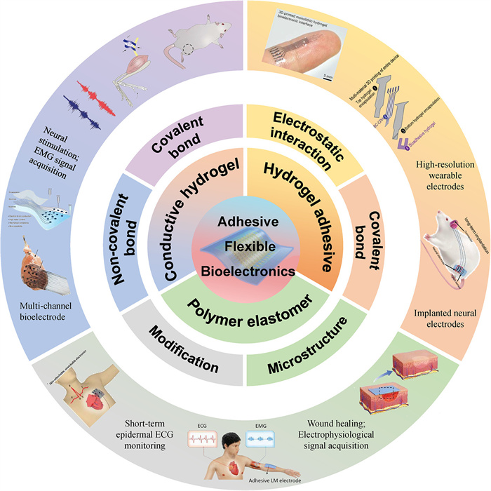
Figure 1.
Overview of adhesive flexible bioelectronics.

Adhesive flexible bioelectronics: Current status and future challenges
Jianhui Chen , Renping Ma , Dan Yang , Ruiwen Tian , Longlong Cui , Wang Guan , Yan Liu , Dianpeng Qi , Gongwei Tian , Chengyu Wang


In recent years, flexible bioelectronics have developed rapidly due to their excellent stretchability and electrical stability [1-4]. These excellent performances ensure that flexible bioelectronics maintain stable electrical performance during stretching and transmit electrophysiological signals accurately. Traditional flexible bioelectronics are mainly composed of hydrophobic polymer elastomer substrates (such as polydimethylsiloxane (PDMS), polyurethane (PU), Ecoflex elastomers) and rigid conductive materials (such as gold, conductive polymers, carbon-based materials) [4-7]. Flexible bioelectronics are fabricated through methods such as evaporation, 3D printing and doping on polymer elastomer substrates [3,4,8-10]. Although these flexible bioelectronics can maintain stable mechanical and electrical properties during tissue deformation, they cannot possess adhesion to tissues (especially on wet tissue surfaces), which makes them easy to drift or detach from the tissue surface when tissues deform, resulting in the inability of flexible bioelectronics to transmit signals.
With the rapid development of hydrogel adhesives, researchers have utilized hydrogel adhesives to enhance the adhesion between flexible bioelectronics and tissues to address the issue of flexible bioelectronics drifting or falling from the tissue surface during tissues deformation [11-15]. These hydrogel adhesives greatly enhance the adhesion between flexible bioelectronics and tissues through physical and covalent bonding with tissues [3,14,16-21], such as electrostatic and hydrogen bonding, and covalent bonding with Schiff base, amide and disulfide bonds. Simultaneously, hydrogel adhesives can enhance the conductivity by directly mixing rigid conductive nanomaterials or polymerizing conducting polymers, which is directly used as flexible bioelectronics to transmit physiological signals [16,22-25]. In addition, traditional polymer elastomer substrates (without adhesives) are fabricated into micro-nano structure and endowed with hydrophilic property [26-29], and hydrogel adhesives are directly used as the substrate of conductive materials (without considering the conductivity of hydrogels) [16,18,30], thereby endowing flexible bioelectronics with adhesion.
At present, adhesive flexible bioelectronics can be classified into three categories (Fig. 1). The first type is composed of the polymer elastomer substrate with adhesion and conductive materials. These polymer elastomer substrates obtain adhesion by designing the micro nano structure and endowing hydrophilic properties. The second type is that hydrogel adhesives are directly used as substrate materials for flexible bioelectronics. This type of hydrogel adhesive does not need to consider electrical conductivity because it only used to enhance the adhesion to tissues, and does not play a role in signal transmission. The third type is conductive hydrogel adhesive, which is directly used as flexible bioelectronics or the adhesive layer of flexible bioelectronics. The hydrogel adhesive here should not only consider the conductivity, but also consider the swelling characteristics, anti-adhesion, and self-healing properties, thereby endowing the conductive hydrogel adhesive with more stable adhesion, self-healing, and excellent biocompatibility performance. Therefore, we will discuss the polymer elastomer with adhesion as substrate of flexible bioelectronics, hydrogel adhesive as substrate of flexible bioelectronics and conductive hydrogel directly used as flexible bioelectronics, respectively.
There are two ways to endow polymer elastomer substrate with adhesion. The first method is constructing micro nano structures on polymer elastomer substrate through biomimetic strategies, which can generate adhesive forces on the tissue surface. The second method is synthesizing polymer elastomer substrates with hydrophilic property, which can generate adhesion to tissue by physical interactions on tissues, such as hydrogen bonding and electrostatic interactions. Next, polymer elastomer substrates with micro nano structures and the polymer elastomer substrates with hydrophilic property will be discussed respectively.
Traditional polymer elastomer substrates are difficult to directly adhere to dynamic tissue surfaces due to their hydrophobic property and high specific surface energy. Currently, polymer elastomer substrates have been fabricated with micro-nanostructures using biomimetic strategies to enhance adhesion between flexible bioelectronics and tissue. These strategies include gecko-inspired (Figs. 2a and b) [27,31,32], octopus-inspired (Figs. 2c and d) [28,33-35], and diving beetle-inspired (Figs. 2e and f) [36,37]. The adhesion mechanism of gecko-inspired strategy lies in the microscopic hairs on their feet (each foot is composed of approximately 500,000 bristles), which with a length of several tens of micrometers and a very thin diameter, about one tenth the size of a human hair. Each bristle contains hundreds of small shovel shaped branches that terminate at the submicron level. When a gecko attaches to a wall, tens of millions of scrapers extending from its bristles contact with the wall, and van der Waals forces act on the interface. Although van der Waals forces are relatively weaker than other bonding forces, the formation of van der Waals forces gradually increases as the number of molecules attracted to each other increases [38]. Similar to how geckos’ setae induce van der Waals force, tree frogs’ toe pads also have unique structure hexagonal columns. The intermolecular interactions between columns and skin, as well as the capillary forces caused by mucus, contribute to their significant wet adhesion [2]. To mimic the characteristics of geckos, the gecko inspired adhesive with micro pillars is usually manufactured on the surface of polymer elastomers using mold printing technology (Fig. 2a) [32], generating adhesion on skin surface (Fig. 2b).
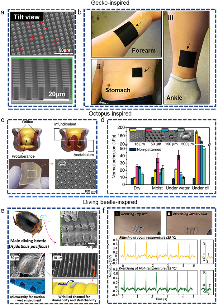
Another biomimetic bonding strategy is the octopus inspired strategy (Fig. 2c). This strategy first seals the entire chamber by contracting the radial muscles in the funnel, thinning the funnel wall, while expanding the flat surface radially. After the chamber is tightly sealed, the radial muscles in the acetabulum contract and the walls become thinner, thereby increasing the volume of the chamber. By relying on the high bulk modulus of water in the chamber to resist this expansion, the pressure inside the water decreases like a vacuum state. This reason forms a pressure difference between the inner suction cup and the outer surface, thereby stably adsorbing on the surface of the object and generating adhesive force [39-42]. Nano suction cup arrays were fabricated on the surface of polymer elastomers by the template method to mimic octopus inspired adhesive (Figs. 2c and d) [28,33,43]. Additionally, the adhesion mechanism generated by diving beetle-inspired is as follows [37]: (ⅰ) Enhancing adhesion force by preventing the generation of microstructural cracks, (ⅱ) enhancing capillary adhesion force by maximizing surface area and drainage, (ⅲ) suction stress caused by pressure difference between the internal and external states of the cavity due to volume changes. During the separation process along the tensile direction, due to stress concentration at the center of the micro column, the tip of the micro column exerts strong adhesive force by preventing crack propagation [44]. Compared to flat surfaces, microstructures exhibit enhanced surface area. The surface area of the adhesive is a key parameter for enhancing capillary interactions on wet surfaces and van der Waals forces on dry surfaces (Fig. 2e) [37,45]. Inspired by diving beetle adhesion system, a regular arrangement of mushroom-shaped pillars with microcavities and micro wrinkles of PDMS surface was fabricated by using techniques such as replica-molding and the selective-transfer method. Additionally, due to the capillary effect, micro washers with grooves can discharge excess residual water on rough surfaces, thereby forming capillary bridges of appropriate height between the adhesive and the bonding surface (Fig. 2f), successful recording EEG (Electroencephalogram) signals during relaxing and exercising [37,46]. In addition to constructing micro-nano structures on the surface of polymers, there are also methods to reduce the thickness of the polymer substrate, such as, polyimide (PI), polypropylene (PP), polyurethane (PU) and paper [47-50]. Enabling the substrate to fully adapt to rough skin surface and generate adhesion to tissues through mechanical interlocking and van der Waals forces.
Although the construction of micro-nano structure on the polymer elastomer substrate can enhance the adhesion strength between flexible bioelectronics and skin, sweating and large deformation of skin will seriously weaken the physical adhesion, and even the electrode will fall off. In addition, the micro-nano structures on the surface of the elastomer substrate will also greatly weaken the tensile ability of flexible bioelectronics, because the stress will be concentrated on the surface of these micro-nano structures, resulting in the weakening of the tensile ability of the electrode. Therefore, the adhesion stability of micro-nano structure to skin can be enhanced by increasing the perspiration ability of micro nano structure or adding waterproof layer in the future. At the same time, highly stretchable electrodes can be integrated onto micro-nanostructured substrates. In this way, the electrode can be stably fixed in the target area to monitor the electrophysiological signal.
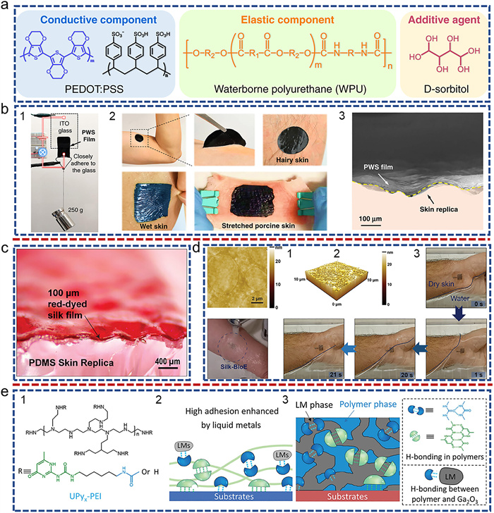
Subsequently, researchers have also developed various polymer substrates with hydrophilic properties to prepare adhesive flexible bioelectronics [47,51-54], such as waterborne polyurethane (WPU) (Fig. 3a) [51], silk protein (Figs. 3c and d) [52], and supramolecular polymer ureidopyramidinone (UPy)-modified cationic polymer (Fig. 3e) [47,53]. These water-soluble polymer substrates can interact with tissues, which exert adhesion to tissues through hydrogen bonding. For example, Jianyong Ouyang et al. prepared an adhesive, stretchable, highly conductive dry electrode (PWS) by using a blend of poly(3,4-ethylenedioxythiophene):poly(styrene sulfonate) (PEDOT:PSS), WPU, and D-sorbitol [51]. It exhibits high electrical conductivity and skin compliance, and good adhesion under dry and wet skin conditions (Fig. 3b). The adhesion of the electrode may come from WPU and D-sorbitol, since the PEDOT:PSS film cannot possess adhesion. The rich -OH, -NH, -C=O groups on the surface of WPU and D-sorbitol can strongly physical absorption the outermost cuticle, which is mainly composed of keratin and lipids [5,55,56]. In addition, the soft PWS electrode can adapt well to the gaps in the skin, which not only increases the contact area between PWS and the skin, but also increase adhesion between them [57]. However, weak conductivity of PEDOT:PSS during stretching cause unstable conductivity of PWS electrode. Subsequently, silk protein based adhesive bioelectrodes and UPy-modified polyethyleneimine (PEI) based bioelectrodes were prepared using inkjet printing Ag nanoparticles (Ag NPs) method and liquid metal (LM) doping method, respectively. Among them, the intrinsic tensile properties of LM enable the UPy-PEI based bioelectrodes to have superior tensile conductivity stability. In addition, UPy-PEI forms outstanding adhesion with tissues through hydrogen bonding (Fig. 3e). Meanwhile, the addition of liquid metal significantly improved the adhesion of UPy-PEI. This is due to the significant increase in cohesiveness caused by non-covalent crosslinking between LMs and polymer networks. In addition, conductive ink formed by blending MXene nanosheets, liquid metal, and sodium alginate aqueous solution is sprayed onto the skin surface to form electronic tattoos [50]. Sodium alginate forms micro gel shells around liquid metal droplets by chelating Ga3+ with its carboxyl group, thereby reducing oxygen permeability and enhancing chemical stability [58]. The conformal coverage of electronic tattoos on textured skin full of wrinkles and folds, maintaining strong skin adhesion and intact form, successful using for recording ECG signals, electromyography (EMG) signals and wound healing.
Whether traditional polymer substrates or hydrophilic polymer substrates, they are suitable for skin surfaces and do not cause foreign body sensation to human skin because flexible electronics based on these substrates do not undergo chemical reactions with the skin. Compared to traditional polymer elastomer substrates, these hydrophilic polymer substrates can form stronger and more stable hydrogen-bond-mediated adhesion to the skin, and have similar hydrophilicity to tissues, making them the best material for attaching bioelectronic devices to the body surface. However, these hydrophilic polymer substrates are difficult to be applied in the preparation of multi-channel microelectrode arrays because the substrates absorb water, resulting in signal crosstalk between channels. In addition, these substrates are difficult to be used in vivo because the hydration film contained on the moist tissue surface makes it difficult for these substrates to contact with the tissue surface. Even if adhesion is generated on the surface of the tissue, the tissue fluid continuously seeping of the wet dynamic tissue will continuously weaken physical adhesion of these bioelectronics [59,60]. Therefore, in the future, adhesion groups that can form covalent bonds with tissues can be modified on the surface of hydrophilic polymer substrates, which can enhance the adhesion stability between hydrophilic polymer substrates and tissues. At the same time, an insulating layer can be introduced on the surface of hydrophilic polymer elastomer and conductive material to prevent the problem of signal crosstalk between channels. In addition, whether the hydrophilic polymer elastomer will swell in vivo needs further exploration, because swelling will weaken the adhesion stability and reduce the biocompatibility of the bioelectrode.
Polymer elastomer substrates can play a critical role in various flexible bioelectronic applications through different adhesion strategies. By biomimetically structuring micro- and nanoscale features on the elastomer surface, mechanical interlocking with tissue can be achieved without chemical modification, enabling stable attachment to skin or soft tissues, which is particularly suitable for short-term epidermal monitoring and wearable sensors [61,62]. Pang et al. fabricated wrinkled suction cups on polymer elastomers to produce sweat-drainable, high-adhesion flexible patches, integrated with stretchable electrodes based on multiwalled carbon nanotube/PDMS nanocomposites [37]. This design results in flexible bioelectronic devices with high conductivity, mechanical compliance, and stable electrical performance under wet conditions, capable of long-term, real-time ECG and body temperature monitoring during dynamic physiological activity, highlighting the potential of adhesive polymer elastomers for multi-signal sensing. Alternatively, hydrophilic elastomers can achieve mild, reversible adhesion through physical-chemical interactions such as hydrogen bonding, electrostatics, or van der Waals forces. Yao et al. introduced UPy-modified polymers into liquid metal electrodes to enable tunable adhesion; high-adhesion modes provide reliable electrical interfaces with rigid components, whereas low-adhesion modes are suitable for skin interfaces and wound healing [53]. Meanwhile, noncovalent crosslinking between liquid metal particles and the polymer network enhances UPy-PEI cohesion, improving overall electrode adhesion and mechanical stability. By combining these adhesion strategies, researchers can flexibly tune adhesion strength, reversibility, and comfort of polymer elastomer substrates according to application requirements, thereby expanding their potential in skin-wearable devices, soft tissue interfaces, and dynamic organ monitoring in flexible bioelectronics.
Compared with the traditional hydrophobic and hydrophilic polymer substrates, hydrogel adhesives have better biocompatibility and can generate stronger adhesion to tissues. Directly using it as a substrate for conductive materials not only eliminates the need to consider conductivity, but also enables the construction of microelectrode arrays.
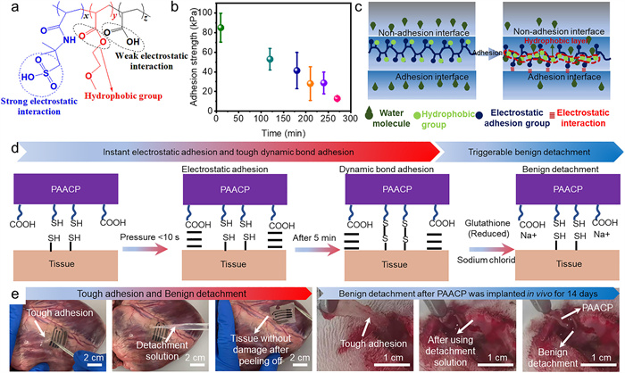
We prepared the hydrogel adhesive of electrostatic adhesion in the previous work (Fig. 4a) [30]. The strong electrostatic adhesion was mainly contributed to the sulfonic acid groups, which exhibit strong electron-withdrawing ability due to the conjugation effect and inductive effect. Tough and stable electrostatic adhesion also promotes the robust integration of the flexible bioelectronics on wet tissue surface, accurately recording the electrophysiological signals. Simultaneously, hydrophobic groups in 2-methoxyethyl acrylate and the addition of the hydrophilic gelatin greatly enhanced the electrostatic adhesion stability of the hydrogel (Figs. 4b and c) [30,63,64]. Because the hydrophobic group prevents the diffusion of water molecules at the nonadherent interface to the adherent interface, gelatin can continuously adsorb water molecules at the adherent interface, thereby greatly enhancing electrostatic adhesion stability. The electrostatic adhesion time was increased to 24 h. At the same time, dynamic bond adhesion hydrogel was fabricated by employing chitosan-grafted N-acetyl-L-cysteine (CS-NAC) with sulfhydryl groups to form dynamic disulfide (S-S) bonds with tissues (Fig. 4d) [18]. The obtained hydrogel can stably adhere to the surface of wet dynamic tissue for a long time through covalent bonds, and can achieve on-demand detachment through reduced glutathione at different time scales without any damage to the tissue (Fig. 4e) [18].
To achieve high stretchability of the electrode, Zhao et al. prepared a hydrogel adhesive hydrogel adhesive by grafting WPU to polyacrylic acid while introducing NHS ester groups. NHS ester group can generate tough adhesion by forming covalent cross-linking with tissue. Simultaneously, carboxyl groups of polyacrylic acid form hydrogen crosslinking with carboxyl groups of tissue. These excellent performances endow adhesive bioelectronics based on hydrogel substrate with tough adhesion and conductive stability during stretching [16].
Flexible bioelectronics based on adhesive hydrogels offer advantages such as softness, good biocompatibility, and stable tissue adhesion. To impart functionality, the hydrogel electrodes are commonly modified on the surface with conductive materials to enhance electrical performance [65], typically by coating or depositing conductive polymers. For example, Qi et al. used photolithography and electropolymerization of pyrrole on indium tin oxide (ITO) glass to fabricate bioelectrodes based on electrostatically adhered hydrogel adhesives [30]. These electrodes achieved tight contact with biological tissue and successfully recorded EMG signals from mice, ensuring stable electrode performance and effective signal transduction. However, the stretchability of such electrodes is limited by the pre-strain applied during fabrication. To solve this problem, Zhao et al. prepared PEDOT:PSS inks with high conductivity and high stretchability by using the phase separation principle [16]. By utilizing the moderate phase separation characteristics of hydrophilic polyurethane and PEDOT:PSS in a mixed solvent containing 70% (v/v) ethanol and 30% (v/v) water, the prepared ink exhibits unique mechanical and conductive ink grade phase separation without significant aggregation and precipitation. At the same time, during the solvent evaporation process, the stretched network phase and conductive network phase separated in the ink are compacted separately, forming a dual continuous phase, that achieved high conductivity and stretchability. The conductive polymer exhibited fully recoverable elastic deformation exceeding 200% strain, simultaneously, achieving high tensile strength (100% strain) while maintaining stable conductivity (~11 S/cm). The conductive polymer ink is printed on the surface of the hydrogel adhesive layer and encapsulated with a non-conductive hydrogel, thereby fabricating a fully hydrogel-based bioelectronic device with high conductivity. This device enables long-term and efficient electrical stimulation and signal recording of multiple tissues and organs in rat models, offering a versatile tool and platform for hydrogel bioelectronics research.
The Young’s modulus of the hydrogel substrate is significantly lower than that of the traditional polymer elastic substrate, resulting in superior compatibility to tissues. The ionic conductivity caused by hydrogel absorption water will lead to signal crosstalk between multi-channel electrodes. In our previous work, we have also introduced the contact part between multi-channel electrode and substrate into the insulation layer, which can avoid the problem of signal crosstalk between channels caused by hydrogel ion conduction, and greatly promote the rapid development of bioelectronic devices on hydrogel substrates. In addition, hydrogel as a substrate also needs to consider the influence of swelling, degradation of the substrate, contact stability of conductive material-biological tissue interface and other factors on the long-term stability of adhesive flexible bioelectronics. This will provide an important reference for the construction of highly stable adhesive flexible bioelectronics.
Conductive hydrogel adhesives, whether used as an adhesive layer of flexible electronics or directly as flexible bioelectronics, possess not only tough adhesion but high conductivity. Because their conductivity, including electronic conductivity and ionic conductivity, directly impact the quality of the transmitted physiological signal. Simultaneously, the swelling characteristics, anti-adhesion characteristics and self-healing properties of conductive hydrogel adhesive can influence adhesion stability and conductive stability of adhesive flexible bioelectronics. Therefore, the adhesion mechanism, conductivity, swelling characteristics (Section 4.4 in Supporting information), anti-adhesion (Section 4.5 in Supporting information) and self-healing (Section 4.6 in Supporting information) of conductive hydrogels adhesive are reviewed.
The adhesion strategies of currently developed conductive hydrogels are mainly divided into non-covalent adhesion strategy and covalent adhesion strategy. The non-covalent adhesion strategy is that the hydrogel forms adhesion on the wet dynamic tissue surface through physical interaction without any chemical reaction. The covalent adhesion strategy involves many active groups of the hydrogel, such as carboxyl groups, amino groups, sulfhydryl groups and hydroxyl groups, forming chemical bonds with the groups on the tissue surface to generate tough and stable adhesion. Following sections elaborate on these two adhesion strategies in detail.
Conductive hydrogel adhesives can generate adhesion by non-covalent bond adhesion strategy, without any chemical crosslinking with tissues, such as physical interactions. The biggest advantage of physical interactions is that they can enable hydrogel to quickly adhere to the tissue surface and form adhesion through electrostatic and hydrogen bonding. However, the adhesion formed by physical interactions is weak (<50 kPa) and exhibits poor stability. Because the hydration layer weakens the hydrogen bonds or electrostatic interactions at the adhesive interface [66,67]. Currently, biomimetic strategies are commonly used to increase electrostatic adhesion stability [66,67]. The prerequisite for using these strategies is to ensure that the hydrogel has high tensile strength (Fig. 5a), large fracture stress (σc) and fracture strain (εc), and the interface bonding strength (σa) is greater than the yield stress (σy), thereby ensuring that the hydrogel can withstand large deformation in the debonding process, thus dissipating a lot of energy. At the same time, its electrostatic adhesive group must contact the surface of the object, which means the hydrogel is able to break the hydration layer on the surface of the object and fully contact the object surface to form a strong physical interaction (high σa). Notably, many solid surfaces, including rocks, glass, and metals, carry negative charges in aquatic environments, which can be leveraged for electrostatic adhesion [68,69].
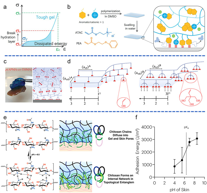
Inspired by barnacle (Fig. 5b), a strategy was adopted to fabricate hydrogels with strong electrostatic adhesion by radical copolymerization of cationic 2-(acryloyloxy)ethyl trimethylammonium chloride (ATAC) and aromatic 2-phenoxyethyl acrylate (PEA) monomers in dimethyl sulfoxide (DMSO) solution. Strong π-π and cation-π interactions act as dynamic crosslinking to enhance the toughness and cohesive strength of the hydrogel (high σc). Aromatic groups can disrupt the hydration layer on the surface of objects, providing regions with low dielectric constants to enhance the electrostatic interactions of cationic groups. The prepared hydrogel achieves strong electrostatic adhesion (high debonding stress σa), achieving adhesion strength of 180 kPa to inorganic materials, which can last for 100 days underwater. Although these strategies may provide new design concepts for underwater adhesion of hydrogel adhesives [69]. The hydrogel adhesive prepared by this design principle is difficult to adhere to the wet tissue surface. Unlike inorganic materials, the water molecules contained in the tissue will continuously diffuse from various directions towards the adhesive interface, thereby seriously weakening the electrostatic adhesion. To solve this problem, a hydrogel was prepared by copolymerizing N-[tris(hydroxymethyl)methyl]acrylamide (THMA) with N-(3-aminopropyl)methacrylamide hydrochloride (APMA) [70]. The prepared p(APMA-co-THMA) hydrogel possesses a high density of hydroxyl groups, which can strongly adhere to the tissue surface through hydrogen bonding without any chemical reaction (Figs. 5c and d). Although physical bonding strategies can quickly adhere to wet dynamic tissue surfaces, their stability is highly susceptible to the influence of surrounding tissue fluids, making the hydrogel difficult to ensure long-term stable adhesion of flexible bioelectronics to wet dynamic tissue surfaces. In addition, the adhesion is limited by the number of groups at the tissue interface because hydrogel generate adhesion by chemical reaction with groups of tissue interface [70,71]. Subsequently, another non-covalent adhesion strategy-topological adhesion is developed. Chitosan is used as layers of topological adhesion. The reason is that chitosan dissolves as a polyelectrolyte in the solution when pH < 6.5, and form hydrogen bonds when pH > 7 (Fig. 5e). When the pH of the adhesive layer with chitosan is less than 6.5, the chitosan dissolved in the solution will follow the solvent to diffuse to the hydrogel substrate and tissue. With the increase of diffusion depth, the pH of chitosan will also increase. The hydroxyl and amino groups between the chitosan molecular chains will form hydrogen bonds with the tissue and hydrogel respectively, thereby achieving topological adhesion (Fig. 5f). However, the adhesion formed by topological adhesion is heavily dependent on the pH change of tissue and hydrogel substrate, which makes it difficult to ensure the stability of the adhesion [72,73].
In addition, the discussion and analysis of non-covalent bond adhesion mechanisms such as hydrogen bonding, hydrophobic interactions, π-π stacking, cation-π interactions, and host-guest complexation. Although physical interaction can quickly generate tough adhesion, its stability is still affected by tissue fluid, resulting in poor adhesion stability. Chitosan can not only form tough and stable physical topological adhesion within tissues, but also enhance adhesion and fatigue resistance. However, the hydrogen bonds formed between chitosan molecular chains are highly susceptible to changes in pH around the tissue. Therefore, enhancing the stability of physical adhesion and topological adhesion is the key to improve the application of physical adhesion hydrogel adhesives in the field of flexible bioelectronics. Additionally, adhesion failure mechanisms in wet environments, the influence of dynamic bond energies, and bond-exchange kinetics on long-term interfacial stability, are discussed in detail in Section 4.1.1 (Supporting information).
The covalent adhesion strategy requires chemical reactions between hydrogel groups (aldehyde, catechol/quinone, thiol, amino, carboxyl) and tissue surface groups (amino, carboxyl, thiol) to form tough and stable adhesion strength [18,70,74-78]. Aldehyde groups react with amino groups in tissues via Schiff base formation. For example, oxidized dextran (Dex CHO) containing aldehyde group is introduced into the hydrogel, and the aldehyde group of Dex CHO forms a covalent bond with the amino group of the tissue to generate adhesion [70]. However, the covalent crosslinking formed by aldehyde and amino groups is unstable and easily affected by changes in the pH value around the tissue, compromising long-term stability for flexible bioelectronics on dynamic wet tissue. Active catechol/quinone can generate hydrogen bonding with tissues, metal coordination, π-π interactions, and other interactions and form adhesion [79-84]. For example, dopamine containing catechol is usually introduced on the hydrogel to enhance the adhesion strength of the hydrogel. Most dopamine hydrogels show weak adhesion, short-term adhesion and limited reusability due to the poor stability of catechol/quinone [75]. Amino and carboxyl groups will form more stable amide bonds with the carboxyl and amino groups of the tissue, but it is necessary to use (1-(3-dimethylaminopropyl)-3-ethylcarbodiimide and N-hydroxysuccinimide (EDC/NHS) as an adhesive layer to activate carboxyl groups [51,52], thereby forming stable amide bonds. For example, Suo et al. utilized the EDC/NHS to activate the carboxyl groups on the tissue surface (Fig. 6a) [74]. The activating carboxyl groups generate dehydration condensation reaction with the amino groups of chitosan, forming stable amide bonds and increasing adhesion (Fig. 6b). In addition, NHS ester groups on acrylic acid N-hydroxysuccinimide ester can directly react with the amino groups of tissues, thereby forming amide bonds. For example, Zhao et al. used acrylic acid, acrylic acid N-hydroxysuccinimide ester and gelatin crosslinked copolymerization to synthesize hydrogel adhesive (DST) [85], NHS ester groups of obtained hydrogel adhesive directly form amide bonds with the amino groups of tissues, avoiding the activation of EDC/NHS. However, NHS ester groups are unstable and easily hydrolyzed to form carboxyl groups and reduce adhesion. At the same time, the formation of amide bonds still takes about 5 min (Fig. 6c) [17,85]. The carboxyl group on the hydrogel can enhance the initial adhesion through electrostatic and hydrogen bonding, so that it can quickly generate physical adhesion on the tissue surface within a few seconds. Therefore, the combination of non-covalent and covalent adhesion can not only improve the adhesion speed of conductive hydrogel adhesive, but also enhance the adhesion stability. Finally, DST-adhered strain sensor was adhered to porcine heart, and recorded obvious resistance changes (Figs. 6d and e). Simultaneously, non-covalent bond adhesion and covalent bond mechanisms have been discussed in detail in 4.1.1 Non-covalent bond adhesion strategy, 4.1.2 Covalent bond adhesion strategy (Supporting information).
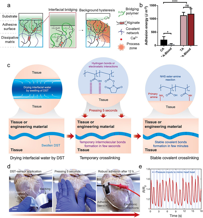
In summary, non-covalent bond adhesion can quickly adhere to wet dynamic tissue surfaces without any chemical reaction to the tissue, but the adhesion stability is poor. The covalent bond adhesion strategy is to generate covalent adhesion through the chemical reaction between the hydrogel and the groups on the tissue surface. Covalent adhesion has better stability than non-covalent bond adhesion. However, the time required to form stable adhesion is relatively long (>5 min). In addition, whether it is non-covalent bond adhesion or covalent adhesion, their adhesion is limited by the number of groups at the tissue interface. This reason leads to weak adhesion and fatigue resistance. Covalent topology adhesion strategy can effectively solve the above problems. Because this topological adhesion relies on bridging polymers diffusing into the tissue and forming covalent topological networks within the tissue, resulting in a tough and fatigue resistant adhesion [51,52]. However, the weak diffusion force of the bridging polymer also limits the speed of its diffusion into the tissue and the hydrogel substrate, resulting in the slow formation of adhesion. Therefore, increasing the diffusion force of the bridging polymer to the hydrogel and the tissue at the same time is an important condition for achieving tough and fatigue resistant adhesion.
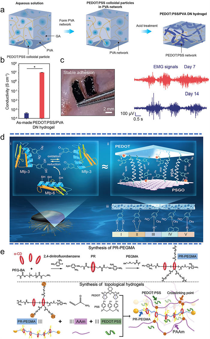
The conductivity of conductive hydrogel adhesive will directly affect the signal quality transmitted between flexible bioelectronics and tissues. Therefore, adjusting the conductivity of these hydrogels is crucial to improve signal quality. At present, current strategies are mainly by doping conductive polymers [16,22,86-88], conductive carbon materials [14,89,90], and conductive polymers/carbon materials [91,92] into hydrogels. Generally, the conductivity of hydrogels is directly proportional to the concentration of rigid conductive materials. For example, the double network conductive hydrogel constructed by using PEDOT:PSS and polyvinyl alcohol (PVA) can possess a high mass ratio of PEDOT:PSS/PVA in the precursor hydrogel by minimizing the content of PVA, achieving high conductivity (~10 S/cm) [22]. By soaking PEDOT:PSS/PVA hydrogel in a large amount of pure acetic acid (HOAc), then HOAc replaces water in the hydrogel (Fig. 7a). The poor solubility of HOAc for PVA and PEDOT: PSS leads to phase separation and aggregation of PVA and PEDOT:PSS. The PVA network constrains the aggregation of PEDOT: PPS into a continuous network. The hydrogel reaches a conductivity of 10 S/cm and exhibits high stretchability (150%) (Fig. 7b). In addition, the adhesive hydrogel with NHS ester groups is further added to the stretchable conductive hydrogel, which can be covalently bonded to the tissue surface and stably integrated with the tissue (high interface toughness above 200 J/m2). These methods greatly increase the conductivity and adhesion of the conductive hydrogel. Obtained conductive hydrogel adhesive was successfully implanted into the subcutaneous muscle of rats, and stable EMG signals were collected (Fig. 7c) [22].
In addition to doping conductive polymers in the conductive hydrogel, rigid carbon materials, such as graphene oxide, graphene sheets, carbon nanotubes, MXene, can be incorporated into hydrogel to achieve high conductivity [14,90,91]. Although rigid carbon materials have good conductivity, if they are directly doped into the hydrogel, the conductivity and stretchability of the hydrogel are very weak due to the discontinuity of these nanomaterials. Therefore, suitable physical crosslinking agents, such as ionic liquids containing imidazole monomers and hyaluronic acid with polyphenol tannin groups, are generally selected to connect and rearrange these carbon nanomaterials through physical interaction with the surface of carbon nanomaterials. This strategy endows hydrogel with good conductivity. For example, the strong cation-π interaction between imidazole cations and the surface of graphene sheets, MXene and single-walled carbon nanotubes (SWCNTs) can be utilized [14,90,91]. After mixing imidazole cation with graphene sheet or single wall carbon nanotubes, a 3D gel network will be formed to obtain physically crosslinked graphene-based hydrogels. In addition, the flat structure of imidazole ring makes most atoms exposed, and can more firmly form cation-π interactions with π-electron molecules of graphene flakes or single-walled carbon nanotubes in aqueous solution.
Conductivity of conductive hydrogels can also be improved by doping the mixture of conductive polymers and carbon nanomaterials [91]. For example, functionalized graphene oxide (GO) templates are used to prepare high conductivity nanosheets (Fig. 7d). The GO surface was introduced sulfonic acid groups to obtain sulfonated graphene oxide (SGO). These sulfonic acid groups serve as doping sites for PEDOT. Then, catechol groups were introduced onto SGO and functionalized with polydopamine (PDA) to form PSGO. This enhances the interaction between PEDOT and PSGO. Simultaneously, PDA increases the conductivity of PSGO. Therefore, PEDOT can be assembled onto PSGO templates to form conductive sandwich shaped nanosheets. Importantly, PSGO templated PEDOT (PSGO-PEDOT) nanosheets possess abundant hydrophilic and reducing catechol groups, thus exhibiting good water dispersibility and redox activity. Finally, PSGO-PEDOT nanosheets were integrated into the polyacrylamide (PAM) hydrogel network to obtain a highly stretchable and highly conductivity hydrogel. Compared to previously reported nanofillers, PSGO-PEDOT nanosheets have several advantages. (1) The nanosheets endow the hydrogel with high conductivity. (2) Due to the hydrophilic groups of GO and PDA, the nanosheets have good water dispersibility to prevent aggregation in the hydrogel. Therefore, the PEDOT nanosheets can form physical cross-linking with PSGO and be evenly dispersed in the chemically cross-linked polyacrylamide (PAM) hydrogel network. The synergistic effect of physical and chemical crosslinking makes the hydrogel have toughness and extensibility. (3) The nanosheets possess redox activity, retaining enough catechol groups and endowing the hydrogel with excellent adhesion. These nanosheets can be used as multifunctional nano fillers to build conductive, super stretched, adhesive hydrogels for wearable and implantable bioelectronics. However, when the hydrogel based on the conducting carbon nanomaterials is stretched, the connection between the carbon nanomaterials fracture more readily than polymeric networks. Because these physical crosslinkers are not stretchable, which destroys the electronic transmission path. The conductivity and stretchable stability of these conductive hydrogels are still lower than conductive hydrogels based on the conductive polymer.
Although these strategies can enhance tensile performance and conductivity of hydrogel, the aggregation of conductive materials will greatly reduce the light transmittance of conductive hydrogels, thus affecting the efficiency of hydrogels to regulate nerves through light control. How to improve the transparency of conductive hydrogels without serious degradation of conductivity is still an important challenge. If the aggregation of conductive polymer is reduced, the transmittance of the hydrogel will be improved, but this will affect the conductivity of the conductive hydrogel. Zhang et al. [87,93] introduced a mechanically interlocked polyrotaxane (PR-PEGMA) into the topological hydrogel as the crosslinking agent of the hydrogel. PR-PEGMA contains a polyethylene glycol (PEG) skeleton and 15 cyclodextrins (CDs) with PEGMA side chains. PEDOT:PSS was used as conductive filler in the hydrogel to obtain good conductivity. Meanwhile, PEDOT:PSS is arranged along PEG chains on CDs to enhance conductivity (Fig. 7e). In addition, the sliding of CDs on the PEG main chain reduces the aggregation of the PEDOT chain, thus reducing the interaction between PEDOT and light waves, improving the transparency of the hydrogel. The conductivity of the obtained hydrogel reaches 33 S/m. And LFP signals were successful evoked by optically controlled. Meanwhile, utilizing this principle, the rearrangement of PEDOT:PSS induced by amylopectin is employed to achieve high transparency [88]. Although the induced rearrangement of conductive polymers can improve the transmittance of conductive hydrogels, their conductivity is much lower than that generated by the induced aggregation of conductive polymers. Therefore, achieving both conductivity and transparency remains a huge challenge.
Additionally, the hydrogel possesses ionic conductive characteristics by doping ions inside the hydrogel [91,94-96]. Compared with hydrogels doped with conductive fillers, the biggest advantage of these ionic conductive hydrogels is that they have great tensile conductivity stability, because the ions in hydrogel depend on the migration of water solvent. Therefore, even under large strain, ionic hydrogels can still maintain stable conductivity. However, ratio of mass of water solvent in the hydrogel will affect its electrical stability. Subsequently, some tensile conductors without water molecules were gradually developed, such as organic gel and ionic elastomer [97-103]. Although these flexible bioelectronics can solve some problems faced by hydrogels, such as dehydration. However, they in vivo will lead to ion leakage, which will seriously affect the biocompatibility of flexible bioelectronics. Therefore, these organic gel and ionic elastomers are only applicable to the body surface. Meanwhile, they generate weak adhesion on tissues through physical interactions. When the skin sweats, the flexible bioelectronics easily drift or detach from the skin surface.
In adhesive flexible electronic devices, conductive hydrogels have evolved from interfacial buffering or encapsulation materials into core electrode components that directly enable signal acquisition and transmission [104-107]. Their tissue-like mechanical compliance and intrinsic adhesive properties allow electrodes to conformally contact and stably adhere to skin and soft tissues, thereby supporting complex electrode architectures and high-quality signal recording [108-112].
From a fabrication perspective, mold casting enables pattern replication from the millimeter to sub-hundred-micrometer scale and is suitable for the rapid construction of multichannel but low-density arrays. As demands for higher resolution and greater channel programmability increase, 3D printing has been introduced into the fabrication of hydrogel-based electronic devices [113]. For example, Liu et al. employed direct ink writing to print PEDOT:PSS-based hydrogels into a 16-channel electrode array and achieved multilayer integration of the conductive layer, adhesive substrate, and encapsulation layer [114]. This enabled the construction of hydrogel bioelectronic devices that can be conformally attached to the dynamic surface of the heart, maintaining stable adhesion and reliable epicardial electrophysiological signal localization under mechanical deformation.
To further meet the demands for higher spatial resolution and higher channel density, photolithography has been introduced into the fabrication of conductive hydrogel electrodes, enabling feature sizes down to the micrometer scale. Lu et al. developed a photosensitive biphasic conductive hydrogel (PB-CH) composed of PEDOT:PSS as the conductive phase and a photosensitive matrix as the mechanical phase [115]. This biphasic structure preserves the continuity of the conductive network during photo-crosslinking, allowing high-resolution patterning (~5 μm) while maintaining mechanical compliance and processing stability. The resulting devices exhibit favorable performance, including a conductivity of ~30 S/cm and a stretchability of ~50%, providing a representative example of material-process co-design for high-resolution manufacturing of high-performance conductive hydrogels.
The construction of micro/nano structures on the surface of polymer elastomers can confer flexible bioelectronics with adhesion capabilities. Notably, this adhesion strategy avoids chemical reactions with tissues and leaves no residual chemical reagents upon device removal, ensuring excellent biocompatibility. Nonetheless, the hydrophobic characteristic of polymer elastomers always makes it difficult to stably adhere to wet dynamic tissue surfaces for long periods. The continuous infiltration of tissue fluid from the wet tissue surface and the surrounding blood will severely weaken the van der Waals, capillary and electrostatic forces generated by the micro/nano structures, thereby reducing the adhesion stability of flexible bioelectronics. Alternatively, the developed water-soluble polymer substrates can adhere to the tissue surface through hydrogen bonding, electrostatic, and mechanical interlocking, and do not require complicated means to fabricate micro/nano structure on these polymer substrates. However, these physical interactions are still weakened by tissue fluids. Thus, the adhesive stability of flexible bioelectronics is affected. Consequently, improving the physical adhesion stability of micro-nano structured elastomers and water-soluble elastomers remains a critical challenge in advancing flexible bioelectronics.
Hydrogel adhesives as substrates for flexible bioelectronics exhibit great advantages in the field of adhesive flexible electronics. This method avoids doping conductive materials inside the hydrogel adhesive, because the hydrogel adhesive only acts as a substrate for the conductive material. So, this strategy does not affect the mechanical properties and adhesion properties of the hydrogel adhesive. The conductivity of the obtained bioelectronics is much better than that of the conductive hydrogels because the conductive materials are directly used as electrodes without the need to be doped into any polymer substrate. But there are still some problems to be solved. The hydrogel substrate has ionic conductivity after water absorption, which will lead to signal crosstalk between different channels, so it is necessary to introduce an insulating layer at the interface between the conductive material and the hydrogel adhesive. Then it is necessary to increase the interfacial adhesion of the conductive hydrogel-insulating layer and the insulating layer-conductive material, preventing the electrode material disengaging from the hydrogel adhesive substrate surface. The swelling of the hydrogel substrate will also weaken electrical stability, because swelling will weaken the adhesion stability of the hydrogel adhesive to the insulating and conductive layers. Therefore, it is crucial to limit the swelling of the hydrogel adhesive substrate.
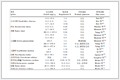
Conductive hydrogel adhesives as flexible bioelectronics possess both the adhesion to tissues and the electrical conductivity. Since the conductivity of hydrogels directly influences the efficiency of flexible bioelectronics in transmitting signal. Several methods have been developed to enhance the conductivity of conductive hydrogel adhesives, such as phase separation, cross-linkers enhancing bonding between carbon nanomaterial, and conductive polymers binding carbon materials. But the electrical conductivity of these conductive hydrogel adhesives is still much lower than that of the conductive material. Although these methods improve high continuity of conductive materials and increase conductivity, the non-stretchable or weak stretchable ability of conductive materials will still limit the electrical stability of hydrogel adhesives during stretching. In addition, the conductive hydrogel adhesive cannot achieve high conductivity, anti-adhesion, high adhesion, non-swelling and self-healing properties at the same time. This limits the development of conductive hydrogel adhesives in the field of flexible bioelectronics. The performance comparison of flexible electronics based on different adhesion strategies is shown in Table 1 [16,18,30,32,37,51,53,116,117].
 DownLoad:
CSV
| Materials | Adhesive strength | Electrical conductivity | Stretchability (%) | Biocompatibility | Preparation strategy | Application | Ref. |
| Polymer elastomer | ~1.3 N/cm2 | ~0.01 S/cm | 30 | - | Template method | ECG | [32] |
| 2.2 N/cm2 | ~0.87 S/cm | 30 | - | Replica-molding and selective-transfer | ECG; body-temperature | [37] | |
| 0.43 N/cm | 72–545 S/cm | 30 | - | Template method | ECG; EMG; EEG | [51] | |
| 1.5–8.9 MPa | ~4000 S/cm | 100 | Noncytotoxic | Stencil-printing | ECG; EMG; wounds treated | [53] | |
| Hydrogel adhesive | 85 kPa | - | 100 | Noncytotoxic | Electropolymerization and prestretched | ECG; EMG | [30] |
| 211.4 kPa | - | 30 | Noncytotoxic | Electropolymerization and prestretched | ECG; EMG; strain sensors | [18] | |
| 290 kPa | - | 100 | Noncytotoxic | Electropolymerization and prestretched | ECG; EMG | [116] | |
| 3300 J/m2 | 11 S/cm | 400 | Low cytotoxicity | 3D printing | ECG; neural stimulation | [16] | |
| Conductive hydrogel | 200 J/m2 | ~10 S/cm | 100 | High biocompatibility | In-situ polymerization | EMG; neural stimulation | [22] |
| - | 0.27 S/cm | - | Excellent biocompatibility | Template method | Brain neural signal recording; optogenetic modulation | [117] |
In fact, different types of adhesive flexible bioelectronics have distinct advantages and disadvantages, making it difficult to achieve perfection. Therefore, it is necessary to select suitable adhesive flexible bioelectronics according to different application fields and regulate their shortcomings to meet our needs. (1) For short-term epidermal monitoring, the interfaces are typically weakly moist or nearly dry, and the primary requirements electrodes are comfort, breathability, reversible adhesion and minimal impact on the skin barrier. Biomimetic microstructures and weak physicochemical interactions (e.g., van der Waals forces and hydrogen bonding) are thus most suitable, providing sufficient adhesion while allowing gentle, non-damaging removal. (2) Long-term implantable devices require stable interfaces adhesion force in fluid-rich environments over extended periods. Chemical bonding and topological networks are preferable because they enhance the stability of interface adhesion. (3) Dynamic organ interfaces experience high hydration and continuous cyclic deformation, demanding both strong adhesion and high compliance. Single adhesion mechanisms are generally insufficient to meet these dual requirements, making multi-mechanism coupling-such as combining microstructural adhesion with physicochemical interactions or topological interlocking-more effective. Such synergistic designs enable stable adhesion while maintaining mechanical compatibility under dynamic conditions (Table 2) [16,18,28,30,32,37,51,52,72,74,85,91,117]. Therefore, a comprehensive and in-depth understanding of the preparation strategies of flexible bioelectronics, as well as their respective advantages and disadvantages, is more conducive to finding suitable methods and strategies to compensate for these shortcomings, thereby improving the stability of adhesive flexible bioelectronics.
DownLoad:
CSV
| Materials | Mechanical | Electrical | Adhesion | Advantages | Limitations | Application scenario | Ref. |
| Polymer elastomers | Low modulus, high extensibility | Non-conductive | Non-covalent adhesion | Mechanical match to tissue, allowing gentle, non-damaging removal | Low conductivity, limited adhesion | Short-term epidermal monitoring, | [28,32,37,51,52] |
| Hydrogel adhesive | Modulus adjustable, stretchable | Low conductive | Non-covalent and covalent adhesion | Good biocompatibility, strong adhesion, high compliance | Poor conductivity, prone to drying or degradation, swelling easily in humid environment. | Long-term implantable | [16,18,30] |
| Conductive hydrogel | Modulus adjustable, stretchable | Electronically or ionically conductive | Non-covalent and covalent adhesion | Supporting high-resolution signal acquisition, stable interface adhesion | Limited mechanical strength, complex fabrication, limited long-term stability | Long-term implantable, dynamic humid environment | [72,74,85,91,117] |
Jianhui Chen: Writing – review & editing, Writing – original draft, Conceptualization. Renping Ma: Writing – review & editing. Dan Yang: Writing – original draft. Ruiwen Tian: Writing – review & editing. Longlong Cui: Writing – original draft. Wang Guan: Writing – original draft. Yan Liu: Project administration, Funding acquisition. Dianpeng Qi: Supervision, Project administration, Funding acquisition. Gongwei Tian: Writing – review & editing, Supervision, Funding acquisition, Conceptualization. Chengyu Wang: Supervision, Project administration, Funding acquisition, Conceptualization.
The authors declare that they have no known competing financial interests or personal relationships that could have appeared to influence the work reported in this paper.
The authors acknowledge the funding support from the National Natural Science Foundation of China (Nos. 52473255, 52173237). The Fundamental Research Funds for the Central Universities (Nos. HIT.OCEF.2022018, HIT.NSRIF 202315). Natural Science Foundation of Heilongjiang Province, China (Nos. LH2022E051, LH2021B009). Research Start-up Funding Project of Zhengzhou Research Institute of Harbin Institute of Technology (No. CUGD0200501623). Key Laboratory of Scientific Technology and Intelligent Equipment for Prevention and Control of Major Chronic Diseases, Ministry of Industry and Information Technology (No. MCD-2025-2-12). Financial assistance under Heilongjiang Postdoctoral Fund (No. LBH-Z25053). Heilongjiang Postdoctoral Research Start-up Fund (No. 415995). Youth Foundation of Natural Science of Henan Province (No. 252300423366).
Supplementary material associated with this article can be found, in the online version, at doi:
Q. Tian, H. Zhao, X. Wang, et al., Adv. Mater. 35 (2023) 2211236. doi: 10.1002/adma.202211236
J. Park, H.W. Kim, S. Lim, et al., Adv. Funct. Mater. 34 (2024) 2313728. doi: 10.1002/adfm.202313728
Q. He, Y. Cheng, Y. Deng, et al., Adv. Funct. Mater. 34 (2024) 2308974. doi: 10.1002/adfm.202308974
D. Qi, K. Zhang, G. Tian, B. Jiang, Y. Huang, Adv. Mater. 33 (2021) 2003155. doi: 10.1002/adma.202003155
F.E. Golling, R. Pires, A. Hecking, et al., Polym. Int. 68 (2019) 848–855. doi: 10.1002/pi.5665
S. Biswas, S.W. Lee, Y. Lee, et al., Small Sci. 4 (2024) 2300148. doi: 10.1002/smsc.202300148
G. Balakrishnan, J. Song, C. Mou, C.J. Bettinger, Adv. Mater. 34 (2022) 2106787. doi: 10.1002/adma.202106787
K.A. Deo, M.K. Jaiswal, S. Abasi, et al., ACS Nano 16 (2022) 8798–8811. doi: 10.1021/acsnano.1c09386
E. Song, J. Li, S.M. Won, W. Bai, J.A. Rogers, Nat. Mater. 19 (2020) 590–603. doi: 10.1038/s41563-020-0679-7
E. Cuttaz, J. Goding, C. Vallejo-Giraldo, et al., Biomater. Sci. 7 (2019) 1372–1385. doi: 10.1039/c8bm01235k
Y. Yang, S. Zhong, X. Wang, Y. Gao, X. Cui, Chem. Eng. J. 474 (2023) 145891. doi: 10.1016/j.cej.2023.145891
Z. Jin, L. Yang, S. Shi, et al., Adv. Funct. Mater. 31 (2021) 2103391. doi: 10.1002/adfm.202103391
Q. Yang, T. Wei, R.T. Yin, et al., Nat. Mater. 20 (2021) 1559–1570. doi: 10.1038/s41563-021-01051-x
J. Deng, H. Yuk, J. Wu, et al., Nat. Mater. 20 (2021) 229–236. doi: 10.1038/s41563-020-00814-2
H. Choi, Y. Kim, S. Kim, et al., Nat. Electron. 6 (2023) 779–789. doi: 10.1038/s41928-023-01023-w
T. Zhou, H. Yuk, F. Hu, et al., Nat. Mater. 22 (2023) 895–902. doi: 10.1038/s41563-023-01569-2
X. Chen, H. Yuk, J. Wu, et al., Proc. Natl. Acad. Sci. U. S. A. 117 (2020) 15497–15503. doi: 10.1073/pnas.2006389117
G. Tian, D. Yang, C. Liang, et al., Adv. Mater. 35 (2023) 2212302. doi: 10.1002/adma.202212302
Y. Fu, Z. Wang, K. Wu, et al., Chin. Chem. Lett. 36 (2025) 110479. doi: 10.1016/j.cclet.2024.110479
X. Jing, F. Liu, Y. Li, et al., Nano Energy 142 (2025) 111182. doi: 10.1016/j.nanoen.2025.111182
X. Jing, L. Chen, Y. Li, et al., Small 20 (2024) 2405587. doi: 10.1002/smll.202405587
G. Li, K. Huang, J. Deng, et al., Adv. Mater. 34 (2022) 2200261. doi: 10.1002/adma.202200261
T. Dai, X. Qing, H. Zhou, et al., Synth. Met. 160 (2010) 791–796. doi: 10.1016/j.synthmet.2010.01.024
Y. Zhao, B. Zhang, B. Yao, et al., Matter 3 (2020) 1196–1210. doi: 10.1016/j.matt.2020.08.024
H. Wei, M. Lei, P. Zhang, et al., Nat. Commun. 12 (2021) 2082. doi: 10.1038/s41467-021-21869-y
D.M. Drotlef, M. Amjadi, M. Yunusa, et al., Adv. Mater. 29 (2017) 1701353. doi: 10.1002/adma.201701353
H. Park, C. Song, S.W. Jin, et al., Nano Energy 83 (2021) 105837. doi: 10.1016/j.nanoen.2021.105837
S. Baik, D.W. Kim, Y. Park, et al., Nature 546 (2017) 396–400. doi: 10.1038/nature22382
F. Liu, X. Wang, M. Wang, et al., Chin. Chem. Lett. 34 (2023) 108353. doi: 10.1016/j.cclet.2023.108353
G. Tian, Y. Liu, M. Yu, et al., ACS Appl. Mater. Interfaces 14 (2022) 4852–4861. doi: 10.1021/acsami.1c18983
K. Autumn, Y.A. Liang, S.T. Hsieh, et al., Nature 405 (2000) 681–685. doi: 10.1038/35015073
T. Kim, J. Park, J. Sohn, et al., ACS Nano 10 (2016) 4770–4778. doi: 10.1021/acsnano.6b01355
Y.C. Chen, H. Yang, ACS Nano 11 (2017) 5332–5338. doi: 10.1021/acsnano.7b00809
B. Mazzolai, A. Mondini, F. Tramacere, et al., Adv. Intell. Syst. 1 (2019) 1900041. doi: 10.1002/aisy.201900041
M.K. Choi, O.K. Park, C. Choi, et al., Adv. Healthc. Mater. 5 (2016) 80–87. doi: 10.1002/adhm.201500285
Y. Chen, M.C. Shih, M.H. Wu, et al., J. R. Soc. Interface 11 (2014) 20140273. doi: 10.1098/rsif.2014.0273
H. Min, S. Baik, J. Kim, et al., Adv. Funct. Mater. 32 (2022) 2107285. doi: 10.1002/adfm.202107285
A.P. Russell, A.Y. Stark, T.E. Higham, Integr. Comp. Biol. 59 (2019) 101–116. doi: 10.1093/icb/icz032
A.M. Smith, J. Exp. Biol. 157 (1991) 257–271. doi: 10.1242/jeb.157.1.257
A.M. Smith, J. Exp. Biol. 199 (1996) 949–958. doi: 10.1242/jeb.199.4.949
W.M. Kier, A.M. Smith, Integr. Comp. Biol. 42 (2002) 1146–1153. doi: 10.1093/icb/42.6.1146
F. Tramacere, L. Beccai, M. Kuba, et al., PLoS One 8 (2013) e65074. doi: 10.1371/journal.pone.0065074
S. Chun, D.W. Kim, S. Baik, et al., Adv. Funct. Mater. 28 (2018) 1805224. doi: 10.1002/adfm.201805224
S. Baik, J. Kim, H.J. Lee, et al., Adv. Sci. 5 (2018) 1800100. doi: 10.1002/advs.201800100
S. Nagashima, A. Nakatani, Langmuir 37 (2021) 5282–5289. doi: 10.1021/acs.langmuir.1c00347
D.W. Kim, S. Baik, H. Min, et al., Adv. Funct. Mater. 29 (2019) 1807614. doi: 10.1002/adfm.201807614
Y. Cheng, Y. Zhou, R. Wang, et al., ACS Nano 16 (2022) 18608–18620. doi: 10.1021/acsnano.2c07097
J. Gao, M. Hu, H. Sun, et al., npj Flex. Electron. 9 (2025) 6. doi: 10.1117/12.3083176
J.H. Shin, J.Y. Choi, K. June, et al., Adv. Mater. 36 (2024) 2313157. doi: 10.1002/adma.202313157
L. Wang, Y. Lin, C. Yang, et al., Chem. Eng. J. 500 (2024) 157504. doi: 10.1016/j.cej.2024.157504
L. Zhang, K.S. Kumar, H. He, et al., Nat. Commun. 11 (2020) 4683. doi: 10.1038/s41467-020-18503-8
S.S. Mirbakht, A. Golparvar, M. Umar, et al., Adv. Sci. 12 (2025) 2405988. doi: 10.1002/advs.202405988
C. Cao, C. Hou, X. Wang, et al., Adv. Funct. Mater. 34 (2024) 2403671. doi: 10.1002/adfm.202403671
M. Wang, H.S. Tsai, C. Zhang, et al., Chin. Chem. Lett. 33 (2022) 2807–2816. doi: 10.1016/j.cclet.2021.11.060
S. Laurichesse, L. Avérous, Prog. Polym. Sci. 39 (2014) 1266–1290. doi: 10.1016/j.progpolymsci.2013.11.004
A. Kihara, Prog. Lipid Res. 63 (2016) 50–69. doi: 10.1016/j.plipres.2016.04.001
B.N.J. Persson, O. Albohr, U. Tartaglino, et al., J. Phys. Condens. Matter 17 (2005) R1–R62. doi: 10.1088/0953-8984/17/1/R01
X. Li, M. Li, L. Zong, et al., Adv. Funct. Mater. 28 (2018) 1804197. doi: 10.1002/adfm.201804197
Y. Ahn, Y. Jang, N. Selvapalam, et al., Angew. Chem. Int. Ed. 52 (2013) 3140–3144. doi: 10.1002/anie.201209382
M.V. Rapp, G.P. Maier, H.A. Dobbs, et al., J. Am. Chem. Soc. 138 (2016) 9013–9016. doi: 10.1021/jacs.6b03453
E. Kizilkan, S.N. Gorb, ACS Appl. Mater. Interfaces 10 (2018) 26752–26758. doi: 10.1021/acsami.8b06686
P. Rao, T.L. Sun, L. Chen, et al., Adv. Mater. 30 (2018) 1801884. doi: 10.1002/adma.201801884
C. Cui, C. Fan, Y. Wu, et al., Adv. Mater. 31 (2019) 1905761. doi: 10.1002/adma.201905761
X. Liu, Q. Zhang, G. Gao, ACS Nano 14 (2020) 13709–13717. doi: 10.1021/acsnano.0c05932
L. Shan, Y. Xue, X. Chen, et al., Adv. Mater. 38 (2026) e11014. doi: 10.1002/adma.202511014
L. Han, M. Wang, L.O. Prieto-López, X. Deng, J. Cui, Adv. Funct. Mater. 30 (2020) 1907064. doi: 10.1002/adfm.201907064
J. Sun, L. Xiao, B. Li, et al., Angew. Chem. Int. Ed. 60 (2021) 23687–23694. doi: 10.1002/anie.202100064
J.N. Israelachvili, Intermolecular and Surface Forces, Academic Press, 2011.
H. Fan, J. Wang, J.P. Gong, Adv. Funct. Mater. 31 (2021) 2009334. doi: 10.1002/adfm.202009334
J. Chen, D. Wang, L.H. Wang, et al., Adv. Mater. 32 (2020) 2001628. doi: 10.1002/adma.202001628
Z. Ma, C. Bourquard, Q. Gao, et al., Science 377 (2022) 751–755. doi: 10.1126/science.abn8699
J.A. Cintron-Cruz, B.R. Freedman, M. Lee, et al., Adv. Mater. 34 (2022) 2205567. doi: 10.1002/adma.202205567
J. Yang, R. Bai, Z. Suo, Adv. Mater. 30 (2018) 1800671. doi: 10.1002/adma.201800671
J. Li, A.D. Celiz, J. Yang, et al., Science 357 (2017) 378–381. doi: 10.1126/science.aah6362
L. Han, X. Lu, K. Liu, et al., ACS Nano 11 (2017) 2561–2574. doi: 10.1021/acsnano.6b05318
J. Wu, H. Yuk, T.L. Sarrafian, et al., Sci. Transl. Med. 14 (2022) eabh2857. doi: 10.1126/scitranslmed.abh2857
Z.P. Zhou, X. Wei, M. Yan, et al., Chin. Chem. Lett. 36 (2025) 111400. doi: 10.1016/j.cclet.2025.111400
L. Chen, H. Yin, F. Liu, et al., Cellulose 31 (2024) 11015–11033. doi: 10.1007/s10570-024-06226-8
Y. Wang, E.J. Jeon, J. Lee, et al., Adv. Mater. 32 (2020) 2002118. doi: 10.1002/adma.202002118
Y. Gao, J. Chen, X. Han, et al., Adv. Funct. Mater. 30 (2020) 2003207. doi: 10.1002/adfm.202003207
Y. Lee, H.J. Chung, S. Yeo, et al., Soft Matter 6 (2010) 977–983. doi: 10.1039/b919944f
J.H. Ryu, Y. Lee, W.H. Kong, et al., Biomacromolecules 12 (2011) 2653– 2659. doi: 10.1021/bm200464x
C. Xie, X. Wang, H. He, et al., Adv. Funct. Mater. 30 (2020) 1909954. doi: 10.1002/adfm.201909954
Y. Wang, H. Zhang, Y. Zhao, et al., Chin. Chem. Lett. 36 (2025) 111483.
H. Yuk, C.E. Varela, C.S. Nabzdyk, et al., Nature 575 (2019) 169–174. doi: 10.1038/s41586-019-1710-5
F. Lin, W. Yang, B. Lu, et al., Adv. Funct. Mater. 35 (2025) 2416419. doi: 10.1002/adfm.202416419
Z. Shen, Q. Liang, Q. Chang, Y. Liu, Q. Zhang, Adv. Mater. 36 (2023) 2310365.
Y. Qin, H. Zhao, Q. Chang, et al., Adv. Mater. 37 (2025) 2416926. doi: 10.1002/adma.202416926
I.K. Han, K.-I. Song, S.-M. Jung, et al., Adv. Mater. 35 (2023) 2203431. doi: 10.1002/adma.202203431
W. Wang, H. Zhou, Z. Xu, et al., Adv. Mater. 36 (2024) 2401035. doi: 10.1002/adma.202401035
D. Gan, Z. Huang, X. Wang, et al., Adv. Funct. Mater. 30 (2020) 1907678. doi: 10.1002/adfm.201907678
C. Yu, M. Shi, S. He, et al., Nat. Commun. 14 (2023) 6226. doi: 10.1038/s41467-023-42008-9
Q. Liang, Z. Shen, X. Sun, et al., Adv. Mater. 35 (2023) 2211159. doi: 10.1002/adma.202211159
D. Lu, Z. Zhu, M. Zhu, et al., J. Mater. Chem. A 13 (2025) 427–440. doi: 10.1039/d4ta05293e
M. Xu, F. Liu, L. Chen, et al., Energy Storage Mater. 80 (2025) 104373. doi: 10.1016/j.ensm.2025.104373
Y. Xie, L. Chen, F. Liu, et al., Polymer 337 (2025) 128966. doi: 10.1016/j.polymer.2025.128966
C. Sun, J. Luo, T. Jia, et al., Chem. Eng. J. 431 (2022) 134012. doi: 10.1016/j.cej.2021.134012
L. Wang, Y. Wang, S. Yang, et al., Nano Energy 91 (2022) 106611. doi: 10.1016/j.nanoen.2021.106611
X. Yan, R. Zhao, H. Lin, et al., Adv. Funct. Mater. 35 (2025) 2412244. doi: 10.1002/adfm.202412244
O. Hu, M. Lu, M. Cai, et al., Adv. Mater. 36 (2024) 2407129. doi: 10.1002/adma.202407129
J. Shen, M. Cai, G. Li, et al., Adv. Funct. Mater. 35 (2025) 2413597. doi: 10.1002/adfm.202413597
S. Wang, H. Cheng, B. Yao, et al., ACS Appl. Mater. Interfaces 13 (2021) 20735–20745. doi: 10.1021/acsami.1c04671
X. Li, Y. Sun, S. Wang, et al., Chem. Eng. J. 498 (2024) 155195. doi: 10.1016/j.cej.2024.155195
Z. Li, L. Chen, F. Liu, X. Liu, Colloids Surf. A 726 (2025) 138095. doi: 10.1016/j.colsurfa.2025.138095
X. Liu, L. Chen, A. Sufu, F. Liu, Int. J. Biol. Macromol. 293 (2025) 138900. doi: 10.1016/j.ijbiomac.2024.138900
H. Yin, L. Chen, F. Liu, et al., Mater. Today Phys. 47 (2024) 101537. doi: 10.1016/j.mtphys.2024.101537
H. Yin, F. Liu, T. Abdiryim, et al., Carbohydr. Polym. 327 (2024) 121677. doi: 10.1016/j.carbpol.2023.121677
J. Lao, Y. Jiao, Y. Zhang, et al., ACS Nano 19 (2025) 7755–7766. doi: 10.1021/acsnano.4c12823
D. Li, T.R. Cui, J.H. Liu, et al., Nat. Commun. 16 (2025) 3259. doi: 10.1038/s41467-025-58390-5
H. Sun, S. Wang, P. Wang, et al., Adv. Sci. 12 (2025) e13487. doi: 10.1002/advs.202513487
R. Wan, J. Yu, Z. Quan, et al., Chem. Eng. J. 490 (2024) 151454. doi: 10.1016/j.cej.2024.151454
P. Zhang, Y. Yang, Z. Li, et al., Adv. Funct. Mater. 35 (2025) 2422869. doi: 10.1002/adfm.202422869
J. Yu, R. Wan, F. Tian, et al., Small 20 (2024) 2308778. doi: 10.1002/smll.202308778
F. Wang, Y. Xue, X. Chen, et al., Adv. Funct. Mater. 34 (2024) 2314471. doi: 10.1002/adfm.202314471
W. Wang, J. Liu, H. Li, et al., Adv. Sci. 12 (2025) 2414834. doi: 10.1002/advs.202414834
G. Tian, M. Zhu, J. Chen, et al., Bioact. Mater. 53 (2025) 178–187.
Z. Shen, Q. Liang, Q. Chang, et al., Adv. Mater. 36 (2024) 2310365. doi: 10.1002/adma.202310365

Figure 2 Polymer elastomer substrates with micro nano structure. (a) Tilt (top) and cross-sectional (bottom) SEM images of micro-nano structure PDMS with gecko inspired. (b) Digital images of conductive dry adhesives based on micro-nano structure PDMS conformally attached on human skin: (i) Wrist, (ii) stomach, (iii) ankle. Reprinted with permission [32]. Copyright 2016, American Chemical Society. (c) Detailed illustrations of the octopus sucker structure, Photographs and SEM image of octopus-inspired architectures. The scale bar in the inset represents 15 μm. (d) Adhesion forces for octopus-inspired architectures of different radii and for a non-patterned sample. Reprinted with permission [28]. Copyright 2017, Springer Nature. (e) Photograph of a male diving beetle foreleg, and SEM images of the microsuction cup and microwrinkles on the forelegs. Schematic illustration of microwrinkles and microcups the enhance suction force with liquid drainability and stretchability. (f) Photographs of the APSE patch with diving beetle-inspired microstructure and measuring electrocardiogram (ECG) signal. Reprinted with permission [37]. Copyright 2021, Wiley.

Figure 3 Polymer elastomer substrates with hydrophilic property. (a) Chemical structures of PEDOT:PSS, WPU, and D-sorbitol. (b1) A PWS film attaches tightly on the ITO glass and pulls up a weight of 250 grams, enabling the LED light in the conducting circuit. (b2) A PWS film attaches skin surface. (b3) The SEM image of a PWS film conformed on skin replica. Reprinted with permission [51]. Copyright 2020, Springer Nature. (c) The interface image of red-dyed 100 µm silk substrate on PDMS skin replica. (d1, d2) The surface topology and 3D topography of the silk substrate. (d3) Evaluation of the adhesion quality of skin-like silk bioelectronics (Silk-BioE) on skin under water. Reprinted with permission [52]. Copyright 2025, Wiley. (e1) Molecular structure of the polymer. (e2) Schematic illustrations of high adhesion by enhanced by liquid metals. (e3) Schematic structure of the liquid metals/UPy8-PEI electrode. Reprinted with permission [53]. Copyright 2024, Wiley.

Figure 4 The adhesion mechanism of hydrogel adhesives to tissues. (a) The chemical structure of electrostatic adhesion hydrogel adhesive. (b) Stability of electrostatic adhesion. (c) Adhesion mechanism of electrostatic adhesion hydrogel adhesive. Reprinted with permission [30]. Copyright 2022, American Chemical Society. (d) Adhesion and detachment mechanism of dynamic bond adhesion hydrogel. (e) On-demand detachment of dynamic bond adhesion hydrogel at different time scales. Reprinted with permission [18]. Copyright 2023, Wiley.

Figure 5 Non-covalent bond adhesion strategy of conductive hydrogel adhesive. (a) An illustration of the mechanical principle for a strong adhesive. The blue curve represents the bulk tensile stress-strain curve of soft material. (b) Schematic of the molecular and adhesion mechanism to fabricated adhesive hydrogels. Reprinted with permission [69]. Copyright 2021, Wiley. (c) Adhesion of the adhesive hydrogel (mixed with a blue food dye) to a mouse liver. (d) The load sharing of homogeneously distributed hydrogen bonding of the adhesive hydrogel enhancing the adhesion at the interface. Reprinted with permission [70]. Copyright 2020, Wiley. (e) The mechanism of topological entanglement by interchain interactions of chitosan through hydrogen bonds. (f) The effect of skin pH on the adhesion energy to skin tissue. Reprinted with permission [72]. Copyright 2022, Wiley.

Figure 6 Covalent bond adhesion strategy of conductive hydrogel adhesive. (a) A hydrogel containing both ionically (calcium; red circles) cross-linked and covalently cross-linked (black and blue lines), and an adhesive surface composed by chitosan with primary amines (green lines). The chitosan penetrates into hydrogel (dissipative matrix) and the substrate (light green region). (b) Comparison of tough adhesives (TA, consisting of hydrogel chitosan) versus cyanoacrylate (CA) placed on porcine skin with and without exposure to blood. Reprinted with permission [74]. Copyright 2017, The American Association for the Advancement of Science. (c) The adhesion mechanism of DST hydrogel adhesive. (d) DST-adhered strain sensor adhesion porcine heart. (e) Electrical resistance (R/R0) of the DST-adhered strain sensor over time. Reprinted with permission [85]. Copyright 2019, Springer Nature.

Figure 7 Conductivity strategy of conductive hydrogel adhesive. (a) Schematic illustration of the fabrication hydrogel during the glutaraldehyde (GA) crosslinking and the acid treatment process. (b) Electrical conductivity of the as-made hydrogel and the PEDOT:PSS/PVA DN hydrogel with acid treatment. (c) Photograph of the conductive hydrogel electrodes adhered on muscle and EMG signals recorded by the electrodes. Reprinted with permission [22]. Copyright 2022, Wiley. (d) The adhesion mechanism of the obtained conductive hydrogel. Reprinted with permission [91]. Copyright 2019, Wiley. (e) Synthesis of PR-PEGMA and topological hydrogels. Reprinted with permission [87]. Copyright 2023, Wiley.
Table 1. Performance comparison of flexible electronic devices with different adhesions.
| Materials | Adhesive strength | Electrical conductivity | Stretchability (%) | Biocompatibility | Preparation strategy | Application | Ref. |
| Polymer elastomer | ~1.3 N/cm2 | ~0.01 S/cm | 30 | - | Template method | ECG | [32] |
| 2.2 N/cm2 | ~0.87 S/cm | 30 | - | Replica-molding and selective-transfer | ECG; body-temperature | [37] | |
| 0.43 N/cm | 72–545 S/cm | 30 | - | Template method | ECG; EMG; EEG | [51] | |
| 1.5–8.9 MPa | ~4000 S/cm | 100 | Noncytotoxic | Stencil-printing | ECG; EMG; wounds treated | [53] | |
| Hydrogel adhesive | 85 kPa | - | 100 | Noncytotoxic | Electropolymerization and prestretched | ECG; EMG | [30] |
| 211.4 kPa | - | 30 | Noncytotoxic | Electropolymerization and prestretched | ECG; EMG; strain sensors | [18] | |
| 290 kPa | - | 100 | Noncytotoxic | Electropolymerization and prestretched | ECG; EMG | [116] | |
| 3300 J/m2 | 11 S/cm | 400 | Low cytotoxicity | 3D printing | ECG; neural stimulation | [16] | |
| Conductive hydrogel | 200 J/m2 | ~10 S/cm | 100 | High biocompatibility | In-situ polymerization | EMG; neural stimulation | [22] |
| - | 0.27 S/cm | - | Excellent biocompatibility | Template method | Brain neural signal recording; optogenetic modulation | [117] |
 下载: 导出CSV
Table 2. Performance comparison of different adhesion strategies.
| Materials | Mechanical | Electrical | Adhesion | Advantages | Limitations | Application scenario | Ref. |
| Polymer elastomers | Low modulus, high extensibility | Non-conductive | Non-covalent adhesion | Mechanical match to tissue, allowing gentle, non-damaging removal | Low conductivity, limited adhesion | Short-term epidermal monitoring, | [28,32,37,51,52] |
| Hydrogel adhesive | Modulus adjustable, stretchable | Low conductive | Non-covalent and covalent adhesion | Good biocompatibility, strong adhesion, high compliance | Poor conductivity, prone to drying or degradation, swelling easily in humid environment. | Long-term implantable | [16,18,30] |
| Conductive hydrogel | Modulus adjustable, stretchable | Electronically or ionically conductive | Non-covalent and covalent adhesion | Supporting high-resolution signal acquisition, stable interface adhesion | Limited mechanical strength, complex fabrication, limited long-term stability | Long-term implantable, dynamic humid environment | [72,74,85,91,117] |
下载: 导出CSV

 扫一扫看文章


扫一扫关注我们

 下载: