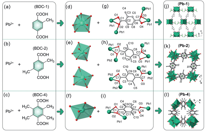
Figure 1.
Precursor composition (a-c), coordination modes of Pb2+ ion (d-f), coordination modes of the organic ligands (g-i), and three-dimensional (3D) framework structures (j-l) of Pb-1, Pb-2, and Pb-4.

Tuning band structures in metal-organic frameworks via ligand engineering strategy enables high-performance radio-photoluminescence dosimeters
Haoming Qin , Tianping Wang , Qian Yang , Deli Wu , Yumin Wang , Nannan Shen , Yanlong Wang , Zhifang Chai , Hanzhou Liu , Xing Dai , Shuao Wang


Radio-photoluminescence (RPL) is a promising passive radiation dosimetry technique for personal and environmental dose monitoring, owing to its ability to quantitatively store radiation information, the stability of its luminescent centers, and its nearly non-destructive readout capability [1–5]. The RPL principle involves the generation of radiation-induced luminescent centers that exhibit photoluminescence (PL) characteristics [6,7]. It enables the quantitative assessment of radiation exposure by measuring the RPL intensity of the material upon ultraviolet (UV) light excitation [6]. Current research efforts in RPL materials have primarily focused on metal-ion-doped inorganic materials such as Ag-doped phosphate glass [8,9], LiF [10,11], Mg2SiO4 [12], and Ag2O3:C,Mg [13]. Doping and co-doping strategies are commonly employed to enhance RPL performance [4]. However, achieving precise control over the dopant concentration and distribution within the crystal lattice of inorganic hosts remains a huge challenge. This often leads to the formation of undesired luminescent sites and obscures the clear establishment of structure-property relationships, thereby impeding the rational design of RPL materials tailored to specific application needs [3]. Therefore, there is an urgent need to develop novel RPL materials that allow for more precise control over their structure and RPL properties.
Metal-organic frameworks (MOFs), composed of metal ions or clusters coordinated with organic ligands, are promising candidates for next-generation RPL materials due to their exceptional designability, high structural stability, and efficient radiation absorption [14–19]. The first MOF-based RPL material, SCU-200 by us, demonstrating selective recognition of high-energy radiation and enabling an intersystem crossing (ISC) process wherein excited electrons transition from the singlet to the triplet state [20]. This transition results in distinct red triplet luminescence. However, there remains considerable room for improvement in the RPL performance of MOFs, particularly in prolonging the duration of RPL signals and lowering detection limits. To address these challenges, two primary strategies have proven effective in the broader field of organic phosphorescent materials, which share the same triplet-state-based luminescence mechanism. First, constructing a rigid molecular environment can suppress the nonradiative relaxation pathways, thereby enhancing phosphorescence efficiency [21]. Second, introducing electron-donating groups (e.g., CH3, NH2) or heavy atoms (e.g., As, Sb, Bi, Pb) can enhance spin-orbit coupling, significantly improving phosphorescence quantum yields [22–24]. The intrinsic structural tunability of MOFs provides a versatile platform for applying these strategies to modulate RPL properties with high precision [25]. Despite this potential, the precise tuning of RPL performance in MOFs remains largely unexplored. Therefore, developing a simple and effective approach to control RPL performance, while elucidating the underlying structure-property relationships, is essential for advancing the rational design of high-performance MOF-based RPL dosimeters.
This study aims to precisely tune the band structures of MOFs through ligand engineering and to investigate their influence on RPL performance. We synthesized three Pb-based MOFs using 1,4-benzenedicarboxylate (BDC) ligands functionalized with varying numbers of methyl groups on the benzene rings. The increasing number of methyl groups introduces greater steric hindrance, thereby influencing the overall framework conformation. In situ RPL measurements reveal that this simple ligand modification not only enhances the radiation response but also prolongs the RPL decay time. Electron paramagnetic resonance (EPR) analysis reveals that the radical species are retained consistently across the series of functionalized ligands. Meanwhile, photoluminescence measurements show that the intrinsic fluorescence of the ligands is unaffected by structural variations in the MOFs, whereas the RPL peak positions shift in correlation with the bandgap width, as verified by diffuse reflectance spectroscopy. Integrating these experimental findings with density functional theory (DFT) calculations, we constructed band structure models for the three MOFs and proposed the design principles for enhancing RPL performance.
As illustrated in Figs. 1a-c, Pb-1, Pb-2, and Pb-4 were synthesized using similar organic ligands (BDC-1, BDC-2, and BDC-4), differing only by the number of methyl groups attached to the benzene ring. Single crystal X-ray diffraction (SC-XRD) analysis revealed distinct coordination patterns for each MOF, as detailed in Tables S1-S5 (Supporting information). Pb2+ ions in these MOFs adopt multiple coordination geometries due to their large ionic radii and flexible coordination environments. In Pb-1, each Pb2+ ion coordinates with six carboxylate oxygens from four BDC-1 ligands and a carbonyl oxygen from an N, N-dimethylformamide (DMF) molecule (Fig. 1d and Fig. S1a in Supporting information). Pb-2 adopts two configurations (Fig. 1e): One involves six carboxylate oxygens from four BDC-2 ligands and a DMF carbonyl oxygen (Fig. S1b in Supporting information), and another involves five carboxylate oxygens from four BDC-2 ligands and a DMF carbonyl oxygen (Fig. S1c in Supporting information). Pb-4, in contrast, coordinates with four carboxylate oxygens from three BDC-4 ligands and two water molecules (Fig. 1f and Fig. S1d in Supporting information).
The ligand coordination models also differ among these MOFs. Pb-1 adopts a (κ1-κ2-
The varying coordination patterns of Pb2+ ions and the ligands result in distinct asymmetric units for each MOF (Table S5): Pb-1 includes one Pb2+ ion, one BDC-1 ligand, and one DMF molecule; Pb-2 comprises two Pb2+ ions, two BDC-2 ligands, and two DMF molecules; Pb-4 consists of one Pb2+ ion, one BDC-4 ligand, one H2O molecule, and a half-shared H2O molecule. As a result, the steric effect induced by the methyl groups leads to significantly different chemical structures in these MOFs despite their similar organic ligands (Figs. 1j-l).
Further characterization by powder X-ray diffraction (PXRD), N2 adsorption analysis, thermogravimetric analysis (TGA) and Fourier transform infrared (FT-IR) confirmed the structural differences. PXRD patterns align with those simulated from SC-XRD data (Fig. S2a in Supporting information), verifying the pure phase of these MOFs. PXRD results further confirm the excellent radiation stability of all three MOFs, as their crystal structures remain intact even after exposure to 100 kGy γ-ray irradiation (Fig. S2b in Supporting information). Nitrogen adsorption-desorption measurements show that Pb-1, Pb-2, and Pb-4 exhibit low BET surface areas of 8.16, 4.87, and 9.56 m2/g, respectively (Fig. S2c in Supporting information), indicating their densely packed frameworks. Such compact structures, together with the presence of heavy Pb atoms, are beneficial for efficient X-ray attenuation [28]. TGA analysis reveals differing solvent expulsion behaviours (Fig. S2d in Supporting information), with Pb-4 showing about 10% less weight loss compared to Pb-1 and Pb-2. This is due to the coordinated H2O in Pb-4 instead of the DMF in Pb-1 and Pb-2. DMF expulsion occurs at 230 ℃ for Pb-1 and 200 ℃ for Pb-2, indicating weaker coordination bond strength in Pb-2. FT-IR spectra (Fig. S2e in Supporting information) further highlight these differences. Pb-4 shows stronger C—H stretching vibrations of the methyl group and O—H stretching vibrations of H2O in the 2800–3600 cm-1 range [29]. The C═O stretching vibrations of the carboxylate group around 1650 cm-1 are suppressed or shifted due to the steric hindrance [30]. In contrast, Pb-1 and Pb-2 exhibit distinct N—H and C—H stretching vibrations around 2900 cm-1 [31]. They also exhibit similar vibrational peaks in the fingerprint region (1300–400 cm-1), indicating DMF coordination, confirmed by the C—N stretching vibrations at 1540 cm-1 [32,33].
The structural differences among these three MOFs are expected to influence their RPL performances. To investigate this, we compared their photoluminescence (PL) and RPL spectra before and after X-ray irradiation, as depicted in Figs. 2a-c. Initially, all samples exhibit a prominent emission band around 430 nm, which is ascribed to the π → π* transition of the benzene ring [20]. The initial emission intensity decreases under X-ray and ultraviolet-visible excitation, attributed to quenching effects and non-radiative transitions [34,35]. Upon X-ray irradiation, a new RPL emission band emerges within the 500–600 nm range. Notably, this RPL emission band shows a blue shift from 574 nm in Pb-1 to 563 nm in Pb-2, and further to 520 nm in Pb-4. To substantiate that the blue shift is primarily influenced by the organic ligands rather than Pb2+ ions, we synthesized two Ba-based MOFs (Ba-2 and Ba-4) using the same ligands as the Pb-based MOFs, but with Ba2+ as the metal ion. As shown in Fig. S3 (Supporting information), a similar blue shift is observed in the Ba-based MOFs, confirming that the ligand is responsible for this colour tunability. For the specific intensity of the new emission, the observed order under the same dose is Pb-4 > Pb-2 > Pb-1. This indicates that Pb-4 exhibits significantly higher efficiency in generating RPL centers compared to Pb-1 and Pb-2, suggesting that an increased number of methyl groups enhances RPL performance by promoting the formation of RPL luminescent centers.
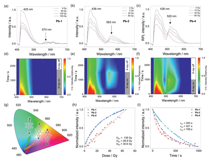
To eliminate potential errors caused by temperature fluctuations, position changes, environmental lighting, and dose, an in-situ RPL test system (Scheme S1 in Supporting information) was developed to track the luminescence behaviour of the materials. As illustrated in Figs. 2d-f, the RPL intensity increases with ongoing X-ray exposure, indicating a dynamic energy-charging process and the formation of new RPL emission centers. Under X-ray excitation, the decrease in the origin peak luminescence and the increase in RPL luminescence result in dynamic colour shifts on the Commission Internationale de l'Eclairage (CIE) diagram, as shown in Fig. 2g. Once the X-ray source is turned off, the RPL intensity decreases over time. We further analysed the specific data from Figs. 2d-f to quantitatively assess the RPL response and decay behaviours of these three materials. The RPL response data reflect a dynamic equilibrium between carrier generation and recombination processes, and they were well-fitted using a single saturating exponential function (Eq. S1 and Figs. S4a-c in Supporting information). The corresponding dose-response constant (DR) is 138, 28.9, and 30.6 Gy for Pb-1, Pb-2, and Pb-4, respectively (Fig. 2h). A lower DR suggests that the material has a quicker response and a higher RPL efficiency upon X-ray irradiation. In specific, RPL intensity shows a linear correlation with dose accumulation in low-dose ranges (Figs. S4d-f in Supporting information), indicating that these materials can function effectively as RPL dosimeters. The detection threshold (DT) is calculated to be 0.147 Gy and 0.202 Gy for Pb-2 and Pb-4, respectively, representing a significant improvement of more than an order of magnitude over Pb-1 (2.360 Gy) (Eq. S2 and Figs. S4d-f). Given that the DT is the minimum dose that can be reliably detected by RPL material, a much lower DT of Pb-2 and Pb-4 indicates their higher sensitivity to X-ray irradiation. For an RPL dosimeter, low DR and DT are particularly valuable, especially for low-dose applications. In addition, RPL decay intensity was monitored in real-time after cessation of X-ray irradiation. The decay time constant (τD) was obtained by fitting the decay data with a single decay exponential function (Eq. S3 in Supporting information). As a result, Pb-4 shows the longest τD (708 s) compared to Pb-1 and Pb-2 (203 and 321 s, respectively) (Fig. 2i and Figs. S4g-i in Supporting information). These results demonstrate that Pb-4 exhibits the most stable radiation-induced RPL centers, as a higher τD reflects a slower rate of decay and longer retention of the excited state, which is essential for accurate dose information readout [36,37]. Importantly, all three MOFs exhibit excellent recyclability, maintaining consistent baseline and RPL intensities over five repeated cycles (Fig. S5 in Supporting information), demonstrating their high stability and reusability for dosimetry applications.
In short, the introduction of methyl groups in the organic ligand significantly improves RPL performance. Compared to Pb-1, which has only one methyl group in its organic ligand, Pb-2 and Pb-4 exhibit a lower detection threshold, a quicker response to X-ray irradiation, and more stable radiation-induced RPL centers. These two materials show great potential for applications requiring high sensitivity and precision in X-ray detection.
To elucidate the significantly different RPL performances of these MOFs, we investigated their electron transition processes and electron-hole recombination behaviors using diffuse reflectance spectroscopy. As illustrated in Fig. 3a, Pb-2 and Pb-4 exhibit higher energy bandgaps (3.76 and 3.81 eV, respectively) compared to Pb-1 (3.43 eV). PL and RPL spectra (Figs. 2a-c) show that while the initial emission band remains unchanged, the new RPL emission undergoes a noticeable blue shift. This suggests that the increased energy bandgap is primarily associated with the process from the triplet state to the ground state, rather than the process from the singlet state to the ground state.
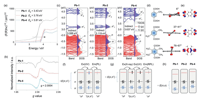
Electron paramagnetic resonance (EPR) is an effective method for identifying the existence of radicals and triplet excitons in materials [22]. EPR measurements under X-ray irradiation reveal that all three MOFs produce carboxylic acid oxygen radicals, each exhibiting a consistent g value of 2.0004 (Fig. 3b). This observation indicates a common radical formation process across the three MOFs: X-ray irradiation breaks the C═O bonds in carboxylic acid groups, leading to the formation of carboxylic acid oxygen radicals [20,38]. The conjugated structure of the benzene ring in these MOFs is conducive to stabilizing the radicals [38], while the presence of methyl groups modulates the fine structure of the radicals through interactions with conjugated π-orbitals [35,39]. These effects account for zero-field splitting observed in the EPR spectral profiles among the three MOFs (Fig. 3b). In contrast, EPR spectra obtained under UV irradiation show no evidence of radical formation (Fig. S6 in Supporting information), indicating that the energy of UV light is insufficient to induce the generation of radicals.
To elucidate the impact of ligand methyl groups on RPL performance, we conducted DFT calculations to analyse the band structures and density of states of Pb-1, Pb-2, and Pb-4. As shown in Fig. 3c, the valence bands of Pb-1 and Pb-2 are primarily contributed by the n electrons from the carboxyl groups, while the valence band of Pb-4 is dominated by both of the π electrons of the benzene rings and n electrons of the carboxyl groups. The conduction bands for all three MOFs predominantly consist of π* electrons. The calculated indirect bandgaps are 3.088, 3.119, and 3.637 eV for Pb-1, Pb-2, and Pb-4, respectively, consistent with the increasing trend observed in the diffuse reflectance spectra (Fig. 3a). The introduction of methyl groups on the ligand widens the bandgap, highlighting their key role in tuning MOF band structures. SC-XRD analysis reveals that the dihedral angles between the carboxylic acid group and the benzene ring increase from 6° in BDC-1 to 37°−41° in BDC-2 and 80°−87° in BDC-4 (Fig. 3d), demonstrating progressive steric hindrance due to methyl substitution. This distortion tunes the ground-state electronic structure and gives rise to significant n-π spin-orbit coupling in BDC-42- (Fig. 3e).
Combining experimental and computational insights, we propose a mechanism elucidating how ligand methyl groups modulate RPL performance in these Pb-based MOFs (Figs. 3f-h). The luminescence of these RPL-MOFs arises from two distinct components. The first component is a PL peak at around 430 nm, arising from the S1(1(π,π*)) → S0(1n2) radiative transition of the benzene ring under UV excitation (Fig. 3f) [20]. Since this emission is dominated by the π → π* transition, its wavelength remains nearly constant across the series MOFs. The second emission component corresponds to lower-energy RPL. Under X-ray irradiation, the high-Zeff Pb2+ ions efficiently absorb and transfer energy to the ligands, promoting an n → π* transition and enabling ISC to the triplet state (Fig. 3g) [40]. Occupation of the π* orbital by n-electrons suppresses the π → π* transition, thereby reducing PL intensity. Under UV excitation, π* electrons gain sufficient energy to undergo spin reversal and return to the n orbital, giving rise to RPL emission. The RPL wavelength, governed by the ΔE(n, π*) gap, blue-shifts across the series due to a progressive rise in π* orbital energy (Fig. 3h).
Ligand engineering enhances RPL performances through two distinct yet complementary mechanisms: First, accelerated triplet generation via enhanced ISC. Methyl substitution modulates the orbital energies, reducing the energy gap between the singlet excited state 1(π,π*) and the triplet state 3(n, π*), denoted as
|
|
(1) |
The enhanced ISC leads to improved RPL performance, including lower detection thresholds (Figs. S4d-f) and larger dose-response constants (Fig. 2h and Figs. S4a-c). Second, suppressed non-radiative decay via structural rigidification. Methyl-induced steric hindrance rigidifies the framework (Fig. 3d), effectively suppressing non-radiative recombination and quenching pathways [44]. The increased stability of the RPL centers is evidenced by slower decay kinetics (Fig. 2i and Figs. S4g–i). In summary, ligand methyl groups promote both the formation and the stability of RPL centers, resulting in superior sensitivity and durability for Pb-2 and Pb-4 compared to Pb-1.
In this work, three Pb-based MOFs were synthesized by modifying the organic ligands with varying numbers of methyl groups on the benzene rings. We found that the steric hindrance induced by methyl groups plays a critical role in tuning the band structures, with energy bandgaps varying from 3.43 eV to 3.81 eV. Correspondingly, the RPL emission colours can be tuned from orange (574 nm) to green (520 nm). Compared to Pb-1 with only one methyl group on the ligand, Pb-2 and Pb-4 exhibit significantly improved RPL performance, including lower detection thresholds (0.147 Gy and 0.202 Gy vs. 2.360 Gy), smaller dose-response constants (28.9 Gy and 30.6 Gy vs. 138 Gy), and longer decay time constants (321 s and 708 s vs. 203 s). In light of both the experimental results and DFT simulations, we propose a schematic model illustrating the electron transitions in these RPL MOFs. Our investigation highlights the potential of MOFs as a versatile platform for the rational design of highly sensitive, stable, and colour-tunable RPL dosimeters. Expanding the library of functional ligands and heavy metal nodes offers a promising route to tailor the RPL properties of MOFs for customized detection across different radiation dose regimes.
Haoming Qin: Writing – review & editing, Writing – original draft, Methodology, Investigation, Data curation, Conceptualization. Tianping Wang: Writing – review & editing, Supervision. Qian Yang: Investigation, Data curation. Deli Wu: Investigation, Data curation. Yumin Wang: Data curation. Nannan Shen: Supervision. Yanlong Wang: Supervision. Zhifang Chai: Supervision. Hanzhou Liu: Writing – review & editing, Supervision, Resources, Project administration, Methodology, Funding acquisition, Conceptualization. Xing Dai: Supervision. Shuao Wang: Supervision, Resources, Project administration, Funding acquisition.
The authors declare that they have no known competing financial interests or personal relationships that could have appeared to influence the work reported in this paper.
We appreciate the financial support from the National Natural Science Foundation of China (Nos. 22276132, U24B20194, 22376153, 22406135), Gusu Innovation and Entrepreneurship Leading Talent Program Project (Nos. ZXL2024403, ZXL2024406), Science Foundation of the Higher Education Institutions of Jiangsu Province (No. 22KJA150006) and the Postdoctoral Science Foundation of China (Nos. GZC 20231888, 2024M762296), and the Priority Academic Program Development of Jiangsu Higher Education Institutions (PAPD).
Supplementary material associated with this article can be found, in the online version, at doi:
L. Yuan, Y. Jin, Y. Su, et al., Laser Photonics Rev. 14 (2020) 2000123. doi: 10.1002/lpor.202000123
T. Kron, J. Lehmann, P.B. Greer, Phys. Med. Biol. 61 (2016) R167–R205. doi: 10.1088/0031-9155/61/14/R167
G. Okada, J. Ceram. Soc. Jpn. 129 (2021) 419–424. doi: 10.2109/jcersj2.21056
Z. Yang, H. Vrielinck, L.G. Jacobsohn, et al., Adv. Funct. Mater. 34 (2024) 2406186. doi: 10.1002/adfm.202406186
T. Yanagida, G. Okada, T. Kato, et al., Radiat. Meas. 158 (2022) 106847. doi: 10.1016/j.radmeas.2022.106847
T. Yanagida, G. Okada, N. Kawaguchi, J. Lumin. 207 (2019) 14–21. doi: 10.1016/j.jlumin.2018.11.004
H. Kawamoto, Y. Fujimoto, M. Koshimizu, et al., Jpn. J. Appl. Phys. 57 (2018) 062401. doi: 10.7567/jjap.57.062401
J.H. Schulman, R.J. Ginther, C.C. Klick, et al., J. Appl. Phys. 22 (1951) 1479–1487. doi: 10.1063/1.1699896
T. Kurobori, Y. Miyamoto, N. Takashima, et al., Radiat. Meas. 181 (2025) 107376. doi: 10.1016/j.radmeas.2025.107376
J. Nandha Gopal, B. Sanyal, A. Lakshmanan, Radiat. Meas. 109 (2018) 24–34. doi: 10.1016/j.radmeas.2017.12.007
P. Bilski, B. Marczewska, Y. Zhydachevskii, Radiat. Meas. 97 (2017) 14–19. doi: 10.1016/j.radmeas.2016.12.014
G. Okada, T. Kojima, J. Ushizawa, et al., Curr. Appl. Phys. 17 (2017) 422–426. doi: 10.1016/j.cap.2017.01.004
L.F. Nascimento, M. Karampiperi, J.P. Oliveira, et al., Radiat. Meas. 106 (2017) 218–224. doi: 10.1016/j.radmeas.2017.02.006
J.X. Wang, J. Yin, O. Shekhah, et al., ACS Appl. Mater. Interfaces 14 (2022) 9970–9986. doi: 10.1021/acsami.1c24759
Z. Wang, C.Y. Zhu, S.Y. Yin, et al., Angew. Chem. Int. Ed. 58 (2019) 3481–3485. doi: 10.1002/anie.201812708
P.Y. Liao, J.X. Li, J.C. Liu, et al., Angew. Chem. Int. Ed. 63 (2024) e202401448. doi: 10.1002/anie.202401448
X. Yu, J. Mi, Y. Han, et al., Chin. Chem. Lett. 35 (2024) 109233. doi: 10.1016/j.cclet.2023.109233
Y. Cui, H. Lu, H. Hou, et al., Angew. Chem. Int. Ed. 63 (2024) e202410453. doi: 10.1002/anie.202410453
Y. Cui, J. Lin, Chem. Commun. 61 (2025) 5395–5409. doi: 10.1039/d5cc00711a
H. Liu, H. Qin, N. Shen, et al., Angew. Chem. Int. Ed. 59 (2020) 15209–15214. doi: 10.1002/anie.202006380
X. Ma, J. Wang, H. Tian, Acc. Chem. Res. 52 (2019) 738–748. doi: 10.1021/acs.accounts.8b00620
G. Bao, R. Deng, D. Jin, et al., Nat. Rev. Mater. 10 (2024) 28–43. doi: 10.1038/s41578-024-00704-y
X. Wu, R. Zhang, J. Liu, J. Solid State Chem. 301 (2021) 122298. doi: 10.1016/j.jssc.2021.122298
S. Arunkumar, G.R. Kumar, J. Organomet. Chem. 1001 (2023) 122849. doi: 10.1016/j.jorganchem.2023.122849
L. Ma, S. Guo, Z.-M. Zhang, et al., Chin. Chem. Lett. 35 (2024) 108661. doi: 10.1016/j.cclet.2023.108661
C. Chen, J.K. Sun, Y.J. Zhang, et al., Angew. Chem. Int. Ed. 56 (2017) 14458–14462. doi: 10.1002/anie.201707290
O. Tayfuroglu, A. Kocak, Y. Zorlu, Phys. Chem. Chem. Phys. 24 (2022) 11882–11897. doi: 10.1039/d1cp05973d
Q. Yang, H. Qin, L. Chen, et al., ACS Mater. Lett. 5 (2023) 1619–1626. doi: 10.1021/acsmaterialslett.3c00326
C. Hu, Y.T. Zhai, L.L. Song, et al., Polym. Compos. 41 (2019) 1418–1427.
S.R. Mathis Ii, S.T. Golafale, J. Bacsa, et al., Dalton Trans. 46 (2017) 491–500. doi: 10.1039/C6DT03755K
Y.K. Ren, S.D. Liu, B. Duan, et al., J. Alloys Compd. 705 (2017) 205–210. doi: 10.1016/j.jallcom.2017.01.035
Y. Zhang, L. Chen, J. Guan, et al., Dalton Trans. 49 (2020) 3676–3679. doi: 10.1039/c9dt03871j
X. Wang, X. Dai, C. Shi, et al., Nat. Commun. 10 (2019) 2570. doi: 10.1109/cac48633.2019.8996820
H.Y. Li, H. Xu, S.Q. Zang, et al., Chem. Commun. 52 (2016) 525–528. doi: 10.1039/C5CC08168H
S. Han, J. Hu, G. Wang, Coord. Chem. Rev. 452 (2022) 214304. doi: 10.1016/j.ccr.2021.214304
Z. Yang, J. Hu, D. Van der Heggen, et al., Laser Photonics Rev. 17 (2023) 2200809. doi: 10.1002/lpor.202200809
T. Kurobori, A. Takemura, Y. Miyamoto, et al., Radiat. Meas. 83 (2015) 51–55. doi: 10.1016/j.radmeas.2015.04.017
H. Lu, M. Xu, Z. Zheng, et al., Inorg. Chem. 60 (2021) 18629–18633. doi: 10.1021/acs.inorgchem.1c03182
Z. Yue, J. Liu, M. Baumgarten, et al., J. Phys. Chem. A 127 (2023) 1565–1575. doi: 10.1021/acs.jpca.2c06648
W. Zhao, Z. He, B.Z. Tang, Nat. Rev. Mater. 5 (2020) 869–885. doi: 10.1038/s41578-020-0223-z
M.A. El-Sayed, J. Chem. Phys. 38 (1963) 2834–2838. doi: 10.1063/1.1733610
J.M. Jacob, M.K. Ravva, Mater. Adv. 3 (2022) 4954–4963. doi: 10.1039/d2ma00039c
I.E. Serdiuk, M. Monka, K. Kozakiewicz, et al., J. Phys. Chem. B 125 (2021) 2696–2706. doi: 10.1021/acs.jpcb.0c10605
X. Yang, D. Yan, Adv. Opt. Mater. 4 (2016) 897–905. doi: 10.1002/adom.201500666

Figure 1 Precursor composition (a-c), coordination modes of Pb2+ ion (d-f), coordination modes of the organic ligands (g-i), and three-dimensional (3D) framework structures (j-l) of Pb-1, Pb-2, and Pb-4.

Figure 2 Photoluminescence and RPL spectra of Pb-1 (a), Pb-2 (b), and Pb-4 (c) before and after X-ray irradiation. Time-dependent persistent RPL spectra of Pb-1 (d), Pb-2 (e), and Pb-4 (f). (g) CIE chromaticity coordinates from in-situ RPL analysis at varying doses. (h) The RPL intensity-response curves under X-ray exposure. (i) The RPL intensity decay curves after cessation of X-ray irradiation.

Figure 3 Diffuse reflectance spectra (a) and EPR spectra (b) of Pb-1, Pb-2, and Pb-4. (c) Calculated band structures and density of states for Pb-1, Pb-2, and Pb-4. (d) Bond angles between the carboxylic acid group and the benzene ring in BDC-1, BDC-2, and BDC-4. (e) DFT-calculated molecular orbitals of the ground state for BDC-12-, BDC-22- and BDC-42-. Schematic diagrams of the orbital structures for the PL process (f), the RPL process (g), and the effect of ligand methyl groups on the three MOFs (h).

 扫一扫看文章


扫一扫关注我们
 DownLoad:
 下载:
 下载: