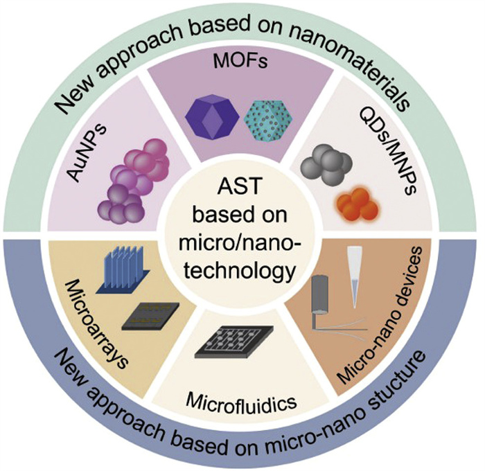
Figure 1.
AST based on micro/nano technology.

Micro/nano technology-driven diagnostic platforms: Addressing the speed dilemma in antimicrobial susceptibility testing
Ling Sun , Xiaoqing Wei , Yingping Li , Junzhen Deng , Jingya Zhao , Shaobing Zhou


The global threat of antimicrobial resistance is escalating. According to a recent study, antimicrobial-resistant bacterial infections may result in over 39 million fatalities worldwide in the next 25 years [1,2]. Crucially, a substantial number of deaths could be avoided by improving infection control, ensuring antibiotic accessibility and quality, and strengthening surveillance systems [3]. Antimicrobial susceptibility testing (AST) is a cornerstone intervention in this effort, providing essential data for antibiotic stewardship, epidemiological monitoring, and resistance pattern assessment [4]. In clinical practice, AST guides the selection of effective therapeutic regimens and predicts patient outcomes by identifying bacterial susceptibility profiles [5]. However, the utility of conventional AST is severely limited by its prolonged turnaround time, which stems from reliance on labor-intensive culture-based techniques. Therefore, the development of novel AST technologies must prioritize overcoming this time bottleneck while simultaneously improving detection sensitivity [6].
As an interdisciplinary frontier, micro/nano technology has exhibited transformative impacts across scientific and industrial domains, significantly accelerating technological advancements in recent years [7,8]. In bacterial detection, it has revolutionized diagnostic capabilities by enabling biosensors with ultra-low detection limits as low as 10 CFU/mL, thereby facilitating rapid pathogen identification and improving early infection management [9-11]. Furthermore, in therapeutic applications, drug delivery systems mediated by nanomaterials offer novel avenues for precision antimicrobial therapy by targeting pathogens directly and minimizing systemic side effects [12-20].
In addition to its applications in bacterial detection and treatment, micro/nano technology also exhibits considerable potential for revolutionizing AST [21]. Traditional AST methods rely on lengthy bacterial culturing and response assessment. Given this limitation, micro/nano technology-based ASTs are emerging as powerful tools to significantly shorten turnaround time while improving operational convenience and detection sensitivity. In this review, we summarize current advancements in AST technologies and outline the advantages of micro/nano technology-based AST (Fig. 1). We also discuss persistent challenges and potential avenues for future AST development.
Currently, clinical AST technologies can be broadly divided into two main categories: Phenotype-based and genotype-based detection strategies [22]. Among these, phenotypic AST is the mainstream approach in clinical microbiology laboratories. Its principle involves directly evaluating bacterial responses under exposure to antimicrobial agents to determine the inhibitory effects of the drugs on bacterial strains. Certain phenotypic AST methods utilize biochemical indicators, such as pH indicators, fluorescent substrates, or redox dyes to indirectly reflect bacterial metabolic activity. As bacteria grow, they metabolize sugars and amino acids or produce specific enzymes, leading to detectable changes in the chemical environment of the culture medium. By monitoring these alterations, bacterial viability can be assessed, thus allowing the determination of drug susceptibility. Such methods are relatively simple to operate, but are time-consuming and unable to detect the mechanisms of drug resistance [23].
Most ASTs assess susceptibility by comparing the minimum inhibitory concentration (MIC) against established breakpoints. MIC is defined as the lowest drug concentration achieving complete bacterial growth suppression. In addition to their comparative function, MIC values provide quantitative characterization of resistance levels. Globally recognized breakpoint standards are upheld by two organizations: The Clinical and Laboratory Standards Institute (CLSI) and the European Committee on Antimicrobial Susceptibility Testing (EUCAST). These standards provide an essential foundation for evaluating bacterial susceptibility to antibiotics, thereby facilitating the rational use of these medications and mitigating the emergence of resistant strain [24,25]. In traditional AST, bacterial susceptibility is determined by co-incubating bacteria with antibiotics and observing the resulting inhibitory effects on bacterial growth. Traditional ASTs do not require specialized instruments or equipment, relying solely on visual assessment of the MIC. These methods typically encompass broth dilution, disk diffusion, and E-test (Fig. 2).
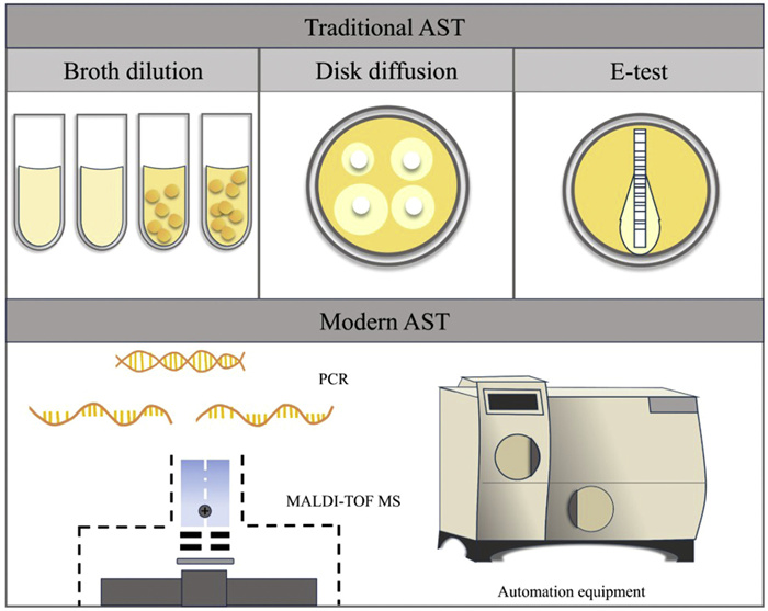
The broth dilution method is a fundamental AST technique that involves co-culturing bacteria (typically at 5 × 10⁵ CFU/mL) with serially diluted antibiotics [26]. This method is widely applicable to various microbes and drugs. A limitation, however, is substantial reagent consumption. To overcome this, a miniaturized microdilution technique was developed using 96-well plates, which reduces volumes of 100 µL per test while maintaining reliability [27].
The disk diffusion method evaluates susceptibility by measuring the zone of inhibition around a drug-impregnated paper disk placed on an agar plate inoculated with the bacteria. As the drug diffuses into the medium, it creates a concentration gradient. The diameter of the clear zone reflects the inhibitory effect of the drugs [28]. However, this method is less quantitative than the broth dilution method, as inhibition zone measurement can be subjective and does not directly yield a MIC. Nevertheless, its key advantage is the capacity to test multiple antibiotics simultaneously on a single plate [26].
The E-test method combines the principles of disk diffusion and broth dilution. It utilizes a strip impregnated with a predefined antibiotic gradient [29]. The MIC is read directly at the point where the elliptical zone of inhibition intersects the strip, thereby enabling precise quantitative results. Despite the relatively high cost of the strips, the method is valued for its excellent reproducibility and accuracy.
Modern AST encompasses recently developed automated and semi-automated systems that leverage advanced technologies. Unlike traditional methods, which rely on manual, repetitive pipetting and dilution steps, processes prone to human error and contamination, modern AST integrates automation to minimize manual intervention, thereby enhancing operational consistency and result reliability. Modern phenotype-based AST systems, such as MicroScan WalkAway and VITEK 2, predominantly employ highly automated microbroth dilution as their technical basis [30]. These systems utilize precisely designed reaction plates containing antimicrobial agents at varying concentration gradients. After inoculating bacterial suspensions and incubating for a defined period, highly sensitive photometers are used to monitor real-time changes in the turbidity of the bacterial solutions in each well. These systems can detect subtle differences in bacterial growth, thereby accurately determining the MIC and providing quantitative data for clinical treatment decisions.
Polymerase chain reaction (PCR) is one of the most widely used and essential genotype-based detection technologies. PCR is based on the semi-conservative replication mechanism of DNA, simulating the natural process of DNA replication in vitro and enabling the efficient synthesis and amplification of specific nucleic acid fragments through enzymatic reactions. By screening known antibiotic resistance genes, PCR can rapidly and accurately identify potentially resistant bacteria, providing critical support for the diagnosis and treatment of infectious diseases [31]. Additionally, gene chips, as an emerging genotypic detection technology, are increasingly being applied in AST. This approach relies on high-throughput solid-phase platforms, where a large number of sequence-specific molecular probes (such as DNA or RNA probes) are immobilized on the chip surface to allow for the parallel detection of multiple target nucleic acid sequences. After hybridization with nucleic acids extracted from clinical samples, gene chips can detect various resistance genes simultaneously through signal capture and analysis, offering advantages in terms of efficiency, throughput, and integration. They are not only suitable for identifying known resistance genes but also hold potential for discovering new resistance mutations, highlighting their broad application prospects in both research and clinical diagnostics.
Matrix-assisted laser desorption/ionization time-of-flight mass spectrometry (MALDI-TOF MS) offers a unique approach to AST by integrating phenotypic and genotypic detection modes while maintaining the speed of molecular methods [32]. This is achieved through two distinct mechanisms. In the phenotypic mode, resistance is detected either by identifying modification/hydrolysis of antimicrobial agents caused by bacterial enzymes (e.g., β-lactamases) or by establishing standard models based on the distinct protein fingerprint profiles of resistant versus susceptible strains. In the genotypic mode, it directly targets and detects specific resistance-associated gene sequences, thereby linking their presence directly to the resistance phenotype and providing an alternative pathway for rapid AST.
In recent years, a variety of nanomaterials and micro/nano-structures have been extensively explored as promising candidates for advancing AST. Functional nanomaterials, such as gold nanoparticles (AuNPs) and magnetic nanomaterials, enable instrument-free visual readout and rapid target separation, substantially enhancing detection sensitivity while streamlining the AST process [33]. The application of micro/nano-structures, especially microfluidic chips, has further revolutionized AST capabilities [34]. These highly integrated platforms enable both phenotypic and genotypic AST at the single-cell level, significantly reducing detection time from days to just hours. These advancements in nanomaterials and micro/nano-structures have collectively addressed the critical bottleneck of prolonged turnaround time in conventional AST while providing enhanced detection sensitivity and operational flexibility. Herein, we mainly discuss the role of micro/nano-technology in AST, focusing on two key aspects. The first is nanomaterial-based AST, which enhances sensitivity by leveraging unique material properties. The second is micro/nanostructure-based AST, in which structural engineering of devices enables low sample consumption and single-cell analysis.
By leveraging the distinctive properties of nanomaterials, such as their high surface area-to-volume ratio as well as their unique optical and electrical characteristics, new opportunities for rapid AST have emerged. AuNPs, metal-organic frameworks (MOFs), fluorescent nanomaterials, and magnetic nanomaterials are extensively utilized for rapid AST (Table 1). These nanomaterials enable visual detection and facilitate rapid separation, which can significantly enhance the sensitivity and efficiency of AST while reducing reliance on large-scale equipment or instruments.
 DownLoad:
CSV
| Material | Property | Biological indicator | Ref. |
| AuNPs | Visualization | LPS, peptidoglycan, 16 s RNA, metabolic activity, catalase | [36,37,42-44] |
| MOFs | Large specific surface area | Bacterial membrane, ATP, LPS | [47,48] |
| QDs | Visualization | Lipid A in LPS, DNA | [52,53] |
| Magnetic nanoparticles | Magnetic separation | Pathogen-associated molecular patterns, genomic DNA (gDNA) | [58] |
While elemental gold exhibits chemical inertness, its nanoscale counterparts, AuNPs, acquire unique optical properties through the surface plasmon resonance effect. This phenomenon manifests as changes in solution color that depend on particle size and morphology. Moreover, the high stability and good biocompatibility of AuNPs have facilitated their widespread application in bacterial imaging and detection. The unique optical properties have also been leveraged to develop novel diagnostic platforms for AST [35].
Wang et al. designed concanavalin A modified AuNPs (ConA-AuNPs) that facilitate targeting of both Gram-positive and Gram-negative bacteria for broad-spectrum AST (Fig. 3a) [36]. They initially incubated Staphylococcus aureus (S. aureus) and Escherichia coli (E. coli) with different concentrations of antibiotics for 2 h, followed by further incubation with the nanoparticles. The differential antibacterial efficacy of the antibiotics resulted in concentration-dependent bacterial survival profiles, which were observed as quantifiable color changes due to interactions between nanoparticles and bacteria. Thus, the MIC can be determined by visually observing the point at which color change occurs (Fig. 3b). In addition, they developed vancomycin-modified AuNPs (Van-AuNPs) and polymyxin B-modified Prussian blue NPs (PMB-PBNPs) for the identification of Gram-positive and Gram-negative bacteria, respectively. This detection strategy enables direct assessment of antibiotic susceptibility, and the results obtained align well with those from traditional methods.
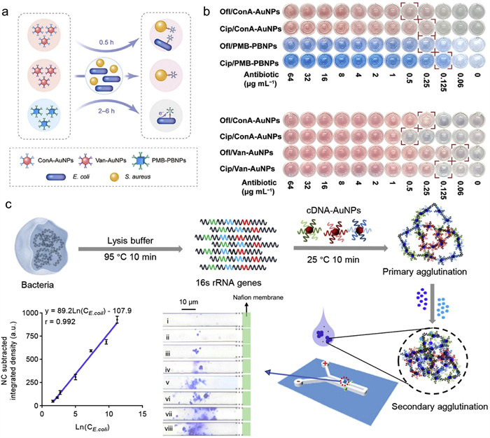
Bacterial lysis products can also serve as targets for AST. Recently, a cation dye-triggered electrokinetic AuNP agglutination strategy has been applied to AST. Wei et al. modified the surface of AuNPs with cDNA [37]. Upon release from lysed bacteria, siRNA induced primary aggregation of the AuNPs, which is further enhanced under an electric field, leading to secondary aggregation and the formation of blue AuNP agglutinates (Fig. 3c). Receiver operating characteristic analysis of urine samples demonstrated that this AST method achieved a sensitivity of 100% and a specificity of 98%. This approach innovatively leverages bacterial lysis products for AST, offering a novel strategy for rapid AST.
Bacterial metabolism has been proven to be associated with antibiotic resistance [38]. Therefore, investigating the metabolic responses of bacteria to antibiotics provides new insights for shortening the turnaround time of AST [39-42]. Zhang et al. designed cysteine-modified AuNPs, which were coupled with the metabolism of bacteria towards Cu2+ for bacterial detection and AST [43]. In the absence of bacteria, the cysteine on AuNPs chelated with Cu2+, leading to aggregation of NPs. Conversely, in the presence of bacteria, the bacteria could metabolize Cu2+, thereby altering the aggregation degree of AuNPs. Based on this, bacterial antibiotic sensitivity can be determined within approximately 3 h by monitoring the colorimetric changes in the solution. Experimental results showed that when testing 4 clinical isolates against 6 antibiotics, this method achieved an accuracy of 95.8% compared to traditional assays.
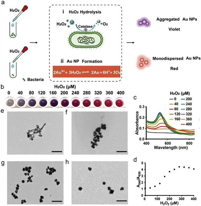
Huang et al. developed a rapid AST platform by combining gold ions and H2O2 (Fig. 4a) [44]. The reduction of gold ions by H2O2 resulted in the production of AuNPs, which led to a visible color change of the solution from gray to red (Fig. 4b). UV absorption spectroscopy and transmission electron microscopy (TEM) analyses of AuNPs generated at increasing H2O2 concentrations confirmed the color transition and revealed that higher H2O2 levels led to the formation of larger and more dispersed AuNPs (Figs. 4c-h). Upon the addition of catalase-positive bacteria, the enzymatic degradation of H2O2 occurred, leading to a reduction in the availability of H2O2 for the reduction of gold ions. However, when the inhibitory effect of antibiotics was not significant, this resulted in the formation of aggregated and irregularly shaped AuNPs, causing the solution to appear gray. This method allowed visual assessment of bacterial susceptibility and shortened the AST time to within 2 h. When evaluated against traditional AST methods, the accuracy for detecting susceptibility in E. coli and K. pneumoniae exceeded 90%.
MOFs are a relatively new class of porous nanomaterials, consisting of metal ions or clusters connected by organic ligands [45]. Unlike traditional nanomaterials, MOFs exhibit tunable porosity, high surface areas, structural flexibility, and versatile functionality. Their enhanced surface area contributes significantly to the high sensitivity and rapid response of MOF-based biosensors. Extensive studies have been conducted on the application of MOFs in bacterial detection and the treatment of bacterial infections [45,46]. More recently, MOFs have been integrated with AST, providing a promising strategy to accelerate AST processes.
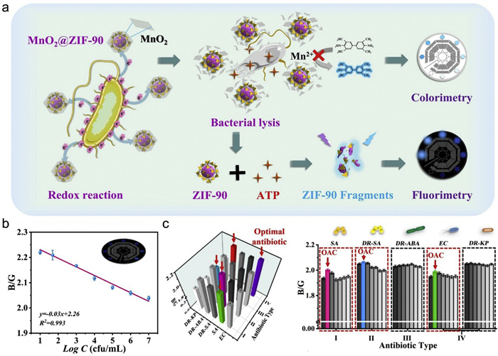
Liu et al. developed a dual-mode AST platform based on zeolitic imidazolate framework-90 (ZIF-90), which achieved a detection limit of 10 CFU/mL and an assay time of just 5 min [47]. Upon interaction with bacterial membrane, the MnO2 coating on the ZIF-90 surface decomposed into Mn2+ ions. These ions then reacted with the substrate 3,3′,5,5′-tetramethylbenzidine, leading to a reduction in the colorimetric signal and allowing the assessment of the bacterial activity and antibiotic resistance. Concurrently, adenosine triphosphate (ATP) released through bacterial lysis competed with imidazole ligands for Zn2+ in ZIF-90, leading to the disintegration of the ZIF-90 frameworks and the release of fluorescent intermediates (Fig. 5a). The fluorescence signals were quantitatively assessed through RGB analysis, demonstrating a robust linear correlation between the B/G values and bacteria concentration (Fig. 5b). When applied to five bacterial strains, the MnO2@ZIF-90 nanomaterial-based AST platform showed high accuracy compared to conventional methods (Fig. 5c).
Owing to their large specific surface area, MOFs provide extensive anchoring planes and abundant pore structures, which are advantageous for biomolecule immobilization. Leveraging this property, Sun et al. fabricated a MOF-based electrochemiluminescence (ECL) biosensor for rapid AST [48]. The immense surface area of the MOFs significantly facilitated the loading of ruthenium complexes onto the electrode, thereby greatly enhancing the ECL signal intensity and achieving a detection limit as low as 16 CFU/mL.
Fluorescent nanomaterials, particularly quantum dots (QDs), have been widely used in biodetection [46,49] and in vivo imaging [50,51]. Owing to their exceptional visualization properties, these nanomaterials also hold significant potential in advancing rapid AST. For example, Jiang et al. constructed a CsPbBr3 QDs-DNA nanoprobe to detect the drug resistance genes of Mycobacterium tuberculosis using Förster resonance energy transfer (FRET) [52], which provides a new strategy for genotypic AST. Additionally, QDs have been applied in phenotypic AST. Wang et al. designed fluorescein isothiocyanate and QD-labeled peptide probes that specifically target mcr-1-modified lipid A [53]. These probes enabled the detection of mcr-1-induced resistant strains through an immunofluorescence assay.
Magnetic nanoparticles are widely used for bacterial separation to streamline AST [54,55]. A recent research focus has been the direct application of AST to blood samples [56,57]. However, the high complexity of blood and the low bacterial concentration present major obstacles. The high capture efficiency and excellent magnetic properties of magnetic nanoparticles position them as ideal candidates for such applications, enabling effective AST directly from blood. Kim et al. developed a rapid platform based on magnetic nanoparticles for direct AST from blood samples [58]. By modifying magnetic nanoparticles with β−2-glycoprotein Ⅰ peptides, they achieve a broad-spectrum pathogen capture, with efficiencies of 96.23% ± 3.31% for E. coli and 91.54% ± 4.35% for S. aureus. After magnetic separation and resuspension in PBS, the bacteria were subjected to a short period of cultivation and microplate identification, followed by AST on a customized 96-well plate. This approach shortens the culture duration and simplifies the AST process.
Microfluidic technology, renowned for advantages such as low sample consumption, fast detection speed, ease of operation, multifunctional integration, and portability has been extensively leveraged to advance point-of-care AST [59,60]. In addition to microfluidics, microarrays and other micro/nano devices also play a vital role in enabling rapid AST. This section summarizes the applications of microarrays, microfluidics and micro/nano devices in AST.
Microelectrode arrays can identify drug-resistant bacteria by detecting changes in the dielectric properties around the bacteria. Chung et al. developed a quadruple electrode array for AST of E. coli and K. Pneumoniae [61]. After treatment with antibiotics, the bacteria underwent morphological changes, making them more polarized than the surrounding medium. Consequently, the bacteria were attracted to regions of high electric field gradient (Fig. 6a). In contrast, resistant bacteria did not undergo these antibiotic-induced morphological changes and exhibited a lower polarization rate than the surrounding medium, causing them to be repelled toward weak regions of the electric field (Fig. 6b) [62]. Therefore, the non-uniform electric field formed by the microelectrode arrays selectively attracted bacteria according to their susceptibility, facilitating the determination of AST profiles. The MIC values obtained by this method showed 95.6% consistency with the broth dilution method.
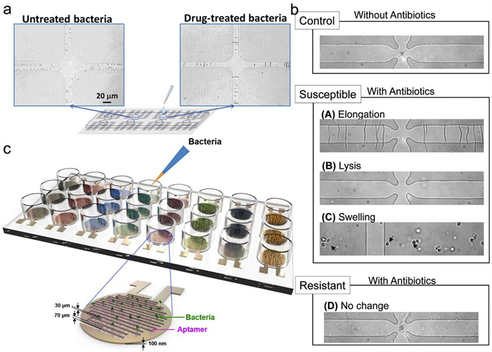
Jo et al. designed a capacitive sensor functionalized with aptamers, which detected bacteria by recognizing them as dielectric particles [63]. The binding of bacteria to the aptamers induced an increase in capacitance (Fig. 6c). Conversely, when bacteria were killed by antibiotics, they underwent morphological changes and detached from the sensor surface, leading to a decrease in capacitance. By monitoring the changes in capacitance, this platform enabled the assessment of antibiotic susceptibility and the determination of the MIC. The results obtained with this sensor showed good agreement with those from conventional methods.
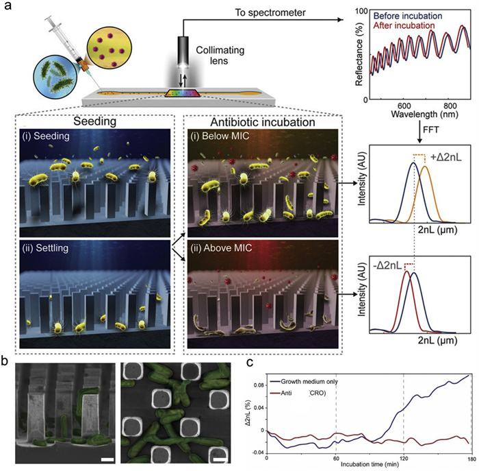
The microcolumn structure offers a unique advantage for immobilizing bacteria in fluids [64]. This platform employs phase-shift reflectance interference spectroscopy (PRISM) to detect subtle changes induced by bacteria by monitoring the phase-shift characteristics of light waves interacting with a silicon micropillar array (Fig. 7a). Bacterial growth within the arrays alters the local refractive index, generating measurable optical path differences that enable rapid assessment of antibiotic susceptibility. For example, wheat germ agglutinin (WGA) was immobilized on silicon micropillars for capturing bacteria (Fig. 7b). Critically, the micropillar dimensions were designed to account for the size variations among different bacterial species, ensuring effective entrapment while providing sufficient space for growth and minimizing mechanical deformation. By comparing the optical path difference before and after antibiotic exposure, the susceptibility can be determined (Fig. 7c). The AST results obtained through PRISM were consistent with those from the broth dilution method.
The integrated characteristics of microfluidic chips make it possible to construct comprehensive platforms that consolidate sample separation, reaction, and detection for AST. These microsystems typically operate within a confined space, which can accelerate bacterial growth and biochemical reactions. They generally consume minimal amounts of samples and reagents, making them well-suited for high-throughput, highly parallel, and single-cell analyses. Furthermore, their compact size facilitates integration into low-cost, portable devices for point of care (POC) bacterial detection. Overall, microfluidic chips have significant advantages as integrated systems for bacterial AST, featuring reduced analysis time, low cost, and high adaptability for POC applications.
During the AST process, the primary workload involves the manual procedures of antibiotic preparation and bacterial inoculation. The utilization of microfluidic chips for rapid dilution and precise distribution can greatly reduce manual workload and shorten detection time. Recently, a ready-to-use microfluidic chip preloaded with antibiotics has been developed to streamline this process [65]. With this design, only a mixture of bacterial suspension, resazurin dye, and oil (for sample separation) needed to be injected. This mixture then flowed into the chambers pre-filled with 12 different antibiotics. Bacterial growth was indicated by the reduction of blue resazurin to pink resorufin, visually revealing bacterial susceptibility. Although pre-loading antibiotics can streamline operational procedures and mitigate contamination risk, the variety and concentrations of antibiotics available are limited [66]. The following technology offers a better balance between antibiotic flexibility and operational convenience. A 3D concentration gradient array with micro/nano channels facilitates the diffusion of antibiotics into culture chambers, thereby establishing a continuous gradient [67]. This platform also supports combination AST by simply introducing two high-concentration antibiotics into the generator.
Single-cell detection techniques based on microfluidic chips have been employed for both phenotypic and genotypic AST [68]. These techniques allow for the analysis of individual bacterial cells, enabling critical distinctions that are obscured in bulk population studies [69]. For example, by comparing the luminescence changes of oxygen-sensing nanoprobes in bacterial growth chambers with different concentrations of antibiotics, resistant and susceptible bacteria were distinguished within 2–3 h [70]. This method rapidly detects variations in respiratory metabolism of individual bacteria. Moreover, Pitruzzello et al. reported an alternative approach by integrating microelectrodes into a microfluidic chip to monitor the metabolic activity of individual bacteria through their electrical responses [71].
In addition, microfluidic single-cell analysis is uniquely suited for mixed infections, as it can simultaneously identify constituent bacterial species and their respective resistance profiles. This capability is essential for managing complex infections and formulating personalized treatment strategies. Kandavalli et al. developed a phenotypic AST method that integrated fluorescence in situ hybridization on a microfluidic chip. This platform successfully determined bacteria susceptibilities in over 100 mixed samples within 2 h, effectively distinguishing resistant from susceptible bacteria [72].
Athamanolap et al. combined microfluidics with machine learning to develop digital polymerase chain reaction (dPCR) and digital high-resolution melting (dHRM) techniques for the quantification and identification of bacterial DNA [73]. Compared with conventional PCR, dPCR partitioned DNA molecules into a large number of tiny reaction units by limiting dilution, with each unit containing either one or zero DNA molecule. This approach allowed the precise quantification of DNA molecules. dHRM was able to accurately distinguish bacterial species by analyzing their distinct melting curves. Furthermore, the application of deep learning algorithms for the analysis of image data can improve both the efficiency and accuracy of AST. For example, Riti et al. utilized deep learning algorithms to analyze microscope images of bacteria in droplets, which allowed for the high-throughput processing of multiple data and effectively avoided false-positive results [74].
Capillaries are characterized by their needle-like shape, whose diameter expands from a few nanometers or micrometers at the orifice to several millimeters at the distal end. This unique property makes them particularly well-suited for collecting samples from microenvironments and conducting biological analyses. For example, Cui et al. designed a chitosan/polyethylene pyrrolidone (CS/PVP) hydrogel-filled nanocapillary for AST of acid-producing bacteria [75]. The incorporated hydrogel served as a sensor by responding to pH changes resulting from bacterial metabolism, allowing bacterial growth monitoring. The MIC values obtained with this nanocapillary were consistent with those from the traditional broth dilution method. Although currently limited to acid-producing bacteria, this strategy of integrating hydrogels into capillaries provides a feasible method to enhance the sensitivity of sensors in high-salt culture media containing complex metabolic byproducts. Bacteria in the nanocapillaries exhibited a faster growth compared to those grown in traditional 96-well plates, resulting in a reduction of detection time by about 25% [76]. Therefore, the cultivation of bacteria in capillaries may be a novel approach for accelerating AST.
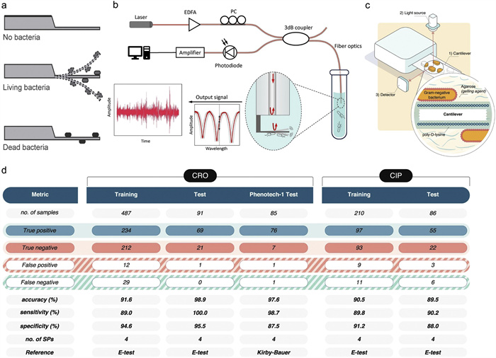
The cantilever, a core component of the atomic force microscope (AFM), can detect the activity and susceptibility of bacteria through its nanoscale vibrations (Fig. 8a). Moreover, this technology is particularly advantageous for monitoring slow-growing bacteria, allowing the AST to be completed within several hours [77]. For example, Zhou et al. developed a highly sensitive optical fiber-based nanomotion sensor that provides AST results within minutes by detecting nanoscale oscillations (Fig. 8b) [78]. Sturm et al. developed a cantilever-based platform to perform AST on 223 strains, accurately predicting susceptibility and resistance with accuracies between 89.5% and 98.9% (Figs. 8c and d) [79].
The rise of antimicrobial resistance necessitates the prudent use of antibiotics, for which rapid AST is essential [80,81]. However, traditional AST methods, despite their low per-test cost, involve significant instrument investments and, more critically, requrie two to three days to yield results. For patients with severe infections, such a delay can lead to irreversible organ damage or death, underscoring the necessity of developing rapid AST technologies. Recently, micro/nano technologies have been fully employed in the development of rapid AST, driving remarkable advancements in this field. This review has outlined key advances, highlighting how the unique optical properties of AuNPs and QDs facilitate equipment-free AST and visible readouts. The structural and compositional tunability of MOFs allows for multimodal detection strategies. Meanwhile, highly integrated microfluidic chips enable both phenotypic and genotypic AST at the single-cell level. The cost of emerging AST methods varies significantly, from approximately $0.69 to hundreds of dollars per test [11,36,58,63,74]. Methods utilizing nanomaterials (e.g., AuNPs, QDs) are generally low-cost, as they often rely on visual readouts that avoid the need for expensive instruments. In contrast, platforms based on micro/nano structures, such as microfluidic chips, entail higher expenses due to complex fabrication processes, custom-made components, and the requirement for skilled operation. However, this higher cost is frequently justified by their superior detection sensitivity and multiplexing capabilities. Overall, micro/nano technologies have shortened the AST turnaround time to just a few hours while also providing greater flexibility in terms of cost options.
Although significant progress has been made in micro/nano technology-based AST, some challenges still need to be addressed prior to their clinical application. First, most current AST methods rely on bacterial proliferation. However, this approach cannot be applied to non-proliferating yet metabolically active bacteria, which may lead to inaccurate discrimination between truly susceptible and resistant phenotypes [82]. This requires the strategic development of highly specific nanosensors capable of monitoring bacterial metabolic activity in real time.
Second, the key to minimizing the AST turnaround time lies not in further reducing the detection time itself, but in shortening the sample preprocessing steps such as bacterial culture and ultimately establishing an integrated rapid testing platform that spans from sample collection to result analysis. Future research must focus on reducing or even eliminating sample preprocessing. For example, by leveraging nanomaterial-based separation technologies to directly isolate and enrich pathogens from complex clinical samples and combining these with highly sensitive genotypic detection or single-cell AST approaches, it would be possible to obtain results more rapidly.
Clinically, polymicrobial infections are frequently encountered and pose severe threats to human health. Therefore, future micro-nano platforms should extend beyond single-drug testing. We envision high-density microarrays or droplet-based microfluidic systems capable of simultaneously screening hundreds of antibiotic combinations. Such platforms would not only identify effective antibiotics but also reveal the most potent synergistic drug combinations for specific infections, paving the way for truly personalized antibiotic therapies. Given the rapid advances in micro/nano technologies and the advantages of new AST methods, such as rapid speed, low cost, and operational simplicity, we anticipate their clinical translation in the near future.
Ling Sun: Writing – original draft, Formal analysis, Data curation, Conceptualization. Xiaoqing Wei: Investigation, Formal analysis. Yingping Li: Investigation, Formal analysis. Junzhen Deng: Formal analysis. Jingya Zhao: Writing – review & editing, Supervision, Funding acquisition, Conceptualization. Shaobing Zhou: Supervision.
The authors declare no competing financial interest.
This work was supported by the National Natural Science Foundation of China (No. 32371466), the Sichuan Science and Technology Program (No. 2024NSFTD0002) and the Fundamental Research Funds for the Central Universities (No. 2682026ZTPY033).
G.A.R. Collaborators, Lancet 404 (2024) 1199–1226. doi: 10.1016/S0140-6736(24)01867-1
R.C. MacLean, A.S. Millan, Science 365 (2019) 1082–1083. doi: 10.1126/science.aax3879
H. Zhao, L. Wei, H. Li, et al., Lancet Infect. Dis. 21 (2021) 847–857. doi: 10.1016/S1473-3099(20)30596-X
D. Nathwani, D. Varghese, J. Stephens, et al., Antimicrob. Resist. Infect. Control. 8 (2019) 35. doi: 10.1186/s13756-019-0471-0
F. Baquero, B.R. Levin, Nat. Rev. Microbiol. 19 (2021) 123–132. doi: 10.1038/s41579-020-00443-1
A. Bolotsky, R. Muralidharan, D. Butler, et al., Biosens. Bioelectron. 172 (2021) 112615. doi: 10.1016/j.bios.2020.112615
E. Ruiz-Hitzky, M. Darder, B. Wicklein, et al., Adv. Healthc. Mater. 9 (2020) e2000979. doi: 10.1002/adhm.202000979
W. Gao, L. Zhang, Nat. Rev. Microbiol. 19 (2021) 5–6. doi: 10.1038/s41579-020-00469-5
J. Shen, X. Zhou, Y. Shan, et al., Nat. Commun. 11 (2020) 267. doi: 10.1038/s41467-019-14135-9
L. Li, M. Liu, S. Deng, et al., Small 19 (2023) e2208249. doi: 10.1002/smll.202208249
M. Agar, M. Laabei, H.S. Leese, P. Estrela, Biosens. Bioelectron. 272 (2025) 117136. doi: 10.1016/j.bios.2025.117136
X. Huang, L. Li, Z. Chen, et al., Adv. Mater. 35 (2023) e2302431. doi: 10.1002/adma.202302431
Z. Ma, H. Wang, Z. Shi, et al., ACS Nano 17 (2023) 5740–5756. doi: 10.1021/acsnano.2c12165
J. Wang, B. Hao, K. Xue, et al., Adv. Mater. 34 (2022) e2205653. doi: 10.1002/adma.202205653
Y. Wang, F. Wu, Y. Li, et al., Mater. Horiz. 11 (2024) 4781–4790. doi: 10.1039/d4mh00642a
L. Yu, Y. Sun, Y. Niu, et al., Adv. Healthc. Mater. 12 (2023) e2202596. doi: 10.1002/adhm.202202596
J. Zeng, C. Gu, X. Geng, et al., Small 20 (2024) e2307096. doi: 10.1002/smll.202307096
Z. Zhang, J. Xie, J. Xing, et al., Adv. Healthc. Mater. 12 (2023) e2201565. doi: 10.1002/adhm.202201565
Z. Zhang, H. Yan, B. Qiu, et al., Small 18 (2022) e2200813. doi: 10.1002/smll.202200813
W. Zhong, S. Handschuh-Wang, U.T. Uthappa, et al., ACS Nano 18 (2024) 32335–32363. doi: 10.1021/acsnano.4c11430
K.P. Survanshu Saxena, Nadim Ahamad, Subhasini Singh, Prachi Bendale, Rinti Banerjee, ACS Biomater. Sci. Eng. 8 (2022) 2232–2257. doi: 10.1021/acsbiomaterials.1c01516
Z. Liang, Z. Zeng, Y. Liao, et al., Front. Microbiol. 15 (2024) 1501204. doi: 10.3389/fmicb.2024.1501204
Z. Zhang, Y. Wang, Y. Pang, C. Liu, Antimicrob. Agents Chemother. 58 (2014) 5632–5635. doi: 10.1128/AAC.02778-14
T.P. Cusack, E.A. Ashley, C.L. Ling, et al., Clin. Microbiol. Infect. 25 (2019) 910–911. doi: 10.1016/j.cmi.2019.03.007
A. Baraz, M. Chowers, D. Nevo, U. Obolski, Clin. Microbiol. Infect. 29 (2023) 390 e391-390 e394.
G. Palladini, C. Garbarino, A. Luppi, et al., J. Microbiol. Methods 212 (2023) 106796. doi: 10.1016/j.mimet.2023.106796
T.P. Lazou, S.C. Chaintoutis, J. Microbiol. Methods 204 (2023) 106649. doi: 10.1016/j.mimet.2022.106649
G.M. Carvalho, B.A. Silva, R.G.C. Xavier, et al., Anaerobe 80 (2023) 102720. doi: 10.1016/j.anaerobe.2023.102720
L.A. Arroyo, A. García-Curiel, M.E. Pachón-Ibañez, et al., J. Clin. Microbiol. 43 (2005) 903–905. doi: 10.1128/JCM.43.2.903-905.2005
M. Vading, O. Samuelsen, B. Haldorsen, A.S. Sundsfjord, C.G. Giske, Clin. Microbiol. Infect. 17 (2011) 668–674. doi: 10.1111/j.1469-0691.2010.03299.x
A. Garrido-Maestu, M. Prado, Compr. Rev. Food Sci. Food Saf. 21 (2022) 1913–1939. doi: 10.1111/1541-4337.12902
F.R. Neuenschwander, B. Groß, S. Schubert, Antibiotics 12 (2023) 1042. doi: 10.3390/antibiotics12061042
X. Lin, K. Zhai, B.M. Liu, J. Chen, Anal. Chem. 97 (2025) 17040–17049. doi: 10.1021/acs.analchem.5c02762
K. Syal, R. Iriya, Y. Yang, et al., ACS Nano 10 (2015) 845–852.
H.W. Cheng, H.M. Tsai, Y.L. Wang, Anal. Chem. 95 (2023) 16967–16975. doi: 10.1021/acs.analchem.3c03259
M. Zhao, F. Cao, J. Chen, et al., Biosens. Bioelectron. 221 (2023) 114902. doi: 10.1016/j.bios.2022.114902
S. Wei, Q. Tang, X. Hu, et al., ACS Sens. 9 (2024) 325–336. doi: 10.1021/acssensors.3c02014
P. She, Y. Wang, Y. Liu, et al., Microbiologyopen 8 (2019) e933. doi: 10.1002/mbo3.933
C. Chen, W. Hong, Antibiotics 10 (2021) 10030311.
J. Gao, J. Guo, J. Chen, et al., Adv. Healthc. Mater. 11 (2022) e2101736. doi: 10.1002/adhm.202101736
J. Gao, J. Qin, C. Ding, et al., RSC Chem. Biol. 3 (2022) 1314–1319. doi: 10.1039/d2cb00163b
X. Gao, M. Li, M. Zhao, et al., Anal. Chem. 94 (2022) 6957–6966. doi: 10.1021/acs.analchem.1c05006
J. Zhang, M. Wang, J. Xiao, et al., ACS Sens. 9 (2024) 379–387. doi: 10.1021/acssensors.3c02144
R. Huang, X. Cai, J. Du, et al., ACS Nano 16 (2022) 19229–19239. doi: 10.1021/acsnano.2c08532
Y. An, X. Fang, J. Cheng, et al., RSC Med. Chem. 15 (2024) 380–398. doi: 10.1039/d3md00581j
J.H. Park, H.R. Kim, M.J. Kim, et al., ACS Appl. Mater. Interfaces 15 (2023) 56702–56717.
X. Liu, X. Chen, S. Yin, et al., Chem. Eng. J. 499 (2024) 156506. doi: 10.1016/j.cej.2024.156506
L. Sun, Y. Chen, Y. Duan, F. Ma, ACS Appl. Mater. Interfaces 13 (2021) 38923–38930. doi: 10.1021/acsami.1c11949
L.J. Zhang, L. Xia, H.Y. Xie, Z.L. Zhang, D.W. Pang, Anal. Chem. 91 (2019) 532–547. doi: 10.1021/acs.analchem.8b04721
A.M. Saeboe, A.Y. Nikiforov, R. Toufanian, et al., Nano Lett 21 (2021) 3271–3279. doi: 10.1021/acs.nanolett.1c00600
L.L. Chen, L. Zhao, Z.G. Wang, S.L. Liu, D.W. Pang, Small 18 (2022) e2104567. doi: 10.1002/smll.202104567
X. Jiang, H. Zeng, C. Duan, et al., Dalton Trans. 51 (2022) 3581–3589. doi: 10.1039/d1dt03624f
C. Wang, B. Tang, J. Wu, et al., Int. J. Antimicrob. Agents 62 (2023) 106898. doi: 10.1016/j.ijantimicag.2023.106898
A. Matsui, H. Niimi, Y. Uchiho, et al., Sci. Rep. 9 (2019) 13565. doi: 10.1038/s41598-019-49358-9
J. Ki, I.H. Kwon, J. Lee, et al., J. Hazard. Mater. 460 (2023) 132398. doi: 10.1016/j.jhazmat.2023.132398
D.R. Giacobbe, T. Giani, M. Bassetti, et al., Clin. Microbiol. Infect. 26 (2020) 713–722. doi: 10.1016/j.cmi.2019.09.023
R. Aloni-Grinstein, O. Schuster, S. Yitzhaki, et al., Front. Microbiol. 8 (2017) 312.
T.H. Kim, J. Kang, H. Jang, et al., Nature 632 (2024) 893–902. doi: 10.1038/s41586-024-07725-1
M.P. Luyao Ma, Xiaonan Lu, Appl. Environ. Microb. 86 (2020) e00096-00020.
A. Arienzo, L. Murgia, V. Cellitti, et al., PLoS One 18 (2023) e0284746. doi: 10.1371/journal.pone.0284746
C.C. Chung, I.F. Cheng, H.M. Chen, et al., Anal. Chem. 84 (2012) 3347–3354. doi: 10.1021/ac300093w
I.H. Su, W.C. Ko, C.H. Shih, et al., Anal Chem 89 (2017) 4635–4641. doi: 10.1021/acs.analchem.7b00220
N. Jo, B. Kim, S.M. Lee, et al., Biosens. Bioelectron. 102 (2018) 164–170. doi: 10.1016/j.bios.2017.11.010
H. Leonard, S. Halachmi, N. Ben-Dov, O. Nativ, E. Segal, ACS Nano 11 (2017) 6167–6177. doi: 10.1021/acsnano.7b02217
F.E. Chen, A. Kaushik, K. Hsieh, et al., Anal. Chem. 93 (2021) 1260–1265. doi: 10.1021/acs.analchem.0c04095
P.J.W. He, I.N. Katis, A.J.U. Kumar, et al., Biosens. Bioelectron. 152 (2020) 112008. doi: 10.1016/j.bios.2020.112008
W.B. Lee, C.C. Chien, H.L. You, et al., Biosens. Bioelectron. 176 (2021) 112890. doi: 10.1016/j.bios.2020.112890
C. Chen, Y. Wang, F. Wu, W. Hong, Anal. Chem. 95 (2023) 15556–15565. doi: 10.1021/acs.analchem.3c02243
H. Li, K. Hsieh, P.K. Wong, et al., Nat. Rev. Methods Primers 3 (2023) 6. doi: 10.1038/s43586-022-00190-y
P. Juskova, S. Schmitt, A. Kling, et al., ACS Sens 6 (2021) 2202–2210. doi: 10.1021/acssensors.1c00020
G. Pitruzzello, S. Johnson, T.F. Krauss, Biosens. Bioelectron. 224 (2023) 115056. doi: 10.1016/j.bios.2022.115056
V. Kandavalli, P. Karempudi, J. Larsson, J. Elf, Nat. Commun. 13 (2022) 6215. doi: 10.1038/s41467-022-33659-1
P. Athamanolap, K. Hsieh, C.M. O’Keefe, et al., Anal. Chem. 91 (2019) 12784–12792. doi: 10.1021/acs.analchem.9b02344
J. Riti, G. Sutra, T. Naas, et al., Biosens. Bioelectron. 257 (2024) 116301. doi: 10.1016/j.bios.2024.116301
H.S. Cui, Z.R. Wu, X.Y. Shi, et al., ACS Sens 9 (2024) 3540–3548. doi: 10.1021/acssensors.4c00381
R. Wang, S. Vemulapati, L.F. Westblade, et al., Anal. Chem. 92 (2020) 2731–2738. doi: 10.1021/acs.analchem.9b04991
M.I. Villalba, P. Stupar, W. Chomicki, et al., Small 14 (2018) 1702671. doi: 10.1002/smll.201702671
J. Zhou, C. Liao, M. Zou, et al., Nano Lett 24 (2024) 2980–2988. doi: 10.1021/acs.nanolett.3c03781
A. Sturm, G. Jozwiak, M.P. Verge, et al., Nat. Commun. 15 (2024) 2037. doi: 10.1038/s41467-024-46213-y
U. Theuretzbacher, S. Gottwalt, P. Beyer, et al., Lancet Infect. Dis. 19 (2019) e40-e50. doi: 10.1016/S1473-3099(18)30513-9
E.M. Darby, E. Trampari, P. Siasat, et al., Nat. Rev. Microbiol. 21 (2023) 280–295. doi: 10.1038/s41579-022-00820-y
Y.F. Tao, Y. Wang, S. Huang, et al., Anal. Chem. 89 (2017) 4108–4115. doi: 10.1021/acs.analchem.6b05051

Figure 3 (a) Strategy for AST based on functionalized AuNPs. (b) AuNPs for E. coli (top) and S. aureus (bottom) AST. Reprinted with permission [36]. Copyright 2023, Elsevier. (c) Detection of bacterial lysis products based on AuNPs for AST. Reprinted with permission [37]. Copyright 2024, American Chemical Society.

Figure 4 AuNPs respond to the proliferation of bacteria for AST. (a) Principle of the H2O2-GNP platform. (b) Photograph of GNP solutions with different concentrations of H2O2. (c, d) UV−vis absorption spectra of GNP solutions and corresponding A540/A650. TEM images of GNP grown with H2O2 at concentrations of (e) 0, (f) 80, (g) 160, and (h) 240 μmol/L, respectively. Scale bar: 200 nm. Reprinted with permission [44]. Copyright 2022, American Chemical Society.

Figure 5 MOFs with fluorescence properties for AST. (a) Strategy for AST based on MnO2@ZIF-90. (b) Calibration curve of the fluorescence signal for MnO2@ZIF-90. (c) AST results for five bacterial strains based on MnO2@ZIF-90 nanomaterials. Reprinted with permission [47]. Copyright 2024, Elsevier.

Figure 6 Microelectrode arrays for AST. (a, b) Bacteria distribution in a quadruple electrode array and their morphology before and after treated with antibiotics. Reprinted with permission [61,62]. Copyright 2012, 2017, American Chemical Society. (c) Schematic diagram of aptamer-functionalized electrode array. Reprinted permission [63]. Copyright 2018, Elsevier.

Figure 7 Microcolumn arrays for AST. (a) Principle of AST platform based on microcolumn arrays. (b) False-colored, cross-sectional HR-SEM micrographs (left) and plan-view HR-SEM images (right) of E. coli immobilized on silicon micropillars. Scale bars: 1 μm. (c) PRISM curves of micropillar gratings. Reprinted with permission [64]. Copyright 2017, American Chemical Society.

Figure 8 Cantilevers for AST. (a) Scheme of AST based on cantilevers. Reprinted with permission [77]. Copyright 2017, Wiley. (b) Scheme of optical fiber-based nanomotion sensor for AST. Reprinted with permission [78]. Copyright 2024, American Chemical Society. (c, d) Machine learning-assisted nanomotion technology platform for AST. Reprinted with permission [79]. Copyright 2024, Springer Nature.
Table 1. Properties of namomaterials and biological indicators used in the nanomaterial-based AST.
| Material | Property | Biological indicator | Ref. |
| AuNPs | Visualization | LPS, peptidoglycan, 16 s RNA, metabolic activity, catalase | [36,37,42-44] |
| MOFs | Large specific surface area | Bacterial membrane, ATP, LPS | [47,48] |
| QDs | Visualization | Lipid A in LPS, DNA | [52,53] |
| Magnetic nanoparticles | Magnetic separation | Pathogen-associated molecular patterns, genomic DNA (gDNA) | [58] |
 下载: 导出CSV

 扫一扫看文章


扫一扫关注我们


 下载: