Citation:
Xiaoxiong Wang, Hu Li, Peng Sun, lingling Li, Shoufeng Tang, Tiecheng Wang. Facilitating the spread of antibiotic resistance genes: The role of low carbonate radical anion concentrations in aquatic and intestinal environments[J]. Chinese Chemical Letters,
2026, 37(11): 112648.
doi:
10.1016/j.cclet.2026.112648
Facilitating the spread of antibiotic resistance genes: The role of low carbonate radical anion concentrations in aquatic and intestinal environments
English
Facilitating the spread of antibiotic resistance genes: The role of low carbonate radical anion concentrations in aquatic and intestinal environments
Department of Gastroenterology, General Hospital of Ningxia Medical University, Ningxia Province, Yinchuan 750021, China
b.
School of Ecology and Environment, Ningxia University, Yinchuan 750021, China
c.
College of Natural Resources and Environment, Northwest A&F University, Yangling 712100, China
d.
State Key Laboratory of Metastable Materials Science and Technology, Hebei Key Laboratory of Heavy Metal Deep-Remediation in Water and Resource Reuse, Yanshan University, Qinhuangdao 066004, China
e.
Ningxia Sports Science and Technology Center, Yinchuan 750021, China
Received Date:
26 October 2025 Accepted Date:
19 March 2026 Revised Date:
31 December 2025 Available Online:
15 November 2026
Abstract:
Antibiotic resistance genes (ARGs) have emerged as a critical global threat to public health and environmental sustainability. Carbonate radical anion (CO3•−), a potent reactive species, plays a significant role in both aquatic environments and biological systems, contributing to various oxidative biochemical processes. However, the effects of CO3•− on the transfer of ARGs in aquatic ecosystems and the human intestinal microbiome remain poorly understood. In this study, we investigated the conjugative transfer of the RP4 plasmid, harboring ARGs, in both water environments and the human intestinal microbiome under CO3•− exposure. Our findings revealed that low concentrations of CO3•− in water significantly enhanced the transfer of ARGs, while higher concentrations suppressed this process. The inhibitory effects at elevated CO3•− concentrations were linked to a reduction in the SOS response, decreased ATP synthesis through the electron transport chain and tricarboxylic acid cycle, and diminished intercellular contact. Machine learning analysis further confirmed that SOS response, cellular contact, and energy supply are pivotal factors in regulating ARGs transfer under CO3•− exposure. Additionally, low CO3•− levels markedly altered the human intestinal microbiome, influencing pathogenic bacteria, ARGs, and transport proteins, resulting in a 47.5% increase in the conjugative transfer of ARGs. This study elucidates the mechanisms by which CO3•− facilitates ARGs spread in aquatic and intestinal environments, offering novel insights into the potential health risks associated with ARG dissemination.
Antimicrobial resistance (AMR) has emerged as a global health crisis, contributing to increased mortality rates and escalating healthcare costs [1-5]. In recent decades, the spread of antibiotic resistance genes (ARGs) has amplified the threat posed by microorganisms to human health, challenging the foundational concept of "One Health" [6-8]. ARGs are now commonly detected across diverse water sources, including drinking water, groundwater, surface water, and wastewater, further intensifying the problem [9,10]. Horizontal gene transfer (HGT) is a primary mechanism for the propagation of ARGs and predominantly relies on mobile genetic elements such as plasmids, integrons, and transposons [5,11,12]. Among these, plasmid-mediated conjugation is particularly significant, facilitating the transfer of ARGs not only between bacterial species but also among bacteria, other organisms, and even plants. This process plays a pivotal role in the widespread dissemination of ARGs within ecosystems [13,14]. Notably, ARGs can enter the human body through food and drinking water, where they may evolve, posing substantial health risks [3,14]. Currently, antimicrobial resistance results in approximately 1.3 million global deaths annually, with projections indicating this number may reach 10 million by 2050 [15]. Thus, the transfer of ARGs represents a significant public health concern.
Our previous work demonstrated a drastic 99.97% reduction in the conjugative transfer frequency (CTF) of ARGs in aqueous solutions following exposure to reactive oxygen species (ROS) such as hydroxyl radicals (•OH) [16]. Chlorination disinfection, which produces reactive oxygen species like hypochlorite (ClO•), has been shown to significantly degrade ARGs, with high reaction rate constants (4.29 × 10¹⁰ L mol-1 s-1) [17]. Similarly, sulfate radicals (SO4•−) have been found to reduce mobile genetic elements that facilitate HGT of ARGs by up to 76.1% [18]. While much of the research has focused on the effects of free radicals on ARG transfer in environmental media, little is known about their influence on ARG transfer within the human body. In the human gut, HGT is a well-documented process involving various molecular mechanisms, with transduction and conjugative transfer being the primary modes of genetic exchange [19]. Thus, understanding the role of free radicals in the transmission of ARGs within the human body is crucial for mitigating ARG risks.
Carbonate radical anion (CO3•−, reduction potential was 1.59 V) is an electrophilic reagent, and its lifetime is three orders of magnitude longer than those of •OH and •Cl [20]. The CO3•− can react selectively with electron-rich sites of compounds with reaction rate constants of 109–1010 L mol−1 s−1 [21]. Significant amounts of CO3•− are generated in organisms during normal metabolic activities or in inflamed and pathological tissues under conditions of oxidative stress [22,23]. CO2 can react with peroxynitrite anion (ONOO−) to generate an extremely unstable intermediate (ONOOCO2−), which undergoes homolysis at its O-O bond, releasing CO3•− and NO2 [24,25]. Additionally, •OH can effectively oxidize some of the CO32− or HCO3− present in the body, producing CO3•− with reaction rate constants of 4.2 × 10⁸ and 8.5 × 10⁶dm3 mol−1 s−1, respectively [24]. The concentration of HCO3− in intracellular fluids can reach 10 mmol/L, and it is particularly higher in inflamed tissues [26]. Previous studies established that CO3•− influenced proteins, lipids, nucleic acids, and other cellular components during cell metabolism, and participated in the activities of cell synthesis [22,23]. CO3•− is a significant active substance in the human body, and ARGs can be ingested through the food chains, allowing them to coexist for extended periods, particularly within the intestinal system [27,28]. However, the underlying impacts of CO3•− on ARGs transfer in the water environment and in the human body remains unclear.
Herein, we investigated the effects of CO3•− exposure on the conjugative transfer of the RP4 plasmid (which encodes ARGs) in both water and in vitro colon simulation systems. We employed the bicarbonate-activated hydrogen peroxide process, a cost-effective and environmentally friendly method, to generate CO3•− [29,30]. The CTF of RP4 plasmids was first assessed in a water environment under varying concentrations of CO3•−. Subsequently, we explored the metabolic responses of cells involved in conjugative transfer, focusing on energy metabolism, intercellular interactions, membrane permeability, and oxidative stress. Using transcriptomic analysis and gene expression profiling, we examined the underlying mechanisms of CO3•−-induced regulatory changes in conjugation. Additionally, a machine learning-based random forest model was applied to assess the contribution of cellular metabolism to conjugation. Finally, the effects of CO3•− exposure on the CTF of RP4 plasmids in the human intestinal microbiome were evaluated in a colon simulation system.
Prof. Barth F. Smets from the Technical University of Denmark provided the Escherichia coli MG1655 (lacIq-Plpp-mCherry) strain carrying the RP4 plasmid with antibiotic resistance genes (ApR, KmR, TcR) and GFP labeling. E. coli ETEC, sensitive to these antibiotics, was obtained from Baiou Bowei Biotech Co., Ltd. (Beijing), and chemicals were sourced from Sigma-Aldrich (USA). The in vitro intestinal simulator setup (Fig. S1 in Supporting information) consisted of a fecal collection container, a feeding vessel with nutrient medium, and a fermenter maintained at 37 ℃, each containing 300 mL of fermentation broth. Intestinal microbiota was collected from healthy adult stool samples, with no antibiotic use in the past three months. Two microbial models were used: Model-1 (E. coli MG1655 and human intestinal microbiome) and Model-2 (E. coli ETEC), both exposed to different concentrations of NaHCO3 and H2O2 to study the effect of CO3•− on ARGs transfer. Bacterial colonies were collected after CO3•−-exposed conjugation, DNA and RNA were extracted and sequenced by Shanghai Personal Biotechnology Co., Ltd. and Sangon Biotech (Shanghai). Further details can be found in Supporting information.
To evaluate the effect of CO3•− exposure on CTF, a 10 mL PBS-based system containing 108 CFU/mL of donor and recipient cells (1:1 vol ratio) was prepared. Sodium bicarbonate (NaHCO3, SBC) was added at concentrations of 4, 8, 16, 32, 64, and 128 mg/L, followed by adding 1 μL of 30% H2O2 to initiate the reaction. At low levels of CO3•− (4 and 8 mg/L SBC), the CTF of ARGs from E. coli (MG1655) to E. coli (ETEC) was significantly promoted, while high concentrations of CO3•− (16–128 mg/L SBC) inhibited the transfer (P < 0.05) (Figs. 1a and b). Similarly, the number of transconjugants significantly decreased by 0.71-fold under high-level CO3•− exposure (128 mg/L SBC) (P < 0.05), as shown in Fig. 1c. This result indicated that high levels of CO3•− significantly inhibited ARGs conjugation transfer in aquatic systems. It is noteworthy that the densities of both recipient and donor strains were not significantly affected by 1 mmol/L H2O2 or NaHCO3 concentrations (16–64 mg/L) (Fig. S2 in Supporting information). However, E. coli numbers slightly decreased as NaHCO3 concentration increased to 128 mg/L. These results suggested that appropriate CO3•− exposure dosage could promote the spread of ARGs in the water environment.
Figure 1
Figure 1.
Changes in conjugation transfer frequency of plasmid RP4 after CO3•− exposure in water. (a) Conjugation transfer frequency. (b) Fold change of CTF. (c) Number of transconjugants. (d) Changes in number of E. coli. Significant differences between different experimental duration spans were tested with an independent-sample t-test and marked with * for P < 0.05, data were presented as mean ± SD (n = 3). Letters a-g indicate significant differences at P < 0.05. [cell density] = 108 CFU/mL, [H2O2] = 1 mmol/L.
The population density of donor and recipient cells is a critical factor in the CTF of ARGs [31,32]. Our previous work demonstrated that long-term exposure to free radicals significantly inhibited CTF between E. coli due to reduced cell densities [16]. Similarly, E. coli density decreased with increasing CO3•− concentrations (Fig. 1d), particularly at higher levels. This decrease in density corresponded to a reduction in CTF, suggesting a direct link between CTF and microbial population decline [33]. Guo et al. [31] also observed that UV disinfection altered the numbers of donor and recipient strains, leading to changes in ARGs conjugative transfer.
Genome-wide RNA sequencing was employed to identify differentially expressed genes (DEGs) between the CO3•− exposure group and control group. Greater effects on down-regulated genes were detected under high levels of CO3•− exposure (Figs. S3 and S4 in Supporting information). The significantly down-regulated genes in the Kyoto Encyclopaedia and Genomes (KEGG) terminology were mainly involved in cellular processes, environmentally-informed processes, genetically-informed processes, metabolism, and organismal systems (Fig. S5 in Supporting information).
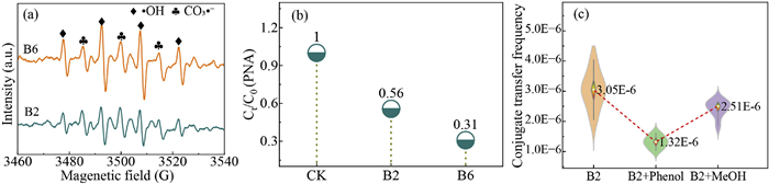
As shown in Fig. 2a, •OH and CO3•− were detected and their electron paramagnetic resonance (EPR) intensities increased with the HCO3− concentration [34]. p-Nitroaniline (PNA) was employed as the probing molecule to quantify CO3•− due to its high reaction rate constant (7.3 × 10⁷ L mol−1 s−1) with CO3•− [35]. In the intestinal simulator, nearly 69.0% of PNA was converted as the HCO3− concentration reached 128 mg/L (Fig. 2b), demonstrating the production of CO3•−. Additionally, phenol and MeOH inhibited the CTF by 56.7% and 17.7%, respectively (Fig. 2c). Thus, CO3•− emerged as the primary reactive oxygen species responsible for altering ARGs transfer in the intestinal simulator system.
Figure 2
Figure 2.
Free radical detection. (a) EPR spectra. (b) The conversion of PNA. (c) Quenching experiments of •OH and CO3•−. •OH was quenched using 30 mmol/L MeOH, and CO3•− was quenched using 2.4 mg/L phenol. B2 and B6 represent 8 and 128 mg/L bicarbonate concentrations, respectively. [cell density] = 108 CFU/mL, [H2O2] = 1 mmol/L.
ROS is a by-product of cellular respiration and redox cycles and can induce oxidative stress in cells at high concentrations, leading to the destruction of intracellular biomacromolecules (such as nucleic acids, membrane lipids, and cellular proteins) and ultimately triggering apoptosis [3,36]. The levels of ROS and enzymes are important signals and factors affecting the conjugative transfer of ARGs [37]. A previous study reported that ROS affected ARGs horizontal transfer mainly by changing membrane permeability and inducing SOS response [38]. Figs. S6 and S7 (Supporting information) depict the antioxidant stress in E. coli strains under exposure to CO3•−. Similar to the CTF, low levels of CO3•− exposure promoted intracellular ROS generation in the E. coli strains (P < 0.05) (Fig. S6a). SOD and CAT are known to be the most important antioxidant enzymes for scavenging ROS [39], and influence the conjugative transfer of ARGs [40]. The SOD and CAT activities increased at relatively low levels of CO3•− exposure and significantly decreased at high levels (Figs. S7a and b). To further determine whether the generation of intracellular ROS induced by CO3•− influenced the conjugative transfer of ARGs, thiourea was added to the conjugation system together with the CO3•−. Notably, the addition of the scavenger thiourea significantly reduced the CTF in the CO3•− groups (P < 0.05) (Fig. S7c) compared to the thiourea-free group. These results suggested that the levels of ROS played a significant role in regulating ARGs conjugation transfer [41]. Our previous study also found that long-time exposure to free radicals inhibited the ARGs conjugative transfer by reducing the intracellular ROS contents and enzyme activities [16]. In comparison with the control, the genes associated with SOS response (sodA, sodB, sodC, katG, yebY, and yebE) were significantly up-regulated under low levels of CO3•− exposure (Fig. S6d). This result was consistent with the CTF of ARGs under CO3•− exposure. Consequently, the reduced CTF of ARGs under high levels of CO3•− exposure could be partially due to suppression of SOS responses. Exposure to high CO3•− environments reduces ROS levels, weakening the oxidative stress response, membrane protein activity, and fluidity, which in turn inhibits conjugative transfer by reducing cell contact, shortening conjugation duration, and decreasing plasmid transfer efficiency [3,11].
Yu et al. [39] reported that ROS accumulation destroyed the cell membranes under high doses of CeO2 exposure, which enhanced the conjugative transfer of ARGs. The cell membrane permeability under CO3•− exposure was measured by malondialdehyde (MDA), scanning electron microscope (SEM), and flow cytometer (FCM) (Figs. S6b, S6g-i, and Fig. S8 in Supporting information). Before the CO3•− exposure, the E. coli strains maintained complete blue fluorescence; however, a dense population of purple and green fluorescent cells was observed after exposure to CO3•− (Figs. S8a-c). Similarly, a wrinkled morphology was observed after exposure to low levels of CO3•−, and strong rupture of the cells at high levels of CO3•− (Figs. S6g-i). Images of LB plates and laser confocal scanning microscopy further demonstrated this point (Figs. S8d-i). MDA was an important indicator of cell destruction [42]. The MDA content first increased and then decreased with the CO3•− exposure dosage (Fig. S6b). Meanwhile, under high levels of CO3•− exposure, the levels of genes associated with cell membrane were down-regulated (Fig. S6d). Thus, cell membrane permeability played a crucial role in influencing the conjugative transfer of ARGs in the presence of CO3•−.
Polysaccharides (PS), proteins (PN), and lipids affect cell surface properties and play an important role in intercellular adhesion and adsorption [16,43]. The PN/PS ratio effectively reflects the relative composition of protein and polysaccharides in extracellular polymeric substances (EPS) and is widely used to evaluate matrix structural properties, adhesion capabilities, and biofilm maturation status [36,44]. The PN/PS first exhibited a remarkable increase in the E. coli strains and then dramatically decreased with the CO3•− exposure (Fig. S6c). Compared with the control, the hydrodynamic size and zeta potential also decreased by 45.1% and 34.0% under high levels of CO3•− exposure, respectively (P < 0.05) (Fig. S9 in Supporting information). The SEM images of the strains further verified the intercellular physical contact (Fig. S6h). High polysaccharide or protein content in EPS enhances cell contact and adhesion stability, promotes conjugation pili formation, prolongs contact time, and increases plasmid transfer success [44]. Additionally, changes in the protein-to-polysaccharide ratio affect key microenvironmental factors, regulating the expression of conjugation-related genes and conjugation efficiency [45]. Flemming et al. reported that EPS could significantly affect the ability of cells to adhere to each other by changing the hydrophobicity and surface charges of the cell membranes [46].
In addition, low ROS levels can lead to a decrease in EPS [47-50]. Fig. S10 (Supporting information) describes the effects of CO3•− on metabolic pathways of protein export and lipopolysaccharide biosynthesis in the E. coli strains, and the genes involved are shown with KEGG pathways. The expression levels of genes related to prokaryotic type translocation channels and related proteins (secA, secE, secG, secM, yidC, and ffh) and tat (twin arginine translocation) system (tatE) were up-regulated under low levels of CO3•− exposure, and then down-regulated at high levels of CO3•− exposure (Fig. S10a). These results proved that there was a reduction in the production of the proteins (membrane). In addition, the absorption and conversion of extracellular UDP-N-acetyl-d-glucosamine, lipid A disaccharide, UDP-3–0-(3-hydroxy-tetradecanoyl)-N-acetyI-d-glucosamine, KDO-Lipid IV A, CMP-3-deoxy-d-manno-octulosonate, 3-deoxy-d-manno-octulosonate, d-glycerol-β-d-manno-Heptose-7P, ADP-d-glycerol-β-d-manno-Heptose, and KDO2-Lipid IV A were reduced in E. coli under high levels of CO3•− exposure (Fig. S10b). Given that EPS could affect intercellular contact, the changes in the EPS contents in the presence of CO3•− played an important role in the ARGs conjugation transfer.
ROS can influence EPS production in bacteria by stimulating the expression of genes associated with metabolic processes [48]. As shown in Fig. S6e, the gene expression levels related to polysaccharides were first up-regulated and then down-regulated with the CO3•− exposure. Similar phenomena were also observed for the quorum sensing-related genes. Quorum sensing affects biofilm formation and ARGs conjugation transfer [49]. Thus, high levels of CO3•− exposure decreased the quorum sensing of E. coli strains and intercellular contact, thereby inhibiting the CTF of ARGs.
ATP serves as the primary energy source for cellular metabolism, facilitates the exchange of cellular information, and plays a crucial role in inducing conjugation transfer mechanisms [51]. The changes in the electron transfer chains and energy diving force of E. coli strains under CO3•− exposure are shown in Fig. S11a (Supporting information). The ATP content was first enhanced and then decreased with the CO3•− exposure dosage (Fig. S11b in Supporting information). Additionally, the transcription levels of ATP-coding genes, which regulate cellular energy production, were also up-regulated under low levels of CO3•− exposure (Fig. S11c in Supporting information). The main energy source for cellular growth and adhesion primarily comes from the synthesis of flavin adenine dinucleotide (FADH2) and nicotinamide adenine dinucleotide (NADH), which are generated through the process of biological oxidation of substances via the electron transport chain (ETC) located on the plasma membrane [52,53]. ETC is mainly composed of complexes Ⅰ-Ⅳ, among which complexes Ⅰ and Ⅲ are the main ROS production sites (Fig. S11a) [54]. The electrons and hydrogen ions released by NADH and FADH2 are transferred through four enzyme complexes, while two removable electron carriers facilitate the transfer of electrons to oxygen [55]. This process releases energy to generate ATP by supplying ADP with phosphoric. The expression levels of genes related to complexes Ⅰ, Ⅱ, and Ⅳ were upregulated under low levels of CO3•− exposure (P < 0.05) (Fig. S11c). Glycolysis and tricarboxylic acid cycle (TCA cycle) are the most effective ways for cells to gain energy (ATP) [32]. Glucose undergoes glycolysis to produce pyruvate, which is then used in the TCA cycle [55]. Stacpoole et al. [56] reported that 1.0 mol of acetyl CoA underwent four dehydrogenations to produce 12.0 mol of ATP. A complete TCA cycle can generate 3.0 mol of NADH, which subsequently enters the ETC for oxidative phosphorylation to yield a substantial quantity of ATP [57]. As shown in Fig. S11c, the expression levels of most genes participating in glycolysis and TCA cycle were down-regulated under high levels of CO3•− exposure. These results suggested that high levels of CO3•− could reduce the initial utilization of glucose by inhibiting the glycolysis and TCA cycle. As a result, ARGs conjugation transfer decreased due to a lack of energy supply.
Plasmid gene expression plays an important role in determining specific mating pairs and facilitating efficient mobilization and transfer channel formation [39]. The conjugation process of the RP4 plasmid is influenced by crucial plasmid-borne factors, including global regulatory genes [58]. The mRNA expression levels of key genes involved in DNA binding transcriptional regulation were significantly down-regulated at high levels of CO3•− exposure (Fig. S6f). In summary, the modulation of conjugative gene expression in the RP4 plasmid by high levels of CO3•− exposure also emerged as an important strategy to mitigate the dissemination of ARGs.
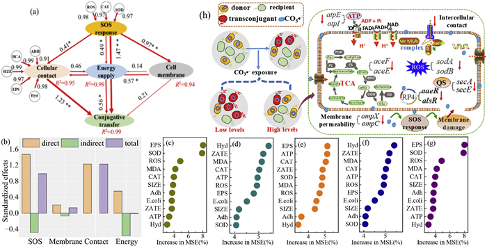
Conjugation transfer of ARGs is influenced by lots of factors, including intracellular ROS, MDA, physical contact between cells, and ATP in E. coli strains [59,39]. Pearson correlation coefficient and structural equation model were applied to establish correlations among various cellular metabolism actions under CO3•− exposure. As shown in Fig. S12 (Supporting information), ROS, CAT, SOD, MDA, zeta potential, ATP, EPS, and adhesion had significantly positive correlations with CTF of ARGs. Notably, the CO3•− exposure significantly and directly drove the changes in ROS, MDA, EPS, and ATP, which were the predominant factors for the CTF of ARGs (P < 0.05) (Figs. 3a and b). Previous studies also reported that various physiological parameters, such as SOS response, cell membrane permeability, physical contact, and energy generation, directly affected the conjugation transfer of ARGs [60,61]. Furthermore, a random forest model in machine learning was used to comprehensively analyze significant physiological and biochemical indicators influencing the CTF of ARGs [62]. Through simulation and testing on independent samples to assess and validate the model's predictive accuracy, EPS, SOD, ROS, MDA, CAT, zeta potential, and ATP were the primary factors influencing the CTF (Figs. 3c-g and Fig. S13, Tables S4 and S5 in supporting information). These results indicated that SOS response, cellular contact, and energy supply made significant contributions, suggesting their pivotal roles in determining CTF.
Figure 3
Figure 3.
Contribution parsing among various E. coli strains parameters and underlying mechanisms on CTF after CO3•− exposure in water. (a, b) Structural equation models. Numbers adjacent to the arrows are path coefficients (λ), and the width of the arrows is proportional to the strength of the path coefficients. (c-g) Variable importance of random forest model based on 5 training sample sets. (h) Promotion mechanisms. CTF was conjugation transfer frequency, ROS was reactive oxygen species, MDA was malondialdehyde, EPS was extracellular polymeric substances, ATP was adenosine triphosphoric. Adh: adhere; Hyd: hydrophobic.
Fig. 3h depicts the potential mechanisms of CO3•− exposure on the ARGs conjugation transfer. CO3•− exposure could accelerate the ARGs conjugation transfer by directly interacting bacterial oxidative stress and SOS response, membrane permeability, physical contact between cells (inhibiting polysaccharides in extracellular polymeric substances), ATP content, and binding energy (electron transport chain and TCA cycle). High levels of CO3•− exposure significantly reduced these phenotypic and genotypic analyses. Simultaneously, transcription analysis revealed significant decreases in the expression of genes related to physiological and biochemical markers under high levels of CO3•− exposure.
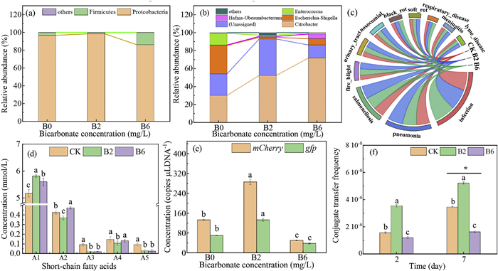
CO3•− can be continuously generated in organisms, particularly in inflamed tissues under oxidative or nitrosative stress conditions, thus establishing it as a crucial reactive oxygen species in biological systems [22,23]. CO3•− participates in oxidation reactions during cellular metabolism and plays a significant role in the synthesis and decomposition of cellular substances [27]. The complex microbial community in the intestine is essential for digestion, immune function, and overall human health [63]. Fig. 4 depicts the changes in the human intestinal microbiome and CTF after CO3•− exposure in the intestinal simulator. At the genera level, Citrobacter (30.2%) and Escherichia-Shigella (31.6%) were the two predominant genera in the control; under exposure to a low level of CO3•−, the abundance of Citrobacter significantly rose from 30.2% to 52.3%, while that of Escherichia Shigella decreased notably from 31.6% to 1.8% (Figs. 4a and b). Similar trends were also observed at the phyla level. Escherichia Shigella and Citrobacter promoted the mineralization of spherical carbonate, participated in the decomposition and cycling of organic matter, and contributed to the pathogenesis of inflammatory bowel diseases [64]. The abundance of pathogenic bacteria, ARGs, and transport proteins significantly increased under exposure to CO3•−, particularly at high levels (Fig. 4c and Fig. S14 in Supporting information). Thus, CO3•− could disrupt the microbial community and lead to dysbiosis. Short-chain fatty acids (SCFAs) were detected to evaluate the intestinal microbiota metabolism. The metabolism of SCFAs exhibited significant alterations under the exposure of CO3•− compared to the control group, potentially impacting human health adversely (Fig. 4d). A previous study reported that SCFAs directly activated G protein-coupled receptors, inhibited histone deacetylases, and served as energy substances to influence ATP generation [65].
Figure 4
Figure 4.
Human intestinal microbiome and conjugate transfer frequency after CO3•− exposure in the intestinal simulator. (a) Major bacterial phyla. (b) Major bacterial genera. (c) Major pathogenic bacteria. (d) Short-chain fatty acids. A1: acetic acid, A2: propionic acid, A3: butyric acid, A4: isobutyric acid, A5: valeric acid. (e) Copy numbers of specific genes. (f) Conjugation transfer frequency in Model-2. Significant differences between different experimental duration spans were tested with an independent-sample t-test and marked with * for P < 0.05, data were presented as mean ± SD (n = 3). Annotation letters a-c indicate significant differences at P < 0.05. B2 and B6 represent 8 and 128 mg/L bicarbonate concentrations, respectively. [cell density] = 108 CFU/mL, [H2O2] = 1 mmol/L.
Additionally, the specific genes mCherry and gfp carried by the RP4 plasmid initially increased and then decreased with rising CO3•− concentration (Fig. 4e). These findings indicated that CO3•− at a low concentration significantly enhanced the abundance of the RP4 plasmid, potentially due to an increase in ARGs horizontal transfer. To demonstrate it, we introduced only the E. coli ETEC and E. coli MG1655 into the fermenter and evaluated the impacts of CO3•− exposure, as depicted in Fig. 4f. The CTF exhibited a significant increase with the exposure time (P < 0.05), and it increased by approximately 0.52-fold as the exposure time extended from 2 days to 7 days under a low level of CO3•− exposure. In addition, the CTF first increased and then decreased with the CO3•− exposure dosage, and it decreased by 52.9% under a high level of CO3•− exposure compared with the control. The intestinal microbiome hosts trillions of organisms that play crucial roles in human health and diseases, and these bacteria wield significant influences over the efficacy and properties of drugs employed for treatment [66]. The accelerated dissemination of ARGs by CO3•− would heighten the likelihood of intestinal bacteria acquiring drug resistance, thus posing serious threats to human health [23,66].
CO3•−, owing to its electrophilic properties, is a highly reactive species that selectively interacts with electron-rich sites in compounds. We investigated the conjugative transfer of the RP4 plasmid carrying ARGs under CO3•− exposure both in water and an in vitro colon simulation model. We found that the transfer of the RP4 plasmid in water was significantly enhanced under low CO3•− exposure conditions, and this promoting effect became more pronounced over time. The promoting effects of CO3•− on conjugative transfer were mainly attributed to significant increases in the SOS response, ATP production via the electron transport chain and tricarboxylic acid cycle, and enhanced intercellular communication through upregulated polysaccharide synthesis. A random forest machine learning model also confirmed that the key drivers of this facilitation included the SOS response, cellular contact, and energy production, which together enabled CO3•− to enhance the conjugative transfer of ARGs in water. Additionally, CO3•− at a low concentration significantly enhanced the abundance of the RP4 plasmid in the human intestinal microbiome. This study revealed the underlying mechanisms of promoted ARGs spread in water and human intestinal microbiome induced by CO3•− stimulation, providing new insights into health risks caused by ARGs.
The authors declare that they have no known competing financial interests or personal relationships that could have appeared to influence the work reported in this paper.
Acknowledgments
The authors thank the National Natural Science Foundation of China (No. 42377388), Ningxia Key Research and Development Program (No. 2024BEG02001), and Natural Science Foundation of Ningxia Province (No. 2023AAC05004)
Supplementary materials
Supplementary material associated with this article can be found, in the online version, at doi:10.1016/j.cclet.2026.112648.
Figure 1
Changes in conjugation transfer frequency of plasmid RP4 after CO3•− exposure in water. (a) Conjugation transfer frequency. (b) Fold change of CTF. (c) Number of transconjugants. (d) Changes in number of E. coli. Significant differences between different experimental duration spans were tested with an independent-sample t-test and marked with * for P < 0.05, data were presented as mean ± SD (n = 3). Letters a-g indicate significant differences at P < 0.05. [cell density] = 108 CFU/mL, [H2O2] = 1 mmol/L.
Figure 2
Free radical detection. (a) EPR spectra. (b) The conversion of PNA. (c) Quenching experiments of •OH and CO3•−. •OH was quenched using 30 mmol/L MeOH, and CO3•− was quenched using 2.4 mg/L phenol. B2 and B6 represent 8 and 128 mg/L bicarbonate concentrations, respectively. [cell density] = 108 CFU/mL, [H2O2] = 1 mmol/L.
Figure 3
Contribution parsing among various E. coli strains parameters and underlying mechanisms on CTF after CO3•− exposure in water. (a, b) Structural equation models. Numbers adjacent to the arrows are path coefficients (λ), and the width of the arrows is proportional to the strength of the path coefficients. (c-g) Variable importance of random forest model based on 5 training sample sets. (h) Promotion mechanisms. CTF was conjugation transfer frequency, ROS was reactive oxygen species, MDA was malondialdehyde, EPS was extracellular polymeric substances, ATP was adenosine triphosphoric. Adh: adhere; Hyd: hydrophobic.
Figure 4
Human intestinal microbiome and conjugate transfer frequency after CO3•− exposure in the intestinal simulator. (a) Major bacterial phyla. (b) Major bacterial genera. (c) Major pathogenic bacteria. (d) Short-chain fatty acids. A1: acetic acid, A2: propionic acid, A3: butyric acid, A4: isobutyric acid, A5: valeric acid. (e) Copy numbers of specific genes. (f) Conjugation transfer frequency in Model-2. Significant differences between different experimental duration spans were tested with an independent-sample t-test and marked with * for P < 0.05, data were presented as mean ± SD (n = 3). Annotation letters a-c indicate significant differences at P < 0.05. B2 and B6 represent 8 and 128 mg/L bicarbonate concentrations, respectively. [cell density] = 108 CFU/mL, [H2O2] = 1 mmol/L.



 DownLoad:

 下载:


 下载:

