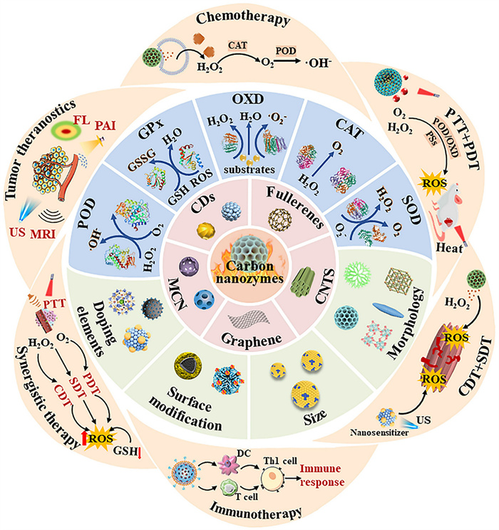
Scheme 1.
The classification, characteristics and application in tumor therapy of carbon-based nanozymes.

Advances in carbon-based nanozymes for photothermal-enhanced tumor theranostics
Na Lin , Lu Zou , Yitan Fang , Jinya Xiong , Qiuling Deng , Zefang Liu , Xueyi Hao , Qinfu Zhao , Xiaofan Wang , Long Wan


Recently, tumor has become one of the greatest threats to human health and life. Traditional treatments such as surgery, radiotherapy, and chemotherapy struggle to completely eradicate tumors and often carry significant side effects. Recently, novel tumor therapies have continuously emerged, among which the catalytic therapy of nanozymes has attracted the interest of researchers [1]. Nanomaterials improve the unique characteristics of the tumor microenvironment (TME) by mimicking the catalytic capabilities of natural enzymes, thereby significantly enhancing the efficacy of other tumor therapies and becoming a core driving force for comprehensive treatment strategies. However, limited by catalytic efficiency, catalytic therapy fails to achieve the expected effects in vivo. In recent years, the synergistic use of catalytic therapy and photothermal therapy (PTT) has surprisingly been found to possess remarkable tumor inhibitory effects. Under laser irradiation, the local high temperature generated by PTT can accelerate electron transfer, while nanozymes with excellent photothermal conversion effects can also generate reactive oxygen species (ROS), thereby exhibiting the enhanced catalytic activity. PTT has garnered widespread attention due to its advantages of low cost, broad applicability, and minimally invasive nature. PTT mainly works by photothermal agents (PTAs) accumulated in tumor tissue converting light energy into thermal energy under light of specific wavelengths, generating local high temperatures to thermally ablate the tumor [2]. Due to the high permeability and complex vascular network of tumors, PTAs can be significantly enriched in the tumor tissue, forming localized hyperthermia while reducing the impact on surrounding normal tissues. Furthermore, the penetration capability of lasers enables PTT to effectively target deep tissues. The therapeutic efficacy of PTT depends on the photothermal conversion efficiency (PCE) of the PTAs. Therefore, selecting PTAs with high PCE and good biosafety is a prerequisite for PTT. In recent years, carbon-based nanomaterials have been discovered to possess strong light absorption characteristics in the conventional near-infrared (NIR) window (NIR-Ⅰ window, 750-1000 nm) [3]. Their unique conjugated structures and plasmonic effects at specific wavelengths not only enhance light absorption capacity in the NIR region but also efficiently convert light energy into thermal energy output, positioning them as highly promising PTAs. Additionally, the rapid conversion of light energy into thermal energy and the rapid dissipation of heat can generate PA waves through thermoelastic expansion [4,5], enabling carbon-based nanomaterials to achieve photoacoustic imaging (PAI) in tumor treatment, thereby playing a key role in tumor therapy.
Carbon is one of the most plentiful elements discovered on earth. Nanoscale carbon allotropes can be classified into fullerenes, carbon dots (CDs), carbon nanotubes (CNTs), mesoporous carbon nanoparticles (MCNs), etc., based on their distinct structures [6]. These carbon allotropes with varying structures exhibit dimensionalities ranging from zero-dimensional (0D) to three-dimensional (3D). Carbon nanomaterials have exhilarating properties, such as (1) high specific surface area, favorable for drug loading; (2) supramolecular π-π stacking force and strong adsorption for high drug-loading capacity and sustained drug release; (3) easy-to-modify surfaces for easy control and targeted drug delivery [7]; (4) excellent biocompatibility and physicochemical stability; and (5) unique optical properties for different synergistic therapeutic and diagnostic strategies [8]. In recent years, carbon nanomaterials have been extensively studied and applied in the field of biomedicine. Nanomaterials, whose dimensions are comparable to those of numerous essential molecules sustaining fundamental life processes in the human body, serve as a pivotal bridge connecting nanoscience and biology. This size-matching characteristic eliminates the dimensional mismatch between artificial nanoscale materials and endogenous biological components, enabling carbon nanomaterials to interact with biological systems at the molecular and subcellular levels with high biocompatibility, and thus establishing an intrinsic link between the engineered design principles of nanoscience and the structural and functional features of biological systems. Nanomaterials ranging from 1 nm to 1 µm in size are comparable to the scale of biological macromolecules; for instance, globular proteins typically range from 1 nm to 100 nm, and the diameter of the DNA double helix is approximately 2 nm. These dimensions allow carbon nanomaterials to interact effectively with biological barriers within the human body, such as the several-nanometer-sized ion exchange channels and the glomerular filtration barrier of the kidneys [9].
Certain natural pro-oxidant enzymes are utilized for their selectivity toward specific substrates, as they are capable of stimulating ROS formation in vivo and consuming oxygen in the local microenvironment. However, natural enzymes are subject to harsh reaction conditions and tend to be inactivated under non-physiological conditions, thus imposing greater limitations in cancer therapy [10]. Nanozymes are a class of catalytic nanomaterials with enzyme-like properties [11]. Nanozymes harness the combined benefits of enzymes and nanomaterials, including both catalytic activity and physicochemical features. Nanozymes possess several advantages over natural enzymes, including ease of preparation, recyclability, customizable functionality, and exceptional stability [12,13]. Consequently, numerous researchers are investigating the development of nanozymes to mitigate the negative therapeutic effects induced by the TME. Nanozymes have the potential to partially substitute natural enzymes to achieve synergistic cancer treatment [14]. Since carbon-based materials have unique sp2 and sp3 carbon atoms, they can be easily doped with different heteroatoms and metal elements in the backbone of carbon-based materials, and the doping facilitates the formation of more active sites, which further enhances the various enzymatic activities of carbon-based materials. Currently, nanozymes exhibit many enzyme-like activities such as oxidase (OXD), peroxidase (POD), superoxide dismutase (SOD), catalase (CAT), and multi-enzyme activities, which have made great contributions to the biomedical field, particularly in the theranostics of tumors.
Due to their favorable electronic structure and particle geometry, carbon nanozymes (CNs) have been studied in areas where chemical drugs are less effective, such as tumor therapies. However, the therapeutic efficacy of many approaches is compromised by the constraints of the complex TME. Low oxygen, low pH, hydrogen peroxide (H2O2) and glutathione (GSH) excess, high osmolarity and immunosuppressive microenvironment are prominent features of solid tumors, which enhances tumor cell proliferation, migration and immune escape [15]. CNs can use their POD-like activity to catalyze the direct generation of ROS from H2O2 in the TME to kill tumor cells. Some CNs can use their POD-like or SOD-like enzyme properties to provide oxygen to the tumor site to ensure that some kinetic therapies that require oxygen can be carried out successfully [16]. In addition, under light irradiation, the surface of carbon-based nanomaterials is activated to expedite the process of enzyme-like reactions (POD-like/OXD-like, etc.), which not only kills the tumor directly by generating high levels of ROS, but also disturbs the redox homeostasis and maintains the sensitivity of the tumors to the anticancer drugs and the damage caused by ROS [17]. Despite their promise, the in vivo application of carbon nanomaterials faces scrutiny regarding biosafety. High aspect ratio materials like CNTs may induce frustrated phagocytosis and chronic inflammation, whereas spherical 0D materials like CDs generally exhibit better biocompatibility and renal clearance. Surface engineering remains a critical strategy to mitigate these risks.
In view of the unique advantages of CNs and the great progress has made, this paper focuses on a systematic review of the individual properties and catalytic mechanisms of the CNs family, especially their applications in tumor theranostics. Besides, it creatively reviews the classification and characteristics of CNs and their latest applications in tumor theranostics in the last decade, including tumor therapy such as enhanced chemotherapy, PTT, photodynamic therapy (PDT), chemodynamic therapy (CDT), sonodynamic therapy (SDT), immunotherapy, synergistic therapy and tumor theranostic such as PAI, ultrasound imaging (USI), magnetic resonance imaging (MRI), fluorescence imaging (FI), and multimodal imaging guidance (Scheme 1). In addition, this review provides an outlook on carbon-based nanozymes, as well as the key issues that need to be addressed. It is hoped that this review will be applicable for the research in the fields of medicine and pharmacology and contribute to the development of carbon-based nanozymes.
The easily modifiable surfaces of carbon nanomaterials, along with their specific targeting of tumor cells, photothermal properties, and enzyme-like activities, are making them increasingly popular in cancer therapy. Carbon nanomaterials can be categorized based on their dimensional structures (0D, 1D, 2D, 3D) and atomic hybridization states. The common sp2 carbon structure in materials like graphene and CNTs endows them with similar optical and electrical properties, while surface defects and edge states in 0D materials like CDs provide abundant active sites for catalysis. We also compiled a table to summarize the characteristics, advantages and applications of different carbon-based nanozymes as shown in Table S1 (Supporting information).
CDs, also known as carbon quantum dots (CQDs), consist of ultrafine, scattered, quasi-spherical carbon nanoparticles (NPs) with diameters less than 10 nm. Compared with other carbon-based nanozymes, 0D CDs have a larger specific surface area, which leads to more active sites, while they are endowed witheasy functionalization characteristics because of abundant surface oxygen-containing functional groups, such as carbonyl and hydroxyl groups. These features make CDs easy to be doped with metal elements and exhibit excellent enzyme-like activity [18]. Additionally, CDs possess an inner sp2 and outer sp3 hybrid structure, along with controllable size, excellent optical properties, strong chemical inertness, low toxicity, good biocompatibility, low cost, and large-scale synthesis capability, among many other advantages [19]. These extraordinary properties make them suitable for cell labeling, biological imaging, drug delivery, sensors, and energy-related equipment, among other fields [20,21]. Geng et al. [22] designed N-doped CDs deposited on Nb2C nanosheets to construct a nanocatalytic platform with mild thermotherapy enhancement (Fig. S1A in Supporting information). These compounds exhibited excellent photothermal effects in the NIR-Ⅱ window, and promoted ROS accumulation through POD-like, CAT-like and GPx-like activities, thereby inhibiting tumor growth. Furthermore, their optical qualities allow them to be used in bioimaging and biosensing, both of which have several applications in nanomedicine and pharmacology. CDs exhibit excitation-absorption dependent photoluminescence that varies with structure, surface groups, defects, and environment, and they have been employed in "treatment-diagnosis integration" systems for sensing and diagnostics [23–25].
Fullerenes are the third isoforms in which elemental carbon has been found, with possible five-or six-membered ring structures due to the presence of C5-C5 single and C5-C6 double bonds. Discovered in 1985, fullerenes have been considered a fascinating carbon cage due to their photo-responsiveness and free radical trapping capabilities, thus widely used in photothermal, photodynamic tumor therapy, antioxidant therapy, and drug carrier design [26].
Nanomaterials with photothermal effects are widely used to enhance PTT, in which fullerenes have high photothermal efficiency and superior stability and are ideal candidates for PTT [27]. Kawasaki et al. [28] reported a therapeutic diagnostic agent of hybrid NPs containing fullerene nanocrystals and gold NPs (FGNPs) for PTT and PAI. Fullerenes efficiently transfer energy from their long-lived triplet excited states to oxygen molecules, resulting in the production of ROS. Fullerenes generate ROS in the presence of light opens up a promising path for the photodynamic destruction of tumor cells, with stronger photostability than traditional photosensitizers (PSs) [29,30]. Li et al. [31] constructed FTCD-SRGD modified and tirapazamine (TPZ) conjugated fullerene (C70), which could be used to treat deep hypoxic tumors through multimodal therapy (Fig. S1B in Supporting information). FTCD-SRGD could target tumors and achieve responsive release of GSH. Under light irradiation, O2 was consumed by C70 while a large amount of ROS was generated, which killed tumors and aggravated hypoxia, thus activating TPZ to produce toxic substances and further ablate tumors, enabling hypoxia-activated PDT.
Recent research on fullerene applications has concentrated on developing dispersible fullerenes to increase their insolubility, designing tumor-targeted fullerenes for highly enhanced PDT, and developing synthetic biocompatible fullerenes for tumor therapy [32].
CNTs are a tubular structure composed of helically arranged graphene layers, which can be classified into single-walled CNTs (SWCNTs) and multi-walled CNTs (MWCNTs) based on the number of graphene layers [33]. SWCNTs are formed by curling graphene sheets with diameters ranging from 0.4 nm to 40 nm, while MWCNTs have diameters ranging from 2 nm to 100 nm and are made up of concentric cylinders with a spacing of 0.35 nm, which is equivalent to the basal spacing in graphite. Nanotubes are often closed at the ends, possessing a semi-fullerene molecular structure, and a tip with pentagonal flaws [34].
CNTs have an excellent ability to penetrate the cell membrane (CM), and all sp2 hybrid carbon can almost be combined with some biological molecules or compounds, such as polymer, protein, DNA, and RNA, to achieve functionalization, allowing it to target cells under appropriate environmental stimulation for a variety of treatments as a diagnostic reagent [35]. The facile modifiability of CNTs makes them widely used in tumor therapy. Wang et al. [36] constructed multilayered Fe single-atom nanozymes (SAzymes) with axially coordinated O-Fe-N4 activity centers, which possessed OXD-like, POD-like, CAT-like, and GPx-like activities to rapidly generate generous ROS (Fig. S1C in Supporting information). In addition, tumor-associated macrophage phenotypes could be repolarized by these nanozymes, which could alleviate the immune suppression and inhibit the growth of tumors.
The tubular structure of 1D CNTs gives them a higher specific surface area and more active sites compared to 0D CDs and fullerenes, resulting in higher drug loading and functionalization capabilities, which enables multifunctional synergistic therapies with multiple enzyme activities in tumor therapy. However, the superior transmembrane ability of CNTs, which increases drug permeability, also exhibits higher cytotoxicity. Therefore, in order to facilitate the clinical development of CNTs, a number of issues need to be addressed, such as poor water solubility, low biodegradability, and dispersion, of which toxicity is the most important [37].
Graphene is the basic building block of graphite materials, strictly defined as a single layer of sp2 hybridized carbon atoms arranged in a honeycomb lattice, with a theoretical thickness of approximately 0.34 nm. However, in the context of biomedical applications, the term “graphene-family nanomaterials” is often used to encompass a broader range of structures, including few-layer graphene (2-10 layers) and graphene oxide nanosheets. Although monolayer graphene exhibits unique electronic properties, such as high carrier mobility, thicker graphene sheets or stacks (>10 nm) exhibit properties closer to bulk graphite. The remaining π electrons of multiple carbon atoms can form delocalized large π bonds through conjugation, facilitating the free movement of electrons within the conjugated system, thereby endowing graphene with excellent electrical conductivity [38,39].
The graphene family contains a variety of materials, including graphene, graphene alkyne, graphene methane, graphene oxide (GO), reduced graphene oxide (rGO), graphene quantum dots (GQDs) [40]. Graphene and its derivatives with unique physicochemical properties are widely used for the development of new anticancer therapeutic agents [41]. First, by causing tumor cell death or preventing angiogenesis, it can have a direct anticancer impact [42]. Second, functionalizing certain receptor-bound targeting groups on cancer cells can increase the drug's selectivity for cancer cells [43]. Additionally, because of its large surface area, it may be employed as anticancer drug carriers, resulting in graphene-based drug delivery [44–46]. Additionally, the optical characteristics of graphene and its derivatives can be used for PTT, FI, and in vivo tracing [47,48]. However, when the synthesized graphene materials have certain defects, it will affect the ability to catalyze the generation of ROS, resulting in a decreasing therapeutic effect on tumors. Zhuang et al. [49] found that the growth of surface defects would increase the active sites on the graphene surface and structural asymmetry (Fig. S1D in Supporting information). Therefore, defect engineering g-C3N4 with enhanced piezoelectricity and acoustic catalytic performance was designed and found to be effective in generating ROS in tumor cells, inducing apoptosis under ultrasound stimulation with enhanced catalytic properties. In addition, these substances promoted immune responses in TME and activated anti-tumor immune responses, thus enhancing the therapeutic effects.
MCNs are spherical, porous nanomaterials made of carbon with pores that range in size from 2 nm to 50 nm. They can be separated into solid MCNs nanospheres, hollow MCNs, yolk-eggshell MCNs, and core-shell MCNs nanospheres based on their structural characteristics. Due to the three-dimensional structure with high pore volume and relatively large specific surface area, MCNs stand out among other carbon nanomaterials in clinical applications such as tumor therapy. For example, Feng et al. [50] designed polyethylene glycol (PEG)-modified 3D MCNs loaded with ICG (ICG/MC-PEG) demonstrated superior PCE, POD-like activity, and tumor inhibition rate compared to GO under identical conditions. Therefore, MCNs have gradually come into researchers' focus during the last few decades.
Compared to other nanomaterials, MCNs possess a number of advantages. First, the MCNs’ hydrophobic surface and the huge specific surface area provided by the porous open structure make them easier to load and transport hydrophobic chemotherapeutic medicines in a responsive manner [51]. Second, the surface of MCNs can be loaded with a range of pharmaceuticals to enable structural modification, thus improving drug release responsiveness [52,53]. Third, compared with other nanomaterials, MCNs have a broader absorption range and outstanding PCE in NIR, which can serve as PTAs for thermal ablation of tumor cells by PTT at the tumor site, achieving tumor PTT and photothermal-based combined tumor therapies. Additionally, MCNs have good biocompatibility and lower toxicity compared with other carbon-based materials, which facilitates biological agent development [54]. Based on the prominent properties of MCNs, such as easy modification and PTT, targeted multimodal synergistic therapy for tumor theranostics can be achieved through their drug loading and modification. Song et al. [55] designed biomimetic NPs (EV@Gd-MCNs-R837), with Gd doped and carbon filling while retaining enough pore space for loading the immune adjuvant R837, which not only allowed tumor MRI, but also demonstrated the potential of biomimetic based Gd-MCNs NPs for targeted PTT/immune-enhanced tumor synergistic therapeutic diagnosis.
Numerous carbon-based nanozymes exhibit elevated enzymatic activities while showing excellent prospects in the treatment of tumors. The physicochemical properties of the nanozymes are among the most significant elements determining catalytic activity and biological applications. Natural enzymes fall into seven groups based on their functions: Oxidoreductases, hydrolases, isomerases, ligases, transferases, transposases, and lyases [56]. Most nanozymes, particularly those carbon-based ones with demonstrated tumor diagnostic efficacy, mimic the biocatalytic functions of the natural oxidoreductase enzyme family. Oxidoreductase-mimetic nanozymes, such as POD-like, OXD-like, CAT-like, and SOD-like, have been investigated in cancer diagnostics and treatment.
Catalyzing biological processes is a capability of POD-like enzymes. Catalytic reactions break down peroxides, especially lipid and H2O2 [56]. As the POD-like activity has been studied, numerous carbon-based nanomaterials, including GO, CNTs oxides, carboxylated derivatives of fullerenes, and C61(COOH)2, have been found to possess intrinsic POD-like activity.
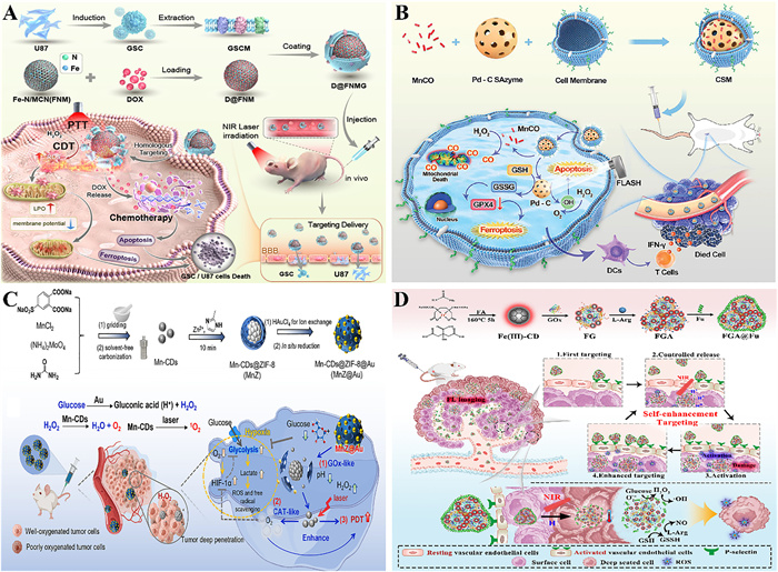
Within TME, the abnormal accumulation of endogenous H2O2 provides a unique substrate advantage for the catalytic reactions of nanozymes exhibiting POD-like activity. These carbon-based nanozymes can adsorb H2O2 molecules through surface active site specificity, subsequently breaking the O-O bond of H2O2 by regulating their own electronic structure, thereby efficiently generating highly oxidative hydroxyl radicals (•OH) [57]. DNA, proteins, and lipids in tumor cells are oxidized by •OH groups, which leads to tumor cell apoptosis. Lin et al. [58] designed doxorubicin (DOX)-loaded, glioma stem cell membrane (GSCM)-camouflaged and iron-nitrogen doped mesoporous carbon nanorods (FNM) to achieve synergistic therapies for PTT and CDT (Fig. 1A). D@FNMG could target glioma cells. In TME, FNM exerted POD-like activity to catalyze H2O2 into ROS and converted light energy to thermal energy under NIR irradiation. Meanwhile, the released DOX could also generate ROS. In addition, efforts have been made to understand the catalytic mechanism of POD-like carbon-based nanozymes. Sun et al. [59] studied the effect of three functional groups on the POD-like activity of GQDs and found that the −O=C−O− group was the substrate-binding site, the −C=O group served as the catalytic active site, and the −C−OH group could undermine this activity. Although GQDs and CNTs both exhibit POD-like activity, differences in morphology and structure may affect the mechanism by which POD-like activity is generated. Therefore, there are also studies on CNTs that reveal the POD-like mechanism of nanozymes. Han et al. [14] synthesized a novel nanozyme (CNTs/Fe-NC/DOX/CM) wrapped DOX in mesoporous CNT/Fe-NC with the breast cancer CM and showed that the single-atom iron-containing active site was the crucial component for the POD-like activity of CNTs. Besides, this nanozyme was found to have photothermal effect and could be used in PTT-enhanced tumor therapy.
Although POD-like nanozymes can catalyze the formation of toxic •OH to kill tumor cells, the generation of •OH is greatly limited by the low affinity between the nanozymes and H2O2, and the relative insufficiency of H2O2 in the TME. To solve the problem of low affinity between nanozymes and H2O2, researchers can design carbon-based nanozymes with high pyrrole nitrogen content to improve the activity of POD-like enzymes [63].
Due to the high content of GSH in TME, which can scavenge ROS in tumor cells under the action of the GPx4 protein family. The efficacy of therapies such as CDT relying on ROS to kill tumors is not as expected. Many carbon-based nanozymes have GPx-like activity, which can catalyze GSH into oxidized glutathione (GSSG). The massive depletion of GSH can lead to the inactivation of GPx4, thereby increasing the level of ROS in tumor cells, which can induce ferroptosis to a certain extent and greatly improve the tumor inhibition rate [64].
Ferroptosis is caused by lipid peroxidation, and inhibiting GPx4 can lead to increased ROS levels and lipid peroxidation within cells, thereby promoting cell ferroptosis. Cancer cells experiencing ferroptosis are immunogenic and activate an immune response [64,65]. Therefore, GPx4 enzyme activity can be inhibited by targeting and depleting GSH to induce ferroptosis and activate an immune response to kill tumor cells. For example, Yao et al. [66] prepared CQDs derived from chlorogenic acid (ChA), which activated the immune response to treat the tumor. The CQDs exhibited promising GPx-like activity, which significantly depleted the highly expressed GSH in the TME, while triggering ferroptosis and immune response to treat tumors. In addition to the GPx-like activity of carbon-based nanozymes directly used for tumor therapy, they can be co-doped and surface modified to achieve multi-enzyme activity and synergistic therapy due to their easy modification and high loading. For instance, Lyu et al. [60] designed cancer CM-encapsulated, carbon monoxide donor (MnCO)-loaded, porous Pd-C SAzyme (CMS) for tumor therapy (Fig. 1B). Carbon-nanozymes and Mn2+ together exhibited GPx-like activity, significantly reducing GSH level, thereby preventing ROS clearance and enhancing ferroptosis in tumor cells, which showed that NPs loaded with dual GPx-like activity triggered a robust cellular immune response and inhibited tumor growth significantly, prolonging the survival of mice.
The carbon-based nanozymes with OXD-like activity catalyze the oxidation of substrates to generate H2O2, water, or superoxide radicals (•O2−) [67]. This catalytic process holds significant biological importance: The ROS it generates serve as core drivers for multiple critical functions, most notably bactericidal activity and selective cell-killing effects, both particularly crucial in tumor therapy. When integrated into carbon-based nanozyme systems, the H2O2 generated by OXD-like activity exhibits dual functions. It not only supplies reaction substrates for POD-like nanozymes but also enhances their catalytic efficiency. This ultimately promotes the generation of additional ROS, such as •OH, thereby amplifying the cytotoxic effects on tumor cells [68].
Furthermore, by producing •O2− directly, carbon-based nanozymes with OXD-like activity can also destroy cells. An excellent illustration is single-atom catalysts (SACs). Atom-dispersed metal active sites anchored to the nitrogen-doped carbon structure (M-N-C) are a significant feature of SACs. Zhu et al. [69] created a nitrogen-doped carbon structure called Mn/PSAE, which trapped Mn atoms inside nitrogen-rich porous carbon. As a result of its OXD-like activity, Mn/PSAE could mediate the transfer of electrons from O2 to form large quantities of deadly •O2− and trigger apoptosis. Furthermore, Mn/PSAE exhibited excellent photothermal properties and could enable PTT-enhanced enzyme catalytic therapy. Furthermore, the ultra-small size of CDs, the abundant unsaturated chemical groups on their surfaces, and the high specific surface area make them more amenable to be anchored by active metals and results in higher catalytic activity. For example, Wang et al. [70] constructed Ru single atoms loaded on biocompatible CDs. This composite exhibited excellent OXD-like activity and PCE, capable of catalyzing the generation of a large number of •OH and •O2–, which can be utilized in PTT to kill tumors.
The nanozymes with CAT-like activity mainly stimulate the production of O2 in tumors by decomposing H2O2 into water [71], which is utilized as a sensitizer for chemotherapy, PDT, and radiation therapy for cancer. By addressing the O2 shortage at the tumor site, nanozymes with CAT-like activity can improve the efficacy of aerobic therapy such as radiotherapy and PDT. The process of PDT requires continuous consumption of O2, which is scarce at the tumor site. Therefore, CAT-like nanozymes that can convert H2O2 to O2 are the best candidates for combining PDT [72,73]. Luo et al. [61] constructed a hybrid material (MnZ) with a core of Mn2+-doped CDs and a shell of zeolitic imidazolate framework-8 (ZIF-8), and modified the surface of MnZ with gold NPs (AuNP) to achieve enhanced PDT through a cascade enzyme reaction (Fig. 1C). The AuNPs catalyzed the degradation of glucose and produced a large amount of H2O2, while triggering the degradation of ZIF-8. The released Mn-CDs catalyzed the conversion of H2O2 into O2 through CAT-like activity, thereby improving the hypoxia and enhancing the efficacy of PDT.
Additionally, surface modifications can be applied to carbon-based nanomaterials, allowing for the loading of PSs, which can create a ROS generation storm in TME, thereby enhancing the tumor-killing effects of PDT. Zhao et al. [74] designed a copper doped CNs with multi-enzyme activity and integrated it with the PS chlorin e6 (Ce6) to prepare a ROS amplifier (CCC). CAT-like activity of CNs decomposes H2O2 to produce O2, which provided raw materials for PDT. The CNs with CAT-like activated not only alleviated hypoxia at the tumor site, but also enhanced the effect of PDT for tumor treatment.
Carbon-based nanozymes with SOD-like activity can generate H2O2 and O2 by catalyzing the disproportionation reaction of •O2−. The reduction of •O2− can decrease the oxidative stress in the cells. Carbon-based nanozymes can not only serve as carriers for antitumor drugs, but their SOD-like activity can also act on tumor cells to enhance tumor therapy. For example, Kepinska et al. [75] fabricated DOX-loaded fullerenes (DOX-C60), achieving enhanced chemotherapy. The SOD-like activity of C60 depleted •O2−, reducing oxidative stress in tumor cells, thereby inhibiting the overexpression of SOD enzyme activity in tumor cells. This result demonstrated that the DOX-C60 composite not only mitigated the ROS-induced side effects of DOX, but also significantly inhibited tumor proliferation. Additionally, carbon-based nanozymes with SOD-like and POD-like activity can also be combined with other groups to generate more potent •OH, achieving enhanced synergistic tumor therapy [76].
POD-like activity is one of the most common and extensively studied activities in carbon-based nanozymes. Defect sites or oxygen-containing functional groups (e.g., carboxyl and hydroxyl groups) on the surface of carbon-based materials serve as catalytic active centers, facilitating electron transfer. By catalyzing the decomposition of H2O2, highly toxic •OH are generated, which subsequently oxidize substrates to induce colorimetric reactions. POD-like activity typically exhibits peak activity under acidic conditions, which aligns with the acidic characteristics of the TME. Distinct from POD-like activity, OXD-like activity does not require H2O2 as a co-substrate but directly utilizes dissolved oxygen. Carbon nanomaterials activate molecular oxygen, converting it into ROS to oxidize substrates. This process typically relies on the sp2 hybridized carbon structure on the material surface or specific doping elements (such as nitrogen doping). The other two activities, CAT-like and SOD-like, utilize active sites on the surface of carbon-based nanozymes to capture and adsorb reductive substrates such as H2O2 and •O2−, generating harmless species like H2O, and O2 through disproportionation reactions. Compared with a single activity, nanozymes with multienzyme-like activities have significant advantages in terms of catalytic reaction efficiency and persistence and better tumor treatment effects can be achieved through the synergistic action of multiple enzymes.
The combined effects of OXD-like activity and POD-like activity can consistently export ROS to kill tumor cells. This system achieves the effect of continuous ROS generation depending on OXD-like activity to produce •O2− and POD-like activity to produce •OH. N-doped carbon nanomaterials (N-CNMs) hold great potential as metal-free electrocatalysts. ROS production and consumption are regulated by N-CNMs, which also have significant enzyme-like activity. Quaternary and pyridine N have been identified as potential active sites [77]. The pore space of these materials also enhances their activity, given that the porous interface provides a significant number of active sites that support large-scale transport during catalysis. Fan et al. [78] created an N-PCNS system that had four enzyme-like functions: OXD, POD, CAT, and SOD. The strategy of cancer therapy with this system was to use an OXD-like and POD-like mechanism to produce ROS in the acidic lysosomal milieu while restricting CAT-like and SOD-like activities to eliminate ROS in an alkaline pH environment. In addition, carbon-nanozymes with multiple enzyme-like activities can make the product of one enzyme-catalyzed reaction become the substrate of another enzyme-catalyzed reaction, and achieve efficient cascade reaction. Song et al. [62] prepared iron-doped red light CDs nanozyme (Fe(Ⅲ)-CDs) and GOx complex nanoreactor (FG) to realize the enzymatic cascade catalysis. Then fucoidan (Fu) and fucoidan (Fu) were further coated on the surface of FG to form FGA@Fu for self-enhanced targeting of tumors (Fig. 1D). FGA@Fu was depolymerized under weak acid/NIR conditions, and the released GOx consumed glucose and produced H2O2, while the POD-like activity of Fe(Ⅲ)-CDs could catalyze the production of •OH from H2O2. This nanoplatform achieved stronger tumor suppression through enzyme cascade catalysis and PTT combined therapy.
Many researchers have demonstrated that an optimized enzyme-like activity of CNs is accompanied with rational designs and strategies targeting various factors which affect their biomedical applications [79]. In the subsequent sections, we will investigate the influencing factors of CNs, which encompass physic-chemical properties, doping elements, size, surface modification, pH and temperature [80]. In addition, we also summarized the factors influencing catalytic properties of CNs in Table 1 [78,81–91].

 DownLoad:
CSV
DownLoad:
CSV
| Factors | Key modification | Mechanism of catalytic regulation | Impact on catalytic performance | Ref. |
| Doping elements | (1) Single non-metal (2) Dual non-metals (3) Dual atoms |
(1) Enhance response performance (2) Reduce the reaction energy barrier (3) Increase active sites/charge density affinity |
N-doping: Improve OXD-/SOD-like activity, ROS production in acidic TME Non-metals (S/N): Smaller Michaelis-Menten constant, higher specific activity than single N-doping Dual metals: The POD-like activity better than single atom |
[78,81,82] |
| Size | Optimum size range | (1) Increase specific surface area and active centers (2) Achieve effective substrate contact |
Smaller size: Enhance POD-like activity Excessively small size: Diminish activity due to agglomeration |
[83–85] |
| Surface modification | (1) Functional modification (2) Targeting modification |
(1) Improve solubility and biocompatibility (2) Increase the active site and partial charge density (3) Enhance tumor targeting |
HA modification: Utilizing the specific binding of the CD44 receptor on tumor cell surfaces to the modifying molecules Anion and purine modification: Enhance the dispersion stability of the substance in aqueous solutions and biological media |
[86,87] |
| Morphology | (1) 0D (2) 1D (3) 2D |
(1) Increase specific surface area and porosity (2) Accelerate electron transfer |
0D: Superior substrate affinity 1D: Accelerated electron transfer 2D: Provide voids to generate more active centers |
[83,88] |
| pH and temperature | (1) Optimum pH range (2) Temperature stability |
(1) Acidity enhances the generation of ROS (2) Thermal activation of catalytic sites |
Optimal conditions: pH 4.0, 40 ℃ Maintain structural integrity within a wide pH range (covering acidic, neutral, and weakly alkaline conditions) |
[89–91] |
In enzyme mimic design methods, doping is commonly used to produce CNMs which involves the incorporation of a range of ions or molecules into the structure to enhance performance, leading to the improvement of catalytic efficiency [92]. The catalytic activity of CNs is intrinsically linked to their electronic structure. The introduction of heteroatoms (e.g., nitrogen) creates charge redistribution. Pyridinic-N and pyrrolic-N sites, in particular, act as active centers by modifying the adsorption energy of intermediates like H2O2, thereby significantly enhancing POD-like activity.
Heteroatom N-doping CNs are among the most widely researched dopants in the non-metal-doped approach. Increasing enzymatic activity is displayed along with four microenvironment-responsive enzyme-like behaviors under physiological conditions. Additionally, under acidic TME, they can specifically modulate the production of ROS and substantially undermine tumor growth [78].
Interestingly, nanozymes doped with dual non-metals exhibit better activity. Chen et al. [81] constructed hierarchically porous CNs with co-doping of sulfur (S) and nitrogen (N) (SNC). The findings suggested that S-doping in SNC nanozymes could boost their catalytic activity and affinity towards substrates, as they exhibited a smaller Michaelis-Menten constant and higher specific activities in relation to nitrogen-doped carbon (NC) nanozymes.
Meanwhile, nanozymes doped with dual metals exhibit superior activity. Zeng et al. [82] manufactured nanozymes consisting of dual-atom metal-nitrogen-carbon (M = Fe, Co, Ni, Mn, Ru, Cu). These nanozymes displayed POD-like activities, surpassing those of single-atom metal NC nanozymes. The experimental results showed that the doping of heteronuclear metal atoms removed the reaction energy barrier of the whole catalytic reaction path and made the catalytic process easier to complete. These results suggested that incorporating dual atoms into the CNs could enhance nano-catalytic therapeutics in the treatment of tumors.
Typically, smaller carbon nitrides exhibit a higher specific surface area and more active centers for the nanozyme, enabling effective substrate contact [83]. Zheng et al. [84] found that graphene dots (GDs) exhibited remarkably efficient POD-like catalytic activity, which surpassed that of larger GO NPs.
Furthermore, Zhang et al. [85] reported that the GQDs/Au electrode assembled through covalent bonding displayed exceptional activity and stability when detecting H2O2 due to their excellent POD-like activity and small lateral size. It was beneficial for monitoring changes in H2O2 level fluctuations in biological systems. Interestingly, the nanozyme has an optimum size range, if the size decreases below the smallest size, the specific surface area will decrease due to agglomeration which weakens its enzymatic performance [83].
Surface modification is a common method for rational design and regulation of the activity of carbon nanomaterials, which exhibit diverse surface chemical properties. Therefore, the functionalization of carbon nanomaterials can be achieved by surface modification to improve their basic properties. For example, CNTs can be surface modified with anions, purines, and other substances to enhance their solubility, biocompatibility, biodegradability, and reduce toxicity [93,94]. More importantly, surface modification can improve the enzymatic activity of carbon nanomaterials. For instance, Dang et al. [86] prepared CNTs modified with bimetallic organic frameworks MOFs (Fe/Mn) and Au NPs, which boosted the POD-like activity of CNTs by increasing the active site and partial charge density of CNTs and promoting electron transfer between the metal-organic frameworks and NPs.
In addition, carbon nanomaterials can not only achieve multi-enzyme activity through surface modification, but also improve their dispersion and biocompatibility, the ability to specifically target tumor cells, and promote their accumulation in tumor cells to enhance anti-tumor activity. Zhong et al. [87] designed nitrogen-doped porous carbon loaded with bismuth nanoclusters modified with hyaluronic acid (Bi-NC@HA). Due to the biodegradability, colloidal stability and tumor-targeting ability of HA, this composite not only exhibited good dispersion, tumor responsiveness, and accumulation in tumor cells, but also showed outstanding POD-like and GPx-like dual-enzyme activities and photothermal conversion performance under irradiation of NIR, achieving tumor catalysis and PTT.
The morphology of nanozymes is closely linked to the active center and the specific surface area of nanozymes, thus affecting their catalytic performance. However, limited research has documented the influence of morphology on the catalytic efficacy, it is reported that Heo et al. [88] synthesized Rosette-graphitic carbon nitride (GCN) possessing POD-like activity. The catalytic performance of Rosette-GCN was about 10 times better than that of traditional bulk GCN. It was believed that the enhanced activity was due to the fact that Rosette-GCN had a considerably bigger surface area and greater porosity whilst retaining its distinctive graphitic framework.
In addition, nanomaterials with different dimensions represent various kinds of activities. 0D CNs, e.g., fullerenes, GQDs, and CDs owing to the properties of surface energy and surface area-to-volume ratio, tend to react with the surroundings, therefore the 0D materials display superior enzymatic activity and affinity for substrates. 1D carbon nanomaterials have a strongly anisotropic structure, which confers them with fine tunable electronic characteristics, thus the enzymatic activity of GNRs could be improved thanks to the accelerating transfer of electrons. 2D carbon materials, such as graphene, can provide more voids or defects due to their unique geometry, which is advantageous to generate more active centers. In conclusion, morphology is a key factor for the catalytic activities of CNs. Besides, many factors that can influence the control of morphology, including the facet type, the valence and the surface strain effects, must be taken into account as well [83].
Similar to the natural enzyme, the catalytic activity of the CNMs is closely related to pH and temperature, with pH 4.0 and 40 ℃ usually being the optimal reaction conditions. However, unlike natural enzymes that are inactivated in specific environments, CNMs can show relatively stable and high activity over a wide pH range, even within the natural pH range [89], as in the case of hybrid catalyst, where the synergistic effect of GO-AuNCs hybrid expands its application in biological systems requiring a near-neutral pH [90]. Additionally, Lin et al. [91] showed that the natural enzyme HRP experienced significant suppression of enzymatic activity beyond 60 ℃. In contrast, the N-doped GQDs (N-GQDs) maintained good catalytic activity above this temperature.
Due to the special conditions of TME, such as hypoxia, acidity, and high expression of endogenous H2O2, natural enzymes and most other nanozymes are difficult to accurately target and effectively treat tumors. However, carbon-based nanozymes provide a new approach to precisely target and treat tumor cells with characteristics such as stability in a wide range of pH, excellent PCE, enzyme-like catalytic ability Zhong et al. [95] designed nitrogen-doped carbon loaded with Ce SAzymes (Ce SAs@NC), which exhibited excellent POD-like activity and cascade enzyme activities of CAT-like and OXD-like under the weakly acidic conditions of TME, generating a large amount of ROS. Additionally, under NIR excitation, the composite could increase and maintain temperature, enhancing catalytic efficiency, thus achieving TME-responsive photothermal-enhanced tumor nano-catalytic therapy.
CNs possess a wide range of applications in the field of treating cancer with drug therapy, such as CDT, PTT, PDT, SDT, immunotherapy and synergistic therapy based on CNs. With the assistance of various high-quality properties of CNs, the efficacy of traditional therapies has been significantly enhanced. The recent applications of carbon-based nanozymes for photothermal-enhanced tumor theranostics are shown in Table S2 (Supporting information).
One of the main methods for treating cancer is chemotherapy, which primarily involves the systemic or local use of chemical medicines to eradicate tumor cells [96,97]. However, many chemotherapeutic drugs are far from ideal because they do not specifically target systemic biomolecules. And low tumor accumulation may prevent complete tumor eradication at low safe doses [91]. In addition, many chemotherapy patients may develop multiple drug resistance, which ultimately results in subpar treatment outcomes [98,99]. As nanotechnology has matured, new biocompatible materials called CNs have been created that can be functionalized with medications to deliver anticancer medicines. Through the use of a nanozyme-based drug delivery system, chemotherapeutic effectiveness has been seen along with increased drug delivery efficiency [100].
Chemotherapeutic medicines frequently require CM penetration to accomplish their effects. CDs, leveraging their ultrasmall size (generally <10 nm), abundant surface functional groups (e.g., -COOH, -NH2, -OH), and excellent biocompatibility, could efficiently load chemotherapeutic drugs through covalent conjugation, π-π stacking, or electrostatic adsorption for drug delivery. Their surface chemical properties (e.g., charge, hydrophilicity/hydrophobicity) modulated interactions with CMs, enabling CDs to carry therapeutic molecules into cells primarily via energy-dependent endocytic pathways (such as clathrin-mediated endocytosis and caveolae-mediated endocytosis) to achieve efficient cellular uptake. For instance, Lu et al. [101] constructed a zeolitic imidazolate framework-8 (ZIF-8) nanoassembly integrated with coordinated CDs (CCDs) loaded with paclitaxel (PTX) and coated with rutin (Ru), termed Ru/CCDs-PTX@ZIF, for synergistic catalysis/chemotherapy. The CCDs exhibited exceptional POD-like activity and functioned as SAzymes to generate substantial •OH. Facilitated by GLUT receptor-mediated endocytosis, the Ru/CCDs-PTX@ZIF system enabled targeted tumor killing and suppressed migration, thereby inhibiting tumor progression.
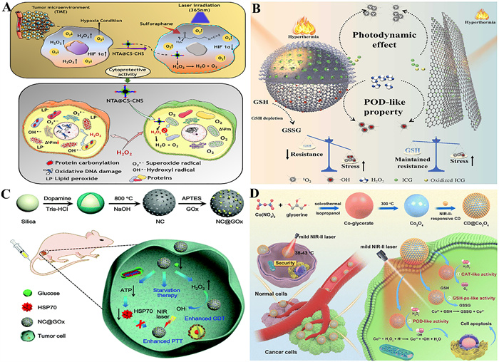
Hypoxia is a significant feature of TME. To adapt to the hypoxic environment, cancer cells show a series of changes, such as the increase of P-gp expression, inhibition of apoptosis and cell senescence, promotion of cell cycle arrest, reduction of oxidative metabolism and autophagy, which endow tumor with strong resistance to all kinds of stress, leading to the occurrence of multiple drug resistance. Tumor hypoxia by promoting chemotherapy resistance, seriously weakens the occurrence of tumor therapy [102–106]. To alleviate tumor hypoxia, nanozymes are found to contribute to regulating chemoresistance. Cai et al. [107] constructed cobalt single-atom nitrogen-doped porous carbon (Co-SAs@NC) as a dual-functional nanozyme. Through a cascade catalytic reaction (similar to CAT-/POD-like activity), H2O2 within the tumor was converted into O2 to alleviate hypoxia, and further generated highly toxic •O2−; in combination with the loading of chemotherapeutic drug DOX, a large amount of ROS was produced, thus achieving synergistic tumor treatment. Beyond single enzyme-mimicking therapies, CN-based delivery systems significantly expand the application strategies of nanozymes in overcoming tumor hypoxia and drug resistance by carrying combined therapies, such as CDT/PDT. Among these, nanozyme-catalysis-enabled chemotherapy represents a safe and promising strategy, utilizing catalytic reactions to convert excess H2O2 in the TME into O2, thereby alleviating hypoxia and enhancing therapeutic efficacy. For instance, Shukla et al. [108] successfully synthesized carbon nanospheres (CNS) using Camellia sinensis plant extract as a precursor and achieved surface functionalization via nitrilotriacetic acid (NTA) coupling technology (NTA@CS-CNS) (Fig. 2A). Stability studies demonstrated that this carbon-based nanoassembly remained stable under various cations, anions, and across five different pH conditions. Crucially, NTA@CS-CNS was able to catalyze the conversion of H2O2 in the TME into O2 efficiently. Due to its dual enzyme-mimicking activities (CAT-like and POD-like), NTA@CS-CNS not only helped address CDT resistance stemming from hypoxia, but enabled the sustained catalytic production of ROS and O2 from H2O2, effectively combating hypoxic tumor cells.
In view of the current limitation in tumor treatment such as the non-specific toxicity of chemotherapy drugs, low accumulation of tumors and multi-drug resistance, CNs-based synergistic tumor treatment provides a strategy for anti-tumor therapy more precisely. These studies indicate that the CNs-based platforms are moving from single drug delivery to intelligent responsive multi-functional platforms, which has opened up new paths to remove the limitations of traditional chemotherapy by precisely regulating drug release, improving the TME and cytotoxic effects.
PTT refers to the use of high tissue penetration with strong NIR absorption reagent. After NIR wavelength light irradiation, local heat is generated at the tumor site, which causes irreversible solid tumor damage by changing gene expression, causing protein degeneration in turn, which leads to ideal therapeutic effects with minimal side effects [111–114]. Recently, significant progress has been made in the method of treating tumors by catalyzing enzymatic reactions in the TME, however, the poor stability of some physicochemical factors in the TME and the requirement for suitable environmental conditions hinder the application of natural enzymes or enzyme mimetics. With the gradual maturity of nanotechnology, new nanomaterials can, under controllable conditions, simulate the functions of natural enzymes to exhibit enzyme-like activity and regulate related enzymatic reactions [78,115]. Among them, carbon nanomaterials have been reported to mimic the catalytic activity of CAT-like, OXD-like, POD-like, and SOD-like activities [116–118]. Photothermal therapeutic methods rely on the use of photothermal conversion agents, and CNs enable a photosensitive material with higher photothermal conversion capacity, with better photothermal effect and lower toxicity at low power irradiation [119]. Therefore, the disadvantages of PTT can be overcome by integrating PTT reagents with favorable photothermal properties and incorporating CNs with high multi-enzyme catalytic activity into a single nanoplatform [120]. Feng et al. [50] designed 3D MCNs and 2D GO respectively loaded with indocyanine green (ICG) and modified with PEG (Fig. 2B). It was found that the mesoporous structure of ICG/MC-PEG accommodated and protected ICG, resulting in higher photostability and lower phototoxicity. Additionally, ICG/MC-PEG exhibited higher PCE and POD-like activity, leading to a higher tumor inhibition rate than GO under the same conditions.
Although PTT has high specificity and negligible invasion, NIR light has limited penetration depth in tumor tissue, and tumors beyond the laser radiation range cannot be completely ablated, leading to tumor recurrence and metastasis [121]. Therefore, a single PTT often does not completely cure the tumors, especially for larger tumors, and for lesions with distal metastatic and spreading properties. It is encouraging that the PTT-mediated thermal effects have some functional utility, such as controlling drug release, adjusting intracellular gene expression and enzyme activity, triggering anti-tumor immune response and increasing chemical response in targeted tissues [122]. Therefore, combining PTT with other treatments provides an effective strategy for synergistic enhancement of antitumor efficacy. Zhang et al. [123] prepared γ-polyglutamic acid (γ-PGA) coated, GOx modified and Mn, Cu-doped CDs to achieve NIR-triggered combined of starvation therapy/PTT/PDT/immunotherapy. After the intracellular uptake of compound was promoted by γ-PGA coating, H2O2 was generated through glucose was consumed by GOx, which was decomposed by CDs with POD-like activity to generate O2 and alleviate tumor hypoxia, thereby improving the efficiency of PDT. Additionally, the compound exhibited superior PCE, significantly enhancing the tumor treatment efficiency of PDT and PTT. The enzyme-like catalytic system constructed by integrating PTT and CDT effectively addressed the limitations of monotherapy. Xu et al. [109] decorated N-doped carbon (NC) NPs with GOx as biomimetic nanozymes (NC@GOx NPs), which could promote starvation therapy to enhance PTT and CDT against tumors (Fig. 2C). Both in vitro and in vivo experiments demonstrated that NC@GOx NPs could effectively kill cancer cells and eliminate tumors. This design provided a strategy for the synergistic cancer treatment using biomimetic nanozymes.
Tumor nanocatalytic therapy (NCT) faces two major bottlenecks currently, the limited efficiency of single-modal treatment and the restriction of traditional PTT clinical application caused by insufficient tissue penetration depth and overheating damage. To address this challenge, Wang et al. [110] have designed a mild NIR-Ⅱ photothermal enhanced nanocatalysis therapy (NCT) nanoplatform based on the CDs@Co3O4 heterojunction (Fig. 2D). Compared to the single-component CDs and the Co3O4 nanospheres, it possessed a stronger photothermal performance of NIR-Ⅱ, and multi-enzyme-mimic catalytic activities such as POD-like, CAT-like, and GPx-like, which showed high effectiveness for tumor therapy, providing a new paradigm for the development of efficient and low-toxicity synergistic anti-tumor strategies.
Due to tumor resistance to heat shock proteins and insufficient light penetration depth, PTT is faced with therapeutic limitations. By integrating the multi-enzyme catalytic activity of CNs (such as alleviating hypoxia and enhancing ROS generation) with photothermal materials, a synergistic treatment platform can be constructed to break through the bottleneck of single PTT and provide a new path for deep tumor ablation and metastasis inhibition.
PDT is a newly developed photoactivated therapeutic approach that has drawn a lot of interest in the treatment of cancer. When PSs accumulated at the tumor site are exposed to light and become activated, they release energy that converts oxygen in the surrounding tissues into ROS, resulting in irreversible cell destruction. PDT is thought to be a more secure and efficient method of tumor destruction since only certain wavelengths can activate PSs to produce ROS. However, light, oxygen and PSs are three main factors that undermine the effectiveness of PDT [124]. The following will be thorough discussions of these limitations and how CNs overcome the difficulties and increase the efficacy of PDT.
First, the efficacy of PDT is influenced by the intrinsic properties of PSs, including their hydrophobicity, phototoxicity, and facile aggregation. One of the best ways to increase hydrophobicity is to load PSs into carbon-based nanomaterials that hold a high surface area and low toxicity. Hematoporphyrin (HP) PSs were encapsulated inside CQDs to create HP-CQDs, which exhibited superior water solubility while retaining all of the inherent optical and chemical characteristics of HP [125]. Additionally, the goal of lowering PSs phototoxicity and raising patient compliance can be achieved by packing PSs into carbon-based nanomaterials with significant light absorption. Li et al. [126] synthesized a carrier called PMC-MnO2-iRGD (iPMC-MnO2) by modifying polyethylene glycol (PEG) on the surface of MC-MnO2 and introduced iRGD in its outermost layer. IPMC-MnO2 took full advantage of its favorable photothermal effects and could skillfully combine PTT and PDT to reduce the dose of therapeutic drugs and enhance the synergistic therapeutic effect. Furthermore, increasing the hydrophilicity of the PSs can prevent aggregation and hence improve the PDT. A nanosystem (TPFcNP) was created by mixing a hydrophobic PS with numerous carbon-carbon double bonds and a ferrocene-containing amphiphilic block copolymer (PEG-b-PMAEFc) [127]. The POD-like activity of this compound could catalyze H2O2 into •OH, thus catalyzing the addition reaction of GSH and hydrophobic PSs. This process improved the hydrophilicity of PS, reduced aggregation, and simultaneously enhanced the production of 1O2, thereby improving the efficacy of PDT.
Second, the generation of ROS is restricted in hypoxic TME. The depletion of O2 during the process of PDT-induced microvascular injury causes further hypoxia of tumor cells, which drastically reduces the anticancer efficacy of PDT [106,128,129]. Hence, controlling hypoxia and raising the differential oxygen pressure in the TME is crucial for improving the therapeutic result of PDT. Researchers have worked diligently in the past few years to overcome hypoxia and improve the efficiency of PDT. One of the strategies is the creation of nanoplatforms that can produce oxygen in tumor tissues. CAT-like and SOD-like enzymes are common enzymes that produce O2. Sahu et al. [130] fabricated Pluronic coated nanographene oxide (NGO) encapsulating heme and Ce6 to achieve enhanced PDT. The overexpressed H2O2 in tumor cells was catalyzed by the nanoplatform with CAT-like activity to produce O2, which not only alleviated hypoxia, but also facilitated the production of light-induced ROS from the PS Ce6. These results strongly proved that the anti-tumor platform combined with carbon-based nanozymes enhanced PDT tumor treatment, solving the problem that the anti-cancer efficacy of PDT was limited due to hypoxia.
Third, the tissue penetration depth of light is highly connected to its wavelength, with the majority of PS excitation wavelengths occurring in the UV or visible region. However, the limited penetration of light makes PDT difficult to treat deep cancers [131]. NIR-responsive nanomaterials have recently demonstrated tremendous potential for broadening the use of phototherapy, owing to the deep tissue penetration of NIR light [132]. Gulzar et al. [133] acquired NGO-UCNP-Ce6 (NUC) nanocomposites by covalently implanting core-shell structured upconversion NPs (UCNPs) with NGO and consequently loading Ce6 onto the surface of NGO. When exposed to laser stimulation at 808 nm for PDT, the NUCs could produce ROS. In addition, NUCs could rapidly convert the 808 nm photon into the thermal energy of PTT, realizing a synergistic therapy of PDT and PTT.
Even though PDT can produce ROS which kills tumor cells, under continual internal oxidative stress, tumor cells may adapt and resist the damaging effects of ROS. For example, over-expressed GSH in tumors can remove the resulting ROS, lower the level of ROS in the body, and significantly affect the effect of PDT [134]. Lu et al. [135] designed CM-coated MC-COOH modified with MnO2 and Ce6, exhibiting excellent PCE. After entering tumor cells, this composite not only generated O2 to relieve hypoxia, but also MnO2 and the GPx-like activity of MC-COOH itself rapidly consumed GSH, which led to the accumulation of ROS produced by the PS and the OXD-like activity of carbon nanomaterials in tumor cells, thereby achieving enhanced PTT-PDT synergistic therapy.
In conclusion, carbon-based nanozymes have been extensively applied in PDT tumor treatment and have been significant in advancing the course of PDT tumor treatment. In addition to being highly stable and reasonably priced, the diversified PDT system based on carbon-based nanozymes can overcome issues with hypoxia and inadequate light source tissue penetration during PDT treatment and effectively reduce GSH levels in tumor tissues. Furthermore, the combination of PDT and other therapies can also be better used for PDT tumor treatment. It is anticipated that as nanotechnology develops, more novel materials with superior biosafety and synergistic tumor treatment will be employed in PDT tumor treatment, thereby broadening the therapeutic application of PDT tumor treatment and advancing the development of carbon-based nanozymes for PDT tumor treatment in clinical settings.
CDT, an innovative approach to eliminate cancer cells in situ, has drawn lots of attention recently. Higher H2O2 concentrations, weak acidity (pH 6.5–7.0), and higher GSH concentrations are seen in TME compared to normal tissues [136]. The foundation of CDT is the Fenton/Fenton-like reactions, which uses metal ions to transform endogenous H2O2 into ROS [137]. In contrast to PDT, CDT offers superior positioning and selectivity, endogenous stimulation, low multi-drug resistance, and the ability to overcome both the phototoxicity and oxygen reliance of PDT [138].
There are several main factors that influence the effectiveness of CDT: First, even if tumor cells contain a lot of H2O2, it is still insufficient to provide long-term and efficient treatment because of limited H2O2 in the body naturally [139]. Second, at the tumor location, it is challenging to achieve the ideal acidity (pH 2.0–4.0) of the Fe2+ Fenton reaction due to the shortage of acidity [140]. Third, by quickly eliminating ROS created by CDT, up-regulation of GSH levels at the tumor site shields tumor cells from oxidative damage with the fact of high levels of endogenous GSH [139]. What is more, the majority of CDT reagents today have low catalytic efficiencies and are unable to produce the best catalytic results in intricate physiological microenvironments [109]. It is challenging for CDT to get the best clinical application and therapeutic impact because of these issues. Therefore, it is imperative to build nanoscale systems that can enhance acidity, consume GSH, and produce H2O2 in tumor cells.
Many approaches have been investigated in recent years to deal with the problem that CDT faces. Because of their enormous surface area, distinct optical qualities, and physical structure, carbon-based materials can function as both PTAs and Fenton delivery vehicles in multimodal synergistic therapy. Furthermore, carbon-based nanozymes that exhibit high biocompatibility also possess enzyme-like activity, leading to increased anti-tumor efficacy with few side effects. For example, Wibrianto et al. [63] prepared Cu, N, and S-doped CQDs functionalized with GOx and camptothecin (CPT), obtaining a multifunctional nanocatalyst. This nanocatalyst could directly catalyze the generation of •OH and H2S under acidic conditions. Additionally, this composite could also catalyze glucose to produce H2O2, which was highly selectively catalyzed to •OH by its POD-like activity, promoting the accumulation of ROS in tumor cells. This result demonstrated its potential in achieving tumor CDT therapy/gas therapy.
Despite demonstrating considerable therapeutic potential in tumor intervention due to their enzyme-mimetic activity, excellent biocompatibility, and photothermal conversion capabilities, carbon-based nanozymes face two major limitations in practical tumor treatment applications: Relatively low inherent catalytic activity and insufficient substrate specificity. Notably, a growing body of research confirms that elemental doping and surface modification represent the two most widely applied and technically mature approaches capable of significantly enhancing the catalytic performance of carbon-based nanozymes. For example, Han et al. [140] designed an ultra-small Fe single-atom nanozyme (ph-CDs-Fe SAzyme) with a high pyrrole nitrogen content to enhance the efficiency of PDT. Elevating the pyrrole N atom content by a ligand-assisted phenanthroline-mediated approach might improve the POD-like activity of Fe SAzymes. Density functional theory (DFT) computations and steady-state kinetic studies revealed that ph-CDs-Fe SAzyme had greater POD-like activity than that of CDs and CDs-Fe SAzyme.
It is noteworthy that this strategy of enhancing catalytic efficiency through atomic-level precision modulation exhibits deep complementarity with the emerging paradigm of computation-driven material design. While traditional experimental approaches focus on optimizing doping of specific elements, artificial intelligence technology has pioneered new pathways for deciphering complex structure-activity relationships. Active learning (AL), though a powerful method for accelerating novel material discovery, faces significant challenges in extracting physical significance. Li et al. [141] innovatively applied an interpretable AL strategy to effectively optimize the PCE of CDs in PTT (Fig. S2A in Supporting information). The resulting Fe-CDs demonstrated multi-enzyme-like activities responsive to the TME. Both in vitro and in vivo experiments confirmed that Fe-CDs could significantly enhance ferroptosis through synergistic CDT and PTT, yielding remarkable anti-tumor efficacy.
In cancer treatment, CDT and immunotherapy represent distinct therapeutic approaches, yet each suffers from inherent limitations. CDs effectively address these shortcomings by boosting ROS production required for CDT while enabling immunotherapy to precisely target tumors, ultimately achieving synergistic and potent anti-cancer outcomes. Immune checkpoint blockade (ICB) leverages antibodies to block negative immunoregulatory pathways, yet its efficacy is limited by weak immunogenicity in most patients. To address these challenges, Su et al. [142] engineered red-emissive CD-doped copper metal-organic framework NPs (Cu-MOF@RCD) as smart nanoreactors (Fig. S2B in Supporting information). Dual TME/NIR-responsive platform substantially amplified ROS levels to potentiate combined PDT/CDT. When integrated with anti-PD-L1 antibodies, the quintuple-modal PDT/PTT/CDT/GSH-depletion/ICB strategy eradicated primary tumors while suppressing growth of untreated distal metastases.
The therapeutic efficacy of CDT includes limitations such as insufficient H2O2, weak acidity, high GSH content and low catalytic efficiency. However, carbon-based nanozymes can synergistically enhance catalytic function through element doping (such as Cu, N, S) and structural optimization (such as Fe monometallic anchoring). The above series of examples provide efficient and precise nano-catalytic solutions for breaking through the clinical translation barriers of CDT through multi-dimensional regulation of acidity response, GSH depletion and H2O2 self-circulation.
SDT utilizes deep-penetrating ultrasound to activate sonosensitizers for ROS generation and targeted tumor cell apoptosis [143]. Relying on the interaction between ultrasonic and sonosensitizers, SDT induces apoptosis of cancer cells. Carbon-based nanozymes have emerged as exceptional sonosensitizers or synergists to overcome the low acoustic sensitivity of traditional agents, achieving noninvasive and highly precise tumor ablation (for detailed SDT mechanisms and recent applications, please refer to Section S5.5 and Figs. S2C–F in Supporting information).
By efficiently remodeling the immunosuppressive tumor microenvironment and inducing immunogenic cell death, carbon-based nanozymes can robustly activate the host's innate immune defenses to overcome the unresponsiveness of solid tumors. These nanozymes offer a highly specific and promising platform for targeted catalytic immunotherapy (for detailed mechanisms and recent applications, please refer to Section S5.6, Table S2, and Figs. S3A–D in Supporting information).
To overcome the inherent limitations of monotherapies-such as thermoresistance in PTT or hypoxia-restricted ROS generation-carbon-based nanozymes seamlessly integrate multiple modalities into a single nanoplatform. This rational design achieves powerful synergistic anti-tumor effects, such as combining PTT with PDT, CDT, or immunotherapy, to maximize therapeutic efficacy (for detailed applications and synergistic mechanisms, please refer to Section S5.7, Table S2, and Figs. S3E and F in Supporting information).
Beyond direct catalytic therapy, carbon-based nanozymes inherently possess unique optical and physical properties that enable real-time, high-resolution visual guidance for precise tumor intervention [144]. They effectively integrate multi-modal imaging techniques-including PAI, MRI, FI, and USI-with localized treatments to achieve highly efficient theranostics (for specific imaging-guided tumor therapies and diagnostic mechanisms, please refer to Section S6 and Figs. S4A–F in Supporting information) [145].
In this article, we deeply discuss the application and potential of carbon-based nanozyme in photothermal enhanced tumor diagnosis. Carbon-based nanozyme, especially based on CDs, CNTs, fullerenes, graphene and MCNs materials, have become an important research direction in anti-tumor therapy due to their excellent optical properties, biocompatibility, high drug loading capacity, high specific surface area and easy functionalization. Based on the multiple advantages of CN, this review introduces the physical and chemical properties and mechanism of CN family. As well as the catalytic activity of the enzyme such as OXD, POD, CAT and SOD and several factors affecting the activity of the enzyme such as size, surface modification, morphology and pH and temperature and so on, the application of CNs in tumor theranostics, such as in vitro and in vivo sensing, imaging and drug delivery systems of various carbon nanomaterials, focused on summarizing the current mainstream nanodynamic therapy, including PTT, PDT, CDT, SDT, tumor immunotherapy and collaborative governance. Due to its ideal depth of tissue penetration, non-invasiveness, high specificity, and few toxic side effects, the nanodynamics development has been achieved to fill unmet clinical needs. In addition, PAI-guided tumor therapy, USI-guided tumor therapy, MRI-guided tumor therapy, FI-guided tumor therapy and Multimodal are also fully elaborated on imaging-guided tumor therapy.
However, these CNs based therapeutic platforms face a number of challenges in the clinical conversion process, including the complexity of the system, the high cost of the process, the unknown mechanism of system degradation, and biocompatibility issues. The complexity of CNs is a major bottleneck restricting its large-scale application. In order to achieve accurate theranostics of tumors, CNs often need to be combined with other therapeutic functions, such as PTT, PDT, and antioxidant effects. These multifunctional integrations often require forming multiple groups into a complex nanoplatform. In applications, this complex component integration will lead to the complicated synthesis process of nanoplatform, which increases the difficulty and cost of production. In addition, interactions between different functional components may affect their stability and efficacy in vivo, thus affecting the overall effect of treatment. Nevertheless, optimizing the synergies of these multifunctional components could lead to significant improvements in therapeutic outcomes.
It is noteworthy that the photothermal enhancement strategy demonstrates irreplaceability compared to pure PTT or CDT. Standalone PTT is often constrained by HSP-mediated thermotolerance and insufficient penetration into deep-seated tumors; attempts to boost efficacy by increasing thermal power frequently exacerbate damage to surrounding normal tissues. Conversely, pure CDT is heavily dependent on the TME, often exhibiting low efficiency in Fenton or Fenton-like reactions due to insufficient endogenous H2O2 and suboptimal acidic pH. In this context, photothermal enhancement plays a pivotal role: The localized thermal effect not only directly ablates tumors but also significantly accelerates the catalytic kinetics of nanozymes in accordance with the Arrhenius law, thereby boosting CDT efficiency. Furthermore, mild hyperthermia promotes tumor vasodilation and blood perfusion, alleviating tumor hypoxia and increasing the supply of H2O2 required for CDT. Crucially, ROS generated during the CDT process can significantly downregulate HSP expression, effectively inhibiting thermoresistance and sensitizing tumor cells to thermal ablation. This synergistic “thermo-catalytic” effect overcomes the respective bottlenecks of monotherapies, achieving a “1 + 1 > 2” outcome that pure PTT or pure CDT fails to attain. Therefore, despite the systemic complexity, this strategy offers a unique solution for addressing critical clinical challenges such as drug resistance and incomplete tumor eradication.
One of critical challenge lies in the biocompatibility and long-term toxicity of CNs. When applied in vivo, the biodegradation behavior of nanomaterials directly impacts their biosafety and therapeutic sustainability. Current research indicates that although CNs undergo biodegradation through the weakly acidic and H2O2-rich conditions of the TME, the intermediate products generated during their degradation may raise biocompatibility concerns, the interaction mechanisms between these nano-fragments and biological tissues remain unclear and could potentially disrupt CM integrity or interfere with normal metabolic processes. More importantly, the long-term retention of degradation products in the body may lead to chronic toxic effects: Certain nano-sized fragments may cross biological barriers and accumulate in organs such as the liver and spleen, inducing cellular damage through pathways such as oxidative stress or mitochondrial dysfunction. If the degradation products exhibit immunogenicity, they could also trigger sustained inflammatory responses or autoimmune issues. Therefore, systematically investigating the evolution of biocompatibility during the degradation process of CNs and evaluating the long-term toxicological effects of their degradation products are crucial for advancing clinical translation.
Regarding the biocompatibility of CN-based nanoplatforms, although studies have shown that such materials show good biocompatibility in vitro, their performance in animals is not completely clear. While some studies have demonstrated low toxicity and good biocompatibility of these systems in mouse models, safety assessments in humans are still preliminary. The biocompatibility of nanomaterials is not only dependent on their physical and chemical properties, but also closely related to their long-term distribution in the body, metabolic path and excretion mode. If nanosystems accumulate in the body, they may cause problems such as immune responses, allergic reactions and even organ toxicity. Furthermore, the in vivo fate of CNs is morphology-dependent. Ultrasmall CDs (<6 nm) are predominantly cleared intact via the kidneys. In contrast, larger materials like CNTs and GO rely on enzymatic degradation, where POD-like (e.g., myeloperoxidase (MPO) in neutrophils) attack defect sites on the carbon lattice, breaking them down into smaller carbonaceous fragments and eventually CO2. Enhancing this “biodegradation-by-design” through defect engineering is key to ensuring long-term biosafety. Therefore, evaluating the long-term biocompatibility, immunogenicity, and potential side effects of CN-based nanoplatforms in further preclinical studies and clinical trials remains an important task to ensure their safe and effective application.
The manufacturing and production costs of CNs are also issues that cannot be ignored. Compared with traditional drug therapy methods, the preparation of CNs involves sophisticated nanotechnology, which requires large raw material inputs and complex manufacturing processes. In addition, these nanosystems often require customized production processes, which leads to high economic costs. Especially in mass production, reducing costs and improving production efficiency are urgent issue to be addressed. Furthermore, future development must address the lack of standardization in the field. Establishing a universal “Nanozyme Unit” and standardized kinetic assay protocols is essential for the reproducibility and comparative analysis of different materials. Furthermore, moving beyond trial-and-error synthesis, the field should embrace Artificial Intelligence and Machine Learning to establish structure-activity relationships, enabling the rational design of nanozymes with predicted catalytic efficiency and biological fate. While therapeutic applications are still largely preclinical, carbon nanomaterials have already achieved clinical success in diagnostics. For instance, Carbon Nanoparticles Suspension Injection (CNSI), marketed as Kanalin (Canarin), is clinically approved for lymph node mapping in gastric and thyroid cancers. This precedent demonstrates the clinical safety and manufacturability of carbon nanomedicines. Future research should leverage these approved platforms to develop “interventional theranostics”, exploring whether such diagnostic agents can be engineered for adjuvant PTT to eliminate residual metastatic nodes. In summary, this review provides readers with the latest advances in carbon-based nanozyme for photothermal enhanced tumor therapy and will inspire researchers to develop various tumor treatment strategies.
Na Lin: Writing – review & editing, Writing – original draft, Investigation. Lu Zou: Writing – review & editing, Writing – original draft, Investigation. Yitan Fang: Writing – review & editing, Writing – original draft, Investigation. Jinya Xiong: Resources, Investigation. Qiuling Deng: Software, Resources. Zefang Liu: Writing – review & editing. Xueyi Hao: Writing – review & editing, Investigation. Qinfu Zhao: Writing – review & editing, Writing – original draft, Supervision, Project administration, Conceptualization. Xiaofan Wang: Writing – review & editing, Writing – original draft, Conceptualization. Long Wan: Writing – review & editing, Writing – original draft, Project administration, Conceptualization.
The authors declare that they have no known competing financial interests or personal relationships that could have appeared to influence the work reported in this paper.
Grant from "XingLiao Talent Program" of Liaoning Province (No. XLYC2203156) is greatly acknowledged.
Supplementary material associated with this article can be found, in the online version, at doi:
J. Lu, L. Cai, Y. Dai, et al., Chem. Rec. 21 (2021) 781–796. doi: 10.1002/tcr.202000170
K. Qi, B. Sun, S.Y. Liu, et al., Biomed. Pharmacother. 165 (2023) 115070. doi: 10.1016/j.biopha.2023.115070
T. Zhang, J. Wu, Z. Tang, et al., Mater. Chem. Front. 7 (2023) 2359–2372. doi: 10.1039/d3qm00043e
Y.W. Chen, Y.L. Su, S.H. Hu, et al., Adv. Drug Deliv. Rev. 105 (2016) 190–204. doi: 10.5539/ass.v12n10p190
M.S. Kang, H. Lee, S.J. Jeong, et al., Biomedicines 10 (2022) 1374. doi: 10.3390/biomedicines10061374
K.D. Patel, R.K. Singh, H.W. Kim, Mater. Horiz. 6 (2019) 434–469. doi: 10.1039/c8mh00966j
L. Tang, Q. Xiao, Y. Mei, et al., J. Nanobiotechnol. 19 (2021) 423. doi: 10.1186/s12951-021-01174-y
M.M. Hussain, W.U. Khan, F. Ahmed, et al., Chem. Eng. J. 465 (2023) 143010. doi: 10.1016/j.cej.2023.143010
Y. He, C. Hu, Z. Li, et al., Mater. Today Bio 14 (2022) 100231. doi: 10.1016/j.mtbio.2022.100231
X. Ding, Z. Zhao, Y. Zhang, et al., Small 19 (2023) 2207142. doi: 10.1002/smll.202207142
W.D. Gao, Y.Q. Mu, L. Xiao, et al., Eur. Cells Mater. 52 (2025) 80–97. doi: 10.22203/ecm.v052a06
H. Fu, Y. Wang, B. Huang, et al., J. Control. Release 380 (2025) 892–909. doi: 10.1016/j.jconrel.2025.02.038
Y. Wang, H. Fu, B. Huang, et al., Chem. Eng. J. 497 (2024) 155684. doi: 10.1016/j.cej.2024.155684
N.M. Phan, T.L. Nguyen, J. Kim, Tissue Eng. Regen. Med. 19 (2022) 237–252. doi: 10.1007/s13770-022-00430-y
R.M. Joshi, B. Telang, G. Soni, et al., Oncol. Transl. Med. 10 (2024) 105–109.
D. Zhang, X. Cai, Y. Li, et al., Chin. Chem. Lett. 37 (2026) 111569. doi: 10.1016/j.cclet.2025.111569
X. Cai, Q. Zheng, Y. Wu, et al., Chin. Chem. Lett. 37 (2026) 111670. doi: 10.1016/j.cclet.2025.111670
M. Bacon, S.J. Bradley, T. Nann, Part. Part. Syst. Charact. 31 (2014) 415–428. doi: 10.1002/ppsc.201300252
S. Khan, A. Dunphy, M.S. Anike, et al., Int. J. Mol. Sci. 22 (2021) 6786. doi: 10.3390/ijms22136786
B. Garg, T. Bisht, Molecules 21 (2016) 1653. doi: 10.3390/molecules21121653
F. Ostadhossein, D. Pan, Wiley Interdiscip, Rev. Nanomed. Nanobiotechnol. 9 (2017) e1436. doi: 10.1002/wnan.1436
B. Geng, L. Yan, Y. Zhu, et al., Adv. Healthc. Mater. 12 (2023) 2202154. doi: 10.1002/adhm.202202154
X. Chu, P. Zhang, Y. Liu, et al., J. Mater. Chem. B 10 (2022) 2865–2874. doi: 10.1039/d1tb02717d
W. Chao, Y. Li, X. Sun, et al., Chem. Eng. J. 405 (2021) 126703. doi: 10.1016/j.cej.2020.126703
Y.W. Bao, X.W. Hua, Y.H. Li, et al., ACS Appl. Mater. Interfaces 10 (2018) 1544–1555. doi: 10.1021/acsami.7b15332
N. Gharbi, M. Pressac, M. Hadchouel, et al., Nano Lett. 5 (2005) 2578–2585. doi: 10.1021/nl051866b
A. Chen, S.R. Grobmyer, V.B. Krishna, ACS Omega 5 (2020) 14444–14450. doi: 10.1021/acsomega.0c01018
R. Kawasaki, K. Kondo, R. Miura, et al., Int. J. Mol. Sci. 23 (2022) 4686. doi: 10.3390/ijms23094686
Y. Zhou, M. Zhen, M. Guan, et al., Sci. Rep. 8 (2018) 16573. doi: 10.1038/s41598-018-34967-7
Y. Zhou, J. Li, H. Ma, et al., ACS Appl. Mater. Interfaces 9 (2017) 35539–35547. doi: 10.1021/acsami.7b08348
L. Li, J. Fu, J. Ye, et al., Adv. Mater. 36 (2024) 2310875. doi: 10.1002/adma.202310875
Y.S. Youn, D.S. Kwag, E.S. Lee, J. Pharm. Investig. 47 (2017) 1–10. doi: 10.1007/s40005-016-0282-8
B. Gorain, H. Choudhury, M. Pandey, et al., Biomed. Pharmacother. 104 (2018) 496–508. doi: 10.1016/j.biopha.2018.05.066
A. Galano, Nanoscale 2 (2010) 373–380. doi: 10.1039/b9nr00364a
V. Negri, J. Pacheco-Torres, D. Calle, P. López–Larrubia, Carbon nanotubes in biomedicine, in: A.R. Puente-Santiago, D. Rodríguez-Padrón (Eds.), Surface–Modified Nanobiomaterials for Electrochemical and Biomedicine Applications, Springer International Publishing, Cham, 2020, pp. 177–217.
Q. Wang, X. Zhu, B. Yin, et al., Adv. Funct. Mater. 34 (2024) 2408141. doi: 10.1002/adfm.202408141
H.A. Hassan, S.S. Diebold, L.A. Smyth, et al., J. Control. Release 297 (2019) 79–90. doi: 10.1016/j.jconrel.2019.01.017
D. Maiti, X. Tong, X. Mou, et al., Front. Pharmacol. 9 (2019) 1401. doi: 10.3389/fphar.2018.01401
T.M. Magne, T. de Oliveira Vieira, L.M.R. Alencar, et al., J. Nanostruct. Chem. 12 (2021) 693–727.
Y. Wang, J. Li, X. Li, et al., Bioact. Mater. 14 (2022) 335–349.
D.P. Singh, C.E. Herrera, B. Singh, et al., Mater. Sci. Eng. C 86 (2018) 173–197. doi: 10.1016/j.msec.2018.01.004
Z. Youssef, R. Vanderesse, L. Colombeau, et al., Cancer Nanotechnol. 8 (2017) 6. doi: 10.1186/s12645-017-0032-2
Z. Liu, J.T. Robinson, X. Sun, et al., J. Am. Chem. Soc. 130 (2008) 10876–10877. doi: 10.1021/ja803688x
G. Ban, Y. Hou, Z. Shen, et al., Int. J. Nanomedicine 18 (2023) 1695–1708. doi: 10.2147/ijn.s402954
M.G. Burdanova, M.V. Kharlamova, C. Kramberger, et al., Nanomaterials 11 (2021) 3020. doi: 10.3390/nano11113020
A.M. Itoo, S.L. Vemula, M.T. Gupta, et al., J. Control. Release 350 (2022) 26–59. doi: 10.1016/j.jconrel.2022.08.011
J. Jampilek, K. Kralova, Int. J. Mol. Sci. 23 (2022) 6253. doi: 10.3390/ijms23116253
J. Li, H. Zeng, Z. Zeng, et al., ACS Biomater. Sci. Eng. 7 (2021) 5363–5396. doi: 10.1021/acsbiomaterials.1c00875
Z. Yang, M. Yuan, B. Liu, et al., Adv. Mater. 37 (2025) 2412069. doi: 10.1002/adma.202412069
S. Feng, J. Wang, X. Mu, et al., Colloids Surf. B: Biointerfaces 222 (2023) 113095. doi: 10.1016/j.colsurfb.2022.113095
Y. Kuang, Y. Cao, M. Liu, et al., ACS Appl. Mater. Interfaces 10 (2018) 26099–26107. doi: 10.1021/acsami.8b09709
J. Zhou, W. Wang, Q. Zhang, et al., Chem. Sci. 13 (2022) 6967–6981. doi: 10.1039/d2sc01740g
M.M. Rahman, M.G. Ara, M.A. Alim, et al., Int. J. Mol. Sci. 22 (2021) 4498. doi: 10.3390/ijms22094498
L.M. Russell, C.H. Liu, P. Grodzinski, Biomaterials 242 (2020) 119926. doi: 10.1016/j.biomaterials.2020.119926
X. Song, C. Zhang, M. Xing, et al., J. Mater. Chem. B 11 (2023) 6147–6158. doi: 10.1039/d3tb00595j
L. Yang, S. Dong, S. Gai, et al., Nano-Micro Lett. 16 (2023) 28.
X. Shen, Z. Wang, X.J. Gao, et al., Adv. Mater. 36 (2023) e2211151.
J. Lin, Z. Sun, Y. Huang, et al., Chem. Eng. J. 507 (2025) 160181. doi: 10.1016/j.cej.2025.160181
H. Sun, A. Zhao, N. Gao, et al., Angew. Chem. Int. Ed. 54 (2015) 7176–7180. doi: 10.1002/anie.201500626
M. Lyu, M. Luo, J. Li, et al., Adv. Funct. Mater. 33 (2023) 2306930. doi: 10.1002/adfm.202306930
T. Luo, H. Yang, R. Wang, et al., ACS Nano 17 (2023) 16715–16730. doi: 10.1021/acsnano.3c03169
L. Song, Q. Zhao, S. Feng, et al., Adv. Funct. Mater. 35 (2024) 2414121.
A. Wibrianto, G. Getachew, W.B. Dirersa, et al., Carbon 208 (2023) 191–207. doi: 10.1016/j.carbon.2023.03.052
Y. Jiang, C. Glandorff, M. Sun, Antioxidants 13 (2024) 697. doi: 10.3390/antiox13060697
H. Imai, M. Matsuoka, T. Kumagai, et al., The Future of HIV-1 Therapeutics, Springer, Cham, 2017.
L. Yao, M.M. Zhao, Q.W. Luo, et al., ACS Nano 16 (2022) 9228–9239. doi: 10.1021/acsnano.2c01619
A.M. Eaqub, R.M. Motiar, S.S. M, et al., J. Nanomater. 2014 (2014) 192038. doi: 10.1155/2014/192038
Y. Zheng, M. Du, H. Shi, et al., Chem. Eng. J. 472 (2023) 144952. doi: 10.1016/j.cej.2023.144952
Y. Zhu, W. Wang, J. Cheng, et al., Angew. Chem. Int. Ed. 60 (2021) 9480–9488. doi: 10.1002/anie.202017152
W. Wang, Y. Zhu, X. Zhu, et al., ACS Appl. Mater. Interfaces 13 (2021) 45269–45278. doi: 10.1021/acsami.1c12706
X. Zhang, X. Chen, Y. Zhao, Nano-Micro Lett. 14 (2022) 95. doi: 10.1007/s40820-022-00828-2
Y. Li, J. Lu, J. Zhang, et al., ACS Appl. Bio Mater. 3 (2020) 8705–8713. doi: 10.1021/acsabm.0c01102
Y. Yang, D. Zhu, Y. Liu, et al., Nanoscale 12 (2020) 13548–13557. doi: 10.1039/d0nr02800b
D. Zhao, Y. Deng, J. Shi, et al., Int. J. Biol. Macromol. 276 (2024) 133963. doi: 10.1016/j.ijbiomac.2024.133963
M. Kepinska, R. Kizek, H.J. Milnerowicz, Int. J. Mol. Sci. 19 (2018) 3253. doi: 10.3390/ijms19103253
P. Muhammad, S. Hanif, J. Li, et al., Nano Today 45 (2022) 101530. doi: 10.1016/j.nantod.2022.101530
D. Guo, R. Shibuya, C. Akiba, et al., Science 351 (2016) 361–365. doi: 10.1126/science.aad0832
K. Fan, J. Xi, L. Fan, et al., Nat. Commun. 9 (2018) 1440. doi: 10.1038/s41467-018-03903-8
Z. Wang, R. Zhang, X. Yan, et al., Mater. Today 41 (2020) 81–119. doi: 10.1016/j.mattod.2020.08.020
H. Ding, B. Hu, B. Zhang, et al., Nano Res. 14 (2020) 570–583.
Y. Chen, L. Jiao, H. Yan, et al., Anal. Chem. 92 (2020) 13518–13524. doi: 10.1021/acs.analchem.0c02982
R. Zeng, Y. Li, X. Hu, et al., Nano Lett. 23 (2023) 6073–6080. doi: 10.1021/acs.nanolett.3c01454
Y. Sun, B. Xu, X. Pan, et al., Coord. Chem. Rev. 475 (2023) 214896. doi: 10.1016/j.ccr.2022.214896
A.X. Zheng, Z.X. Cong, J.R. Wang, et al., Biosens. Bioelectron. 49 (2013) 519–524. doi: 10.1016/j.bios.2013.05.038
Y. Zhang, C. Wu, X. Zhou, et al., Nanoscale 5 (2013) 1816–1819. doi: 10.1039/c3nr33954h
X. Dang, H. Zhao, Talanta 210 (2020) 120678. doi: 10.1016/j.talanta.2019.120678
S. Zhong, Z. Zhang, Y. Zhao, et al., Nanoscale 15 (2023) 16619–16625. doi: 10.1039/d3nr03957a
N.S. Heo, H.P. Song, S.M. Lee, et al., Microchim. Acta 187 (2020) 286. doi: 10.1007/s00604-020-04249-z
H. Sun, Y. Zhou, J. Ren, et al., Angew. Chem. Int. Ed. 57 (2018) 9224–9237. doi: 10.1002/anie.201712469
Y. Tao, Y. Lin, Z. Huang, et al., Adv. Mater. 25 (2013) 2594–2599. doi: 10.1002/adma.201204419
L. Lin, X. Song, Y. Chen, et al., Anal. Chim. Acta 869 (2015) 89–95. doi: 10.1016/j.aca.2015.02.024
J. Zhang, J. Liu, Luminescence 35 (2020) 1185–1194. doi: 10.1002/bio.3893
A.R. Sureshbabu, R. Kurapati, J. Russier, et al., Biomaterials 72 (2015) 20–28. doi: 10.1016/j.biomaterials.2015.08.046
K. Adachi, Y. Tsukahara, Curr. Opin. Chem. Eng. 11 (2016) 106–113. doi: 10.1016/j.coche.2016.01.002
M. Jin, Z. Liang, Y. Huang, et al., J. Am. Chem. Soc. 146 (2024) 34092–34106. doi: 10.1021/jacs.4c13573
W.D. Joo, I. Visintin, G. Mor, Maturitas 76 (2013) 308–314. doi: 10.1016/j.maturitas.2013.09.008
Q. Liu, Q. Liao, Y. Zhao, Cancer Cell Int. 17 (2017) 68. doi: 10.1186/s12935-017-0437-3
G. Housman, S. Byler, S. Heerboth, et al., Cancers 6 (2014) 1769–1792. doi: 10.3390/cancers6031769
W.Q. Lim, G. Yang, S.Z.F. Phua, et al., ACS Appl. Mater. Interfaces 11 (2019) 16391–16401. doi: 10.1021/acsami.9b04557
F. Zhou, M. Wang, T. Luo, et al., Biomaterials 265 (2021) 120421. doi: 10.1016/j.biomaterials.2020.120421
M. Lu, J. Ding, Y. Zhang, et al., Int. J. Biol. Macromol. 283 (2024) 137776. doi: 10.1016/j.ijbiomac.2024.137776
Z. Meng, X. Zhou, J. Xu, et al., Adv. Mater. 31 (2019) 1900927. doi: 10.1002/adma.201900927
W. Jiang, Q. Li, L. Xiao, et al., ACS Nano 12 (2018) 5684–5698. doi: 10.1021/acsnano.8b01508
S.Y. Guo, D. Sun, D.L. Ni, et al., Adv. Funct. Mater. 30 (2020) 2000486. doi: 10.1002/adfm.202000486
T.D. Eubank, R.D. Roberts, M. Khan, et al., Cancer Res. 69 (2009) 2133–2140.
Y. Liu, W. Zhen, L. Jin, et al., ACS Nano 12 (2018) 4886–4893. doi: 10.1021/acsnano.8b01893
S. Cai, J. Liu, J. Ding, et al., Angew. Chem. Int. Ed. 61 (2022) e202204502. doi: 10.1002/anie.202204502
A.K. Shukla, S. Randhawa, T.C. Saini, et al., Int. J. Biol. Macromol. 233 (2023) 123466. doi: 10.1016/j.ijbiomac.2023.123466
K. Xu, X. Wu, Y. Cheng, et al., Nanoscale 12 (2020) 23159–23165. doi: 10.1039/d0nr05097k
N. Wang, T. Dong, W. Shi, et al., J. Mater. Chem. B 11 (2023) 6372–6382. doi: 10.1039/d3tb00254c
Q. Miao, K. Pu, Adv. Mater. 30 (2018) 1801778. doi: 10.1002/adma.201801778
D. Zhu, M. Lyu, Q. Huang, et al., ACS Appl. Mater. Interfaces 12 (2020) 36928–36937. doi: 10.1021/acsami.0c09969
Y. Liu, P. Bhattarai, Z. Dai, et al., Chem. Soc. Rev. 48 (2019) 2053–2108. doi: 10.1039/C8CS00618K
L. Jiao, H. Yan, Y. Wu, et al., Angew. Chem. Int. Ed. 59 (2020) 2565. doi: 10.1002/anie.201905645
M.E. Hafez, H. Ma, W. Ma, et al., Angew. Chem. 131 (2019) 6393–6398. doi: 10.1002/ange.201901384
W. Zhu, J. Zhang, Z. Jiang, et al., RSC Adv. 4 (2014) 17387–17392. doi: 10.1039/C3RA47593J
G. Wu, V. Berka, P.J. Derry, et al., ACS Nano 13 (2019) 11203–11213. doi: 10.1021/acsnano.9b04229
X. Ren, J. Liu, J. Ren, et al., Nanoscale 7 (2015) 19641–19646. doi: 10.1039/C5NR04685H
S. Balou, P. Shandilya, A. Priye, Front. Chem. 10 (2022) 1023602. doi: 10.3389/fchem.2022.1023602
S. Zhao, L. Yan, M. Cao, et al., ACS Appl. Mater. Interfaces 13 (2021) 53610–53617. doi: 10.1021/acsami.1c15926
G. Qi, Y. Zhang, S. Xu, et al., Anal. Chem. 90 (2018) 13356–13364. doi: 10.1021/acs.analchem.8b03034
J. Lu, K. Wang, W. Lei, et al., Mater. Sci. Eng. C 122 (2021) 111908. doi: 10.1016/j.msec.2021.111908
M. Zhang, W. Wang, F. Wu, et al., Biomaterials 252 (2020) 120106. doi: 10.1016/j.biomaterials.2020.120106
J. Li, J. Huang, Y. Ao, et al., ACS Appl. Mater. Interfaces 10 (2018) 22985–22996. doi: 10.1021/acsami.8b07090
G. Murali, B. Kwon, H. Kang, et al., ACS Appl. Nano Mater. 5 (2022) 4376–4385. doi: 10.1021/acsanm.2c00443
X. Li, X. Feng, C. Sun, et al., J. Control. Release 319 (2020) 104–118. doi: 10.1016/j.jconrel.2019.12.042
C. Wang, P. Zhao, D. Jiang, et al., ACS Appl. Mater. Interfaces 12 (2020) 5624–5632. doi: 10.1021/acsami.9b21589
S.Y. Li, H. Cheng, B.R. Xie, et al., ACS Nano 11 (2017) 7006–7018. doi: 10.1021/acsnano.7b02533
C. Qian, J. Yu, Y. Chen, et al., Adv. Mater. 28 (2016) 3313–3320. doi: 10.1002/adma.201505869
A. Sahu, K. Min, J. Jeon, et al., J. Control. Release 326 (2020) 442–454. doi: 10.1016/j.jconrel.2020.07.023
K. Deng, C. Li, S. Huang, et al., Small 13 (2017) 1702299. doi: 10.1002/smll.201702299
B. Gu, K.T. Yong, B. Liu, Small Methods 2 (2018) 1700392. doi: 10.1002/smtd.201700392
A. Gulzar, J. Xu, D. Yang, et al., Dalton Trans. 47 (2018) 3931–3939. doi: 10.1039/c7dt04141a
S. Feng, J. Lu, K. Wang, et al., Chem. Eng. J. 435 (2022) 134886. doi: 10.1016/j.cej.2022.134886
J. Lu, Y. Mao, S. Feng, et al., Acta Biomater. 148 (2022) 310–322. doi: 10.1016/j.actbio.2022.06.001
M.Z. Jin, W.L. Jin, Signal Transduct. Target Ther. 5 (2020) 166. doi: 10.1038/s41392-020-00280-x
Z. Tang, Y. Liu, M. He, et al., Angew. Chem. Int. Ed. 58 (2019) 946–956. doi: 10.1002/anie.201805664
D. Jana, Y. Zhao, Exploration 2 (2022) 20210238. doi: 10.1002/EXP.20210238
L. Wang, M. Huo, Y. Chen, et al., Adv. Healthc. Mater. 7 (2018) e1701156. doi: 10.1002/adhm.201701156
Y. Han, K. Ge, Y. Zhao, et al., Small 20 (2023) e2306656.
T. Li, B. Cao, Y. Wang, et al., Aggregate 6 (2025) e70060. doi: 10.1002/agt2.70060
Z. Su, H. Xu, Y. Zhang, et al., J. Mater. Chem. B 11 (2023) 4211–4226. doi: 10.1039/d3tb00384a
S. Son, J.H. Kim, X. Wang, et al., Chem. Soc. Rev. 49 (2020) 3244–3261. doi: 10.1039/c9cs00648f
Y. Chong, Q. Liu, C. Ge, Nano Today 37 (2021) 101076. doi: 10.1016/j.nantod.2021.101076
Q. Huang, Z. Huang, C. Peng, et al., ACS Appl. Bio Mater. 6 (2023) 1906–1914. doi: 10.1021/acsabm.3c00127

Scheme 1 The classification, characteristics and application in tumor therapy of carbon-based nanozymes.

Figure 1 (A) Fabrication process of D@FNMG mimetic nanozymes and synergistic therapy of CDT and PTT through its POD-like activity. Reproduced with permission [58]. Copyright 2025, Elsevier. (B) The preparation process of CSM and the mechanism of exerting enzyme-like activity to achieve tumor therapy. Reproduced with permission [60]. Copyright 2023, Wiley-VCH. (C) The preparation of MnZ@Au nanoplatform and the achievement of enhanced PDT through improving hypoxia and regulating glucose metabolism. Reproduced with permission [61]. Copyright 2023, American Chemical Society. (D) Multi-enzyme activities of FGA@Fu nanoplatform for cascade enzyme catalysis/gas/photothermal combination tumor therapy. Reproduced with permission [62]. Copyright 2024, Wiley-VCH.

Figure 2 Schematic illustration of (A) NTA@CS-CNS with CAT-like and POD-like activity catalyzed H2O2 to supply O2 in combination with CDT and PDT therapy to combat hypoxic tumor cells. Reproduced with permission [108]. Copyright 2023, Elsevier. (B) The similarities and differences in the mechanisms of action of ICG/MC-PEG and ICG/GO-PEG with PTT-PDT synergistic effects. Reproduced with permission [50]. Copyright 2023, Elsevier. (C) Synthesis of NC@GOx NP and mechanism of enhanced PTT and CDT by starvation therapy. Reproduced with permission [109]. Copyright 2023, Royal Society of Chemistry. (D) CD@Co3O4 promoted cascade amplification of ROS for tumor eradication through enhanced PTT and multienzyme activities. Reproduced with permission [110]. Copyright 2023, Royal Society of Chemistry.
Table 1. The influence of different regulatory factors on the catalytic activity of CNs.
| Factors | Key modification | Mechanism of catalytic regulation | Impact on catalytic performance | Ref. |
| Doping elements | (1) Single non-metal (2) Dual non-metals (3) Dual atoms |
(1) Enhance response performance (2) Reduce the reaction energy barrier (3) Increase active sites/charge density affinity |
N-doping: Improve OXD-/SOD-like activity, ROS production in acidic TME Non-metals (S/N): Smaller Michaelis-Menten constant, higher specific activity than single N-doping Dual metals: The POD-like activity better than single atom |
[78,81,82] |
| Size | Optimum size range | (1) Increase specific surface area and active centers (2) Achieve effective substrate contact |
Smaller size: Enhance POD-like activity Excessively small size: Diminish activity due to agglomeration |
[83–85] |
| Surface modification | (1) Functional modification (2) Targeting modification |
(1) Improve solubility and biocompatibility (2) Increase the active site and partial charge density (3) Enhance tumor targeting |
HA modification: Utilizing the specific binding of the CD44 receptor on tumor cell surfaces to the modifying molecules Anion and purine modification: Enhance the dispersion stability of the substance in aqueous solutions and biological media |
[86,87] |
| Morphology | (1) 0D (2) 1D (3) 2D |
(1) Increase specific surface area and porosity (2) Accelerate electron transfer |
0D: Superior substrate affinity 1D: Accelerated electron transfer 2D: Provide voids to generate more active centers |
[83,88] |
| pH and temperature | (1) Optimum pH range (2) Temperature stability |
(1) Acidity enhances the generation of ROS (2) Thermal activation of catalytic sites |
Optimal conditions: pH 4.0, 40 ℃ Maintain structural integrity within a wide pH range (covering acidic, neutral, and weakly alkaline conditions) |
[89–91] |
 下载: 导出CSV

 扫一扫看文章


扫一扫关注我们
 下载:
下载: