State Key Laboratory of Structural Chemistry, and Fujian Key Laboratory of Nanomaterials, Fujian Institute of Research on the Structure of Matter, Chinese Academy of Sciences, Fuzhou 350002, China
b.
University of Chinese Academy of Sciences, Beijing 100049, China
c.
Fujian Science & Technology Innovation Laboratory for Optoelectronic Information of China, Fuzhou 350108, China
Multiplexed fluorescence imaging allows for the simultaneous visualization of multiple analytes using distinct luminescent probes, generating high-content datasets that accelerate discovery from the single-cell to tissue scale [1]. However, achieving real-time multiplexed imaging in vivo remains a significant challenge because of photon scattering and strong tissue autofluorescence in visible to near-infrared (NIR) Ⅰ regions (400-1000 nm). The NIR-Ⅱ (1000-2000 nm) region has recently emerged as a superior optical regime for deep-tissue imaging due to suppressed background noise [2,3].
Nevertheless, probes operating within NIR-Ⅱ region remain scarce, and those available probes (e.g., organic dyes, quantum dots) frequently suffer from severe spectral overlap due to their intrinsically small Stokes shifts or broad emission bands [4,5]. In addition, conventional multiplexed NIR-Ⅱ imaging predominantly relies on multi-emission methods, which have primarily demonstrated two-color resolution owing to the limitation of variable band-pass filters, causing inefficient photon collection and wavelength-dependent signal loss. These limitations have hindered the development of real-time multiplexed imaging in living subjects.
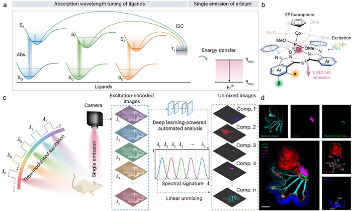
Recently, Zhang and coworkers demonstrated efficient fluorescent multiplexing in live mice based on a versatile spectral palette of lanthanide rainbow (Lanbow), constructed from erbium(Ⅲ)-phthalocyanine complexes [6]. These complexes exhibited narrow-band emission of Er3+ at 1530 nm (4I15/2-4I13/2 transition) sensitized by different phthalocyanines (Fig. 1a). Notably, they created distinct spectral identities by precisely tuning the absorption profile of the phthalocyanine sensitizer instead of altering the emission, which was achieved through systematic substituent engineering at α, β or both positions of the four peripheral aromatic rings (Fig. 1b). Consequently, they acquired nine discrete fluorophores, which exhibited peak absorption wavelengths from 673 nm to 772 nm (corresponding to EP673 to EP772) while maintaining narrow bandwidths (full-width at half-maximum of 20-48 nm).
Figure 1
Figure 1.
(a) Energy diagrams of absorption tuning of ligands and energy transfer path to erbium-centred emission. (b) Chemical structures of EP fluorophores. (c) Schematics illustrating a multispectral workflow with Lanbow, detecting signals in the 1500-1700 nm range via multiple excitation. (d) Unmixed images from each channel, emphasizing specific anatomical structures labelled with different EP fluorophores, along with their overlay. Reprinted with permission [6]. Copyright 2025, Nature Publishing Group.
Leveraging the absorption and excitation tunability of this Lanbow palette, Zhang and coworkers developed a novel multiplexed NIR-Ⅱ imaging strategy based on excitation-encoded and single-emission model (Fig. 1c). To validate this approach, they first evaluated its performance in tissue-mimicking intralipid solution. The spectral signatures of nine fluorophores were acquired by the phasor method, followed by decomposition to multispectral image datasets via a linear unmixing method to identify signals from individual fluorophores. Benefiting from the distinct excitation spectra, the spectral signatures of all nine fluorophores were effectively resolved into separate channels with outstanding image contrast (average > 2.7) and minimal signal bleed-through (average < 1.7%).
They further investigated the Lanbow palette for in vivo multispectral imaging in a colorectal cancer mouse model, demonstrating its potential for intraoperative visualization of multiple anatomical and pathological targets. Five fluorophores were employed to label specific positions (EP679 to intestine, EP699 to vessel, EP725 to tumor, EP737 to colon, EP772 to lymph) via intravenous or intragastric injection, resulting in simultaneous visualization of all five targets (Fig. 1d). Specifically, primary tumors, mesenteric lymph nodes and 16 suspected micronodular sites near the intestinal tract with sufficient signal-to-noise ratio (SNR > 5 dB) were successfully identified. Furthermore, real-time imaging of different channels revealed the synchronized movement of colon-attached tumor metastases and associated vasculature, highlighting the synchronicity of dynamic multiplexed imaging.
To enable real-time analysis of the complex multispectral data streams, they designed and trained a dedicated two-stage neural network of EndmemberNet, which processed a time-series multispectral dataset and initiated with a weighted fusion of images from different excitation wavelengths to enhance wavelength-specific features. Particularly, EndmemberNet exhibited comparably low error (average relative residual < 15%), high correlation (Pearson’s correlation coefficient > 0.96) and fast frame processing time (within 14.3 ms). Such speed is comparable to the deep-learning-based segmentation algorithm (e.g., DeepLabv3+), while approximately 30 times faster than conventional machine learning clustering methods (e.g., Gaussian mixture model and K-means), and orders of magnitude faster than manual analysis. As such, the automated workflow successfully achieved real-time spectral unmixing and provided immediate feedback during colorectal tumor surgery.
Note that the proposed Lanbow strategy can be further extended to other NIR-Ⅱ emission lanthanide ions (e.g., Yb3+, Nd3+, Ho3+, Tm3+), which can be achieved by careful ligand design to ensure energy-level match between the ligand triplet state and Ln3+ emitters. Moreover, the development of highly photostable fluorophores (e.g., StayGold) and the use of lower-irradiance light sources (e.g., LEDs) are also crucial to mitigate photodamage and photobleaching during long-term imaging [7].
The critical challenge for this strategy may lie in signal fidelity. Specifically, luminescence intensity signals usually suffer from unavoidable and inhomogeneous attenuation caused by tissue absorption and scattering, as well as inconsistent probe targeting and aggregation. Such a limitation becomes particularly pronounced when the Lanbow strategy relies on distinct excitation wavelengths. As such, fluorescence lifetime imaging may be a promising alternative approach, which is immune to background interference and provides stable and reliable lifetime signals in biological environments for high-resolution multispectral imaging [8].
In conclusion, this work established a novel excitation-encoded and single-emission strategy for real-time in vivo multispectral NIR-Ⅱ imaging based on a versatile Lanbow spectral palette. Moreover, the integration of an AI-powered automated processing pipeline effectively eliminated the dependency on labor-intensive, expert-driven workflows. Looking ahead, there remains substantial room for optimization of this strategy, especially through the design of fluorophores and the methodology for signal acquisition. It is anticipated that such endeavors will further advance their performance, paving the way for high-fidelity real-time multiplexed imaging in vivo.
Declaration of competing interest
The authors declare that they have no known competing financial interests or personal relationships that could have appeared to influence the work reported in this paper.
L. Hajji, F. Lam, M. Avtodeeva, et al., Adv. Sci. 11 (2024) 2404354. doi: 10.1002/advs.202404354
Figure 1
(a) Energy diagrams of absorption tuning of ligands and energy transfer path to erbium-centred emission. (b) Chemical structures of EP fluorophores. (c) Schematics illustrating a multispectral workflow with Lanbow, detecting signals in the 1500-1700 nm range via multiple excitation. (d) Unmixed images from each channel, emphasizing specific anatomical structures labelled with different EP fluorophores, along with their overlay. Reprinted with permission [6]. Copyright 2025, Nature Publishing Group.

 DownLoad:

 下载:
下载:


 下载:
下载:
