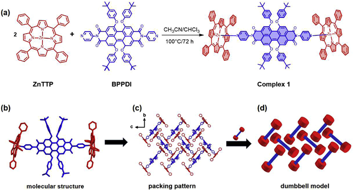
Figure 1.
(a) Synthesis strategy of complex 1. (b) Asymmetric unit of complex 1, omitting disordered CHCl3 molecules for clarity. (c) Packing pattern of complex 1 in bc plane. (d) Dumbbell model of complex 1.

Diradicals induced long-lived charge-separated state in a photo-driven coordination molecular rotor: Crystal structure and photophysical response
Ai Wang , Jun-Fei Bu , Ye Xia , Ming-Xing Chen , Xi Cheng , Feng Pan , Sisi Feng , Bing-Wu Wang , Song Gao


Molecular rotors are commonly employed as microviscosity sensors in liquid environments, providing insights into the dynamics of chemical transport and various biomolecular mechanisms within cellular systems [1-3]. Their capacity for rapid rotational motion enables dynamic switching between molecular geometries and charge transport states, a critical feature for advanced electronic applications. Drawing inspiration from both biological molecular rotors and their macroscopic counterparts, researchers have successfully engineered artificial molecular rotors capable of transducing optical, thermal, or electrical stimuli into controlled mechanical rotation [4,5]. The fundamental design strategy for these molecular rotors focuses on achieving targeted functionalities at the nanoscale, including molecular motors, propellers, switching elements, and sensing components [6-12]. The performance of molecular rotor-based electronics is fundamentally governed by the trade-off between rotational velocity and thermal stability. In particular, understanding how concentration, temperature, and external stimuli (including optical, thermal, and magnetic fields) affect the rotational dynamics and thermal stability of molecular rotors remains a fundamental challenge.
Achieving high yield and long-term stability of charge-separated states (CSS) represents a central objective in artificial photosynthesis systems designed for solar energy capture. Obtaining long-lived, light-driven CSSs in donor-acceptor (D–A) systems remains problematic due to the typically rapid charge recombination (CR) process [13]. Multiple strategies have been proposed to address this limitation, with spin control and D–A distance modulation emerging as the most prevalent approaches. In spin control systems, intersystem crossing (ISC) of the initially formed singlet CSS yields a triplet CSS in polar solvents, thereby rendering CR to the ground state becomes spin-forbidden. The lifetime of this triplet CSS can be further prolonged through magnetic field effects [14]. In the context of D–A distance modification, a great deal of research has been devoted to emulating natural photosynthesis, in which CSSs are generated through multistep, long-range charge transfer from photosynthetic antenna complexes to final electron acceptors [15]. Molecular self-organization in dyads, triads, and complex multicomponent architectures represents another approach for CSS stabilization [16-18]. Proper distance modulation within these assemblies facilitates both charge separation and hopping processes, thereby enhancing CSS longevity. While CSS lifetimes of approximately 1–20 ms have been observed in some systems (the majority being organic molecules), these often result from aggregation-induced charge hopping or D–A distance alterations modulated by electronic interaction [19,20]. However, the realization of long-lived CSS in coordination complexes remains rare, except when incorporating electron-accepting moieties such as C60 fullerene [21]. And the triplet electron transfer between porphyrin and fullerene leads to long-lived charge separation. Furthermore, the synergy of an extremely low CSS energy, lower than the triplet energy of each chromophore is the other strategy to obtain the long lifetime of CSS [22].
Perylenediimide derivatives possess high electron affinity, good charge carrier mobility, and outstanding thermal and oxidative stability. And as an excellent photosensitizer, zinc(Ⅱ) tetraphenylporphyrin (ZnTTP) is widely applied in photodynamic therapy and solar energy conversion. These characteristics render them promising for a range of technologies, including organic electronics, photovoltaics, and flexible displays. Herein, a novel coordination rotor complex 1 [(ZnTTP)2BPPDI·1.75CHCl3] constructed by ZnTTP and a perylenediimide derivative (BPPDI), was synthesized under solvothermal condition. We present the synthesis of complex 1 in Fig. 1. It is important to note that the complex 1 is, to our knowledge, the only example of crystalline coordination molecular rotor by a one-step metal-ligand coordination method. Compared with the previous studies, in our work, the long-lived lifetime of the CCS lies on the generated diradical species for a photo-driven molecular rotor 1. Characterization was performed using single-crystal X-ray diffraction (SCRD), CHN elemental analysis, powder X-ray diffraction (PXRD), IR spectroscopy, thermogravimetric analysis, and solid-state circular dichroism spectroscopy (Figs. S1-S4 in Supporting information). SCRD analyses found that complex 1 crystallizes in the monoclinic system with the chiral space group of C2, and its asymmetric unit consists of 2 ZnTTP, 1 BPPDI, and 1.75 crystalline CHCl3 molecules, forming a dumbbell-shaped structure. As shown in Fig. 1a, two Zn(Ⅱ) cation of ZnTTP are connected by the pyridine groups of BPPDI, resulting in a tetragonal pyramidal coordination geometry (disordered CHCl3 molecules omitted for clarity). The two-fold axis is parallel to the b-axis. Steric hindrance from the four bulky OAr substituents at the bay positions of BPPDI induces the distortion of the central six-membered ring. However, intermolecular interaction may occur between adjacent BPPDI and ZnTTP units. The shortest and longest distances between neighboring BPPDI and ZnTTP are 3.57 and 3.76 Å, respectively (Fig. S5 in Supporting information). In the bc plane, the packing of neighboring molecules in complex 1 can be simplified as close packing of the dumbbell-shaped units (Figs. 1b-d). Detail of single-crystal data and structural refinement results are listed in Tables S1-S3 (Supporting information), and selected bond lengths (Å) and bond angles (°) of complex 1 are displayed in Table S4 (Supporting information).
1H NMR spectroscopy of the complex 1 in CDCl3 confirmed its structure, consistent with the results of single crystal analysis (Fig. S6 in Supporting information). The chemical shift at 8.88 ppm corresponds to the 16 H atoms of the five-membered rings of coordinated ZnTTP (marked as dark-purple spots) [23]. The signal at 8.22 ppm (pink spots) is assigned to the 16 H atoms of the benzene rings of ZnTTP in complex 1. The chemical shifts at 8.04 ppm (blue spots) corresponds to another eight H atoms of benzene rings from coordinated ZnTTP. The signals at δ = 8.04 ppm (blue spots) and δ = 6.67 ppm (orange spots) are attributed to two different types of H atoms from the four OAr groups of BPPDI in complex 1. The signals at δ = 7.11 ppm corresponds to the four H atoms at the ortho-positions of BPPDI, while δ = 6.12 ppm corresponds to the H atoms at meta-positions of the pyridyl rings in the BPPDI ligand. The H atoms of the four tert-butyl groups at the bay positions of BPPDI resonate at 1.13 ppm. The 1H NMR spectrum of complex 1 was collected again after 7 days from the same sample (Fig. S7 in Supporting information). All chemical shifts showed no significant changes, demonstrating that complex 1 remains stable in CDCl3 for at least 7 days. The 1H NMR spectra of BPPDI, the mixture of ZnTTP and BPPDI in a 2:1 ratio and complex 1 are presented in Fig. S8 (Supporting information). Except for an impurity peak at 7.90 ppm, no significant differences in chemical shifts were observed between complex 1 and the ZnTTP and BPPDI mixture, further confirming the stability of complex 1 in CDCl3.
To clarify whether complex 1 remains fully dissociated or partially dissociated, or undergoes structural transformations in CDCl3, 1H NMR titration experiments were conducted (Fig. S9 in Supporting information). As the concentration of ZnTTP increases, the protons located at the meta-positions of the pyridyl rings and ortho-positions (2,5,8,11-positions) exhibit upfield shifts in the NMR spectra due to the formation of coordination bonds. The absence of spectral signals corresponding to non-coordinated, mono-coordinated, or di-coordinated species suggests that the system undergoes rapid exchange on the NMR time scale at room temperature.
To gain deeper insights into the molecular dynamics of complex 1 in solution, the geometric structure of complex 1 was applied to make two ZnTTP fragments undergoing free rotation along the coordination bonds in solution. Through clockwise rotation of one ZnTTP unit from θ = 0° to θ = 240° in 30° increments, the calculated single-point energy differences between configurations were all within 0.26 kcal/mol (Fig. S10 in Supporting information), supporting the potential role of complex 1 as a molecular rotor in CHCl3 [24]. Subsequently, its photochemical and photophysical properties were further analyzed as follows.
As shown in Fig. 2a, compared with the UV spectra of the starting materials ZnTTP (1.0 × 10⁻⁵ mol/L) and BPPDI (1.0 × 10⁻⁵ mol/L), that of complex 1 (1.0 × 10⁻⁵ mol/L) in CHCl3 displays a typical absorption of ZnTTP at 423 nm and two distinct absorption bands of BPPDI between 400 and 600 nm, with prominent peaks at 553 and 593 nm, respectively. Upon excitation at the wavelength maximum (550 nm, Fig. 2b), the fluorescence intensity of complex 1 is much stronger than that of BPPDI and ZnTTP, which may result from more efficient electron transfer in the complex than in the isolated molecules in CHCl3. TD-DFT calculations reveal that the HOMO and LUMO of complex 1 are occupied by the π and the π* orbitals of ZnTTP, respectively (Fig. S11 in Supporting information). The energy gap between the HOMO and LUMO is only 4.28 eV (Table S5 in Supporting information). Moreover, the UV absorptions of complex 1 overlap with its fluorescence emission between 586 nm and 626 nm (Fig. S12 in Supporting information), indicating that resonance energy transfer occurs [25].
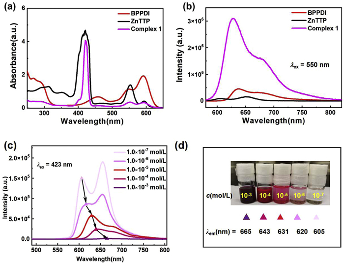
When the maximum absorption wavelength of ZnTTP (423 nm) was used as the excitation wavelength (Figs. 2c and d), the fluorescence spectra at room temperature revealed that complex 1 exhibited ACQ. Furthermore, the fluorescence emission peak showed a red shift with increasing concentration. This concentration-induced phenomenon is attributed to the inner filter effect (IFE) and the formation of different types of aggregates in concentrated solution. When the excitation wavelength was tuned to 550 nm (Fig. S13 in Supporting information), complex 1 demonstrated AIE behavior but only within a low concentration range of 1.0 × 10⁻⁷-1.0 × 10⁻⁵ mol/L. At higher concentration (1.0 × 10⁻³ and 1.0 × 10⁻⁴ mol/L, Fig. S13), the intensity of the maximum emission peak sharply decreased again due to IFE in concentrated solutions. Therefore, with respect to the strongest fluorescence intensity of complex 1, 550 nm was identified as the optimal excitation wavelength to analyze its photoresponsive behavior under different temperatures.
To assess the effect of temperature on the luminescence behavior of molecular rotor 1, fluorescence spectra were recorded from 330 K to 80 K to investigate its photophysical stability. As shown in Fig. S14 (Supporting information), the temperature-dependent fluorescence spectra of complex 1 in CHCl3 (1.0 × 10⁻⁵ mol/L, λex = 550 nm) are displayed as stack plots with Y-offsets. As the temperature decreased from 330 K to 200 K, the emission peak exhibited a red shift (from 625 nm to 654 nm), which can be attributed to the suppression of rotational and vibrational motions of the dumbbell-shaped molecules in solution. This suppression drives aggregation through entropy reduction. Concurrently, the ground-state energy decreases, leading to a narrower energy gap between the excited state and ground state. Below 200 K, however, the dominant emission band showed negligible changes in both peak position and intensity (Fig. S14). Upon further cooling to below 140 K, a distinct phosphorescent feature attributable to ZnTTP fragments emerged at 775 nm. Steady-state fluorescence spectra at 77 K not only revealed a red-shifted maximum emission wavelength (from 625 nm to 654 nm) but also exhibited two new emission peaks at 775 and 873 nm (Fig. S15 in Supporting information) [26]. These near-infrared (NIR) emission bands were preliminarily attributed to triplet-state transitions of the ZnTTP unit within complex 1, consistent with optimization results of the triplet state (T1, Table S6). The experimental phosphorescent lifetimes of complex 1 at 77 K were measured as 26.50 ms (775 nm) and 25.38 ms (873 nm).
To elucidate the excited-state dynamics of rotor 1, its transient absorption spectra in chloroform were systematically characterized, and the lifetimes of the key absorption peaks were analyzed at room temperature (Figs. 3a and b). The differential absorption spectra (ΔOD) of rotor 1 at designated delay times under 550 nm excitation are shown in Fig. 3a. Upon 550 nm nanosecond laser excitation, the absorption peaks at 423 nm and 465 nm decreased in intensity, while a weak, broad absorption band spanning 600–800 nm exhibited a slight enhancement. These observations provide additional evidence for the presence of excited-state energy transfer [27]. Specifically, complex 1 exhibits a positive absorption at 423 nm, corresponding to the singlet excited state ZnTTP(S1)-BPPDI(S0) in rotor 1. Through intersystem crossing (ISC), the ZnTTP•(T1)-BPPDI(S1) state may form from the ZnTTP(S1)-BPPDI(S0) [23], which contributes to a transient absorption maximum at 465 nm [28]. The lifetime of the triplet excited state ZnTTP•(T1)-BPPDI(S1) to ZnTTP•(T1)-BPPDI(S0) was measured as approximately 95 ns, and the lifetime of returning to the ground state ZnTTP(S0)-BPPDI(S0) from ZnTTP•(T1)-BPPDI(S0) was fitted to 746 ns (Fig. S17 in Supporting information). Consistent with the steady-state absorption and PL spectra, a negative ΔOD region between 540 nm and 570 nm is assigned to ground-state bleaching, which corresponds to its optimal excitation wavelength (λex = 550 nm) [29]. The time-resolved absorption spectra of the complex (Fig. 3a) display a broad band around 670 nm, characteristic of the [ZnTTP]+ cation. The absorption band observed at 775 nm is attributed to the BPPDI− anion, suggesting the formation of the ZnTTP•+-BPPDI•− CSS under 550 nm excitation [30]. The CSS exhibited a lifetime of approximately 912 ns, as confirmed by measurements at 775 nm, indicating its potential for artificial energy conversion (Fig. 3b). The CSS lifetime of rotor 1 is significantly longer than that of most other transition metal complexes reported in the literatures (Table S9 in Supporting information) [6,15,22,23,31-42].
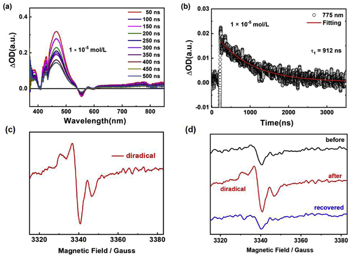
To investigate the origin of the long-lived CSS observed for molecular rotor 1 in chloroform, singlet oxygen generation was examined. As shown in Fig. S18 (Supporting information), the absence of the characteristic 1O2 emission signal at 1270–1280 nm confirms that singlet oxygen was not produced through energy-transfer quenching of the ZnTTP triplet state (T1) by ground-state molecular oxygen (3O2) [43]. Furthermore, the reverse excitation scan failed to detect the characteristic excitation peak of 1O2. These results exclude the formation of 1O2 during the photophysical processes. Furthermore, the EPR spectra at 30 K under xenon lamp irradiation (450 W) is presented in Fig. 3c, the appearance of a diradical signal confirms that complex 1 generates a small amount of diradical species (ZnTTP•+ and BPPDI•−) upon photoexcitation in solution under limited irradiation time [30]. The simulated EPR spectrum of rotor 1 has been applied for the further verification for the photo-induced generation of biradicals at 550 nm, which is presented in Fig. S19 (Supporting information). As shown in Fig. S19, the simulated EPR spectrum is labeled with "Sys1. S = 1"; which suggests a coupled diradical system. It might result from strong spin-spin interaction between the diradical ZnTTP•+-BPPDI•− in rotor 1. Due to the EPR spectrum was collected at 30 K, the frozen chloroform could lead to the local environmental heterogeneity [44,45], and consequently, rotor molecule 1 can adopt different conformations due to the single coordination bonds between Zn of ZnTTP and N of BPPDI. Thus, the weight of strongly coupled double-spin system is 1, while the weakly coupled double-spin system is 0.24 (equivalent to twice the value of a single spin 0.12). The EPR spectra complete recovered to the pre-illumination signal after a 10-minute dark period following irradiation (blue line in Fig. 3d). According to theoretical calculations, the highest occupied and lowest unoccupied molecular orbitals (HOMO and LUMO) involved in the triplet-state transition, are primarily composed of the π orbital of ZnTTP and the π* orbital of BPPDI, respectively, (Fig. S20 in Supporting information), which may contribute to the extended lifetime of the ZnTTP•+-BPPDI•− CSS. A detailed energy-level diagram and the corresponding photophysical pathways of rotor 1 are summarized in Fig. 4a.
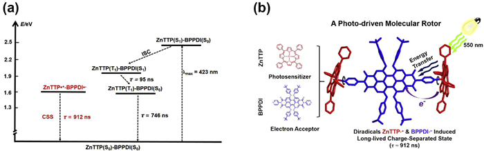
To gain a better understanding of the photochemical and photophysical processes of the rotor 1, we first performed a theoretical analysis of its spin electron density distribution with Multiwfn [46,47]. Spin density plots (light blue ball with black grid in Fig. S21 (Supporting information) and spin density population (Fig. S22 and Table S9 in Supporting information) of triplet states for rotor 1 shows that electron spin density of the diradical is predominantly delocalized over the conjugated framework of ZnTTP and the central core structure of BPPDI, which is consistent with experimental diradical species (ZnTTP•+ and BPPDI•−). Furthermore, optimized structures with HOMO and LUMO of rotor 1 calculated by DFT at the wB97XD/6-31G(d)&Lanl2DZ level of theory. Compared with its chromophores (ZnTTP and BPPDI), the lower triplet energy of rotor 1 together with the coupling between both units, leads to long-lived charge separation [21,22]. The minimal overlap between the HOMO (localized on PDI) and LUMO (localized on ZnTTP) provides a qualitative explanation for the inhibited charge recombination. And in order to visually illustrate the charge transfer process, the hole-electron distribution for rotor 1 was analyzed. Their distributions were displayed in Fig. S24 (Supporting information). The purple meshed spheres are the hole distribution of excitation state, which mainly are occupied on the BPPDI part. While displaying the electron distribution alone, its isosurface appears on the ZnTTP part of rotor 1 (light green). In the triplet state of the rotor 1, electron transfer may occur from the ZnTTP to the BPPDI ligand, which is consistent with the experimental data from transient absorption spectroscopy. The possible CCS formation pathway of rotor 1 under xenon lamp irradiation is depicted in Fig. 4b, photoinduced diradicals dominate the formation of long-lived charge-separated states with lifetime 912 ns.
In summary, we present the preparation of a novel Zn-complex rotor 1 [(ZnTTP)2BPPDI·1.75CHCl3] via a solvothermal method. X-ray crystallographic analysis of a single crystal revealed that BPPDI and ZnTTP form a dumbbell-shaped molecular motor structure in a 1:2 ratio. 1H NMR spectroscopy further confirmed that the binding ratio of the complex in CDCl3 is consistent with the single-crystal structure. Photophysical characterization, including steady-state and time-resolved spectra, demonstrated that rotor 1 is the only example of crystalline coordination molecular rotor with a long-lived CSS with a lifetime exceeding 912 ns. Time-resolved EPR spectroscopy under xenon lamp illumination suggests the formation of a diradical intermediate during photo-induced charge separation, which may contribute to the long-lived CSS. Our work further demonstrates that the generation of diradicals facilitates the attainment of long-lived charge-separated states in complex-rotor molecules. Future efforts will focus on the precise control of the microscopic motion of such molecular rotors under the influence of different field effects (e.g., light irradiation, magnetic fields).
Ai Wang: Writing – review & editing, Writing – original draft, Funding acquisition, Formal analysis, Data curation. Jun-Fei Bu: Investigation. Ye Xia: Formal analysis. Ming-Xing Chen: Methodology, Conceptualization. Xi Cheng: Software. Feng Pan: Software. Sisi Feng: Funding acquisition. Bing-Wu Wang: Writing – review & editing, Project administration, Funding acquisition, Conceptualization. Song Gao: Project administration, Conceptualization.
The authors declare that they have no known competing financial interests or personal relationships that could have appeared to influence the work reported in this paper.
The author gratefully acknowledges stimulating discussions with Professor Ben L. Feringa. The authors thank the financial supports by the Natural Science Foundation of China (Nos. 22573003 and 22475124), the Shanxi Scholarship Council of China (Nos. 2023-004 and 2023-018) and the program of the State Key Laboratory of Quantum Optics Technologies and Devices (No. 202502).
Supplementary material associated with this article can be found, in the online version, at doi:
M.K. Kuimova, G. Yahioglu, J.A. Levitt, K. Suhling, J. Am. Chem. Soc. 130 (2008) 6672–6673. doi: 10.1021/ja800570d
M.A. Haidekker, E.A. Theodorakis, Org. Biomol. Chem. 5 (2007) 1669–1678. doi: 10.1039/B618415D
M. Paez-Perez, M.K. Kuimova, Angew. Chem. Int. Ed. 63 (2024) e202311233. doi: 10.1002/anie.202311233
S. Erbas-Cakmak, D.A. Leigh, C.T. McTernan, A.L. Nussbaumer, Chem. Rev. 115 (2015) 10081–10206. doi: 10.1021/acs.chemrev.5b00146
L. Greb, J.M. Lehn, J. Am. Chem. Soc. 136 (2014) 13114–13117. doi: 10.1021/ja506034n
P. Stacko, J.C.M. Kistemaker, T. Leeuwen, M.C. Chang, E. Otten, B.L. Feringa, Science 356 (2017) 964–968. doi: 10.1126/science.aam8808
J. Shao, W. Zhu, X. Zhang, Y. Zheng, npj Comput. Mater. 6 (2020) 185. doi: 10.1038/s41524-020-00457-6
Z.T. Shi, Y.X. Hu, Z.B. Hu, et al., J. Am. Chem. Soc. 143 (2021) 442–452. doi: 10.1021/jacs.0c11752
J.X. Hou, G.Y. Long, W. Zhao, et al., J. Am. Chem. Soc. 144 (2022) 6851–6860. doi: 10.1021/jacs.2c01060
Q. Zhang, R. Toyoda, L. Pfeifer, B.L. Feringa, J. Am. Chem. Soc. 145 (2023) 6976–6985. doi: 10.1021/jacs.3c00586
A. Singhania, S. Kalita, P. Chettri, S. Ghosh, Nanoscale Adv. 5 (2023) 3177–3208. doi: 10.1039/d3na00010a
J.W. Verhoeven, H.J. van Ramesdonk, M.M. Groeneveld, A.C. Benniston, A. Harriman, ChemPhysChem 6 (2005) 2251–2260. doi: 10.1002/cphc.200500029
V.L. Gunderson, A.L. Smeigh, C.H. Kim, D.T. Co, M.R. Wasielewski, J. Am. Chem. Soc. 134 (2012) 4363–4372. doi: 10.1021/ja211329k
M. Borgström, N. Shaikh, O. Johansson, et al., J. Am. Chem. Soc. 127 (2005) 17504–17515. doi: 10.1021/ja055243b
L. Favereau, A. Makhal, Y. Pellegrin, et al., J. Am. Chem. Soc. 138 (2016) 3752–3760. doi: 10.1021/jacs.5b12650
V.V. Roznyatovskiy, R. Carmieli, S.M. Dyar, K.E. Brown, M.R. Wasielewski, Angew. Chem. Int. Ed. 53 (2014) 3457–3461. doi: 10.1002/anie.201309335
C. Bin, H.W. Song, A. Brnovic, et al., J. Am. Chem. Soc. 145 (2023) 18687–18692. doi: 10.1021/jacs.3c04372
G. Rotas, K. Stranius, N. Tkachenko, N. Tagmatarchis, Adv. Funct. Mater. 28 (2018) 1702278. doi: 10.1002/adfm.201702278
Y.Q. Hou, Q.Y. Liu, X. Zhang, et al., J. Mater. Chem. C 7 (2019) 12048–12074. doi: 10.1039/c9tc04285g
T. Ito, M. Naka, A. Miura, T. Ujiie, H. Nakamura, T. Matsuo, Bull. Chem. Soc. Jpn. 74 (2001) 657–665. doi: 10.1246/bcsj.74.657
C.O. Obondi, G.N. Lim, B. Churchill, et al., Nanoscale 8 (2016) 8333–8344. doi: 10.1039/C6NR01083K
V.M. Blas-Ferrando, J. Ortiz, K. Ohkubo, et al., Chem. Sci. 5 (2014) 4785–4793. doi: 10.1039/C4SC01579G
J. Fernandez-Ariza, R.M.K. Calderon, M.S. Rodríguez-Morgade, D.M. Guldi, T. Torres, J. Am. Chem. Soc. 138 (2016) 12963–12974. doi: 10.1021/jacs.6b07432
C.L.F. van Beekand, B.L. Feringa, J. Am. Chem. Soc. 146 (2024) 5634–5642. doi: 10.1021/jacs.3c14430
M.Y. Fan, B. Sun, X. Li, et al., Inorg. Chem. 60 (2021) 9148–9156. doi: 10.1021/acs.inorgchem.1c01165
A. Harrima, J. Chem. Soc., Faraday Trans. 76 (1980) 1978–1985. doi: 10.1039/f19807601978
W. Danowski, F. Castiglioni, A.S. Sardjan, et al., J. Am. Chem. Soc. 142 (2020) 9048–9056. doi: 10.1021/jacs.0c03063
J. Rodriguez, C. Kirmaier, D. Holten, J. Am. Chem. Soc. 111 (1989) 6500–6506. doi: 10.1021/ja00199a004
L. Flamigni, B. Ventura, C.C. You, C. Hippius, F. Wulrthner, J. Phys. Chem. C 111 (2007) 622–630. doi: 10.1021/jp065394g
I.M. Dixon, J.P. Collin, J.P. Sauvage, L. Flamigni, Inorg. Chem. 40 (2001) 5507–5517. doi: 10.1021/ic010416t
P.J. Griffin, B.J. Charette, J.H. Burke, et al., J. Am. Chem. Soc. 144 (2022) 12116-12126. doi: 10.1021/jacs.2c02580
L. Ma, T.X. Zhang, M.C. Li, et al., Chem. Sci. 15 (2024) 17150-17160. doi: 10.1039/D4SC05592F
A. Sautter, B.K. Kaletas, D.G. Schmid, et al., J. Am. Chem. Soc. 18 (2005) 6719−6729. doi: 10.1021/ja0448216
A.I. Oliva, B. Ventura, F. Würthner, et al., Dalton Trans. (2009) 4023−4037. doi: 10.1039/b819496c
B.J. Charette, S.R. King, J.Q. Chen, et al., J. Phys. Chem. A 127 (2023) 7747−7755. doi: 10.1021/acs.jpca.3c04269
J. Hu, Q. Xiang, X. Tian, et al., J. Am. Chem. Soc. 146 (2024) 10321–10330. doi: 10.1021/jacs.3c11585
K.A. Jolliffe, T.D.M. Bell, K.P. Ghiggino, S.J. Langford, M.N. Paddon-Row, Angew. Chem. Int. Ed. 37 (1998) 915–919. doi: 10.1002/(SICI)1521-3773(19980420)37:7<915::AID-ANIE915>3.0.CO;2-8
G. Ajayakumar, K.R. Gopidas, Photochem. Photobiol. Sci. 7 (2008) 826–833. doi: 10.1039/b717879d
D.I. Schuster, P. Cheng, P.D. Jarowski, et al., J. Am. Chem. Soc. 126 (2004) 7257–7270. doi: 10.1021/ja038676s
S. Chakraborty, T.J. Wadas, H. Hester, R. Schmehl, R. Eisenberg, Inorg. Chem. 44 (2005) 6865–6878. doi: 10.1021/ic0505605
S. Suzuki, R. Sugimura, M. Kozaki, et al., J. Am. Chem. Soc. 131 (2009) 10374–10375. doi: 10.1021/ja904241r
S. Suzuki, Y. Matsumoto, M. Tsubamoto, et al., Phys. Chem. Chem. Phys. 15 (2013) 8088–8094. doi: 10.1039/c3cp50182e
Y. Qin, L.J. Chen, F.Y. Dong, et al., J. Am. Chem. Soc. 141 (2019) 8943–8950. doi: 10.1021/jacs.9b02726
S.Y. Zhang, M. Pink, T. Junghoefer, et al., J. Am. Chem. Soc. 144 (2022) 6059−6070. doi: 10.1021/jacs.2c01141
N. Gallagher, H. Zhang, T. Junghoefer, et al., J. Am. Chem. Soc. 141 (2019) 4764−4774. doi: 10.1021/jacs.9b00558
T. Lu, F.W. Chen, J. Comput. Chem. 33 (2012) 580–592. doi: 10.1002/jcc.22885
T. Lu, J. Chem. Phys. 161 (2024) 082503. doi: 10.1063/5.0216272

Figure 1 (a) Synthesis strategy of complex 1. (b) Asymmetric unit of complex 1, omitting disordered CHCl3 molecules for clarity. (c) Packing pattern of complex 1 in bc plane. (d) Dumbbell model of complex 1.

Figure 2 (a) UV spectra of BPPDI (red line, 1.0 × 10⁻⁵ mol/L), ZnTTP (blank line, 1.0 × 10⁻⁵ mol/L), and complex 1 (pink line, 1.0 × 10⁻⁵ mol/L in CHCl3). (b) Fluorescence spectra of BPPDI, ZnTTP, and complex 1 with λex = 550 nm. (c) Emission spectra of complex 1 in CHCl3 with λex = 423 nm (concentration of 1 are 1.0 × 10⁻⁷, 1.0 × 10⁻⁶, 1.0 × 10⁻⁵, 1.0 × 10⁻⁴ and 1.0 × 10⁻³ mol/L, respectively). (d) Digital photos of different concentrations of complex 1 in CHCl3.

Figure 3 (a) Nanosecond transient absorption spectra of 1 (1.0 × 10⁻⁵ mol/L) in CHCl3 during 550 nm nanosecond laser excitation. (b) Lifetime of the ΔOD spectra at 775 nm and its fitting curve. (c) EPR spectra of complex 1 at 30 K irradiated by a xenon lamp (450 W). (d) EPR spectra of complex 1 at 30 K irradiated by a xenon lamp (450 W) before, after, and recovery after 10 min dark period post-irradiation.

 扫一扫看文章


扫一扫关注我们
 DownLoad:
 下载:

 下载: