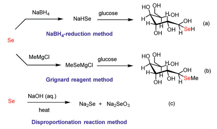
Scheme 1.
Method for activating elemental selenium.

Nano-Se pesticide synthesis from Se(Ⅳ) for industrial chain material balance
Xinrui Xiao , Xingyu Chen , Ming Cao , Xiang Li , Congkui Tian , Hongen Cao , Lei Yu


Selenium is an essential trace element for life, and deficiency of it poses risks to human health [1-3]. Developing selenium-enriched agriculture is a core solution to address the selenium deficiency issue [4-6]. Selenium-enriched agricultural technologies have long focused on optimizing selenium sources: Inorganic selenium like sodium selenite is low-cost but highly ecotoxic [7], while organic selenium such as selenomethionine is eco-friendly but expensive, limiting large-scale application [8]. Our research group has dedicated to developing a method for preparing organic low-valence selenium compounds at low cost for selenium-enriched agriculture, and both NaBH4-reduction method (Scheme 1a) [9] and Grignard reagent method (Scheme 1b) [10] have been developed in recent years. However, these two methods have certain drawbacks that limit their practical application. They are still associated with relatively high costs and require the use of sensitive chemical reagents, which increases operational complexity and expenses. Additionally, the NaBH4-reduction method poses a certain degree of danger, as hydrogen gas is released during the reaction process. Recently, disproportionation reaction of selenium powder with aqueous NaOH has been employed to generate the water-soluble selenium compounds (Scheme 1c) [11]. In comparison with the NaBH4-reduction method and Grignard reagent method, it produces Se(-Ⅱ) (low-valence selenium) at low cost and under mild reaction conditions. Se(-Ⅱ) is a key species for synthesizing organic low-valence selenium fertilizers or antibacterial materials [12-14]. However, the disproportionation reaction inevitably generates Se(Ⅳ) (high-valence selenium) accounting for a 33% molar ratio theoretically. The unutilized Se(Ⅳ) not only violates green chemistry's "complete atomic utilization" principle but also causes material surplus in the industrial chain, which hinders the closed-loop operation and sustainable development. How to consume Se(Ⅳ) is the key to achieving the industrial chain material balance that determines whether the disproportionation reaction can be applied to large-scale production.
Yet, rice blast, caused by Magnaporthe oryzae, is a devastating fungal disease threatening global rice yield and food security [15,16]. Conventional chemical control leads to pesticide residues, environmental pollution, and pathogen resistance, creating an urgent need for eco-friendly alternatives [17-19]. To solve both the Se(Ⅳ) surplus in the selenium industrial chain and the demand for green rice blast control, we tried to convert the underutilized Se(Ⅳ) (in Na2SeO3 form, the disproportionation by-product) into value-added selenium nanoparticles (Se NPs) as the pesticide for rice blast control. This approach not only achieves Se(Ⅳ) recycling to achieve industrial chain material balance and complete atomic utilization but also pioneers an innovative agricultural chemistry concept of 'fertilizer-pesticide dual function': Se NPs, as a selenium source, can enhance rice selenium content (fertilizer function) [20], while simultaneously exhibiting high antifungal activity to control rice blast (pesticide function). This integration aligns with green agricultural development, offering environmental and economic benefits. Herein, we wish to report our findings.
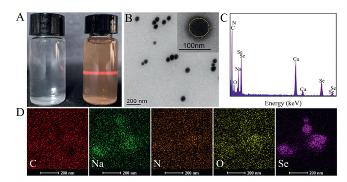
Se NPs were synthesized via an in-situ reduction method, using purified sodium selenite as the selenium source. Gum Arabic (GA), a natural polysaccharide, is rich in hydroxyl groups in its molecular skeleton. These hydroxyl groups could pre-assemble with HSeO3- via hydrogen bonding, thereby effectively anchoring HSeO3- onto the polysaccharide chains. Subsequently, reduced glutathione (GSH) was introduced as a reducing agent. The thiol group (–SH) of GSH provided electrons for the in-situ reduction of the anchored Se4+ to Se0. During this process, the unoxidized GSH in the reaction system was able to adsorb onto the Se0 surface through non-covalent interactions. Simultaneously, the polar functional groups (e.g., amino and carboxyl groups) on GSH molecules formed an extensive hydrogen-bonding network with the hydroxyl groups of GA. Furthermore, the long-chain polysaccharide structure of GA closely enveloped the newly formed Se0. The steric hindrance provided by GA effectively inhibited nanoparticle aggregation, thereby enhancing the stability of the nano system. The successfully synthesized Se NPs were present as a transparent reddish-brown solution with a distinct Tyndall effect upon laser irradiation (Fig. 1A) [21,22]. TEM images illustrated that Se NPs were monodisperse spheres with uniform particle size distribution (Fig. 1B). Energy-dispersive X-ray spectroscopy (EDX), elemental mapping and HAADF-STEM image (Figs. 1C and D, Fig. S1 in Supporting information) elucidated the elemental composition. The core element of the Se NPs was Se. C, mainly derived from GA and GSH, exhibited a distinct circular distribution, while N, derived from GSH, was uniformly distributed throughout the nanoparticles. These results demonstrated that the prepared Se NPs were core-shell-structured spherical nanoparticles with a core of Se0 and a composite organic shell composed of GA and GSH.
Through TEM statistical analysis, the average particle size of Se NPs was 57.94 nm (Fig. 2A). XPS analysis was employed to examine the elemental composition and chemical states of Se NPs (Fig. 2B and Fig. S2 in Supporting information). The survey scan detected characteristic peaks of C, N, O, Na, and Se. The result indicated that the binding energies of Se element were 55.28 eV and 56.03 eV, demonstrating that GSH completely reduced HSeO3⁻ in situ to Se⁰ without oxidation of Se NPs [23]. This result was mainly attributed to the protective effect of the GA/GSH composite shell. The zeta potentials of GA, GSH, Na2SeO3, and Se NPs were determined (Fig. 2C). The zeta potentials of GA and GSH were –18.41 mV and –13.73 mV, which were related to the negative charge properties of carboxyl groups (–COOH) and thiol groups (–SH) in their molecular structures. Compared with the potential of Na2SeO3 (–21.13 mV), the zeta potential of Se NPs was substantially lowered to –32.46 mV. The increased negative charge was mainly attributed to the synergistic assembly of GA and GSH on the outer layer of Se0, which resulted in the high-density enrichment of numerous negatively charged groups, thus enhancing the interfacial charge density. A zeta potential of over ±30 mV typically confers good colloidal stability [24]. X-ray diffraction (XRD) showed that the characteristic diffraction peaks of Se NPs were consistent with those of Se (Fig. 2D). These diffraction peaks appeared at 23.51°, 29.70°, 41.30°, 43.65°, 45.35°, 51.72°, 55.65°, 61.21°, and 65.23°, corresponding to the (100), (101), (110), (102), (111), (201), (003), and (103) crystal planes [25]. The coating of Se⁰ by GA and GSH resulted in a slight decrease in and broadening of the peak intensities. Fourier transform infrared (FT-IR) spectroscopy was shown in Fig. 2E. The spectrum of Se NPs at 3482 cm-1 corresponded to the stretching vibration of O—H bonds in GA, demonstrating that the hydroxyl groups (–OH) on the GA polysaccharide chains coated the surface of Se NPs through hydrogen bonding interactions. The characteristic peaks at 1664 cm-1 and 1585 cm-1 corresponded to the stretching vibration of C═O bonds (amide band Ⅰ) and the bending vibration of N—H bonds (amide band Ⅱ) [26,27], which were mainly derived from the peptide bond structure of GSH. This result verified that GSH formed an extensive hydrogen-bond network with the hydroxyl groups of GA via the amide groups on its peptide chain, thereby confirming the successful preparation of Se NPs.
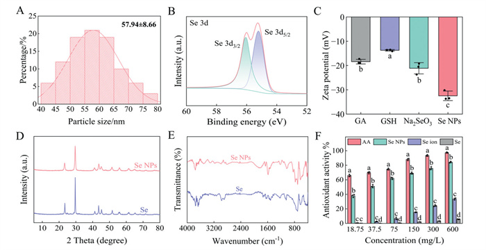
In plant biosystems, the interaction between biomolecules and molecular oxygen generates excessive free radicals, which trigger the accumulation of reactive oxygen species (ROS) [28]. This accumulation ultimately leads to cellular dysfunction, tissue necrosis, and exacerbation of disease symptoms. Antioxidants protect the structural and functional macromolecules of plant cells from free radical damage by blunting the progression of an oxidative chain reaction [29,30]. The DPPH radical is a specific free radical characterized by a nitrogen center and lipophilicity, exhibiting a characteristic absorption peak at 517 nm [31]. It is frequently used to evaluate antioxidant activity. Therefore, ascorbic acid (AA) was used as a positive control to compare the DPPH radical scavenging capacities of Se NPs, Se ion, and commercial Se (referred to as Se, purchased from commercial sources) over a concentration range of 18.75–600 mg/L. As shown in Fig. 2F, at a concentration of 600 mg/L, the antioxidant activities of AA, Se NPs, Se ion, and Se were 97.37%, 84.25%, 33.6%, and 5.21%. Compared with AA, Se NPs exhibited strong DPPH radical scavenging ability, with antioxidant activity exceeding 60% within the 75–600 mg/L concentration range and an IC50 value of 38.17 mg/L. This can be primarily attributed to the nanostructure and surface chemistry of Se NPs. Within a certain range, the antioxidant capacity of nanoparticles increases as their size decreases. The small particle size of Se NPs provides more surface reaction sites. In addition, the abundance of hydrogen-donating groups (–OH, –SH, etc.) in GA and GSH facilitates rapid reduction of DPPH radicals via a hydrogen atom transfer mechanism. In contrast, both Se ion and Se exhibited weak antioxidant activities, with scavenging rates below 35% across the tested concentration range (18.75–600 mg/L). These results indicated that Se NPs possessed considerable antioxidant activity and showed promise for applications in alleviating oxidative damage in plants.
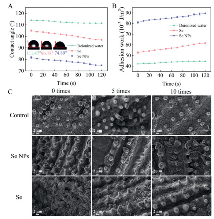
In agricultural production, foliar spraying of fungicides is one of the most common methods for controlling plant diseases [32]. The effective deposition of pesticide droplets on plant leaves can improve the efficiency of pesticide utilization and reduce chemical waste. Contact angle and adhesion work are widely used to evaluate the wettability of materials on leaf surfaces. The contact angle is primarily determined by the surface tension of the leaf-liquid interface [33,34]. Fig. 3A showed the dynamic contact angles of deionized water, Se, and Se NPs (100 mg/L) on rice leaves within 120 s. When the contact time was 120 s, the contact angles of deionized water and Se were 111.47° and 96.78°. The relatively high contact angles indicated that deionized water and Se had poor wettability on rice leaves. In contrast, the contact angle of Se NPs decreased from 81.69° to 74.89° during the same period. Adhesion work depends on the surface tension of the solution and the cosine of the contact angle. A larger adhesion work corresponds to stronger droplet adhesion and better leaf surface wettability. Compared with deionized water and Se, the adhesion work of Se NPs increased from 81.08 mJ/m2 to 89.63 mJ/m2 within 120 s (Fig. 3B). This change could be attributed to the abundant hydrophilic groups present in GA and GSH, which formed a hydrophilic layer on the surface of Se NPs. Through hydrogen bonding interactions, this layer reduced the solid-liquid interfacial tension, thereby verifying the superior wettability of Se NPs on rice leaves. The microstructure of the leaf surface is closely related to its wettability [35]. The surfaces of most plant leaves are covered with a hydrophobic waxy layer, which increases the contact angle of pesticide droplets with the leaf surfaces [36]. Rice leaves possess microstructures such as papillae and trichomes, and are covered with hydrophobic lamellar wax crystals, which further enhance their hydrophobicity [37]. To evaluate the foliar wettability of Se NPs, this study employed a water-washing method to simulate rainwater scouring, and analyzed the deposition morphology of Se NPs and Se on rice leaves using SEM (Fig. 3C) [38]. It could be seen that the surfaces of untreated rice leaves were covered with papillae and stomata along the leaf veins, exhibiting superhydrophobicity. Unwashed Se NPs formed a continuous and dense coating on the rice leaf surfaces, while Se was distributed in discrete sheet-like morphologies. After 5 and 10 washing cycles, a large quantity of Se detached from the leaves, with only a small number of particles remaining in an uneven distribution. In contrast, Se NPs exhibited only partial detachment, formed a thicker deposition layer, and remained more uniformly distributed and firmly attached. These results indicated that Se NPs possessed excellent deposition performance on superhydrophobic leaf surfaces, a property that helps to reduce losses caused by rainwater washing and to improve their practical utilization efficiency.
Rice blast, caused by the ascomycete fungus (Magnaporthe oryzae), is a rapidly spreading and devastating disease [39]. The pathogen is characterized by strong host specificity, multisite infection, frequent outbreaks under high temperature and humidity, rapid spore dissemination, and high risk of fungicide resistance [40]. Following infection, symptoms manifest at different growth stages and on different parts of the rice plant. In severe cases, it leads to leaf wilting, plant lodging, and even plant death. To evaluate the antifungal activity of Se NPs and Se against M. oryzae, in vitro experiments were conducted using the mycelial growth rate method. As shown in Figs. 4A and C, Se NPs exhibited a significant concentration-dependent antifungal activity against M. oryzae, with inhibitory effects becoming more pronounced as the concentration increased. At equivalent concentration, Se NPs showed a significantly stronger inhibitory effect on the mycelial growth of M. oryzae compared with Se. At a concentration of 40 mg/L, the mycelial growth inhibition rates of Se NPs and Se reached 78.42% and 33.34%. At 80 mg/L, mycelial growth was almost inhibited by Se NPs, and the inhibition rate was as high as 97.12%. Furthermore, within the concentration range of 20–80 mg/L, the mycelia treated with Se NPs exhibited a distinct red coloration. This phenomenon could be attributed to the fact that the mycelia of M. oryzae could still maintain a certain metabolic capacity under stress, activate the stress defense system, and sequester Se NPs in organelles via membrane transport proteins. At this point, Se NPs were highly accumulated in the mycelia, thereby showing a red color macroscopically. According to Table S1 (Supporting information), the EC50 values of Se NPs and Se against M. oryzae were 15.21 mg/L and 102.29 mg/L. To further evaluate the biological activity of Se NPs, the mycelial morphology of M. oryzae treated with Se NPs (20 mg/L) was analyzed using SEM. As shown in Fig. 4B, untreated mycelia showed a smooth and regular tubular structure. In contrast, the treated mycelia (Se NPs) showed obvious signs of desiccation, crumpling, twisting, and fragmentation [41]. It is hypothesized that Se NPs initially adhere to the fungal cell wall via electrostatic interactions, leading to structural disruption and increased membrane permeability. This compromised barrier facilitates the internalization of Se NPs into the hyphae [42]. Furthermore, the small particle size of Se NPs allowed them to promote intimate contact with the mycelia, penetrated the mycelial cell wall, and caused local fragmentation and separation, thereby contributing to their antifungal efficacy [43].
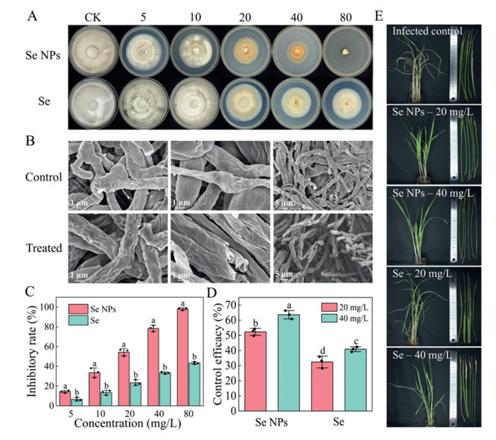
The practical control efficacy of Se NPs against rice blast was investigated through pot experiments. The preventive effects of Se NPs and Se against M. oryzae infection were shown in Figs. 4D and E. Compared with the infected control, rice plants foliar-sprayed with Se NPs at 20 mg/L and 40 mg/L exhibited a reduced number and smaller area of leaf lesions, with pathogenic symptoms were effectively alleviated. The disease control efficiencies were 52.28% and 63.60%, and the rice plants maintained normal growth with green and upright leaves, indicating that Se NPs effectively controlled the disease without causing phytotoxicity to the rice plants. Furthermore, due to their small particle size and excellent leaf wettability, Se NPs could efficiently adhere to leaf surfaces and be absorbed by the plant tissues through stomata and epidermal cells, thereby ensuring that the active selenium components rapidly reached the infection sites to exert their protective effects [44]. In contrast, although Se also showed a certain control efficacy (32.40% and 40.83%), its efficacy was significantly lower than that of Se NPs. Moreover, the corresponding rice plants exhibited symptoms including withering, yellowing, and lodging. These results were consistent with the findings from the in vitro activity assays.
Selenium, as an essential trace element for plants, plays a beneficial role in plant growth [45,46]. To investigate the effects of Se NPs on rice seed germination, we then analyzed the effects of different concentrations of Se NPs and Se on rice seed root and shoot lengths (Figs. 5A-C). After 7 days, the root and shoot lengths of the control group were 6.93 cm and 5.03 cm. At a concentration of 10 mg/L, the root and shoot lengths under Se NPs treatment were 7.12 cm and 6.86 cm, while those treated with Se were 6.01 cm and 5.67 cm. At low concentrations, both Se NPs and Se promoted root and shoot growth, with Se NPs exhibiting a stronger promoting effect than Se. This was mainly attributed to the small particle size and large specific surface area of the nanoparticles, which facilitated their adsorption onto the seed coat. This adsorption enhances hydrophilicity, accelerating the softening of the seed coat and thereby reducing the physical resistance to embryo breakthrough [47]. Additionally, Se NPs can penetrate the seed through micropores on the seed coat and the intercellular spaces of parenchyma tissues, thus enhancing the absorption efficiency of active selenium. As the concentration increased (20–40 mg/L), both materials slightly inhibited root and shoot growth, though this did not affect normal germination.
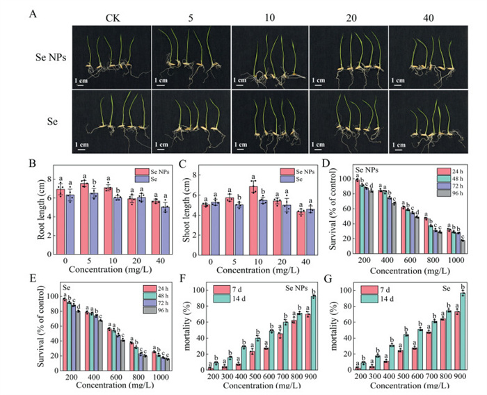
The zebrafish, as an ideal vertebrate model, is widely used to assess the acute toxicity of pesticides to aquatic organisms, providing a critical basis for evaluating pesticide risks in aquatic environments [48]. Figs. 5D and E showed the survival rates of zebrafish treated with different concentrations of Se NPs and Se over different time periods. The survival rate of zebrafish decreased with increasing time and concentration. After 24, 48, 72, and 96 h of exposure, the LC50 values of Se NPs were 739.13, 635.29, 576.64, and 510.50 mg/L. Under the same conditions, the LC50 values of Se were 650.99, 592.11, 530.20, and 442.23 mg/L (Table S2 in Supporting information). The results indicated that both Se NPs and Se were of low toxicity, and the acute toxicity of Se NPs to zebrafish was significantly lower than that of Se. This might have been because Se NPs were more stable in aqueous environments, which reduced the peak exposure concentration of active selenium, thereby enhancing the safety for zebrafish. In contrast, selenium in water was rapidly absorbed by zebrafish, causing oxidative stress and leading to acute poisoning [49]. Earthworms, as classic indicator organisms for assessing soil ecosystem health and pollution levels, are commonly used to evaluate the acute toxicity of pesticides in soil environments [50]. Figs. 5F and G showed the mortality rates of earthworms treated with different concentrations of Se NPs and Se for 7 and 14 days. After 7 days, the LC50 values of Se NPs and Se were 701.65 and 681.18 mg/kg. After 14 days, the LC50 values of Se NPs and Se were 545.93 and 519.71 mg/kg (Table S3 in Supporting information). The results showed that both Se NPs and Se exhibited low toxicity, and the acute toxicity of Se was higher than that of Se NPs.
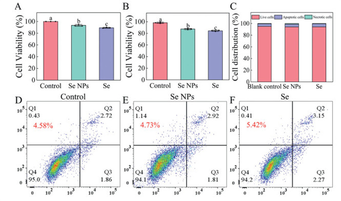
To evaluate the safety of Se NPs to humans, the cytotoxicity of Se NPs and Se on human L02 hepatocytes was assessed using the CCK-8 assay (Figs. 6A and B). At a concentration of 20 mg/L, the cell viabilities of Se NPs and Se were 93.45% and 89.29% after 24 h of culture. After 48 h, the cell viabilities slightly decreased to 87.42% and 84.43%, yet maintained high levels, with Se NPs treatment showing higher cell viability than Se. The cell apoptosis rate was also one of the important indicators for evaluating the safety of Se NPs to humans. As shown in Figs. 6C–F, the apoptosis rates of the control group, Se NPs group, and Se group were 4.58%, 4.73%, and 5.42%. The percentages of apoptotic cells (Q2, Q3), necrotic cells (Q1), and live cells (Q4) in the Se NPs group showed no significant difference from those in the control group. The results indicated that at a certain concentration, Se NPs exerted no significant cytotoxicity to human cells and exhibited good biocompatibility and safety.
The antioxidant enzyme system serves as the core defense mechanism for plants in response to environmental stress, and its activity is directly linked to the capacity to scavenge excess reactive oxygen species (ROS) and maintain redox homeostasis [51,52]. During their growth and development, plants are susceptible to various abiotic stresses such as drought, high temperature, and high salinity, as well as biotic stresses like pathogen infections [53]. These stressors can induce the production of ROS within plant cells. When ROS accumulate excessively, they trigger lipid peroxidation, disrupt protein structure and function, and even cause DNA damage, ultimately impairing plant growth and development [54]. Therefore, investigating changes in the activity of antioxidant enzymes is a key physiological indicator for assessing plant stress resistance [55]. Catalase (CAT), Peroxidase (POD), and Superoxide Dismutase (SOD) perform various functions in plants, including regulating H2O2 levels in plants, indirectly participating in signal transduction, and strengthening cell walls. This study analyzed the effects of foliar spraying with different concentrations of Se NPs and Se (5, 10, 20, 40, 80 mg/L) on the activities of CAT, POD, and SOD. Compared with the control group, both Se NPs and Se at certain concentrations enhanced enzyme activities, with Se NPs exhibiting a significantly stronger enhancing effect than Se. As shown in Fig. 7A, at a concentration of 20 mg/L, the CAT activities of rice plants treated with Se NPs and Se were 812.33 U/g FW and 614.21 U/g FW, with the maximum increases in enzyme activities reaching 44.48% and 9.25%. When the treatment concentration exceeded 20 mg/L, the CAT activity decreased slightly. Fig. 7B indicated that POD activity increased significantly with rising concentration. At 80 mg/L, the POD activities of plants treated with Se NPs and Se were 5701.33 U/g FW and 4796.21 U/g FW, representing maximum increases of 51.67% and 42.55%. At 20 mg/L, the SOD activities treated with Se NPs and Se were 722.12 U/g FW and 479.76 U/g FW, representing maximum increases of 37.41% and 5.79% (Fig. 7C). Selenium can regulate redox signaling pathways in plant cells, enhance the transcription of genes encoding these enzymes and their protein synthesis, protect enzyme active sites, and thereby improve enzyme activities [56]. Compared with Se, Se NPs are more easily absorbed and transformed by rice leaves, thereby efficiently providing nutrients for plant cells and enhancing the synthesis and transformation of antioxidant enzymes.
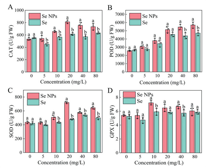
The active centre of Glutathione Peroxidase (GPX) is selenocysteine, and selenium is an essential element for the synthesis of selenocysteine [57]. Selenium deficiency can lead to structural defects in the active center of GPX, resulting in a significant decrease in enzyme activity [58]. Fig. 7D analyzed the effects of foliar spraying with different concentrations of Se NPs and Se (5, 10, 20, 40, 80 mg/L) on GPX activity. At a concentration of 10 mg/L, the GPX activities of rice plants treated with Se NPs and Se were 7.20 U/g FW and 5.93 U/g FW, representing maximum increases of 23.89% and 8.99%. The results indicated that both Se NPs and Se could enhance GPX activity in rice plants, and the enhancing effect of Se NPs was greater than that of Se. Se NPs can efficiently and stably provide selenium for plants, thereby improving the ability of GPX to scavenge peroxides.
In conclusion, we have addressed a critical material balance challenge in the selenium industrial chain by converting Se(Ⅳ), an inevitable by-product (33% molar ratio) of the low-cost disproportionation reaction of selenium powder with aqueous NaOH, into value-added Se NPs. The synthesized Se NPs exhibit favorable physicochemical properties, including uniform spherical morphology, good colloidal stability (zeta potential of −32.46 mV), strong antioxidant activity (DPPH radical scavenging rate of 84.25% at 600 mg/L), and excellent rice leaf wettability (contact angle decreasing to 74.89° within 120 s), which lay a solid foundation for their agricultural application. Importantly, Se NPs demonstrate high efficacy in rice blast control: In vitro, they inhibit the mycelial growth of Magnaporthe oryzae with a low EC50 value, and in vivo pot experiments confirm their ability to reduce lesion formation while enhancing rice antioxidant enzyme activities (catalase, peroxidase, superoxide dismutase, and glutathione peroxidase), thereby boosting the plant's stress resistance.
Beyond solving the Se(Ⅳ) surplus issue, this work pioneers an innovative "fertilizer-pesticide dual function" concept in agricultural chemistry. Notably, extensive studies have demonstrated that Se NPs markedly enhances crop yield, ameliorates nutritional quality, elevates the content and bio accessibility of organic selenium in crops, and mitigates the accumulation of hazardous heavy metals (e.g., arsenic and cadmium) [59-62]. Moreover, Se NPs can control rice blast effectively and show safety toward non-target organisms (low toxicity to zebrafish, earthworms, and human L02 hepatocytes), ensuring environmental and ecological compatibility. This strategy not only achieves closed-loop selenium utilization and industrial chain sustainability but also provides a promising eco-friendly alternative for crop disease management, holding significant potential to advance selenium-enriched agriculture and green pest control practices [63].
Xinrui Xiao: Investigation. Xingyu Chen: Investigation. Ming Cao: Investigation. Xiang Li: Investigation. Congkui Tian: Investigation. Hongen Cao: Writing – original draft, Supervision, Investigation. Lei Yu: Writing – review & editing, Supervision, Funding acquisition, Conceptualization.
The authors declare no conflict of interest.
We thank the Postgraduate Research & Practice Innovation Program of Jiangsu Province (Yangzhou University) (No. SJCX25_2352), the Yangzhou City and Yangzhou University Cooperation Program (No. YZ2023209) and the Priority Academic Program Development of Jiangsu Higher Education Institutions (PAPD) for support.
Supplementary material associated with this article can be found, in the online version, at doi:
M.P. Rayman, Lancet 379 (2012) 1256–1278. doi: 10.1016/S0140-6736(11)61452-9
W. Ding, S. Wang, J. Gu, et al., Chin. Chem. Lett. 34 (2023) 108043. doi: 10.1016/j.cclet.2022.108043
J. Gu, J. Gu, L. Yu, Chin. Chem. Lett. 36 (2025) 110727. doi: 10.1016/j.cclet.2024.110727
J. Huang, Z. Wang, L. Sun, et al., Modern Agriculture 1 (2023) 34–42. doi: 10.1002/moda.2
Y. Ma, F. Guo, H. Zhu, et al., Sci. Total Environ. 950 (2024) 175321. doi: 10.1016/j.scitotenv.2024.175321
X. Zhang, H. He, J. Xiang, et al., J. Agric. Food Chem. 68 (2020) 15061–15073. doi: 10.1021/acs.jafc.0c05594
H. Li, H. Liu, X. Tang, et al., Compr. Rev. Food Sci. F. 24 (2025) e70250. doi: 10.1111/1541-4337.70250
Y. Ma, M. Liu, Y. Zhou, et al., Adv. Synth. Catal. 363 (2021) 5386–5406. doi: 10.1002/adsc.202101227
W. Zhou, P. Li, J. Liu, et al., Ind. Eng. Chem. Res. 59 (2020) 10763–10767. doi: 10.1021/acs.iecr.0c01147
Q. Wang, P. Li, T. Li, et al., Ind. Eng. Chem. Res. 60 (2021) 8659–8663. doi: 10.1021/acs.iecr.1c01437
X. Li, H. Zhou, R. Qian, et al., Chin. Chem. Lett. 36 (2025) 110036. doi: 10.1016/j.cclet.2024.110036
L. Xian, Q. Li, T. Li, et al., Chin. Chem. Lett. 34 (2023) 107878. doi: 10.1016/j.cclet.2022.107878
X. Chen, S. Zhuang, W. Yan, et al., Chin. Chem. Lett. 35 (2024) 109635. doi: 10.1016/j.cclet.2024.109635
X. Xiao, Z. Shao, L. Yu, Chin. Chem. Lett. 32 (2021) 2933–2938. doi: 10.1016/j.cclet.2021.03.047
N. Cruz-Mireles, M. Osés-Ruiz, P. Derbyshire, et al., Cell 187 (2024) 2557–2573. doi: 10.1016/j.cell.2024.04.007
H. Liu, X. Lu, M. Li, et al., Nat. Commun. 14 (2023) 5491. doi: 10.1038/s41467-023-41175-z
L. Rani, K. Thapa, N. Kanojia, et al., J. Clean. Prod. 283 (2021) 124657. doi: 10.1016/j.jclepro.2020.124657
Y. Tan, Q. Liang, Q. Hong, et al., Chem. Eng. J. 520 (2025) 165792. doi: 10.1016/j.cej.2025.165792
W. Wang, M. Adeel Ghafar, L. Liuyang, et al., J. Agric. Food Chem. 73 (2025) 3353–3362. doi: 10.1021/acs.jafc.4c08172
Q. Cheng, Y. Wang, C. Han, et al., J. Agric. Food Chem. 73 (2025) 11495–11514. doi: 10.1021/acs.jafc.5c01293
Y. Ma, P. Yan, Z. Lv, et al., Adv. Funct. Mater. 35 (2025) 2509254. doi: 10.1002/adfm.202509254
X. Zhang, J. Xiao, Y. Huang, et al., Nat. Commun. 16 (2025) 1721. doi: 10.1038/s41467-025-57028-w
W. Li, L. Ma, Y. Ye, et al., Food Chem. 463 (2025) 141439. doi: 10.1016/j.foodchem.2024.141439
Y. Li, R. Chang, L. Lu, et al., Food Chem. 482 (2025) 144212. doi: 10.1016/j.foodchem.2025.144212
S. Jalali, M. Montazer, M. Rad, Carbohyd. Polym. 251 (2021) 117125. doi: 10.1016/j.carbpol.2020.117125
H. Wang, Z. Li, X. Liang, et al., Food Chem. 402 (2025) 134470.
C. Krishnaraj, S. Radhakrishnan, R. Ramachandran, et al., Chemosphere 301 (2022) 134790. doi: 10.1016/j.chemosphere.2022.134790
Q. Liang, Q. Tian, Y. Tan, et al., J. Agric. Food Chem. 73 (2025) 24622–24633. doi: 10.1021/acs.jafc.5c08238
N.A. Ahmed, E.M. Abdelrazek, H. Salaheldin, Int. J. Biol. Macromol. 283 (2024) 137765. doi: 10.1016/j.ijbiomac.2024.137765
Z. Zhang, F. Kong, H. Ni, et al., Carbohyd. Polym. 144 (2016) 106–114. doi: 10.1016/j.carbpol.2016.02.030
B.G. Tarladgis, A.W. Schoenmakers, Nature 210 (1966) 1151–1153. doi: 10.1038/2101151a0
J. Yang, K. Liu, Y. Chen, et al., Nat. Commun. 16 (2025) 2627. doi: 10.1038/s41467-025-57839-x
J. Tang, X. Tong, Y. Chen, et al., Nat. Commun. 14 (2023) 6401. doi: 10.1038/s41467-023-41878-3
J. Yang, H. Ye, H. Xiang, et al., Adv. Funct. Mater. 33 (2023) 2303206. doi: 10.1002/adfm.202303206
K. Mai, S. Yang, X. Zhao, et al., Chem. Eng. J. 479 (2024) 147357. doi: 10.1016/j.cej.2023.147357
S. Wang, W. Wang, D. Xu, et al., Pest Manag. Sci. 81 (2025) 8064–8076. doi: 10.1002/ps.70114
R. Yang, J. Yang, M. Liu, et al., Adv. Sci. 12 (2025) e04225. doi: 10.1002/advs.202504225
L. Shi, W. Yan, L. Sun, et al., Pest Manag. Sci. 78 (2022) 4407–4416. doi: 10.1002/ps.7061
J. Hu, C. Gong, Y. Jia, et al., ACS Appl. Mater. Interfaces 16 (2024) 60842–60855. doi: 10.1021/acsami.4c12611
G. Li, Z. Gong, N. Dulal, et al., Nat. Commun. 14 (2023) 4146. doi: 10.1038/s41467-023-39880-w
B. Liang, S. Lu, J. Hu, et al., ACS Appl. Mater. Interfaces 16 (2024) 15931–15945. doi: 10.1021/acsami.4c00324
F. Ahmed, D. Zhang, X. Tang, et al., Foods 13 (2024) 4026. doi: 10.3390/foods13244026
S. Huo, Y. Jiang, A. Gupta, et al., ACS Nano 10 (2016) 8732–8737. doi: 10.1021/acsnano.6b04207
X. Chen, Y. Jiang, C. Wang, et al., ACS Nano 18 (2024) 13084–13097. doi: 10.1021/acsnano.4c01835
H. Shang, C. Ma, C. Li, et al., ACS Nano 17 (2023) 13672–13684. doi: 10.1021/acsnano.3c02790
Y. Huang, J. Tan, G. Wang, et al., ACS Appl. Nano Mater. 7 (2024) 12881–12890. doi: 10.1021/acsanm.4c01446
J. Singh, S. Kumar, A. Alok, et al., J. Clean. Prod. 214 (2019) 1061–1070. doi: 10.1016/j.jclepro.2019.01.018
L. Sun, C. Hou, N. Wei, et al., Chem. Eng. J. 478 (2023) 147294. doi: 10.1016/j.cej.2023.147294
H. Jia, Y. Zhu, Q. Duan, et al., Control. Release 311 (2019) 301–318.
W. Li, P. Zhang, H. Qiu, et al., Environ. Sci. Technol. 56 (2021) 1138–1148. doi: 10.3724/sp.j.1006.2021.03043
M. Li, P. Zhang, Z. Guo, et al., ACS Nano 17 (2023) 14761–14774. doi: 10.1021/acsnano.3c02783
S. Balusamy, S. Rahimi, J. Sukweenadhi, et al., Carbohyd. Polym. 284 (2022) 119189.
G. Wang, H. Liu, Y. Zhou, et al., Pest Manag. Sci. 81 (2025) 5413–5427. doi: 10.1002/ps.8895
X. Shen, Z. Yang, X. Dai, et al., Adv. Mater. 36 (2024) 2402745. doi: 10.1002/adma.202402745
Y. Li, Y. Liu, D. Yang, et al., J. Hazard. Mater. 394 (2020) 122551. doi: 10.1016/j.jhazmat.2020.122551
Z. Zhu, Y. Chen, G. Shi, et al., Food Chem. 219 (2017) 179–184. doi: 10.1016/j.foodchem.2016.09.138
C. Zhou, D. Li, X. Shi, et al., J. Agric. Food Chem. 69 (2021) 14103–14114. doi: 10.1021/acs.jafc.1c05617
R. Xing, X. Sun, Y. Wang, et al., Environ. Sci. Technol. 58 (2024) 19725–19735. doi: 10.1021/acs.est.4c07121
H. Yu, S. Tang, H. Cheng, et al., ACS Nano 18 (2024) 32145–32161. doi: 10.1021/acsnano.4c11219
Y. Jia, L. Chen, L. Kang, et al., ACS Nano 18 (2024) 20576–20590. doi: 10.1021/acsnano.4c05827
Y. Wang, L. Feng, X. Sun, et al., ACS Nano 17 (2023) 15847–15856. doi: 10.1021/acsnano.3c03739
C. Wang, B. Cheng, Z. Xiao, et al., Proc. Natl. Acad. Sci. U. S. A. 122 (2025) e2508456122. doi: 10.1073/pnas.2508456122
H. Cao, X. Xiao, X. Zhang, L. Yu, Chin. Chem. Lett. 37 (2026) 112197. doi: 10.1016/j.cclet.2025.112197

Figure 1 Preparation and characterization of Se NPs. (A) Appearance morphology. (B) TEM image. (C) EDX and (D) elemental mapping of Se NPs.

Figure 2 (A) Size distribution. (B) X-ray photoelectron spectroscopy spectra. (C) Zeta potential. (D) X-ray diffraction spectra. (E) FTIR spectra of Se NPs. (F) The antioxidant activities of AA, Se NPs, Se ion, Se.

Figure 3 (A) The changes of dynamic contact angles and droplet deposition images at 120 s for deionized water, Se NPs, and Se on rice leaves. (B) Adhesion work. (C) Scanning electron microscopy (SEM) images of rice leaves surfaces treated with control, Se NPs, and Se after 0, 5, and 10 times washing in a simulated rainfastness assay.

Figure 4 (A) Antifungal activity of Se NPs and Se against Magnaporthe oryzae in vitro. (B) SEM images of mycelial morphology in untreated and Se NPs-treated groups. (C) Inhibitory rate of Se NPs and Se on the mycelial growth of Magnaporthe oryzae. (D) Control efficacy of foliar spray with Se NPs and Se against rice blast. (E) Representative images of the control effects.

Figure 5 Germination of rice seeds treated with different concentrations of Se NPs and Se. (A) Growth status. (B) Effect of root length. (C) Effect of shoot length. Survival rates of zebrafish after treatment with different concentrations of Se NPs (D) and Se (E) for 24, 48, 72, and 96 h. Mortality rates of earthworms after treatment with different concentrations of Se NPs (F) and Se (G) for 7 and 14 d.

Figure 6 Cell viability of L02 cells treated by Se NPs and Se after (A) 24 h and (B) 48 h. (C) Accumulation histogram of apoptosis rate. (D–F) Cell apoptosis rate of L02 cells after treatment with Se NPs and Se.

 扫一扫看文章


扫一扫关注我们
 DownLoad:

 下载:

 下载: