
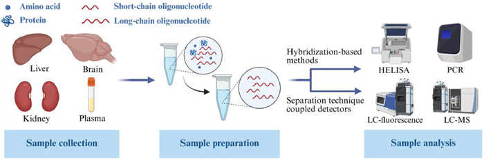
Figure 1.
Bioanalytical workflow depicting key steps in oligonucleotide analysis.


Oligonucleotide therapy utilizes endogenous or synthetic oligonucleotides to modulate gene expression or directly target pathogenic molecules, thereby exerting therapeutic effects [1]. Therapeutic oligonucleotides can be categorized into two primary classes based on their mechanism of action: (1) Nucleic acid-targeting oligonucleotides that regulate protein expression by enhancing or inhibiting translation, including antisense oligonucleotides (ASOs), small interfering RNAs (siRNAs), microRNAs (miRNAs), and small activating RNAs (saRNAs); and (2) protein-targeting oligonucleotides, primarily aptamers, which specifically bind to target proteins analogous to antibodies.
These oligonucleotides allow for precise targeting of disease-related genes or pathogenic molecules through Watson-Crick base pairing, thereby modulating gene expression or directly interfering with pathological processes. They exhibit significant therapeutic potential across a range of diseases, including rare genetic disorders, cancer, cardiovascular conditions, and ophthalmic diseases [1]. Understanding their mechanisms of action is crucial for achieving precise therapeutic outcomes. Furthermore, advances in delivery systems have enhanced the stability and delivery efficiency of oligonucleotides, thereby broadening their clinical applicability [2]. With several oligonucleotide drugs already approved by the Food and Drug Administration (FDA) and a growing number of candidates under clinical investigation, this class of therapeutics has become an integral part of precision medicine and holds promising prospects for future development.
Despite their promising therapeutic potential, safety considerations remain critically important. A retrospective analysis by Henry et al. identified dose-dependent thrombocytopenia in nonhuman primates treated with 2'-O-methoxyethyl modified ASOs [3]. The administration of nusinersen has been associated with nephrotoxicity, including cases of potentially fatal glomerulonephritis. This renal injury is likely attributable to the cumulative effects of the drug following repeated dosing, as ASO-protein complexes undergo glomerular filtration and subsequent proximal tubular reabsorption [4]. Preclinical evaluation of viltolarsen demonstrated nephrotoxicity at elevated injection doses, with concomitant observations of reduced body weight gain and delayed sexual maturation [4]. These findings underscore the critical role of pharmacokinetic studies in evaluating the efficacy, safety, and optimal dosing of therapeutic oligonucleotides [5]. Conducting pharmacokinetic studies on oligonucleotides requires the quantitative analysis of the parent compounds and, occasionally, their metabolites using validated bioanalytical methodologies. Comprehensive bioanalytical approaches for oligonucleotides generally encompass the steps outlined in Fig. 1.
Effective sample preparation is crucial for the bioanalysis of oligonucleotides, given their occurrence in complex biological matrices and their strong binding affinity to plasma proteins. Conventional extraction techniques include protein precipitation, enzymatic digestion, liquid-liquid extraction (LLE), and solid-phase extraction (SPE).
Although sample preparation facilitates the extraction of oligonucleotides from biological matrices, it does not completely eliminate all matrix interferences. Consequently, effective separation prior to detection is crucial for the accurate quantification of oligonucleotides and their metabolites. Liquid chromatography (LC) and capillary electrophoresis (CE) are the most widely used separation techniques in bioanalysis. Commonly employed liquid chromatography-based separation techniques for oligonucleotides include ion-pair reversed-phase liquid chromatography (IP-RPLC), anion-exchange chromatography (AEX), hydrophilic interaction liquid chromatography (HILIC), and size-exclusion chromatography (SEC) [6–9].
Currently, the detection methodologies for therapeutic oligonucleotides mainly include hybridization-based techniques, such as hybridization enzyme-linked immunosorbent assay (HELISA) and quantitative polymerase chain reaction (qPCR), as well as separation techniques coupled with detectors like ultraviolet (UV) spectroscopy, fluorescence, and mass spectrometry (MS) [10–14]. Due to its restricted selectivity and sensitivity, UV detection is excluded from our discussion. Other methodologies such as quantitative nuclear magnetic resonance (qNMR) [15,16], biosensors [17,18], laser-induced fluorescence detectors (LIF) [19,20], and microfluidic systems [21] also show considerable promise for the quantification of oligonucleotides.
This article first outlines the mechanisms and representative drugs of five major therapeutic oligonucleotides, along with a summary of associated delivery systems. Subsequently, we provide a comprehensive overview of the technical advancements and methodological challenges encountered throughout the bioanalytical workflow for therapeutic oligonucleotides, including sample processing, separation, and detection. It is anticipated that this review will offer valuable technical support for pharmacokinetic studies involving oligonucleotides.
Therapeutic oligonucleotides are short, sequence-specific DNA or RNA molecules, such as ASOs, siRNAs, miRNAs, aptamers, and saRNAs. To function at their intended sites of action, they often require the aid of delivery systems. This section provides a concise overview of their mechanisms of action, approved drugs, future development prospects, and the associated delivery systems.
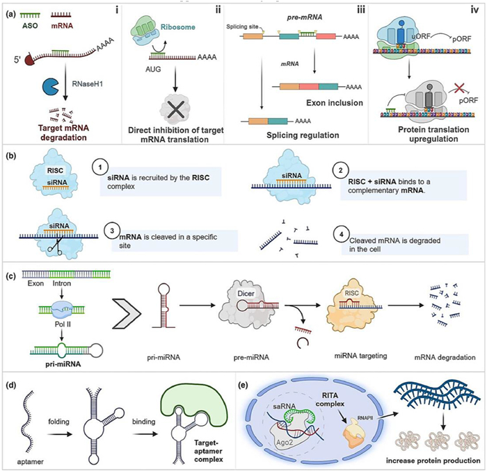
ASOs are short, synthetic, single-stranded oligonucleotides, typically 18-30 nt in length, which specifically bind to target mRNAs to modulate translational processes [22]. Their mechanism of action mainly involves two pathways: RNase H-dependent degradation and the steric block effect (Fig. 2a) [6,23]. The RNase H-dependent mechanism entails ASOs entering cells through endocytosis and binding to target mRNA in the cytoplasm or nucleus, forming an ASO-mRNA hybrid strand [24]. RNase H1 recognizes this hybrid strand and selectively cleaves the target mRNA [25]. In addition, ASOs can bind to miRNAs to form duplex structures, leading to miRNA degradation and subsequent gene expression upregulation [26]. The steric block mechanism involves ASOs binding to specific mRNA sequences, thereby creating a steric hindrance that interferes with the normal function of pre-mRNA or mRNA and modulates gene expression [22]. This mechanism comprises three distinct modes: (1) Direct binding to functional regions of mRNA (e.g., the start codon) to block ribosome recruitment; (2) binding to splice sites to induce exon skipping or inclusion, thereby restoring functional protein expression; and (3) binding to upstream open reading frames (uORFs) to alleviate translational repression of primary open reading frames (pORFs), thereby activating protein expression [23].
Now twelve ASOs have received approval from FDA [27]. The RNase H1-dependent ASOs include inotersen [28] and eplontersen [29], both indicated for hereditary transthyretin amyloidosis (hATTR); fomivirsen (now delisted) [30], which was used for cytomegalovirus retinitis; mipomersen (also delisted) [31], which was indicated for homozygous familial hypercholesterolaemia; tofersen [32] for amyotrophic lateral sclerosis; and olezarsen [33] for familial chylomicronaemia syndrome and severe hypertriglyceridaemia. The splicing-modulating ASOs include eteplirsen [34], golodirsen [35], viltolarsen [36], and casimersen [37], all of which are employed in the treatment of Duchenne muscular dystrophy by inducing exon 51/53/45 skipping, respectively, to restore dystrophin expression. Additionally, nusinersen is used for spinal muscular atrophy by targeting SMN2 pre-mRNA to promote exon 7 inclusion [38], and milasen is approved for a single patient with Batten disease [39].
siRNAs are synthetic double-stranded oligonucleotides, typically 20-25 bp in length, with 3'-terminal dinucleotide overhangs [40]. They facilitate gene silencing via the RNA interference (RNAi) pathway through specific base pairing [41–43]. The process of siRNA-mediated gene silencing involves the incorporation of siRNAs into the RNA-induced silencing complex (RISC), wherein the Argonaute 2 (AGO2) protein binds to the 5'-end of the guide strand, a process determined by thermodynamic stability [44,45]. Subsequently, RISC directs the recognition of the target mRNA, with AGO2 mediating its cleavage, and thereby inhibiting translation (Fig. 2b) [2,23,46].
Seven siRNA therapeutics have received approval from the FDA [47]. These include treatments for rare genetic disorders such as patisiran [48] and vutrisiran [49] for hATTR, givosiran for acute hepatic porphyria [50], and both lumasiran [51] and nedosiran [52] for primary hyperoxaluria type 1. Additionally, fitusiran [53] is approved for the treatment of haemophilia A or B in adults and pediatric patients. For chronic diseases, inclisiran has been approved for the treatment of primary hypercholesterolaemia [54]. Furthermore, several siRNA therapeutics for cancer treatment and other diseases, such as ocular diseases, are undergoing clinical investigation [55].
miRNAs are endogenous non-coding RNAs, typically about 22 nt in length, that regulate gene expression through complementary binding to mRNAs [56]. The mechanism of action of miRNAs is highly intricate (Fig. 2c) [23]. Initially, RNA polymerase Ⅱ/Ⅲ transcribes a primary miRNA (pri-miRNA) with a stem-loop structure. This pri-miRNA is cleaved by the Drosha-DGCR8 complex within the nucleus to produce a precursor miRNA (pre-miRNA) of approximately 70 nucleotides. Subsequently, the pre-miRNA is transported to the cytoplasm via Exportin-5 and further cleaved by the Dicer enzyme into a double-stranded miRNA. One strand of this miRNA is incorporated into the RISC, while the other strand is degraded [57,58]. Typically, a specific region at the 5'-end of the miRNA, known as the "seed region", binds to the 3'-untranslated region (3'UTR) of the mRNA [59]. In cases of full complementarity, the target mRNA is cleaved and degraded, a phenomenon commonly observed in plants and lower eukaryotes. Conversely, in the absence of full complementarity, miRNAs impede ribosome assembly, thereby inhibiting the translation process of the mRNA, a mechanism prevalent in animals [60,61]. miRNAs play dual functional roles in cancer: (1) tumor-suppressive miRNAs, such as let-7 and miR-34, whose downregulation promotes tumorigenesis [62,63], and (2) oncogenic miRNAs, such as miR-21 and miR-221, whose overexpression promotes tumor proliferation, metastasis, and drug resistance [64,65]. Correspondingly, two therapeutic strategies have been developed: miRNA mimics to restore tumor-suppressor function and miRNA inhibitors to silence oncogenic miRNAs [66,67]. However, all miRNA-based therapeutics remain in clinical development, with none having received FDA approval to date [58].
Aptamers are single-stranded DNA or RNA molecules, generally comprising 20-60 nucleotides, derived from a library of nucleic acid molecules through the process of systematic evolution of ligands by exponential enrichment (SELEX) [68,69]. These molecules undergo folding via base pairing and intermolecular forces to adopt a unique three-dimensional conformation, enabling specific binding to their target [70]. Aptamers exhibit high specificity and affinity for a diverse array of targets, including small molecules, proteins, viruses, and even cell surface antigens, such as those present on cancer cells (Fig. 2d) [23,68].
Aptamers can function as therapeutic agents by directly binding to their targets through specific target-binding mechanisms or by being conjugated with drugs to enhance target specificity [71]. To date, two aptamer-based drugs have received approval from the FDA [23]. Pegaptanib, an RNA aptamer previously approved for the treatment of wet age-related macular degeneration (AMD), specifically targets vascular endothelial growth factor (VEGF)-165, thereby inhibiting its interaction with receptors and preventing neoangiogenesis [72]. Although pegaptanib has been delisted, another RNA aptamer, avacincaptad pegol, remains approved for the treatment of geographic atrophy associated with AMD. This aptamer inhibits the cleavage of complement component C5 into C5a and C5b, thereby preventing the formation of membrane attack complexes (MACs) and reducing inflammation and cellular damage [73].
saRNAs are short-chain oligonucleotides typically existing in a double-stranded configuration, with a common length of 21 base pairs. Unlike small interfering RNAs (siRNAs), saRNAs function by activating their target mRNA (Fig. 2e) [23,74]. The antisense strand of saRNA binds to AGO2 to form the saRNA-AGO2 complex, which subsequently recruits RNA helicase A (RHA) and CTR9. This assembly leads to the formation of the RNA-induced transcription activation complex (RITA), which binds to the promoter region and interacts with RNA polymerase Ⅱ, thereby facilitating transcription initiation and elongation [75].
One saRNA-based therapeutic, MTL-CEBPA (CEBPA-51 saRNA), has advanced to clinical trials for the treatment of hepatocellular carcinoma. This drug activates the CCAAT Enhancer Binding Protein Alpha (C/EBPα), resulting in the inhibition of hepatocellular carcinoma cell proliferation, induction of apoptosis, and improvement of liver function. The phase Ⅰ clinical trial for MTL-CEBPA demonstrated both safety and preliminary efficacy [76,77]. Furthermore, numerous saRNA-based therapeutics are currently in preclinical development, showing significant potential for cancer treatment [78].
Therapeutic oligonucleotides exhibit significant potential in the treatment of various diseases, including rare diseases, genetic disorders, and cancers. However, when administered, naked oligonucleotides are easily degraded by nucleases within the body, compromising their stability [79]. Moreover, as exogenous entities, they can trigger immune reactions [80]. Other challenges include poor targeting ability and low cellular uptake efficiency [81]. To address these issues, effective delivery in vivo relies on the use of delivery systems. Currently, commonly used delivery systems include lipid nanoparticles (LNPs), cationic polymeric nanoparticles, N-acetylgalactosamine (GalNAc), exosomes, inorganic nanoparticles, and peptides [82–87].
LNPs are self-assembled structures with a diameter of approximately 100 nm. They encapsulate negatively charged nucleic acid drugs through electrostatic interactions, and their targeting ability can be optimized via surface modification [82]. The ionizable lipids in LNPs remain neutral at physiological pH, which reduces toxicity and immunogenicity; in the acidic environment of endosomes, they become positively charged and can interact with the endosomal membrane structure [88]. Auxiliary lipids further promote the disruption of the endosomal membrane structure and facilitate the escape of therapeutic oligonucleotides from endosomes [88]. Components such as cholesterol confer a structure similar to natural membranes, imparting good biocompatibility, promoting fusion with cell membranes, and enhancing cellular uptake efficiency [89]. PEGylated lipids significantly improve the stability of LNPs, but they may interfere with the binding of apolipoprotein E to LNPs, thereby inhibiting liver uptake [90,91]. Moreover, repeated administration may induce the production of PEG antibodies, which can reduce drug efficacy [92].
Cationic polymeric nanoparticles form complexes with negatively charged nucleic acids through electrostatic interactions, as the polymers themselves possess positive charges. After entering cells via endocytosis, these nanoparticles can disrupt endosomes, release the nucleic acids, and thereby achieve intracellular delivery [83]. Polyethylenimine (PEI) contains amine groups that enhance its electrostatic binding to nucleic acids; however, it suffers from high cytotoxicity and poor biodegradability [93]. Another widely used cationic polymer, poly-L-lysine (PLL), is biodegradable and biocompatible [94]. Despite these advantages, its in vivo application is hindered by high toxicity, low transfection efficiency, and inadequate endosomal escape capability [95].
GalNAc is a high-affinity ligand for the asialoglycoprotein receptor (ASGPR) on the surface of hepatocytes [84]. It enables liver-specific delivery through receptor-mediated endocytosis [96]. After the conjugate enters hepatocytes, ASGPR recycles back to the cell membrane, while GalNAc is degraded, leading to the release of the oligonucleotides [97]. Due to this targeting capability, GalNAc-conjugated systems can be preferentially accumulated in the liver, thereby reducing distribution to non-target tissues and systemic toxicity; however, this liver-specificity also limits their application to extrahepatic tissues [96]. Unlike carrier-encapsulated oligonucleotides, GalNAc-conjugated oligonucleotides are directly exposed to serum and thus rely on chemical modification to resist nuclease degradation [98]. In 2023, nedosiran was approved by the FDA as a GalNAc-siRNA conjugate for the treatment of primary hyperoxaluria type 1 [52].
Exosomes are extracellular vesicles ranging from 40 nm to 160 nm in diameter that are secreted by cells [85]. As natural intercellular delivery carriers, they exhibit excellent biocompatibility, low immunogenicity, and the ability to evade rapid immune clearance [99]. Furthermore, exosomes can cross biological barriers such as the blood-brain barrier, and their targeting specificity can be enhanced through surface modification [100]. However, their natural membrane structure often results in low drug-loading efficiency, necessitating the use of techniques such as electroporation or ultrasound to load oligonucleotides, processes that may compromise the structural integrity of exosomes [101].
Inorganic nanoparticles possess stable physical structures and tunable surface properties, enabling targeted delivery through surface functionalization [86]. Gold nanoparticles (AuNPs) are widely used due to their good biocompatibility and low toxicity. For instance, Shrestha et al. utilized AuNPs to co-deliver doxorubicin and polo-like kinase 1 (Plk1) siRNA [102]. Magnetic nanoparticles (MNPs) can achieve site-specific targeted delivery under the guidance of an external magnetic field, which is particularly advantageous for local tumor therapy [103].
Peptides are composed of amino acids and easy to synthesize. They can be functionally classified into several categories, including targeting peptides, cell-penetrating peptides (CPPs), endosomal escape peptides [104–106]. Owing to their advantages of stable chemical properties, low immunogenicity, and high selectivity, peptides have emerged as highly promising candidates for delivery systems [107,108].
The approval of over 20 drugs by the FDA, covering multiple modalities such as ASOs and siRNAs for diseases ranging from rare genetic disorders to common disorders, reflects the maturation and expansion of this therapeutic platform. The presence of numerous candidates in clinical trials underscores ongoing innovation and a widening scope of applications. Furthermore, advances in delivery systems are progressively improving the stability, targeting, and efficacy of these therapeutics, enabling potential treatment of an even broader range of diseases. Oligonucleotide therapeutics are thus firmly established as a cornerstone of precision medicine with significant future growth potential.
Pharmacokinetic studies of therapeutic oligonucleotides provide a fundamental understanding of their metabolic behavior and aid in the optimization of safe dosing regimens. The bioanalytical techniques employed for pharmacokinetics typically involve sample preparation, separation, and detection. This section outlines the workflow, focusing on key recent technical advances, relative advantages, and remaining challenges for each step, and discuss prospects for achieving highly sensitive and accurate quantification.
Sample preparation involves the extraction of oligonucleotides from complex matrices, which yields a mixture of various chain lengths, and is a prerequisite for subsequent separation and detection of the target analyte. Due to matrix complexity and the strong protein-binding affinity of oligonucleotides, achieving high recovery and purity poses a significant challenge. Therefore, efficient sample preparation constitutes a critical foundation for accurate oligonucleotide analysis. This section elaborates on the primary methods employed, including protein precipitation, enzymatic digestion, LLE, SPE, and magnetic bead hybridization. A summary of these sample preparation methods is provided in Fig. 3.
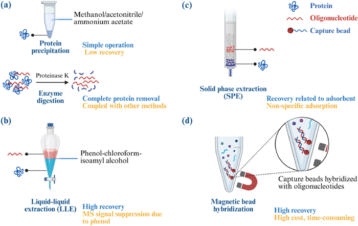
Protein precipitation constitutes the most fundamental method for oligonucleotide extraction, utilizing organic solvents such as methanol and acetonitrile, or salts like ammonium acetate, to disrupt protein-oligonucleotide interactions. This process results in the precipitation of proteins while oligonucleotides remain in the supernatant [109]. Despite its simplicity and rapid execution, this technique is infrequently used due to substantial signal suppression caused by biological matrices and its low recovery [109]. In a comparative study of four sample preparation methods, Studzińska et al. found that acetonitrile-based protein precipitation led to significant sample loss due to the strong binding affinity between oligonucleotides and proteins, which resulted in co-precipitation [110].
Enzyme digestion utilizes proteases, such as Proteinase K, to hydrolyze proteins and release oligonucleotides, with ethylenediaminetetraacetic acid (EDTA) commonly added to inhibit nuclease activity [111]. This approach facilitates the complete removal of proteins and is particularly effective for complex biological matrices [112]. However, as it does not eliminate other interfering substances, it is frequently used in conjunction with additional purification techniques [113]. Chen et al. reported an extraction efficiency of 50%-85% by digesting protein-bound oligonucleotides with Proteinase K, followed by phenol-chloroform extraction [112]. Palaiologou et al. demonstrated that the combination of proteinase K digestion and SPE resulted in higher-purity ASO samples from rat brain tissues [7].
LLE operates on the principle of partitioning, utilizing immiscible solvents such as phenol, chloroform, and isoamyl alcohol to separate oligonucleotides and proteins. In this process, phenol serves to denature proteins and inhibit nuclease activity, while chloroform and isoamyl alcohol facilitate the separation of oligonucleotides from proteins, dissolving them in the upper aqueous phase [114,115]. However, residual phenol in the aqueous phase can suppress mass spectrometric signals. Studzińska et al. successfully removed phenol residues from samples through fivefold LLE with chloroform at a 1:4 ratio, achieving a recovery rate of 95% [110]. Although SPE following LLE has been reported, Ewles et al. observed inconsistent recoveries and peak broadening with this method [116]. They attributed these issues to the saturation of the SPE adsorbent by phenol, leading to either analyte breakthrough or co-elution. To address this, they employed a secondary LLE extraction using dichloromethane (DCM) to eliminate non-polar interferents, including phenol, which proved effective for both single- and double-stranded modified oligonucleotides, albeit with considerable analyte dilution [117]. Yun et al. developed an automated reverse-phase LLE method that demonstrated both shorter processing times than manual extraction and higher recovery than SPE with Clarity OTX columns [118].
SPE has become the leading and most rapidly evolving technique for sample preparation. This method effectively isolates oligonucleotides on adsorbents while eliminating interferences such as proteins and salts, followed by the elution of analytes using optimized solvents. Two primary SPE adsorbents are utilized for extraction of oligonucleotides: reversed-phase materials, such as Oasis HLB [119], which are often used in combination with ion-pairing reagents, and weak anion exchange (WAX) resins, such as Clarity OTX [120]. Comparative studies indicate that Clarity OTX offers superior recovery. Wheeler et al. reported recoveries exceeding 90% for both antisense and sense strands during the extraction of siRNAs from rat plasma using this adsorbent [121]. Another adsorbent, Oasis WAX, which is a mixed-mode adsorbent combining weak anion-exchange and reversed-phase functionalities, demonstrated recoveries ranging from 57.9% to 67.6% [122]. Anand et al. introduced the use of NAX, an aminopropyl-bonded phase, for the extraction of oligonucleotides [123]. Studzińska et al. developed a dispersive solid-phase extraction (dSPE) technique for the extraction of oligonucleotides, employing an innovative adsorbent created by modifying the silica gel surface with an aminopropyl group partially conjugated with aspartic acid. This modification results in an adsorbent characterized by Si-(NH2)COOH, which requires only pH adjustment for effective elution [124]. They subsequently applied this dSPE method to the extraction of nusinersen, demonstrating superior efficiency compared to LLE and hybridization techniques [125]. Additionally, SPE can be integrated with LLE to enhance sample preparation [126,127]. Ewles et al. further refined the extraction process by diluting phenol-chloroform extracts with a water/triethylamine/hexafluoroisopropanol mixture and employing optimized SPE methods, achieving oligonucleotide recoveries between 68% and 78% [116]. However, non-specific adsorption remains a notable limitation, potentially causing sample loss. This issue can be mitigated by minimizing sample transfers, utilizing siliconized glassware, and increasing the concentration of internal standards [128]. Therefore, the development of optimized SPE protocols is imperative to enhance recoveries, reduce analyte loss, and improve analytical sensitivity.
Hybridization-based sample preparation utilizes biotinylated capture probes to selectively bind target oligonucleotides, followed by separation and elution using streptavidin-coated magnetic beads [129]. This technique exhibits high selectivity, sample cleanliness, and recovery, making it highly promising [130,131]. Li et al. achieved recoveries ranging from 89.9% to 109% when extracting target ASOs [132]. Yuan et al. employed proteinase K digestion of biological samples followed by hybridization with magnetic bead-conjugated biotinylated peptide nucleic acids, achieving a recovery efficiency of 90% [133]. Sips et al. compared anion exchange solid-phase extraction (AEX-SPE) with the hybridization method for oligonucleotide extraction, finding that the latter provided higher specificity and effective purification, thereby enhancing sensitivity [134]. However, the requirement for custom probe design poses significant costs and time constraints. Additionally, the affinity of probes for shorter metabolites is limited, although this can be optimized by modifying the melting temperature (Tm), such as by introducing improved capture probes, increasing salt concentration, or reducing binding temperature [132].
Among these, optimized SPE and novel methods such as magnetic bead hybridization have shown superior recovery [128,132,135]. The evolution of sample preparation will continue to prioritize improved recovery to meet analytical demands. Key directions include the optimizing SPE adsorbents, lowering the cost and time requirements of high-specificity approaches such as developing integrated techniques to improve overall analytical performance. Progress in these areas will be essential for enabling highly sensitive and accurate bioanalysis of therapeutic oligonucleotides.
Following sample preparation, high-resolution separation is imperative for distinguishing the target oligonucleotide from metabolic impurities, a process fundamental to reliable quantification. The achievement of such precise separation represents a central challenge in bioanalysis, one that critically dictates the specificity and accuracy of the analytical outcome. This section reviews standard separation methodologies, primarily liquid chromatography and electrophoresis, by addressing their underlying principles, merits, and drawbacks, and concludes by exploring emerging trends. A schematic representation of the operational mechanisms and distinctive characteristics of these methods is provided in Fig. 4.
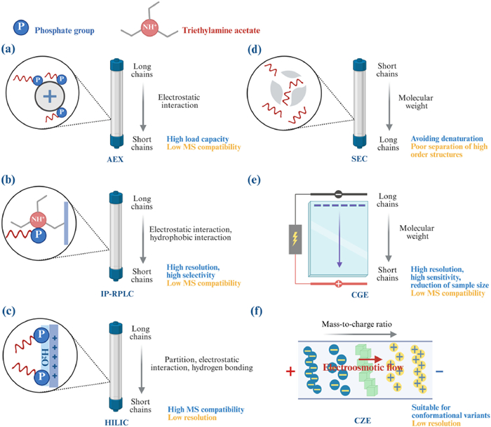
AEX is a separation technique based on the principle of ion exchange, wherein the separation of charged molecules is achieved through their exchange with ions in solution via an ion-exchange resin. Oligonucleotides, characterized by their negatively charged phosphate backbone, interact with the cationic groups present in the stationary phase of AEX through electrostatic attraction [136]. Concurrently, these oligonucleotides compete with increasing concentrations of anions in the mobile phase, allowing for separation based on their negative charge density and phosphate chain length [137]. Typically, the mobile phase comprises a sodium phosphate buffer supplemented with sodium chloride and sodium perchlorate, while the stationary phase consists of an anion exchanger immobilized on a solid support, from which analytes are eluted using a salt gradient [136,138].
Polymer-based strong anion exchange (SAX) columns, utilized in the stationary phase, demonstrate enhanced reproducibility, separation efficiency, and selectivity for the isolation of oligonucleotides from biological samples. This is attributed to the robust electrostatic interactions between the cationic stationary phase and the anionic oligonucleotides. Togawa et al. successfully separated non-denatured siRNA from single-stranded impurities and truncated double-stranded RNA using a DNAPac PA200 RS column under optimized chromatographic conditions [139]. The widespread adoption of WAX columns is noteworthy. Nercessian et al. demonstrated stable oligonucleotide separation utilizing WAX chromatography on a Gen-Pak FAX column, optimized with acetonitrile instead of methanol [9]. AEX is also characterized by a high loading capacity. Enmark et al. conducted a quantitative comparison of the purification of unmodified oligonucleotides using AEX and IP-RPLC under comparable laboratory conditions, revealing that AEX offers a higher loading capacity under specific conditions, which facilitates increased productivity and reduced solvent consumption [140]. The presence of cation interference in the mobile phase, which adversely affects MS compatibility, necessitates desalting [6]. Thayer et al. automated the desalting process using reversed-phase columns following AEX separation, thereby enabling the efficient transfer of oligonucleotides to MS, significantly reducing signal suppression and enhancing detection sensitivity [141].
IP-RPLC has been widely used for the separation of oligonucleotides in recent years. The separation mechanism includes the electrostatic interaction of forming ion pairs and the reversed-phase separation principle of hydrophobic interactions [142]. The specific mechanism is that lipophilic cationic ion-pair reagents in the mobile phase form ion-pairs with the negatively charged phosphate backbone of the oligonucleotide via electrostatic interaction, reducing its net negative charge and increasing its hydrophobicity, which enables separation on a reversed-phase stationary phase [142,143]. Differences in oligonucleotide chain length and base composition affect their separation by IP-RPLC [137].
Initially, triethylamine acetate (TEAA) was used for the separation of oligonucleotides, and the concentration of TEAA significantly influenced the separation. Typically, higher concentrations yield improved separation. However, acetic acid in TEAA can cause ion suppression in MS [144]. Apffel et al. demonstrated that oligonucleotides can be effectively separated using triethylamine/hexafluoroisopropanol (TEA/HFIP) buffers. The fluoroalcohol additive acts as a weak acid to regulate the pH, facilitating better separation at lower pH. Importantly, HFIP is volatile, and its evaporation can lead to pH increase over time, potentially impacting ionization efficiency [145]. Subsequent research on analyzing oligonucleotides has focused on ion-pairing reagents combining alkylamines as the primary reagent with fluorinated alcohols as modifiers [146–151]. Commonly used alkylamine reagents include TEA [152], N-diisopropylethylamine (DIEA) [153], hexylamine (HA) [154], tripropylamine (TPA) [155], dibutylamine (DBA) [156], N,N-dimethylcyclohexylamine (DMCHA) [157], N,N-diethylamine (DEA) [158], diisopropylamine (DIPA) [159], and N,N-dimethylbutylamine (DMBA) [160] among others. Selection depends on the specific modification and type of oligonucleotide for optimal separation. Fluorinated alcohol modifiers include HFIP and hexafluoro-2-methylisopropanol (HFMIP). Studies indicate that HFMIP is more hydrophobic than HFIP, enhancing its suitability for separating highly modified oligonucleotides [161]. The predominant mobile phases are methanol or acetonitrile, while the stationary phases are mostly C18 columns [162]. Both separation efficiency and electrospray ionization efficiency can be optimized by adjusting the types and concentrations of ion-pair reagents and organic solvents.
IP-RPLC offers high retention and resolution for oligonucleotide separation. In an evaluation of the orthogonality of IP-RPLC, AEX and HILIC for the separation of synthetic oligonucleotides, Sorensen et al. demonstrated that IP-RPLC provided superior separation [163]. However, when coupled with MS, this method can exhibit relatively low sensitivity, potentially due to contamination of MS system [164]. Another contributing factor to reduced sensitivity is instability of the aqueous mobile phase over time; freshly prepared mobile phase often restores signal intensity [165].
HILIC is regarded as an alternative technique to IP-RPLC for the separation of oligonucleotides [166,167]. The underlying mechanism of HILIC involves partitioning processes, electrostatic interactions, and hydrogen bonding. The retention of oligonucleotides in HILIC primarily depends on their partitioning within the aqueous layer adsorbed on the stationary phase surface. Additionally, at certain pH levels, the negative charge on the column surface repels the negatively charged oligonucleotides, resulting in the proximity of the bases to the stationary phase surface. This proximity enhances hydrogen bonding interactions between the bases and the stationary phase [168–170].
Commonly employed stationary phases in HILIC include amide (e.g., BEH Amide, XBridge Amide) [171,172], diol (e.g., Luna HILIC) [173], amphoteric (e.g., ZIC-cHILIC) [174], and silica gel (e.g., Kinetex HILIC, BEH HILIC) [175,176]. Lardeux et al. conducted a comparative analysis of seven amphoteric columns, amide columns, poly-hydroxy fructan-functionalized columns, and C18 columns for IP-RPLC, demonstrating that amphoteric columns exhibit high selectivity, particularly for large oligonucleotides [177]. Methanol or acetonitrile is usually used as the organic phase, and the incorporation of buffer solutions, such as ammonium formate and ammonium acetate, enhances the retention and resolution of the chromatographic profile [8,172,178]. Compared with IP-RPLC, HILIC provides the benefit of compatibility with MS but is constrained by limited resolution [179].
SEC separates oligonucleotides based on their hydrodynamic radius through a process of molecular sieving, wherein larger molecules are eluted prior to smaller ones [137]. This technique eliminates the need for oligonucleotide denaturation, a requirement in IP-RPLC, and operates without the use of organic solvents, high-salt mobile phases, or elevated temperatures [180,181]. Consequently, SEC preserves the native conformation of oligonucleotides by avoiding the structural perturbations typically induced by denaturation processes. Noll et al. demonstrated that SEC provides quantification of siRNA duplexes comparable to that of non-denaturing IP-RPLC, thereby confirming the agreement between these methods for duplex content analysis [182]. Seiffert et al. investigated the formation of impurities during the annealing process of siRNA using both methods, finding a slightly higher relative percentage of single-stranded species detected by IP-RPLC compared with SEC [181]. This discrepancy may be attributed to the denaturing conditions inherent in IP-RPLC, which increase the prevalence of single-stranded nucleic acids. Shimoyama et al. demonstrated that SEC is effective in isolating single- and double-stranded oligonucleotides, although it exhibits limited efficacy in separating higher-order structures within complex systems [180].
CE is extensively employed in the analysis of oligonucleotides, with techniques such as capillary gel electrophoresis (CGE) and capillary zone electrophoresis (CZE) being prominent. CZE facilitates the separation of oligonucleotides based on charge mobility, allowing for the analysis of conformational variants. However, it offers lower resolution compared to CGE [183]. Consequently, CZE is not elaborated upon in this discussion. CGE, on the other hand, differentiates oligonucleotides according to their molecular size and is capable of distinguishing between short and long-chain oligonucleotides [184]. The polymer matrices utilized in CGE include linear polyacrylamide (LPA) [185], polyethylene oxide (PEO) [186], and polydimethylacrylamide (pDMA) [187]. Notably, pDMA exhibits lower viscosity than both PEO and LPA, enhancing its efficiency for separation [187]. Furthermore, pDMA demonstrates superior chemical stability under alkaline conditions [185].
Although CGE offers higher resolution for phosphorothioate-modified oligonucleotides compared to LC and requires smaller sample volumes due to its capillary format, it is less reproducible and less robust than LC [188]. Following CGE separation, oligonucleotides are generally analyzed using UV detection [189]. For enhanced sensitivity at low concentrations, LIF detection can be employed after labeling the oligonucleotides with a fluorescent dye [19]. However, when coupled with MS, the most widely used detection method for oligonucleotides, CGE presents technical challenges due to electrospray ionization suppression caused by the CGE buffer [188,189]. Therefore, LC techniques remain the predominant separation techniques.
A summary of the aforementioned separation methods is provided in Table 1. Future advances in separation techniques for oligonucleotide analysis will focus on achieving higher resolution and improving compatibility with MS. Although the refinement of alkylamines and fluorinated alcohols remains an active area for IP-RPLC, concerns regarding potential MS contamination are driving the adoption of more compatible alternatives like HILIC, where enhancing resolution represents a major goal.

 DownLoad:
CSV
DownLoad:
CSV
| Separation methods | Advantages | Drawbacks | Application | |
| LC | AEX | High load capacity | Low MS compatibility | Study on the pharmacokinetics of therapeutic oligonucleotides |
| IP-RPLC | High resolution, high selectivity | Low MS compatibility | ||
| HILIC | High MS compatibility | Low resolution | ||
| SEC | Avoiding denaturation | Poor separation of high order structures | ||
| CE | CGE | High resolution, high sensitivity, reduction of sample size | Low MS compatibility | |
| CZE | Suitable for conformational variants | Low resolution |
The selection of an appropriate detection method is paramount for the accurate quantification of oligonucleotides in t complex matrices, as it directly determines the sensitivity, specificity, and overall success of the analysis. This section focuses on key techniques, ranging from highly sensitive approaches like HELISA and qPCR to highly selective platforms such as LC-MS and fluorescence detection. By comparing their fundamental principles, performance characteristics, and typical applications, this section emphasizes that LC-MS is widely regarded as the current gold standard, owing to its superior selectivity and reliable quantitative capability. Fig. 5 illustrates the mechanisms and characteristics of these detection techniques.
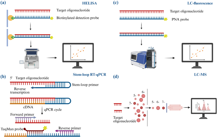
The principle of HELISA for oligonucleotides quantification is based on hybridization. In this method, a capture probe and a detection probe (e.g., digoxigenin-labeled) hybridize to the target oligonucleotide. Specific substrates are then added to generate color or luminescence, enabling quantification via absorbance or luminous intensity [190].
HELISA is highly sensitive, capable of quantifying oligonucleotides at low pg/mL levels, meeting the demand for low-dose oligonucleotide analysis [191]. For plasma samples, complex extraction steps are unnecessary; tissue samples require only simple digestion with protease or LLE to remove bound proteins [192]. Additionally, HELISA supports high-throughput workflows and is suitable for large-scale sample quantification [190,193]. However, traditional HELISA exhibits low selectivity and cannot distinguish between intact oligonucleotides and truncated metabolites [194].
To address this, modified HELISA formats have been developed to improve selectivity, including sandwich HELISA [195], hybrid ligation ELISA [196,197], and competitive HELISA [198]. These methods can differentiate and quantify metabolites with specific 3' and/or 5'-end modifications. Competitive HELISA, for example, quantifies oligonucleotides through competitive binding to biotin-modified nucleic acid primers complementary to their sequence, where the analyte concentration is inversely proportional to the response signal [198]. Lorenson et al. achieved direct, highly sensitive oligonucleotide detection in blood using competitive hybridization. They added an aliphatic spacer group and a poly-T extension to the 5'-end of the capture probe, plus a poly-A extension on the detection probe, reducing steric hindrance and improving hybridization efficiency [199]. Nevertheless, HELISA relies on specific capture probes, and probe design/preparation can be complex, with long development cycles and potential cross-reactivity. Haegele et al. enhanced selectivity by extending the capture probe length and introducing spacers, increasing the unpaired nucleotide region. This facilitates S1 nuclease recognition and degradation of hybridization products with 5'-terminal metabolites. The method also leverages ligase-dependent 3'-end integrity for natural specificity toward 3'-end metabolites. Hybridization efficiency to oligonucleotides were further improved by adding polyethylene glycol (PEG), dimethyl sulfoxide (DMSO), and betaine [200].
qPCR builds upon traditional PCR by quantifying oligonucleotides through real-time monitoring of fluorescent signals. For RNA targets, reverse transcription first converts RNA to cDNA, which is then amplified by PCR. The fluorescent signal is typically generated by either dye-labeled oligonucleotide probes (e.g., TaqMan) or intercalating dyes that bind double-stranded DNA (e.g., SYBR Green) [201].
Stem-loop reverse transcription quantitative PCR (SL RT-qPCR) is the most commonly used method for oligonucleotide quantification, particularly for miRNA and siRNA [202,203]. However, plasma miRNA analysis using SL RT-qPCR requires specific considerations, as detailed by Ban et al. They note that the sample matrix and residual solvents after extraction impact extraction efficiency and reduce RT-qPCR accuracy. Consequently, optimization of the extraction method and assessment of amplification efficiency are necessary before analysis [11].
Several specialized RT-qPCR methods have been developed for quantification, including primer extension qPCR [204], poly A tailing-based RT-qPCR [205], and ligation-based qPCR [206]. These techniques share advantages of high sensitivity, wide dynamic range, and high throughput. A key limitation, however, is that detection efficiency may be compromised for chemically modified oligonucleotides, and they cannot differentiate between intact oligonucleotides and their metabolites [14]. Castellanos-Rizaldos et al. utilized RT-qPCR to detect and quantify chemically modified siRNAs, such as those incorporating glycol nucleic acid (GNA) modifications, supporting the development of RNAi-based therapies [207]. Shin et al. employed ASOs as a bridge to guide the SplintR DNA ligase-mediated ligation of two complementary probes, enabling qPCR quantification of modified ASOs and siRNAs [208].
The development of digital PCR (dPCR) [209] and droplet digital PCR (ddPCR) [210] has attracted significant attention. dPCR partitions the sample into numerous individual reactions. Each partition undergoes independent PCR amplification, and the absolute quantity of target molecules is calculated using Poisson statistics, eliminating the need for a standard curve [209]. ddPCR is a droplet-based implementation of dPCR, which utilizes nanoscale microdroplets on a nanofluidic chip to perform thousands of PCR reactions simultaneously. Quantification is based on detecting the presence or absence of an endpoint signal in each droplet [210]. Turski et al. developed a stem-loop reverse transcription ddPCR method (SL RT-ddPCR) for quantifying siRNAs in mouse plasma and liver extracts. This method demonstrated superior sensitivity and a broader linear range compared to conventional qPCR [211].
Fluorescence-based detection methods are widely employed in oligonucleotide bioanalysis. In addition to the previously discussed HELISA and qPCR, commonly used fluorescence-based techniques include LC-fluorescence, which combines liquid chromatography with fluorescence-labeled peptide nucleic acid (PNA) detection probe for separation and detection, as well as biosensors and capillary electrophoresis with laser-induced fluorescence (CE-LIF), which will be elaborated on later. In summary, although numerous methods employ fluorescence for detection, they share a common fundamental principle: The quantification of oligonucleotides based on fluorescence intensity. The primary distinctions lie in the separation mechanisms and detection platforms employed. This section will focus on LC-fluorescence, one of the most commonly used techniques in current practice.
Fluorescence analysis integrates hybridization techniques with LC and is widely used due to its high sensitivity and reduced limitations associated with modified oligonucleotides compared to PCR [14]. The primary procedure involves the formation of a double-stranded complex through base-pairing between the target oligonucleotide and a fluorescently labeled probe. This complex is subsequently separated via chromatography, and its signal is quantified using a fluorescence detector [14]. Consequently, the quantification of oligonucleotides via fluorescence necessitates the design of corresponding fluorescence-labeled detection probes.
At present, the fluorescence-labeled PNA detection probe is predominantly employed. This probe consists of a base derivative that substitutes the secondary amino group on the backbone glycine chain and is linked via peptide bonds, rendering it more stable as it is resistant to degradation by proteases and nucleases [212]. Furthermore, due to its electrical neutrality, PNA exhibits greater affinity than negatively charged nucleic acid probes, thereby enhancing the hydrophobicity of the complex [213]. Ji et al. investigated the critical role of probe design in the fluorescence-based analysis of oligonucleotides, concluding that the length, sequence, and structural modifications of PNA probes significantly influence subsequent chromatographic separation [213]. Consequently, the primary challenge in the fluorescence analysis of oligonucleotides lies in the design of appropriate probes to enhance sensitivity and separation efficiency, a process that is both time-intensive and costly.
Typically, fluorescence analysis is integrated with AEX for the final separation step, with SAX columns being the most frequently employed due to their ability to effectively distinguish between intact oligonucleotides and shorter metabolites [12,214]. Furthermore, fluorescence analysis can also be combined with CE for effective separation. Hutanu et al. introduced a gel-free hybridization analysis method utilizing CE with fluorescently labeled PNA as an affinity probe. This method facilitates both qualitative and quantitative analysis of oligonucleotides, demonstrating high selectivity in the analysis of single-stranded oligonucleotides and achieving a limit of quantification at the picomolar level [215]. Moreover, Zhan et al. utilized a single-stranded binding (SSB) protein to selectively capture oligonucleotide-functionalized fluorescent probes, facilitating the quantitative detection of nusinersen sodium in human serum [216].
MS is the leading technique for oligonucleotide bioanalysis, providing superior selectivity compared to HELISA and qPCR by directly measuring mass-to-charge ratios and ion abundances [129].
Electrospray ionization (ESI) is the most prevalent ion source for MS analysis of oligonucleotides, demonstrating enhanced sensitivity in negative ion mode because of the efficient formation of gas-phase anions from oligonucleotide phosphate groups [136]. The deprotonation ionization of polyphosphate groups results in ESI-MS spectra showing "envelope peaks" corresponding to (M-nH)n- across multiple charge states, which contributes to increased spectral complexity and, consequently, reduced sensitivity [217,218]. However, these signal peaks can be resolved by acquiring molecular mass information through deconvolution [219]. Additionally, the presence of cations (e.g., Na+, K+) in the sample or reagents used during the separation process can lead to cation addition, as these cations tend to bind to the negatively charged anionic framework [218]. This phenomenon reduces signal the abundance while simultaneously complicating the spectra and further reducing sensitivity [135]. Therefore, reducing the distribution of multiple charges and eliminating cationic adducts can significantly enhance the sensitivity of MS.
Typically, MS is coupled with LC, most commonly IP-RPLC. However, ion-pairing reagents can cause MS contamination [145]. As a result, there has been a notable increase in the application of hydrophilic interaction liquid chromatography-mass spectrometry (HILIC-MS), which offers an alternative to IP-RPLC [179]. Nonetheless, challenges persist when MS is coupled with AEX or CGE due to salt-induced signal suppression [188,220]. Therefore, the sensitivity of liquid chromatography-mass spectrometry (LC-MS) can be enhanced by optimizing ion-pairing reagents or adjusting the separation conditions in HILIC. Furthermore, the ionization of oligonucleotides can be improved by increasing the organic solvent content in the mobile phase and adjusting the solution pH to enhance signal intensity [161,221].
In addition to the commonly employed assays, several other platforms are utilized for the analysis of oligonucleotides.
Among these, biosensors play a significant role in miRNA research. These include electrochemical biosensors that utilize nanomaterials and metal particles to proportionally convert the target biomass into an electrical signal. Optical biosensors, which detect analytes through refractive index changes, include colorimetric and fluorescent methods. Additionally, voltage biosensors, such as the quartz crystal microbalance (QCM), are also employed [17]. Esmaeilzadeh et al. provide a comprehensive description of these technologies. Furthermore, amplification technology has emerged as an effective approach for detecting low-level oligonucleotides. For instance, Li et al. and Yang et al. have developed a CRISPR/Cas12a-based miRNA sensor that offers enhanced sensitivity and accuracy [18,222].
Quantification of oligonucleotides can also be effectively achieved using quantitative nuclear magnetic resonance (qNMR). In particular, Li et al. and Bjørstorp et al. employed 31P qNMR to quantify oligonucleotides, utilizing the internal standard method and the external standard method, respectively [15,16]. This approach is based on the NMR phenomenon, wherein quantitative analysis is achieved by establishing a linear relationship between the intensity of resonance signals from 31P nuclei in the samples and their concentrations. Compared to UV spectroscopy, this method demonstrated superior accuracy.
Capillary Electrophoresis with CE-LIF offers exceptionally high sensitivity and resolution, making it a technique of choice for trace detection of DNA, proteins, and other biomolecules. This method involves the detection and quantification of fluorescence signals emitted upon laser irradiation [223,224]. Ban et al. utilized CE-LIF for the quantitative analysis of miRNAs, demonstrating its high sensitivity and specificity, with the capability to detect miRNA concentrations ranging from femtomolar to nanomolar concentrations [19,20].
Microfluidic systems are miniaturized devices that offer precise fluid control, reduced consumption of samples and reagents, and shorter processing times compared to conventional methods [21]. The systems are designed to integrate the entire analytical workflow on a single chip, with detection typically based on optical, electrochemical, or mass spectrometric methods [225–227]. Li et al. developed a sensitive method for targeted miRNA detection by integrating cyclic enzymatic amplification (CEA) with microfluidic voltage-assisted liquid desorption electrospray ionization tandem mass spectrometry (VAL-DESI-MS/MS) [227]. In this approach, a single-stranded DNA probe complementary to the target miRNA hybridizes to form a DNA-miRNA duplex. This probe is extended at the 3'-end with a two-nucleotide fragments (CpG), which serves as an MS reporter. Duplex-specific nuclease (DSN) is then introduced to selectively cleave the DNA strand within the heteroduplex. Through multiple hybridization-cleavage cycles, numerous CpG reporter molecules are released and accurately quantified using VAL-DESI-MS/MS. The assay exhibited a linear detection range from 2.5 pmol/L to 1.0 nmol/L, with a limit of detection of 0.25 pmol/L.
A summary of the primary detection methods discussed is presented in Table 2. Although HELISA and qPCR offer high sensitivity and broad linear ranges, they are limited by their selectivity, rendering them incapable of distinguishing full-length oligonucleotides from their long-chain metabolites [14,190]. Fluorescence detection, which provides high sensitivity, exhibits moderate selectivity and necessitates the development of costly and time-consuming probes [14]. In contrast, LC-MS offers superior selectivity and sensitivity, facilitating precise quantification of oligonucleotides in biological matrices. Additionally, LC-MS provides structural insights, including base composition and sequence characteristics, which are essential for metabolite identification [228]. Consequently, LC-MS has emerged as the predominant technique for oligonucleotide quantification and metabolite identification. However, the application of LC-MS in oligonucleotide analysis also faces significant challenges due to the inherent acidic and hydrophilic properties of oligonucleotides. These challenges include: (1) Limited ionization efficiency, (2) the formation of cation adducts, and (3) the use of chromatographic reagents that are incompatible with mass spectrometry, all of which contribute to signal suppression and diminished sensitivity. Overcoming these obstacles remains a critical area of research in the field of oligonucleotide bioanalysis.
DownLoad:
CSV
| Detection techniques | Advantages | Drawbacks | Application |
| HELISA | High sensitivity, simple pre-processing, high throughput | Low selectivity | Study on the pharmacokinetics of therapeutic oligonucleotides |
| RT-qPCR | High sensitivity, high throughput | Low selectivity | |
| LC-fluorescence | Medium sensitivity, medium selectivity | High cost, time-consuming, low throughput | |
| LC-MS | High selectivity, time-saving | Low sensitivity, low throughput |
The future development of detection technologies for oligonucleotide bioanalysis will continue to be driven by demands for higher sensitivity and specificity. As the current gold standard, LC-MS will see further refinements aimed at improving ionization efficiency, reducing cation adduct formation, and developing more MS-compatible separation methods. HELISA is expected to progress toward higher selectivity to better distinguish full-length oligonucleotides from their metabolites. dPCR, valued for its ability to achieve absolute quantification without external calibration, is anticipated to find broader application. LC-fluorescence detection may benefit from improved probe design and labeling strategies to enhance both sensitivity and applicability. In addition, emerging technologies including biosensors, CE-LIF, and microfluidic systems offer promising avenues for rapid, highly sensitive detection.
The advancement of bioanalytical methods for oligonucleotides is critical for pharmacokinetics characterization and clinical dosing optimization. This review comprehensively evaluates oligonucleotide bioanalytical techniques, summarizing recent developments, advantages, challenges, and application examples.
Traditional sample preparation methods are often inadequate for achieving high recovery. Consequently, optimized SPE and magnetic bead hybridization have emerged as preferred methodologies. Future developments are expected to focus on: (1) Designing novel SPE adsorbents for efficient oligonucleotide extraction while minimizing nonspecific adsorption; (2) streamlining high-specificity approaches such as magnetic bead hybridization to lower their time and cost requirements; and (3) developing efficient, automated hybrid workflows to achieve high recovery extraction of oligonucleotides from complex biological matrices, thereby providing a reliable foundation for downstream analysis.
Key separation methods exhibit distinct characteristics: (1) AEX offers a high load capacity but is incompatible with MS; (2) IP-RPLC provides high resolution but suffers from gradual sensitivity loss caused by mobile phase degradation or ion source contamination; (3) HILIC is MS-compatible but exhibits suboptimal resolution; (4) CGE offers high resolution and sensitivity but is not MS-compatible. Looking forward, research in separation technology will prioritize: (1) Advancing highly MS-compatible separation methods; and (2) exploring coupled techniques to effectively resolve complex metabolite mixtures.
For detection, LC-MS remains the gold standard, providing high specificity, label-free operation, and cost-effective for rapid analysis. Despite methodological advancements, low sensitivity persists as a major challenge. Optimal LC-MS bioanalysis thus requires: (1) Efficient sample preparation, (2) MS-compatible separation, and (3) minimization of cation adducts to improve sensitivity. Meanwhile, other detection technologies are advancing along distinct trajectories: (1) HELISA will improve discrimination between intact oligonucleotides and metabolites using strategies such as elongated capture probes; (2) fluorescence detection will benefit from the development of high-affinity, stable probes; (3) emerging technologies such as biosensors, CE–LIF, and microfluidic systems are expected to gain importance in trace sample analysis.
Contemporary oligonucleotide bioanalysis relies on integrated workflows, from sample preparation to separation and detection, to maximize both selectivity and sensitivity. This holistic approach enables strategic technique selection tailored to specific analytical requirements. The future evolution of the field will emphasize synergistic combination rather than isolated technological breakthroughs. The seamless incorporation of robust sample preparation, high-resolution separation, and sensitive detection into streamlined systems will be essential to advance oligonucleotide-based pharmacokinetic studies and therapeutic development.
Meichen Li: Writing – review & editing, Writing – original draft, Methodology, Formal analysis. Xiangjun Meng: Writing – review & editing, Supervision, Project administration, Funding acquisition.
The authors declare that they have no known competing financial interests or personal relationships that could have appeared to influence the work reported in this paper.
This work was supported by the National Natural Science Foundation of China (No. 82304443) and Tianjin Natural Science Foundation (No. 24JCQNJC01480).
X. Sun, S. Setrerrahmane, C. Li, J. Hu, H. Xu, Signal Transduct. Target. Ther. 9 (2024) 316. doi: 10.1038/s41392-024-02035-4
T.C. Roberts, R. Langer, M.J.A. Wood, Nat. Rev. Drug Discov. 19 (2020) 673–694. doi: 10.1038/s41573-020-0075-7
S.P. Henry, P. Narayanan, L. Shen, et al., Nucleic Acid Ther. 27 (2017) 197–208. doi: 10.1089/nat.2017.0666
F. Alhamadani, K. Zhang, R. Parikh, et al., Drug Metab. Dispos. 50 (2022) 879–887. doi: 10.1124/dmd.121.000418
M. Herkt, T. Thum, Mol. Ther. 29 (2021) 521–539. doi: 10.1016/j.ymthe.2020.11.008
J. Talap, J. Zhao, M. Shen, et al., J. Pharm. Biomed. Anal. 206 (2021) 114368. doi: 10.1016/j.jpba.2021.114368
A. Palaiologou, M. Naki, M. Pantazopoulou, et al., ACS Pharmacol. Transl. Sci. 8 (2025) 592–601. doi: 10.1021/acsptsci.4c00698
X. Zhang, C. Sha, W. Zhang, et al., Heliyon 10 (2024) e31213. doi: 10.1016/j.heliyon.2024.e31213
L. Nercessian, D. Ozturk, C.I. Butré, et al., J. Chromatogr. A 1736 (2024) 465412. doi: 10.1016/j.chroma.2024.465412
U. Burki, J. Keane, A. Blain, et al., Nucleic Acid Ther. 25 (2015) 275–284. doi: 10.1089/nat.2014.0528
E. Ban, E.J. Song, Genes 13 (2022) 328. doi: 10.3390/genes13020328
Q. Tian, J. Rogness, M. Meng, Z. Li, Bioanalysis 9 (2017) 861–872. doi: 10.4155/bio-2017-0017
Y. Hayashi, Y. Sun, J. Am. Soc. Mass Spectrom. 35 (2024) 2034–2037. doi: 10.1021/jasms.4c00270
L. Wang, C. Ji, Bioanalysis 8 (2016) 143–155. doi: 10.4155/bio.15.234
J. Li, F. Chen, D. Zhang, et al., Anal. Chem. 96 (2024) 16514–16519. doi: 10.1021/acs.analchem.4c03693
S. Bjørstorp, J. Malmstrøm, Anal. Chem. 96 (2024) 11198–11204. doi: 10.1021/acs.analchem.4c00419
A.A. Esmaeilzadeh, M.M. Yaseen, U. Khudaynazarov, et al., Anal. Methods 14 (2022) 4449–4459. doi: 10.1039/D2AY01384C
C. Li, X. Zheng, S. Xie, D. Lin, ACS Omega 10 (2025) 6228–6233. doi: 10.1021/acsomega.4c11417
E. Ban, H. Kwon, E.J. Song, RSC Adv. 10 (2020) 18648–18654. doi: 10.1039/D0RA01213K
E. Ban, H.J. Lim, H. Kwon, E.J. Song, Heliyon 9 (2023) e22809. doi: 10.1016/j.heliyon.2023.e22809
M. Adampourezare, M. Hasanzadeh, F. Seidi, Biomed. Pharmacother. 153 (2022) 113365. doi: 10.1016/j.biopha.2022.113365
A. Kilanowska, S. Studzińska, RSC Adv. 10 (2020) 34501–34516. doi: 10.1039/D0RA04978F
X. Cheng, Y. Wang, T. Zhang, et al., Clin. Pharmacol. Ther. 118 (2025) 308–323. doi: 10.1002/cpt.3678
C.F. Bennett, E.E. Swayze, Annu. Rev. Pharmacol. Toxicol. 50 (2010) 259–293. doi: 10.1146/annurev.pharmtox.010909.105654
M. Hyjek, M. Figiel, M. Nowotny, DNA Repair 84 (2019) 102672. doi: 10.1016/j.dnarep.2019.102672
P.Y. Wang, Y.J. Li, S. Zhang, et al., Mol. Cell Biochem. 339 (2010) 163–171. doi: 10.1007/s11010-009-0380-2
A. Sang, S. Zhuo, A. Bochanis, et al., BioDrugs 38 (2024) 511–526. doi: 10.1007/s40259-024-00665-2
M.D. Benson, M. Waddington-Cruz, J.L. Berk, et al., N. Engl. J. Med. 379 (2018) 22–31. doi: 10.1056/NEJMoa1716793
T. Nie, Drugs 84 (2024) 473–478. doi: 10.1007/s40265-024-02008-5
C.M. Perry, J.A. Balfour, Drugs 57 (1999) 375–381. doi: 10.2165/00003495-199957030-00010
P. Hair, F. Cameron, K. McKeage, Drugs 73 (2013) 487–493. doi: 10.1007/s40265-013-0042-2
H.A. Blair, Drugs 83 (2023) 1039–1043. doi: 10.1007/s40265-023-01904-6
Y.Y. Syed, Drugs 85 (2025) 571–576. doi: 10.1007/s40265-025-02166-0
Y.Y. Syed, Drugs 76 (2016) 1699–1704. doi: 10.1007/s40265-016-0657-1
Y.A. Heo, Drugs 80 (2020) 329–333. doi: 10.1007/s40265-020-01267-2
S. Dhillon, Drugs 80 (2020) 1027–1031. doi: 10.1007/s40265-020-01339-3
M. Shirley, Drugs 81 (2021) 875–879. doi: 10.1007/s40265-021-01512-2
M.K. Claborn, D.L. Stevens, C.K. Walker, B.L. Gildon, Ann. Pharmacother. 53 (2019) 61–69. doi: 10.1177/1060028018789956
J. Kim, C. Hu, C. Moufawad El Achkar, et al., N. Engl. J. Med. 381 (2019) 1644–1652. doi: 10.1056/NEJMoa1813279
J. Guo, Z.G. Lu, R.C. Zhao, B.K. Li, X. Zhang, Chin. Chem. Lett. 36 (2025) 109875. doi: 10.1016/j.cclet.2024.109875
W. Alshaer, H. Zureigat, A. Al Karaki, et al., Eur. J. Pharmacol. 905 (2021) 174178. doi: 10.1016/j.ejphar.2021.174178
R. Li, M. Zhu, X. Hu, et al., Chin. Chem. Lett. 36 (2025) 110736. doi: 10.1016/j.cclet.2024.110736
B. Hu, S. Kong, Y. Weng, et al., Chin. Chem. Lett. 34 (2023) 108210. doi: 10.1016/j.cclet.2023.108210
H.O. Iwakawa, Y. Tomari, Mol. Cell 82 (2022) 30–43. doi: 10.1016/j.molcel.2021.11.026
J. Sheu-Gruttadauria, I.J. MacRae, J. Mol. Biol. 429 (2017) 2619–2639. doi: 10.1016/j.jmb.2017.07.018
P. Ranasinghe, M.L. Addison, J.W. Dear, D.J. Webb, Br. J. Pharmacol. 180 (2023) 2697–2720. doi: 10.1111/bph.15972
B.P. Vinjamuri, J. Pan, P. Peng, J. Pharm. Sci. 113 (2024) 1749–1768. doi: 10.1016/j.xphs.2024.04.021
S.M. Hoy, Drugs 78 (2018) 1625–1631. doi: 10.1007/s40265-018-0983-6
S.J. Keam, Drugs 82 (2022) 1419–1425. doi: 10.1007/s40265-022-01765-5
L.J. Scott, Drugs 80 (2020) 335–339. doi: 10.1007/s40265-020-01269-0
L.J. Scott, S.J. Keam, Drugs 81 (2021) 277–282. doi: 10.1007/s40265-020-01463-0
Y.Y. Syed, Drugs 83 (2023) 1729–1733. doi: 10.1007/s40265-023-01976-4
A. Lee, Drugs 85 (2025) 1073–1077. doi: 10.1007/s40265-025-02203-y
Y.N. Lamb, Drugs 81 (2021) 389–395. doi: 10.1007/s40265-021-01473-6
B. Xiao, S. Wang, Y. Pan, et al., Mol. Ther. Nucleic Acids 36 (2024) 102437.
Y. Li, S. Chen, H. Rao, S. Cui, G. Chen, Adv. Sci. 12 (2025) e2414625. doi: 10.1002/advs.202414625
S. Brillante, M. Volpe, A. Indrieri, Hum. Gene. Ther. 35 (2024) 628–648. doi: 10.1089/hum.2024.113
M.T.D. Martino, P. Tagliaferri, P. Tassone, J. Exp. Clin. Cancer Res. 44 (2025) 126. doi: 10.1186/s13046-025-03391-x
A.A. Seyhan, Int. J. Mol. Sci. 25 (2024) 1469. doi: 10.3390/ijms25031469
L.L. Chen, V.N. Kim, Cell 187 (2024) 6451–6485. doi: 10.1016/j.cell.2024.10.024
D.P. Bartel, Cell 173 (2018) 20–51. doi: 10.1016/j.cell.2018.03.006
S. Roush, F.J. Slack, Trends Cell Biol. 18 (2008) 505–516. doi: 10.1016/j.tcb.2008.07.007
L. Zhang, Y. Liao, L. Tang, J. Exp. Clin. Cancer Res. 38 (2019) 53. doi: 10.1186/s13046-019-1059-5
X. Zhang, C. Liu, H. Li, L. Guo, Exp. Ther. Med. 19 (2020) 2155–2160.
A. Ali, K. Grillone, S. Ascrizzi, et al., Mol. Ther. Nucleic Acids 35 (2024) 102221. doi: 10.1016/j.omtn.2024.102221
H. Mollaei, R. Safaralizadeh, Z. Rostami, J. Cell Physiol. 234 (2019) 12369–12384. doi: 10.1002/jcp.28058
L. Tang, H.Y. Chen, N.B. Hao, et al., Cancer Lett. 407 (2017) 139–147. doi: 10.1016/j.canlet.2017.05.025
X. Xiao, H. Li, L. Zhao, Y. Zhang, Z. Liu, Biomed. Pharmacother. 143 (2021) 112232. doi: 10.1016/j.biopha.2021.112232
C. Zhu, Z. Feng, H. Qin, et al., Talanta 266 (2024) 124998. doi: 10.1016/j.talanta.2023.124998
J. Zhou, J. Rossi, Nat. Rev. Drug Discov. 16 (2017) 181–202. doi: 10.1038/nrd.2016.199
K.S. Allemailem, A. Almatroudi, M.A. Alsahli, et al., 3 Biotech 10 (2020) 551.
E.W. Ng, D.T. Shima, P. Calias, et al., Nat. Rev. Drug Discov. 5 (2006) 123–132. doi: 10.1038/nrd1955
C.J. Danzig, A.M. Khanani, Immunotherapy 16 (2024) 779–790. doi: 10.1080/1750743X.2024.2368342
C.P. Tan, L. Sinigaglia, V. Gomez, J. Nicholls, N.A. Habib, Molecules 26 (2021) 6530. doi: 10.3390/molecules26216530
L.Y. Zhou, Z.Y. He, T. Xu, Y.Q. Wei, Curr. Gene Ther. 18 (2018) 134–142. doi: 10.2174/1566523218666180619155018
V. Reebye, K.W. Huang, V. Lin, et al., Oncogene 37 (2018) 3216–3228. doi: 10.1038/s41388-018-0126-2
D. Sarker, R. Plummer, T. Meyer, et al., Clin. Cancer Res. 26 (2020) 3936–3946. doi: 10.1158/1078-0432.CCR-20-0414
G.L. Gregory, I.M. Copple, Mol. Ther. Nucleic Acids 31 (2022) 211–223.
Z. Zhang, J. Conniot, J. Amorim, et al., J. Control. Release 350 (2022) 80–92. doi: 10.1016/j.jconrel.2022.08.014
N. Bano, C. Ehlinger, T.Y. Yang, M. Swanson, S. Allen, AAPS J. 24 (2022) 93. doi: 10.1208/s12248-022-00741-x
S.F. Dowdy, R.L. Setten, X.S. Cui, S.G. Jadhav, Nucleic Acid Ther. 32 (2022) 361–368. doi: 10.1089/nat.2022.0004
C. Hald Albertsen, J.A. Kulkarni, D. Witzigmann, et al., Adv. Drug Deliv. Rev. 188 (2022) 114416. doi: 10.1016/j.addr.2022.114416
R. Rai, S. Alwani, I. Badea, Polymers 11 (2019) 745. doi: 10.3390/polym11040745
A.J. Debacker, J. Voutila, M. Catley, D. Blakey, N. Habib, Mol. Ther. 28 (2020) 1759–1771. doi: 10.1016/j.ymthe.2020.06.015
H. Xu, C. Liao, S. Liang, B.C. Ye, ACS Appl. Mater. Interfaces 13 (2021) 10760–10767. doi: 10.1021/acsami.1c00016
D.C. Luther, R. Huang, T. Jeon, et al., Adv. Drug Deliv. Rev. 156 (2020) 188–213. doi: 10.1016/j.addr.2020.06.020
G. Yang, Y. Zhao, A. Gong, et al., Mol. Pharm. 18 (2021) 787–795. doi: 10.1021/acs.molpharmaceut.0c00160
Y. Zhang, C. Sun, C. Wang, K.E. Jankovic, Y. Dong, Chem. Rev. 121 (2021) 12181–12277. doi: 10.1021/acs.chemrev.1c00244
S.H. El Moukhtari, E. Garbayo, A. Amundarain, et al., J. Control. Release 361 (2023) 130–146. doi: 10.1016/j.jconrel.2023.07.054
B.L. Mui, Y.K. Tam, M. Jayaraman, et al., Mol. Ther. Nucleic Acids 2 (2013) e139. doi: 10.1038/mtna.2013.66
L.Y. Song, Q.F. Ahkong, Q. Rong, et al., Biochim. Biophys. Acta 1558 (2002) 1–13.
D. Shi, D. Beasock, A. Fessler, et al., Adv. Drug Deliv. Rev. 180 (2022) 114079. doi: 10.1016/j.addr.2021.114079
Y.H. Zhang, Y. Chen, Y.M. Zhang, et al., Sci. Rep. 4 (2014) 7471. doi: 10.1038/srep07471
M. Souri, M.A. Bagherzadeh, M.A. Mofazzal Jahromi, et al., J. Microsc. 287 (2022) 32–44. doi: 10.1111/jmi.13107
J. Li, D. Cheng, T. Yin, et al., Nanoscale 6 (2014) 1732–1740. doi: 10.1039/C3NR05024F
H. Cui, X. Zhu, S. Li, P. Wang, J. Fang, ACS Omega 6 (2021) 16259–16265. doi: 10.1021/acsomega.1c01755
C.R. Brown, S. Gupta, J. Qin, et al., Nucleic Acids Res. 48 (2020) 11827–11844. doi: 10.1093/nar/gkaa670
D.J. Foster, C.R. Brown, S. Shaikh, et al., Mol. Ther. 26 (2018) 708–717. doi: 10.1016/j.ymthe.2017.12.021
M. Mathieu, L. Martin-Jaular, G. Lavieu, C. Théry, Nat. Cell Biol. 21 (2019) 9–17. doi: 10.1038/s41556-018-0250-9
D. Yuan, Y. Zhao, W.A. Banks, et al., Biomaterials 142 (2017) 1–12.
X.M. Xi, S.J. Xia, R. Lu, Pharmazie 76 (2021) 61–67. doi: 10.31083/ph.2021.0128
B. Shrestha, L. Wang, H. Zhang, C.Y. Hung, L. Tang, Int. J. Nanomed. 15 (2020) 8109–8119. doi: 10.2147/IJN.S258625
K. Wu, D. Su, J. Liu, R. Saha, J.P. Wang, Nanotechnology 30 (2019) 502003. doi: 10.1088/1361-6528/ab4241
K.S. Feldman, M.P. Pavlou, M. Zahid, Methods Mol Biol. 2211 (2021) 97–112.
M. Suzuki, K. Iwaki, M. Kikuchi, K. Fujiwara, N. Doi, Biochem. Biophys. Res. Commun. 586 (2022) 63–67. doi: 10.1016/j.bbrc.2021.11.065
Y. Zhao, H. Jiang, J. Yu, L. Wang, J. Du, Angew. Chem. Int. Ed. 62 (2023) e202304692. doi: 10.1002/anie.202304692
J. Lindberg, J. Nilvebrant, P. Nygren, F. Lehmann, Molecules 26 (2021) 6042. doi: 10.3390/molecules26196042
P. Hoppenz, S. Els-Heindl, A.G. Beck-Sickinger, Front. Chem. 8 (2020) 571. doi: 10.3389/fchem.2020.00571
M. Malarvannan, V. Ravichandiran, D. Paul, J. Chromatogr. A 1732 (2024) 465226. doi: 10.1016/j.chroma.2024.465226
S. Studzińska, R. Rola, B. Buszewski, Anal. Bioanal. Chem. 408 (2016) 1585–1595. doi: 10.1007/s00216-015-9266-1
A.C. McGinnis, B.S. Cummings, M.G. Bartlett, Anal. Chim. Acta 799 (2013) 57–67. doi: 10.1016/j.aca.2013.08.040
S.H. Chen, M. Qian, J.M. Brennan, J.M. Gallo, J. Chromatogr. B 692 (1997) 43–51. doi: 10.1016/S0378-4347(96)00499-9
A. Kotorashvili, A. Ramnauth, C. Liu, et al., PLoS One 7 (2012) e34683. doi: 10.1371/journal.pone.0034683
M. Beverly, K. Hartsough, L. Machemer, Rapid Commun. Mass Spectrom. 19 (2005) 1675–1682. doi: 10.1002/rcm.1972
W.D. van Dongen, W.M. Niessen, Bioanalysis 3 (2011) 541–564. doi: 10.4155/bio.11.8
M. Ewles, L. Goodwin, A. Schneider, T. Rothhammer-Hampl, Bioanalysis 6 (2014) 447–464. doi: 10.4155/bio.13.319
M. Ewles, A.R. Ledvina, B. Powers, C.E. Thomas, Bioanalysis 16 (2024) 615–629. doi: 10.4155/bio-2024-0007
C. Yun, H.C. Woo, D. Lovatt, et al., AAPS PharmSciTech 25 (2024) 239. doi: 10.1208/s12249-024-02934-3
R. Erb, K. Leithner, A. Bernkop-Schnürch, H. Oberacher, AAPS J. 14 (2012) 728–737. doi: 10.1208/s12248-012-9381-2
B. Chen, M. Bartlett, AAPS J. 14 (2012) 772–780. doi: 10.1208/s12248-012-9394-x
R. Wheller, S. Summerfield, M. Barfield, Int. J. Mass Spectrom. 345-347 (2013) 45–53.
M. Hemsley, M. Ewles, L. Goodwin, Bioanalysis 4 (2012) 1457–1469. doi: 10.4155/bio.12.117
P. Anand, M. Koleto, D.R. Kandula, L. Xiong, R. MacNeill, Bioanalysis 14 (2022) 47–62. doi: 10.4155/bio-2021-0216
S. Studzińska, Talanta 255 (2023) 124224. doi: 10.1016/j.talanta.2022.124224
S. Studzińska, J. Szymarek, M. Mazurkiewicz-Bełdzińska, Talanta 267 (2024) 125173. doi: 10.1016/j.talanta.2023.125173
A.R. Ledvina, M. Ewles, P. Severin, D. Good, C. Arfvidsson, Bioanalysis 13 (2021) 1343–1353. doi: 10.4155/bio-2021-0134
S. Studzińska, M. Mazurkiewicz-Bełdzńska, B. Buszewski, Int. J. Mol. Sci. 23 (2022) 10166. doi: 10.3390/ijms231710166
Ł. Nuckowski, A. Kaczmarkiewicz, S. Studzińska, J. Chromatogr. B 1090 (2018) 90–100. doi: 10.1016/j.jchromb.2018.05.025
J.M. Sutton, J. Kim, N.M. El Zahar, M.G. Bartlett, Mass Spectrom. Rev. 40 (2021) 334–358. doi: 10.1002/mas.21641
D. Jiang, L. Yuan, Bioanalysis 14 (2022) 1365–1376. doi: 10.4155/bio-2022-0201
P. Li, J.F. Dupuis, V. Vrionis, et al., Bioanalysis 14 (2022) 589–601. doi: 10.4155/bio-2022-0015
P. Li, Y. Gong, J. Kim, et al., Anal. Chem. 92 (2020) 10548–10559. doi: 10.1021/acs.analchem.0c01382
L. Yuan, J.F. Dupuis, K. Mekhssian, Molecules 28 (2023) 1618. doi: 10.3390/molecules28041618
L. Sips, E.N. Ediage, B. Ingelse, T. Verhaeghe, L. Dillen, Bioanalysis 11 (2019) 1941–1954. doi: 10.4155/bio-2019-0117
A. Liu, M. Cheng, Y. Zhou, P. Deng, Int. J. Mol. Sci. 23 (2022) 15474. doi: 10.3390/ijms232415474
S. Studzińska, Talanta 176 (2018) 329–343. doi: 10.1016/j.talanta.2017.08.025
I.C. Santos, J.S. Brodbelt, J. Sep. Sci. 44 (2021) 340–372. doi: 10.1002/jssc.202000833
Q. Zhang, H. Lv, L. Wang, et al., Int. J. Mol. Sci. 17 (2016) 2134. doi: 10.3390/ijms17122134
H. Togawa, T. Okubo, Y. Nonaka, T. Yamaguchi, S. Obika, J. Chromatogr. A 1691 (2023) 463808. doi: 10.1016/j.chroma.2023.463808
M. Enmark, C. Unoson, M. Leśko, et al., J. Chromatogr. A 1746 (2025) 465790. doi: 10.1016/j.chroma.2025.465790
J.R. Thayer, N. Puri, C. Burnett, M. Hail, S. Rao, Anal. Biochem. 399 (2010) 110–117. doi: 10.1016/j.ab.2009.11.009
M. Leśko, K. Kaczmarski, M. Jora, et al., J. Chromatogr. A 1711 (2023) 464446. doi: 10.1016/j.chroma.2023.464446
H. Csaba, M. Wayne, M. Imre, M. Petra, Anal. Chem. 49 (1977) 2295–2305. doi: 10.1021/ac50022a048
Z.J. Lin, W. Li, G. Dai, J. Pharm. Biomed. Anal. 44 (2007) 330–341. doi: 10.1016/j.jpba.2007.01.042
A. Apffel, J.A. Chakel, S. Fischer, K. Lichtenwalter, W.S. Hancock, Anal. Chem. 69 (1997) 1320–1325. doi: 10.1021/ac960916h
T. Takano, C. Aoyama, Y. Terasaki, et al., Anal. Sci. 37 (2021) 1811–1814. doi: 10.2116/analsci.21P224
S.G. Roussis, C. Rentel, J. Chromatogr. B 1220 (2023) 123666. doi: 10.1016/j.jchromb.2023.123666
S. Franzoni, L. Morbioli, A. Turtoro, et al., J. Pharm. Biomed. Anal. 188 (2020) 113451. doi: 10.1016/j.jpba.2020.113451
S.G. Roussis, A.A. Rodriguez, C. Rentel, J. Chromatogr. B 1169 (2021) 122611. doi: 10.1016/j.jchromb.2021.122611
Y. Obata, H. Sakamaki, J. Chromatogr. A 1750 (2025) 465915. doi: 10.1016/j.chroma.2025.465915
Z. Kadlecová, K. Kalíková, E. Tesařová, M. Gilar, J. Chromatogr. A 1676 (2022) 463201. doi: 10.1016/j.chroma.2022.463201
B. Chen, M.G. Bartlett, J. Chromatogr. A 1288 (2013) 73–81. doi: 10.1016/j.chroma.2013.03.003
B. Chen, S.F. Mason, M.G. Bartlett, J. Am. Soc. Mass Spectrom. 24 (2013) 257–264. doi: 10.1007/s13361-012-0509-5
S. Studzińska, R. Rola, B. Buszewski, J. Pharm. Biomed. Anal. 138 (2017) 146–152. doi: 10.1016/j.jpba.2017.02.014
R. Liu, Q. Luo, Z. Liu, L. Gong, J. Chromatogr. A 1629 (2020) 461473. doi: 10.1016/j.chroma.2020.461473
B. Basiri, J.M. Sutton, S. Hooshfar, et al., J. Chromatogr. A 1584 (2019) 97–105. doi: 10.1016/j.chroma.2018.11.029
N.M. Elzahar, N. Magdy, A.M. El-Kosasy, M.G. Bartlett, Anal. Bioanal. Chem. 410 (2018) 3375–3384. doi: 10.1007/s00216-018-1032-8
S.G. Roussis, M. Pearce, C. Rentel, J. Chromatogr. A 1594 (2019) 105–111. doi: 10.1016/j.chroma.2019.02.026
L. Ramanathan, H. Shen, Bioanalysis 11 (2019) 1983–1992. doi: 10.4155/bio-2019-0134
A. Kaczmarkiewicz, Ł. Nuckowski, S. Studzińska, Talanta 196 (2019) 54–63. doi: 10.1016/j.talanta.2018.12.023
B. Basiri, H. van Hattum, W.D. van Dongen, M.M. Murph, M.G. Bartlett, J. Am. Soc. Mass Spectrom. 28 (2017) 190–199. doi: 10.1007/s13361-016-1500-3
Y. Yamauchi, M. Taoka, Y. Nobe, et al., J. Chromatogr. A 1312 (2013) 87–92. doi: 10.1016/j.chroma.2013.09.021
M.J. Sorensen, M.J. Paulines, T.D. Maloney, J. Chromatogr. A 1705 (2023) 464184. doi: 10.1016/j.chroma.2023.464184
L. Gong, Rapid Commun. Mass Spectrom. 29 (2015) 2402–2410. doi: 10.1002/rcm.7409
G.J. Guimaraes, M.G. Bartlett, Future Sci. OA 7 (2021) FSO753. doi: 10.2144/fsoa-2021-0084
A. Kilanowska, B. Buszewski, S. Studzińska, J. Chromatogr. A 1622 (2020) 461100. doi: 10.1016/j.chroma.2020.461100
Z. Vosáhlová, K. Kalíková, M. Gilar, et al., J. Chromatogr. A 1713 (2024) 464535. doi: 10.1016/j.chroma.2023.464535
M.G. Bartlett, J. Chromatogr. A 1736 (2024) 465378. doi: 10.1016/j.chroma.2024.465378
A.J. Alpert, Anal. Chem. 80 (2008) 62–76. doi: 10.1021/ac070997p
G. Greco, T. Letzel, J. Chromatogr. Sci. 51 (2013) 684–693. doi: 10.1093/chromsci/bmt015
R. MacNeill, T. Hutchinson, V. Acharya, R. Stromeyer, S. Ohorodnik, Bioanalysis 11 (2019) 1157–1169.
M. Huang, X. Xu, H. Qiu, N. Li, J. Chromatogr. A 1648 (2021) 462184. doi: 10.1016/j.chroma.2021.462184
S. Studzińska, F. Łobodziński, B. Buszewski, J. Chromatogr. B 1040 (2017) 282–288. doi: 10.1016/j.jchromb.2016.11.001
A. Demelenne, M.J. Gou, G. Nys, et al., J. Chromatogr. A 1614 (2020) 460716. doi: 10.1016/j.chroma.2019.460716
L. Gong, Rapid Commun. Mass Spectrom. 31 (2017) 2125–2134. doi: 10.1002/rcm.8004
H. Lardeux, K. Stavenhagen, C. Paris, et al., Anal. Chem. 96 (2024) 9994–10002. doi: 10.1021/acs.analchem.4c01384
H. Lardeux, D. Guillarme, V. D'Atri, J. Chromatogr. A 1690 (2023) 463785. doi: 10.1016/j.chroma.2023.463785
B. Wei, J. Wang, L. Dai, B. Chen, K. Zhang, J. Chromatogr. A 1710 (2023) 464414. doi: 10.1016/j.chroma.2023.464414
P.A. Lobue, M. Jora, B. Addepalli, P.A. Limbach, J. Chromatogr. A 1595 (2019) 39–48. doi: 10.1016/j.chroma.2019.02.016
A. Shimoyama, A. Fujisaka, S. Obika, J. Pharm. Biomed. Anal. 136 (2017) 55–65. doi: 10.1016/j.jpba.2016.12.036
S. Seiffert, H. Debelak, P. Hadwiger, et al., Anal. Biochem. 414 (2011) 47–57. doi: 10.1016/j.ab.2011.02.040
B. Noll, S. Seiffert, H.P. Vornlocher, I. Roehl, J. Chromatogr. A 1218 (2011) 5609–5617. doi: 10.1016/j.chroma.2011.06.057
V. Dolnik, J. Liu, J.F. Banks Jr, M.V. Novotny, P. Bocek, J. Chromatogr. 480 (1989) 321–330. doi: 10.1016/S0021-9673(01)84301-6
A.S. Cohen, D.R. Najarian, A. Paulus, et al., Proc. Natl. Acad. Sci. U. S. A. 85 (1988) 9660–9663. doi: 10.1073/pnas.85.24.9660
M.N. Albarghouthi, A.E. Barron, Electrophoresis 21 (2000) 4096–4111. doi: 10.1002/1522-2683(200012)21:18<4096::AID-ELPS4096>3.0.CO;2-W
Y. Yamaguchi, Z. Li, X. Zhu, et al., PLoS One 10 (2015) e0123406. doi: 10.1371/journal.pone.0123406
C. Heller, Electrophoresis 20 (1999) 1978–1986. doi: 10.1002/(SICI)1522-2683(19990701)20:10<1978::AID-ELPS1978>3.0.CO;2-G
B. Wei, A. Goyon, K. Zhang, J. Pharm. Biomed. Anal. 219 (2022) 114928. doi: 10.1016/j.jpba.2022.114928
B. Chen, M.G. Bartlett, Biomed. Chromatogr. 26 (2012) 409–418. doi: 10.1002/bmc.1696
S. Kotapati, M. Deshpande, A. Jashnani, et al., Bioanalysis 13 (2021) 931–954. doi: 10.4155/bio-2021-0009
K.K. Chan, Z. Liu, Z. Xie, et al., AAPS J. 12 (2010) 556–568. doi: 10.1208/s12248-010-9214-0
G.A. Tremblay, P.R. Oldfield, Bioanalysis 1 (2009) 595–609. doi: 10.4155/bio.09.66
K. Agrawal, L.K. Calliste, S. Ji, et al., Bioanalysis 16 (2024) 651–667. doi: 10.1080/17576180.2024.2350266
X. Wei, G. Dai, G. Marcucci, et al., Pharm. Res. 23 (2006) 1251–1264. doi: 10.1007/s11095-006-0082-3
S.M. Efler, L. Zhang, B.O. Noll, E. Uhlmann, H.L. Davis, Oligonucleotides 15 (2005) 119–131. doi: 10.1089/oli.2005.15.119
G. Tremblay, G. Khalafaghian, J. Legault, P. Nielsen, A. Bartlett, Bioanalysis 3 (2011) 499–508. doi: 10.4155/bio.11.18
M.B. Thayer, J.M. Lade, D. Doherty, et al., Sci. Rep. 9 (2019) 3566. doi: 10.1038/s41598-019-40187-4
J.R. Deverre, V. Boutet, D. Boquet, et al., Nucleic Acids Res. 25 (1997) 3584–3589. doi: 10.1093/nar/25.18.3584
M.Y. Lorenson, K.E. Chen, A.M. Walker, Biol. Methods Protoc. 4 (2019) bpy014. doi: 10.1093/biomethods/bpy014
J.A. Haegele, R. Boyanapalli, J. Goyal, Nucleic Acid Ther. 32 (2022) 350–359. doi: 10.1089/nat.2021.0100
D. Bong, J. Sohn, S.V. Lee, Mol. Cells 47 (2024) 100141. doi: 10.1016/j.mocell.2024.100141
C. Chen, D.A. Ridzon, A.J. Broomer, et al., Nucleic Acids Res. 33 (2005) e179.
A. Cheng, M. Li, Y. Liang, et al., Oligonucleotides 19 (2009) 203–208. doi: 10.1089/oli.2008.0176
C.K. Raymond, B.S. Roberts, P. Garrett-Engele, L.P. Lim, J.M. Johnson, RNA 11 (2005) 1737–1744. doi: 10.1261/rna.2148705
R. Shi, Y.H. Sun, X.H. Zhang, V.L. Chiang, Methods Mol. Biol. 822 (2012) 53–66.
J. Zhang, Z. Li, H. Wang, et al., Chem. Commun. 47 (2011) 9465–9467. doi: 10.1039/c1cc13466c
E. Castellanos-Rizaldos, C.R. Brown, S. Dennin, et al., Nucleic Acid Ther. 30 (2020) 133–142. doi: 10.1089/nat.2019.0840
M. Shin, P. Meda Krishnamurthy, G. Devi, J.K. Watts, Nucleic Acid Ther. 32 (2022) 66–73. doi: 10.1089/nat.2021.0040
D.W. Dodd, K.T. Gagnon, D.R. Corey, Nucleic Acid Ther. 23 (2013) 188–194. doi: 10.1089/nat.2013.0427
C.M. Hindson, J.R. Chevillet, H.A. Briggs, et al., Nat. Methods 10 (2013) 1003–1005. doi: 10.1038/nmeth.2633
M.K. Turski, M.E. Albertolle, Bioanalysis 16 (2024) 375–388. doi: 10.4155/bio-2023-0228
A. Gupta, A. Mishra, N. Puri, J. Biotechnol. 259 (2017) 148–159. doi: 10.1016/j.jbiotec.2017.07.026
Y. Ji, Y. Liu, W. Xia, et al., Bioanalysis 11 (2019) 1917–1925. doi: 10.4155/bio-2019-0154
R. Xu, E. Njumbe Ediage, T. Verhaeghe, J. Snoeys, L. Dillen, Nucleic Acid Ther. 34 (2024) 199–210. doi: 10.1089/nat.2023.0067
A. Hutanu, C. Signori, B. Moritz, et al., Anal. Chem. 95 (2023) 4914–4922. doi: 10.1021/acs.analchem.2c04813
Y. Zhan, J. Guo, P. Hu, et al., Talanta 275 (2024) 126153. doi: 10.1016/j.talanta.2024.126153
D.C. Muddiman, X. Cheng, H.R. Udseth, R.D. Smith, J. Am. Soc. Mass Spectrom. 7 (1996) 697–706. doi: 10.1016/1044-0305(96)80516-2
C.G. Huber, H. Oberacher, Mass Spectrom. Rev. 20 (2001) 310–343. doi: 10.1002/mas.10011
G. Dai, X. Wei, Z. Liu, et al., J. Chromatogr. B 825 (2005) 201–213. doi: 10.1016/j.jchromb.2005.05.049
K. Cook, J. Thayer, Bioanalysis 3 (2011) 1109–1120. doi: 10.4155/bio.11.66
C.G. Huber, A. Krajete, J. Chromatogr. A 870 (2000) 413–424. doi: 10.1016/S0021-9673(99)00953-X
X. Yang, Y. Luo, C. Su, et al., Anal. Sci. 41 (2025) 867–876. doi: 10.1007/s44211-025-00755-3
E. Ban, E.J. Song, J. Chromatogr. B 929 (2013) 180–186. doi: 10.1016/j.jchromb.2013.04.028
T. Kaneta, Chem. Rec. 19 (2019) 452–461. doi: 10.1002/tcr.201800051
K. Hasegawa, R. Negishi, M. Matsumoto, et al., Anal. Sci. 33 (2017) 171–177. doi: 10.2116/analsci.33.171
R. Bruch, J. Baaske, C. Chatelle, et al., Adv. Mater. 31 (2019) e1905311. doi: 10.1002/adma.201905311
X. Li, P. Rout, R. Xu, et al., Anal. Chem. 90 (2018) 13663–13669. doi: 10.1021/acs.analchem.8b04008
S. Pourshahian, Mass Spectrom. Rev. 40 (2021) 75–109. doi: 10.1002/mas.21615

Figure 2 Mechanisms of action of therapeutic oligonucleotides. (a) Regulatory mechanisms of ASOs. (b) RNAi mechanism utilizing siRNAs. (c) RNAi pathway mediated by miRNAs. (d) Target-binding and functional mechanism of aptamers. (e) Transcriptional activation mechanism of saRNA. Copied with permission [23]. Copyright 2025, Wiley.

Figure 3 Summary of representative sample preparation methods for oligonucleotide analysis. (a) The characteristics of protein precipitation and enzymatic digestion. (b) The characteristics of LLE. (c) The characteristics of SPE. (d) The characteristics of magnetic bead hybridization.

Figure 4 Key separation strategies for oligonucleotide analysis. (a) The mechanism and characteristics of AEX. (b) The mechanism and characteristics of IP-RPLC. (c) The mechanism and characteristics of HILIC. (d) The mechanism and characteristics of SEC. (e) The mechanism and characteristics of CGE. (f) The mechanism and characteristics of CZE.

Figure 5 Detection methodologies for oligonucleotides in complex matrices. (a) The mechanism and characteristics of HELISA. (b) The mechanism and characteristics of stem-loop RT-qPCR. (c) The mechanism and characteristics of LC-fluorescence. (d) The mechanism and characteristics of LC-MS.
Table 1. Separation methods for oligonucleotides.
| Separation methods | Advantages | Drawbacks | Application | |
| LC | AEX | High load capacity | Low MS compatibility | Study on the pharmacokinetics of therapeutic oligonucleotides |
| IP-RPLC | High resolution, high selectivity | Low MS compatibility | ||
| HILIC | High MS compatibility | Low resolution | ||
| SEC | Avoiding denaturation | Poor separation of high order structures | ||
| CE | CGE | High resolution, high sensitivity, reduction of sample size | Low MS compatibility | |
| CZE | Suitable for conformational variants | Low resolution |
 下载: 导出CSV
Table 2. Detection techniques for oligonucleotides.
| Detection techniques | Advantages | Drawbacks | Application |
| HELISA | High sensitivity, simple pre-processing, high throughput | Low selectivity | Study on the pharmacokinetics of therapeutic oligonucleotides |
| RT-qPCR | High sensitivity, high throughput | Low selectivity | |
| LC-fluorescence | Medium sensitivity, medium selectivity | High cost, time-consuming, low throughput | |
| LC-MS | High selectivity, time-saving | Low sensitivity, low throughput |
下载: 导出CSV

 扫一扫看文章


扫一扫关注我们

 下载:
下载: