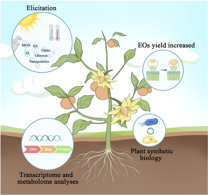
Figure 1.
Synthetic regulation of EOs via techniques such as transcriptomics and metabolomics analysis, plant synthetic biology, and elicitation. This figure was created using Adobe Illustrator 2022.

The evolutionary journey of essential oils: From synthesis to biomedical applications
Xiuxiu Dai , Weisen Lin , Kemeng Wu , Guowang Cheng , Na Wan , Tongkai Chen , Zhenfeng Wu


Essential oils (EOs) are volatile secondary metabolites that impart a distinctive odor and taste to plants. They are stored as droplets in the leaves, stems, flowers, fruits, bark, and roots of plants, within complex secretory structures such as glandular trichomes (GTs) and secretory cavities [1]. EOs largely contain very complex mixtures of terpenes (e.g., α-pinene, β-pinene, and β-Caryophyllene), terpenoids (e.g., linalool, carvacrol, sclareol, and 1,8-cineole), and phenylpropanoids (e.g., eugenol, anethole, and cinnamaldehyde) [2]. Additionally, they may also contain aromatic and aliphatic compounds, as well as nitrogen- and/or sulfur-containing compounds. For example, caper (Capparis spinosa L.) leaf EOs contain dimethyl tetrasulfide, dimethyl trisulfide and methyl isothiocyanate [3], while garlic EOs contain diallyl trisulfide and diallyl disulfide (Fig. S1 in Supporting information) [4].
With their unique combination of chemical components, EOs exhibit a wealth of biological activities and significant health benefits. They are known for enhancing the immune system and possess antimicrobial, antioxidant, anti-inflammatory and anticancer properties [5]. This demonstrates their broad application prospects across areas such as pharmaceuticals, cosmetics, food, and agriculture. However, the limited quality and low yield of EOs restrict their widespread application [6]. Researchers are thus actively seeking solutions to these problems. Genetic engineering, particularly the use of transcription factors to regulate the expression of genes encoding key enzymes involved in the synthesis of EOs, has been employed to address the issue of production scalability [7,8]. Meanwhile, emerging technologies have revolutionized the extraction and separation of EOs, mediating a shift from traditional, time-consuming, and low-yield methods to more diverse, rapid, and efficient approaches. As the global market for EOs continues to expand, the issue of adulteration is becoming increasingly serious. To protect consumer rights and ensure the healthy market growth, comprehensive and efficient methods for EO identification are required [9,10].
In recent years, several encapsulation technologies for EOs (including micro-encapsulation and nano-encapsulation) have been developed to improve the water solubility, stability, and bioavailability of their bioactive constituents [11-13]. More importantly, these strategies have been shown to enhance the biological activity of EOs, enabling controlled, sustained, and targeted release for drug delivery applications and thereby providing innovative solutions for the treatment of various diseases.
To provide a comprehensive understanding of EOs, this review explores the mechanisms that regulate the synthesis and storage of EOs, along with advanced extraction and identification techniques. Furthermore, it emphasizes the diversity of EO bioactivities and their considerable potential in disease prevention and treatment. Finally, it highlights the broad prospects of integrating encapsulation technology with EO science and outlines directions for future research and development.
The biosynthesis of EOs is mediated by several key metabolic pathways. Terpenes and terpenoids are synthesized via the mevalonic acid (MVA) and methylerythritol phosphate (MEP) pathways [14], while phenylpropanoids are derived from the shikimate pathway (Fig. S2 in Supporting information) [15]. The MVA and MEP pathways generate the universal precursors isopentenyl diphosphate (IPP) and dimethylallyl diphosphate (DMAPP). These molecules are subsequently condensed and elongated to produce intermediates such as geranyl diphosphate (GPP) and farnesyl diphosphate (FPP), which are then cyclized or rearranged by terpene synthase (TPS) enzymes to generate diverse terpenoid skeletons [16-18]. The biosynthesis of phenylpropanoids is driven by the shikimate pathway. This pathway converts the primary metabolites phosphoenolpyruvate (PEP) and erythrose 4-phosphate (E4P) into chorismic acid, a key precursor for aromatic amino acids and a wide array of aromatic secondary metabolites [19,20]. Furthermore, the aliphatic compounds in EOs are largely derived from unsaturated fatty acids such as linoleic and linolenic acid via the lipoxygenase (LOX) pathway, ultimately leading to the production of volatile C6 or C9 aldehydes and esters [21]. Meanwhile, nitrogen- and/or sulfur-containing compounds predominantly arise from the enzymatic breakdown of less-volatile precursors such as glucosinolates. For instance, upon cell disruption, the enzyme myrosinase catalyzes the hydrolysis of glucosinolates to yield volatile products such as isothiocyanates [22].
Omics-based approaches can provide systematic insights into the molecular mechanisms of EOs biosynthesis [23]. Transcription factors have been shown to directly modulate the accumulation of EO components by regulating synthase genes [24,25]. In plant synthetic biology, both microbial (e.g., Escherichia coli [26] and Saccharomyces cerevisiae [27]) and plant-derived chassis cells (e.g., Nicotiana benthamiana) are employed to enhance the production of plant terpenes and phenylpropanoids [28,29]. Notably, treatment with elicitors, including phytohormones [30,31] and novel nano-elicitors [32-35], has also been shown to effectively stimulate EO biosynthesis in plants by upregulating biosynthetic genes and promoting compound accumulation, serving as versatile tools for production regulation. These approaches are summarized in Fig. 1.
EOs are crucial for normal plant physiology and environmental interactions, aiding in sun protection, providing antioxidant effects, and enhancing disease resistance. Notably, EOs attract pollinators and seed dispersers, repel herbivores, and summon predators of herbivorous arthropods. Additionally, they mediate information exchange between different plants. The secretory structures responsible for EO production in terrestrial plants first appeared in liverworts [36]. These were called oil bodies. Fig. 2A shows the localization of oil bodies in wild-type (WT-5) and Mptga-1ge mutant gemmae of Marchantia, visualized using the boron dipyrromethene (BODIPY) staining [37]. Liverworts can synthesize and store a variety of terpenoids, especially sesqui- and diterpenoids [38], which contributed to chemical defense during the evolution of bryophytes [39]. Tanaka et al. isolated oil body cells from Marchantia polymorpha using a glass capillary and found the specific localization of sesquiterpenes and marchantin A [40]. Subsequent research revealed that MpC1HDZ and MpERF13 are key transcription factors regulating the differentiation and development of these oil body cells [41], while MpMYB02 modulates oil body formation and marchantin accumulation [42].
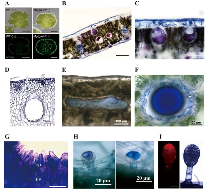
In higher plants, secretory structures are classified based on whether aromatic substances are excreted externally or retained internally. Accordingly, they are divided into endocrine (primarily including secretory cells, secretory cavities, and secretory tubes) and exocrine (primarily including epidermal cells and GTs) structures. Secretory cells, including oil cells, are often found in the thin-walled tissues of the nutritive and reproductive organs of plants. These cells originate from basal meristematic tissues and are capable of secreting and storing aromatic compounds. Mature oil cells are large and contain a single, prominent oil vesicle occupying the cell lumen. Staining with the NADI (alpha-naphthol + dimethyl paraphenylenediamine) reagent produces a bluish-purple coloration in the regions involved in terpene synthesis and storage within plant cells. Figs. 2B and C shows the oil cells of Cinnamomum camphora leaves, with the bluish-purple portion covering almost the entire oil cell in these plants [43]. Meanwhile, secretory cavities and secretory tubes are globular or tubular elongated cellular interstices present in plants. These structures are formed via both schizogeny and lysogeny. Of these, schizogeny refers to the dissolution of the middle lamella between the duct initials, leading to the formation of intercellular spaces. In contrast, lysogeny refers to the programmed death and dissolution of the cells themselves, resulting in the generation of extracellular cavities [44]. In both cases, the surrounding cells transform into highly active epithelial cells that synthesize and secrete EOs into the extracellular cavities [45]. Wu’s group has previously discovered that the genes CisPG21, CisCEL16 [46], CgPBA1 [47], CgPG21 [48], and CgVPE1 [49] regulates secretory cavity formation following programmed cell death in Citrus fruits. Fig. 2D shows a mature secretory cavity in the Citrus grandis 'Tomentosa' fruit [50]. The white arrows in Fig. 2F point to active epithelial cells. Meanwhile, the blue coloration in Figs. 2E and F indicates that the secretory tubes are filled with terpenes [51]. Epidermal secretory cells, which are specialized epidermal cells, form papillae (Fig. 2G) [52]. Their secretions emit characteristic aromas that attract pollinating insects, facilitate pollen adhesion, and regulate pollen germination.
GTs can be found on the surfaces of ∼30% of vascular plant species and are morphologically classified into two main types: Capitate and peltate trichomes [53]. The secretory cells within GTs express isoprenoid biosynthesis genes, which facilitate the synthesis and storage of terpenoids in trichome reservoirs [54]. Fig. 2H shows the capitate GTs of Phlomis herba-venti leaves [55]. Fig. 2I presents the GTs of Rhus typhina L. stained with Neutral Red (red fluorescence) and NADI reagent (purple) for the detection of secretory compounds [56].
In response to the growing demand for EOs, technologies for their extraction and separation have advanced significantly. Currently, EOs extraction and separation strategies often integrate multiple techniques to enhance yield, efficiency, and sustainability while reducing the overall environmental impact. This section systematically reviews conventional and emerging methods, highlighting their comparative advantages, limitations, and industrial applicability.
Traditional methods for extracting EOs, such as steam distillation, hydrodistillation (HD), and solvent extraction, are generally associated with high energy consumption, prolonged processing times, the potential use of hazardous solvents, and the possible degradation of heat-sensitive compounds [57,58]. To address these limitations, studies have increasingly focused on integrating innovative pretreatment techniques with assisted extraction methodologies. The pretreatment strategies mainly include physical, enzymatic, and solvent-based approaches. Beyond methods such as supercritical fluid extraction and subcritical water extraction, several advanced extraction techniques based on HD are still utilized. Their effectiveness is often enhanced through auxiliary energy input, giving rise to methods such as microwave-assisted HD (MAHD), ultrasound-assisted HD, ohmic-assisted HD, and other assisted HD variants. Notably, combining pretreatment with assisted extraction has been found to effectively mitigate the inherent limitations of conventional methods and significantly improve EO yield (Table S1 in Supporting information). This shift from the use of individual treatments to combined treatments highlights a trend towards process intensification, which is crucial for scaling up EOs production for industrial applications. It is now possible to extract the desired EO fraction more effectively, increase the yield, and reduce energy consumption by integrating different methods.
After extraction, EOs are purified to enrich their active components and remove impurities. Terpenes, which are prone to hydrolysis, are often separated from more stable and active terpenoids through deterpenation. At present, several common methods are employed for the separation of functional components from EOs, including crystallization, liquid-liquid extraction, vacuum extraction, batch distillation, molecular distillation, and pervaporation. The improvement of separation methods could increase the concentration of active compounds in EOs, extend their storage time, and enable by-product reuse. See the Supporting information for details.
The chemical composition of EOs varies among plant species and even within different plant parts [59]. Additionally, it depends on the growth period, soil conditions, climate, and other factors as well [60,61]. Finally, extraction and storage conditions including light, heat, oxidation, and other effects, also impact the composition of EOs [62]. Commercially available EOs may be adulterated, leading to quality and safety problems. Therefore, the accurate analysis of EOs composition is crucial for quality control. See the Supporting information for details.
The constituents of EOs not only have their own individual bioactivities but also interact with each other, collectively conferring a diverse range of biological activities to the EOs. For example, oregano and thyme EOs are both rich in carvacrol and thymol, which have antioxidant and antimicrobial activities. However, the content of these molecules differs between these EOs, leading to differences in their antioxidant and antimicrobial activities [63]. Yabuuchi et al. developed a machine learning method to predict the antimicrobial effects of different EOs based on their composition [64]. As shown in Fig. 3, the main biological activities of EOs include their antimicrobial, antioxidant, anti-inflammatory, and anticancer activities.
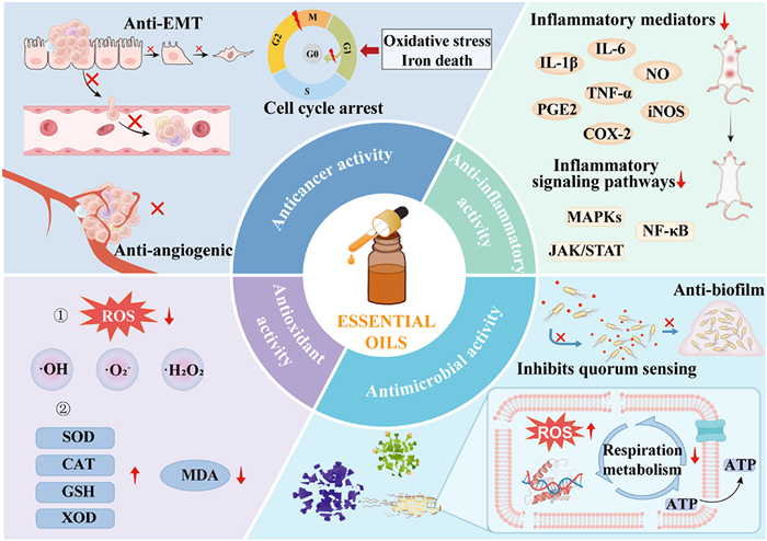
Antimicrobial resistance is a global issue that causes approximately 700,000 deaths annually and has emerged as a threat to both public and veterinary health. EOs, as natural active ingredients of plant origin, demonstrate broad-spectrum antimicrobial activity, unique modes of action, a low risk of drug resistance, and environmental friendliness. These advantages make EOs promising as novel antimicrobial agents for inhibiting the growth of bacteria, fungi, and viruses. The antimicrobial effects of EOs are primarily mediated by their lipophilicity, which allows them to disrupt pathogen membranes and cell walls, interfere with the metabolic processes of pathogens, and inhibit their replication.
The outer membrane of gram-negative bacteria contains a high percentage of lipopolysaccharides, which renders it virtually impermeable to lipophilic compounds. These microbes are often resistant to the active compounds found in EOs and can maintain their cellular integrity even after EO treatment, which is typically more effective against gram-positive bacteria [65]. For fungi, EOs can target key structural components such as chitin in cell walls and ergosterol in membranes. Against viruses, particularly enveloped types, EOs are known to disrupt the viral envelope structure. Beyond structural disruption, EOs can induce the leakage of cellular macromolecules, thereby altering respiration, energy metabolism, and other key physiological processes. Furthermore, they can disrupt the redox balance within microbial cells, leading to the accumulation of reactive oxygen species (ROS) which causes damage to cellular components. Additionally, EOs exhibit anti-biofilm potential by interfering with quorum sensing (QS) systems, thereby inhibiting the cellular communication pathways necessary for biofilm formation. See the Supporting information for details.
ROS play a crucial role in immune responses, not only serving as a core component of the immunoregulatory system but also acting as key signaling elements essential for phagocytosis and apoptosis. However, excessive ROS production triggers oxidative stress, leading to the oxidation of proteins, lipids, and DNA and destroying their structure and function. The chemical constituents of EOs generally contain conjugated carbon-carbon double bonds and hydroxyl groups, which can scavenge free radicals and minimize oxidative stress. Studies have shown that the antioxidant capacity of EOs is strongly linked to their phenolic content, and specific terpene constituents have been identified as potent antioxidants. Furthermore, the antioxidant and ROS-inhibitory effects of EOs and their key components have been validated through chemical assays, molecular docking simulations, and in vivo models, demonstrating their potential in mitigating oxidative damage. See the Supporting information for details.
Inflammation is a natural defense mechanism activated by a host organism in response to potential external threats. However, the mediators involved in immune defense can sometimes have detrimental effects, contributing to the pathogenesis of chronic diseases such as cardiovascular disease, neurodegenerative diseases, diabetes, cancer, obesity, and asthma and inflammatory diseases such as arthritis and periodontal disease [66]. Interestingly, certain EOs have anti-inflammatory properties that may reduce unwanted inflammatory responses and associated symptoms. These EOs typically prevent the release of inflammatory mediators (nitric oxide (NO), prostaglandin E2 (PGE2), tumor necrosis factor-α (TNF-α), interleukin (IL)-1β, IL-6, cyclooxygenase-2 (COX-2), inducible nitric oxide synthase (iNOS), and chemokines), inhibit inflammatory signaling pathways (nuclear factor kappa-B (NF-κB), mitogen-activated protein kinases (MAPKs), and Janus kinase (JAK)/signal transducer and activator of transcription (STAT)), or modulate the activity of the immune system [67]. Furthermore, as EOs exert their anti-inflammatory effects, they also indirectly regulate the immune system. The decrease in inflammation prevents the over-activation of the immune system, thus helping to maintain immune balance, as evidenced by studies showing EOs can inhibit processes like neutrophil chemotaxis and lymphocyte proliferation. See the Supporting information for details.
EOs and their constituents, particularly terpenoids [68] and phenylpropanoids [69], have demonstrated significant anticancer potential. These compounds exhibit cytotoxic effects against a wide range of malignancies, including breast, cervical [70], colon, lung, liver, skin, oral, and prostate cancer as well as glioblastoma [71]. Owing to their lipophilic properties, these compounds can penetrate the membranes of cancer cells [72] and exert powerful anticancer effects through multiple mechanisms, including cytotoxicity, cell cycle arrest, inhibition of cell invasion and migration, inhibition of proliferation, and induction of apoptosis. EOs can interfere with the normal progression of the cell cycle by enhancing oxidative stress in cancer cells, thereby inducing cell cycle arrest and apoptosis. Furthermore, by inhibiting the epithelial-mesenchymal transition (EMT) process, EOs can reduce the migration and invasiveness of tumor cells, thereby suppressing metastasis. Tumor angiogenesis, another critical process for growth and spread, can also be targeted by EOs through the inhibition of pro-angiogenic signaling molecules and pathways. See the Supporting information for details.
The bioactivities of EOs have been validated using in vivo experiments, and EOs have shown favorable therapeutic effects in such disease models. Pathogenic microorganisms, one of the most common causes of respiratory diseases, can invade the respiratory tract, triggering symptoms such as cough, sore throat, and shortness of breath. In some cases, these pathogens can also cause lung infections. However, owing to their antimicrobial and anti-inflammatory activities, EOs can effectively eliminate pathogenic microorganisms and ameliorate the inflammatory responses they induce [73]. α-Pinene is a common monoterpene found in EOs and shows a minimum inhibitory concentration (MIC) of 126 and 172 µg/mL against Haemophilus influenzae and Streptococcus pneumoniae, respectively. Additionally, this compound also inhibits the activity of infectious bronchitis virus (IBV), showing a half-maximal inhibitory concentration (IC50) of 0.98 ± 0.25 mnol/L. Thus, it is often used in the treatment of respiratory tract infections [74]. Helichrysum italicum (Roth) G. Don EO shows antimicrobial and anti-biofilm effects against respiratory pathogens such as Haemophilus influenzae, Haemophilus parainfluenzae, Pseudomonas aeruginosa, and Streptococcus pneumoniae [75]. Furthermore, monoterpenes such as carvacrol, carvone, 1,8-cineol, menthofuran, and pulegone inhibit severe acute respiratory syndrome coronavirus 2 (SARS-CoV-2) replication in infected cells [76]. When the skin barrier is compromised, it becomes susceptible to pathogen invasion, leading to the development of skin diseases. Interestingly, Salvia officinalis EO was found to increase total antioxidant capacity (TAC) levels and decrease the malondialdehyde (MDA) content and IL-1β and TNF-α levels in a mouse wound model infected with Pseudomonas aeruginosa and Staphylococcus aureus (Table S2 in Supporting information) [77]. This indicates that EOs can also be helpful in treating skin infections.
Oxidative stress is caused by an imbalance between the oxidation and antioxidant effects in the body. Meanwhile, inflammation is the body’s defensive response against various external and internal stimuli. Under conditions of oxidative stress, the Keap1/Nrf2/heme oxygenase-1 (HO-1) pathway is activated, promoting the expression of downstream antioxidant genes, such as superoxide dismutase (SOD) and glutathione peroxidase (GPX). This enhances cellular antioxidant capacity and protects cells from oxidative damage. In addition, the induced expression of HO-1 inhibits the production of pro-inflammatory factors such as TNF-α and IL-1β while promoting the expression of anti-inflammatory factors such as IL-10. Notably, Myrrh EO, geraniol, and anethole have been found to modulate the Keap1/Nrf2/HO-1 pathway, enabling the management of isoproterenol-induced myocardial infarction in mice [78,79]. In middle cerebral artery occlusion (MCAO)/R model rats, β-caryophyllene was found to enhance the nuclear translocation of Nrf2, thereby activating the Nrf2/HO-1 pathway [80]. In addition, EOs have also been discovered to regulate the NF-κB/MAPK [81], Toll-like receptor 4 (TLR4)/MAPK [82], TLR4/NF-κB [83,84], extracellular signal-regulated kinase (ERK)/NF-κB [85], and JAK/STAT/NF-κB [86] pathways under conditions of neuroinflammation, colitis, hepatitis, arthritis, and atopic dermatitis.
Owing to their antitumor activity, EOs have yielded remarkable outcomes in tumor treatment. Some EOs have been found to inhibit tumor growth and significantly reduce the mass and volume of tumors. Specifically, in the DA3 murine tumor model, Lippia citriodora EO was found to activate cysteine asparaginase 3 and downregulate the expression of the anti-apoptotic protein survivin in tumor tissues, inducing apoptosis in cancer cells [87]. In addition, Curcuma phaeocaulis EO demonstrated the ability to inhibit tumor angiogenesis in a nude mouse model of colon cancer [88].
However, it is important to note that the use of EOs poses potential health risks, including asthma, the exacerbation of allergic reactions, and skin irritation. The overuse of EOs may lead to liver and kidney damage, seizures, and even death. Zingiber ottensii Valeton EO has demonstrated concentration-dependent embryotoxicity in zebrafish models, showing an half-maximal lethal concentration (LC50) of 1.003 µg/mL, and has been linked to morphological abnormalities, reduced hatchability, and impaired cardiac function [89]. Similarly, lemongrass, thyme, and oregano EOs all exhibit substantial embryotoxic effects in zebrafish, demonstrating LC50 values of 3.7, 14.4, and 5.3 µg/mL, respectively. Exposure to these EOs has been found to cause neurodevelopmental disruptions in zebrafish, including spinal deformation, pericardial edema, ocular hypoplasia, and swimming abnormalities [90]. Thus, understanding the toxicity and adverse effects of EOs is crucial for ensuring their safe use. In this context, further long-term toxicity studies are required to confirm the safety of EOs-based products.
The inherent volatility, poor aqueous solubility, and environmental instability of EOs limit their bioavailability and therapeutic potential. Encapsulation technologies can address these challenges, and delivery systems that protect the active components of EOs, enhance their stability, and enable controlled or targeted release can be developed. Notably, an appropriate encapsulation system needs to be selected based on the physicochemical properties of the EO, the intended route of administration, and the desired release profile.
Encapsulation strategies are broadly divided into microencapsulation (using microcapsules or microemulsions) and nanoencapsulation strategies (employing polymer-based, lipid-based, metal-based, or inorganic nanocarriers, as well as inclusion complexes). The selection of the wall material has a critical influence on encapsulation efficiency, stability, and release behavior. Polysaccharide- and protein-based systems (e.g., gum arabic, maltodextrin, chitosan) are valued for their biocompatibility, film-forming ability, and intrinsic bioactivity (e.g., the antimicrobial properties of chitosan) [91,92]. Lipid-based carriers, such as nanoliposomes and nanostructured lipid carriers (NLCs), effectively encapsulate lipophilic EOs and facilitate their cellular uptake [93]. NLCs, in particular, provide better drug loading capacity and stability than traditional solid lipid nanoparticles (SLNs) [94,95]. Meanwhile, cyclodextrins enhance the stability and solubility of volatile compounds by forming inclusion complexes within their hydrophobic cavities [96]. Metal-based nanocarriers, notably metal-organic frameworks, offer high porosity and enable efficient EO loading, reducing volatilization and oxidation. Their pore structures can be engineered for stimuli-responsive release, with surface functionalization further enhancing their targeting capacity and stability [97]. Inorganic nanocarriers such as mesoporous silica nanoparticles provide high surface areas and tunable pores for high-capacity, protected delivery. Interestingly, EO bioactivity can be demonstrably enhanced through rational formulation design [98]. For instance, a microemulsion of Cymbopogon nardus EO was found to show drastically improved antifungal activity, with its MIC values against Candida albicans strains dropping from ≥500 µg/mL to 31.2–62.5 µg/mL [99]. A d-limonene nanoemulsion stabilized by sodium caseinate exhibited five-fold greater antimicrobial activity against S. aureus than the free EO [100]. Although systems such as chitosan-coated SLNs can successfully deliver EOs to specific sites (e.g., liver tumors), they often face limitations such as low drug loading capacity (typically 1%−5%) and potential long-term instability [101]. In one study, thyme EO encapsulated in dendritic mesoporous silica nanoparticles achieved a 70% inhibition rate against Fusarium oxysporum, compared to ∼40% with the unencapsulated oil [102].
In summary, selecting appropriate encapsulation systems based on the required protection levels, desired release kinetics, and specific treatment targets are essential for developing reliable and effective EO-based products. This is corroborated by the data summarized in Tables S3 and S4 (Supporting information).
The therapeutic potential of EOs has been demonstrated in animal models, offering a foundation for further clinical application. However, animal models cannot fully reflect the physiological and cellular characteristics of the human body. Therefore, the safety and efficacy of EOs need to be verified through rigorous clinical trials before their clinical translation. Some examples of such clinical trials are provided in Table S5 (Supporting information). Nevertheless, the potential clinical applications of a wide variety of EOs still remain unexplored. Furthermore, clinical studies on encapsulated EOs formulations are still in their infancy. Only one such trial is ongoing at present, and it is currently in the recruitment phase. This highlights the need for further research to fully leverage the therapeutic potential of native EOs and EOs-based nanomedicines in clinical practice.
EOs are natural plant-derived products that are rich in active ingredients. With growing attention toward natural remedies and healthy lifestyles, the market demand for EOs has reached unprecedented heights. However, there remains a large gap between EO production and market demand at present. First, the yield of EOs can often be low. EOs typically account for only a small percentage of plant weight, which fundamentally limits their yield. Moreover, given the limitations of current extraction technologies, EOs cannot be efficiently and completely isolated from plants, and extraction processes have high energy requirements, which further increases production costs. Therefore, strategies to improve extraction efficiency and reduce costs are increasingly being explored. Second, the EOs available on the market are frequently adulterated, which not only weakens the actual therapeutic effects of EO products but also severely hampers consumer trust. Although international standards such as ISO have been established to regulate these products, these standards can sometimes be overly stringent, inadvertently excluding genuine high-quality EOs [103]. Given the complex and variable nature of EO components, formulating a standard system that can effectively identify adulterated products without impeding the circulation of authentic EOs has become a significant challenge for the industry. Third, encapsulation technology, as an emerging approach to enhance the bioavailability of EOs, holds great promise for expanding their applications. However, only a handful of encapsulation materials meet current standards, such as those set by the Food and Drug Administration (FDA) for pharmaceutical use, significantly limiting the translation potential of encapsulation technology. Moreover, the quality control standards for encapsulated products are not yet well-established, and a unified and scientific evaluation system to ensure product quality and efficacy is currently lacking. Additionally, the stability of encapsulation technologies in large-scale production scenarios also poses a challenge. Many techniques that have been validated in laboratory settings often fail to yield consistency at scale, further complicating quality control efforts. Finally, despite their potential therapeutic value in multiple pathological conditions, EOs only have an auxiliary role in clinical therapies at present instead of serving as primary treatments. The lack of data from clinical trials featuring EOs as the main intervention not only limits their widespread clinical use but also hampers in-depth research on their potential as standalone therapeutic agents. In response to these aforementioned obstacles, we propose the following potential strategies:
(a) Transcriptomics can be combined with metabolomics to elucidate the biosynthetic processes involved in EO production in a wider range of plant species, identify relevant genes, and develop high-yielding varieties of plants. Additionally, more environmentally friendly and cost-effective extraction technologies should be developed.
(b) Improved quality standards and management norms should be established for the EO industry. A standard system that can effectively identify adulterated products without hindering the circulation of genuine EOs is urgently warranted. A standardized quality control framework should be formulated, covering aspects such as raw material selection, production processes, and final product testing. Investing in advanced production technologies and automation equipment will help to enhance product consistency.
(c) Research and innovation in the field of EO encapsulation should be improved to develop materials that are suitable for pharmaceutical use and meet strict standards, such as those set by the FDA. There is a need to optimize production processes and enhance monitoring and inspection during production. The link between laboratory research and large-scale production needs to be strengthened to ensure that techniques validated in laboratory settings can also maintain consistency when scaled up.
(d) Basic research on EOs should be promoted, and their pharmacological mechanisms should be explored. Through collaboration among researchers, healthcare professionals, and industry stakeholders, clinical trials that focus on EOs and explore their therapeutic efficacy and safety in various diseases should be conducted.
With the ongoing efforts of researchers and the continuous development of technology, we are confident that EOs will be utilized more efficiently and effectively in the coming years. Through targeted strategies, we could leverage the complete potential of EOs as powerful therapeutic agents.
Xiuxiu Dai: Writing – original draft, Validation, Methodology, Investigation, Formal analysis, Data curation, Conceptualization. Weisen Lin: Investigation, Formal analysis, Data curation. Kemeng Wu: Investigation, Formal analysis, Data curation. Guowang Cheng: Investigation, Formal analysis, Data curation. Na Wan: Writing – review & editing, Supervision, Project administration, Funding acquisition, Conceptualization. Tongkai Chen: Writing – review & editing, Supervision, Project administration, Funding acquisition, Conceptualization. Zhenfeng Wu: Writing – review & editing, Supervision, Project administration, Funding acquisition, Conceptualization.
The authors declare that they have no known competing financial interests or personal relationships that could have appeared to influence the work reported in this paper.
This work was financially supported by the National Natural Science Foundation of China (Nos. 82474091 and 82560781) and the Open Project of Key Laboratory of Modern Preparation of Traditional Chinese Medicine, Ministry of Education, Jiangxi University of Chinese Medicine (No. zdsys-202401).
Supplementary material associated with this article can be found, in the online version, at doi:
S. Ivanova, S. Dyankov, D. Karcheva-Bahchevanska, et al., Molecules 28 (2023) 2918. doi: 10.3390/molecules28072918
J. Valverde Sancho, C. Carreño Amate, M.D.M. Caparrós Pérez, et al., Life 13 (2023) 499. doi: 10.3390/life13020499
M. Merlino, C. Condurso, F. Cincotta, et al., Antioxidants 13 (2024) 718. doi: 10.3390/antiox13060718
S. Shah, K.K. Elgizawy, C.M. Shi, et al., Cells 12 (2023) 669. doi: 10.3390/cells12040669
M. Cedillo-Cortezano, L.R. Martinez-Cuevas, J.A.M. López, et al., Pharmaceuticals 17 (2024) 303. doi: 10.3390/ph17030303
J. Wang, F. Wang, Y. Zhang, et al., Front. Microbiol. 16 (2025) 1626646. doi: 10.3389/fmicb.2025.1626646
S.S. Mahmoud, R.B. Croteau, Trends Plant Sci. 7 (2002) 366–373. doi: 10.1016/S1360-1385(02)02303-8
C. Yang, S. Marillonnet, A. Tissier, et al., Plant J. 107 (2021) 1102–1118. doi: 10.1111/tpj.15371
H. Yang, X. Huang, M. Yang, et al., Food Chem. 451 (2024) 139340. doi: 10.1016/j.foodchem.2024.139340
E. Kampasakali, A. Nakas, D. Mertzanidis, et al., Molecules 28 (2023) 1221. doi: 10.3390/molecules28031221
Z. Li, H. Shen, W. Cai, et al., Chin. Chem. Lett. 37 (2026) 111292. doi: 10.1016/j.cclet.2025.111292
R. Nur Fatin Nazurah, M.A. Noranizan, M.A.R. Nor-Khaizura, et al., Int. J. Biol. Macromol. 273 (2024) 132972. doi: 10.1016/j.ijbiomac.2024.132972
M. Cai, Y. Wang, R. Wang, et al., Int. J. Biol. Macromol. 202 (2022) 122–129. doi: 10.1016/j.ijbiomac.2022.01.066
F. Wu, Y. Chen, M. Gao, et al., Int. J. Mol. Sci. 26 (2025) 5690. doi: 10.3390/ijms26125690
N. Sadgrove, G. Padilla-González, M. Phumthum, Plants 11 (2022) 789. doi: 10.3390/plants11060789
S.S. Mahmoud, S. Maddock, A.M. Adal, Front. Plant Sci. 12 (2021) 699157. doi: 10.3389/fpls.2021.699157
P.S. Karunanithi, P. Zerbe, Front. Plant Sci. 10 (2019) 1166. doi: 10.3389/fpls.2019.01166
R. Rehman, M.A. Hanif, Z. Mushtaq, A.M. Al-Sadi, Food Rev. Int. 32 (2015) 117–160.
N.J.H. Averesch, J.O. Krömer, Front. Bioeng. Biotechnol. 6 (2018) 32. doi: 10.3389/fbioe.2018.00032
C.F. Kunz, S. de Vries, J. de Vries, Philos. T. R. Soc. B 379 (2024) 20230358. doi: 10.1098/rstb.2023.0358
M. Ishimori, Plant Cell Physiol. 63 (2022) 1324–1325. doi: 10.1093/pcp/pcac128
M. Shirakawa, T. Oguro, S.S. Sugano, et al., Nat. Plants 11 (2025) 483–504. doi: 10.1038/s41477-025-01921-1
E.R. Lanier, T.B. Andersen, B. Hamberger, Plant J. 114 (2023) 1178–1201. doi: 10.1111/tpj.16177
Q. Wang, V.A. Reddy, D. Panicker, et al., Plant Biotechnol. J. 14 (2016) 1619–1632. doi: 10.1111/pbi.12525
Y. Zhao, Y. Chen, M. Gao, et al., Tree Physiol. 43 (2023) 2150–2161. doi: 10.1093/treephys/tpad111
J. Yang, Z. Li, L. Guo, et al., Renew. Energy 99 (2016) 216–223. doi: 10.1016/j.renene.2016.06.061
R. Li, K. Wang, D. Wang, et al., Green Chem. 23 (2021) 5088–5096. doi: 10.1039/d1gc00917f
B.D. Kwan, B. Seligmann, T.D. Nguyen, et al., Curr. Opin. Plant Biol. 71 (2023) 102330. doi: 10.1016/j.pbi.2022.102330
N. Kitaoka, X. Lu, B. Yang, et al., Mol. Plant 8 (2015) 6–16. doi: 10.1016/j.molp.2014.12.002
F. Kianersi, A. Pour-Aboughadareh, M. Majdi, et al., Int. J. Mol. Sci. 22 (2021) 11124. doi: 10.3390/ijms222011124
P.H. Gorni, A.C. Pacheco, A.L. Moro, et al., Sci. Hortic. 270 (2020) 109436. doi: 10.1016/j.scienta.2020.109436
M.V. Asadollahei, J. Tabatabaeian, M. Yousefifard, et al., Plant Physiol. Bioch. 194 (2023) 722–730. doi: 10.1016/j.plaphy.2022.12.019
T.E. Sayed, E.S.S. Ahmed, Front. Energy Res. 10 (2022) 996253. doi: 10.3389/fenrg.2022.996253
M. Mukarram, M.M.A. Khan, F.J. Corpas, et al., J. Hazardous Mater. 412 (2021) 125254. doi: 10.1016/j.jhazmat.2021.125254
U. Aqeel, R. Parwez, T. Aftab, et al., Environ. Res. 236 (2023) 116851. doi: 10.1016/j.envres.2023.116851
F. Romani, J.R. Flores, J.I. Tolopka, et al., J. Exp. Bot. 73 (2022) 4427–4439. doi: 10.1093/jxb/erac134
N. Gutsche, J. Koczula, M. Trupp, et al., New Phytol. 241 (2023) 1559–1573.
A. Ludwiczuk, Y. Asakawa, Food Chem. Toxicol. 132 (2019) 110649. doi: 10.1016/j.fct.2019.110649
F. Chen, A. Ludwiczuk, G. Wei, et al., Crit. Rev. Plant Sci. 37 (2018) 210–231. doi: 10.1080/07352689.2018.1482397
M. Tanaka, T. Esaki, H. Kenmoku, et al., Phytochemistry 130 (2016) 77–84. doi: 10.1016/j.phytochem.2016.06.008
F. Romani, E. Banić, S.N. Florent, et al., Curr. Biol. 30 (2020) 2815–2828 e2818. doi: 10.1016/j.cub.2020.05.081
H. Kubo, K. Sunhwa, H. Teramori, et al., Planta 260 (2024) 68. doi: 10.1007/s00425-024-04498-9
M. Bottoni, F. Milani, M. Mozzo, et al., Plants 10 (2021) 1008. doi: 10.3390/plants10051008
Y.F. Zhou, S.L. Mao, S.F. Li, et al., Plant Sci. 225 (2014) 147–160. doi: 10.1016/j.plantsci.2014.06.007
V. Todorova, S. Ivanova, Y. Georgieva, et al., Plants 11 (2022) 2061. doi: 10.3390/plants11152061
P. Tong, B. Huai, Y. Chen, et al., Plant Sci. 297 (2020) 110540. doi: 10.1016/j.plantsci.2020.110540
B. Huai, M. Bai, P.P. Tong, et al., Plant Physiol. Bioch. 160 (2021) 306–314. doi: 10.1016/j.plaphy.2021.01.018
M. Bai, P. Tong, Q. Luo, et al., Plant Cell Rep. 42 (2023) 1311–1331. doi: 10.1007/s00299-023-03032-7
B. Huai, M. Liang, J. Lin, et al., Int. J. Mol. Sci. 24 (2023) 11681. doi: 10.3390/ijms241411681
N. Rong, L. Huang, P. Ye, et al., Int. J. Biol. Macromol. 280 (2024) 135671. doi: 10.1016/j.ijbiomac.2024.135671
L.F.P.B. Costa, S.H. Taleb Contini, S.P. Teixeira, et al., Flora 262 (2020) 151496. doi: 10.1016/j.flora.2019.151496
A. Konarska, Micron 158 (2022) 103288. doi: 10.1016/j.micron.2022.103288
M. Sun, L. Zhu, Y. Zhang, et al., BMC Plant Biol. 23 (2023) 13. doi: 10.1186/s12870-022-04029-2
R. Schuurink, A. Tissier, New Phytol. 225 (2019) 2251–2266.
I.N. Gostin, Plants 12 (2023) 2423. doi: 10.3390/plants12132423
A. Konarska, Micron 186 (2024) 103692. doi: 10.1016/j.micron.2024.103692
H. Mushtaq, S. Piccolella, J.A. Mendiola, et al., Foods 14 (2025) 297. doi: 10.3390/foods14020297
J.J. Zhu, J.J. Yang, G.J. Wu, et al., Ind. Crops Prod. 146 (2020) 112206. doi: 10.1016/j.indcrop.2020.112206
B. Lechkova, N. Benbassat, D. Karcheva-Bahchevanska, et al., Molecules 28 (2023) 4906. doi: 10.3390/molecules28134906
N. El Hachlafi, H.N. Mrabti, S.H. Al-Mijalli, et al., Molecules 28 (2023) 5913. doi: 10.3390/molecules28155913
A. Laftouhi, N. Eloutassi, E. Ech-Chihbi, et al., ACS Omega 8 (2023) 46598–46607. doi: 10.1021/acsomega.3c05564
J.M. Alves-Silva, D. Maccioni, E. Cocco, et al., Plants 12 (2023) 1247. doi: 10.3390/plants12061247
M. Chroho, Y. Rouphael, S.A. Petropoulos, et al., Antibiotics 13 (2024) 139. doi: 10.3390/antibiotics13020139
H. Yabuuchi, K. Hayashi, A. Shigemoto, et al., Sci. Rep. 13 (2023) 18947. doi: 10.1038/s41598-023-46377-5
F. Nazzaro, F. Fratianni, L. De Martino, et al., Pharmaceuticals 6 (2013) 1451–1474. doi: 10.3390/ph6121451
C. Zhou, M. Gong, J. Jian, et al., Chin. Chem. Lett. 37 (2026) 112135. doi: 10.1016/j.cclet.2025.112135
M. de Lavor É, A.W.C. Fernandes, R.B. de Andrade Teles, et al., Oxid. Med. Cell. Longev. 2018 (2018) 6468593. doi: 10.1155/2018/6468593
M.A. Mustapa, I. Guswenrivo, A. Zuhrotun, et al., Appl. Sci. 12 (2022) 12738. doi: 10.3390/app122412738
A.Khatkar Neelam, K.K. Sharma, Crit. Rev. Food Sci. 60 (2019) 2655–2675.
J. Ruttanapattanakul, N. Wikan, K. Chinda, et al., Plants 10 (2021) 1419. doi: 10.3390/plants10071419
M. Magalhães, B. Manadas, T. Efferth, et al., Pharmacol. Res. 169 (2021) 105638. doi: 10.1016/j.phrs.2021.105638
M.A. Thalappil, P. Singh, A. Carcereri de Prati, et al., Phytother. Res. 38 (2023) 556–591.
F.O. de Carvalho, É.R. Silva, I.A. Gomes, et al., Phytother. Res. 34 (2020) 2214–2229. doi: 10.1002/ptr.6688
M. Allenspach, C. Steuer, Phytochemistry 190 (2021) 112857. doi: 10.1016/j.phytochem.2021.112857
V.L. Balázs, R. Filep, F. Répás, et al., Molecules 27 (2022) 5518. doi: 10.3390/molecules27175518
S. Ćavar Zeljković, E. Schadich, P. Džubák, et al., Front. Pharmacol. 13 (2022) 893634. doi: 10.3389/fphar.2022.893634
M.R. Farahpour, E. Pirkhezr, A. Ashrafian, et al., Biomed. Pharmacother. 128 (2020) 110120. doi: 10.1016/j.biopha.2020.110120
N.S. Younis, M.E. Mohamed, Chem. Biol. Interact. 360 (2022) 109947. doi: 10.1016/j.cbi.2022.109947
N.S. Younis, M.E. Mohamed, J. Ethnopharmacol. 270 (2021) 113793. doi: 10.1016/j.jep.2021.113793
Q. Hu, T. Zuo, L. Deng, et al., Phytomedicine 102 (2022) 154112. doi: 10.1016/j.phymed.2022.154112
V. Borgonetti, V. López, N. Galeotti, J. Ethnopharmacol. 294 (2022) 115362. doi: 10.1016/j.jep.2022.115362
Y. Hu, Z. He, J. Zhang, et al., Phytomedicine 119 (2023) 155024. doi: 10.1016/j.phymed.2023.155024
E.R. Puppala, S. Jain, P. Saha, et al., Phytomedicine 97 (2022) 153926. doi: 10.1016/j.phymed.2022.153926
R.N. Raka, J. Xiao, H. Wu, et al., Food Res. Int. 164 (2023) 112362. doi: 10.1016/j.foodres.2022.112362
S.Y. Kim, S.D. Han, M. Kim, et al., Antioxidants 10 (2021) 1941. doi: 10.3390/antiox10121941
J. Li, J. Duan, Y. Wang, et al., Biomed. Pharmacother. 168 (2023) 115727. doi: 10.1016/j.biopha.2023.115727
K. Spyridopoulou, T. Aravidou, E. Lampri, et al., Antioxidants 10 (2021) 875. doi: 10.3390/antiox10060875
Y. Feng, L. Deng, H. Guo, et al., Front. Oncol. 11 (2021) 728464. doi: 10.3389/fonc.2021.728464
W. Thitinarongwate, R. Mektrirat, W. Nimlamool, et al., Toxics 9 (2021) 102. doi: 10.3390/toxics9050102
I.I. da Silva Jr., N.P.C. da Silva, J.A. Marrs, et al., Biomedicines 11 (2023) 2821. doi: 10.3390/biomedicines11102821
J. Bajac, B. Nikolovski, I. Lončarević, et al., Food Hydrocoll. 125 (2022) 107430. doi: 10.1016/j.foodhyd.2021.107430
G.P. Sahoo, T.K. Rajwar, J. Halder, et al., Chin. Chem. Lett. 37 (2026) 111797. doi: 10.1016/j.cclet.2025.111797
W. Ji, T. Zhang, Z. Lu, et al., Chin. Chem. Lett. 30 (2019) 739–742. doi: 10.1016/j.cclet.2018.09.008
E. Gomaa, H.A. Fathi, N.G. Eissa, et al., Methods 199 (2022) 3–8. doi: 10.1016/j.ymeth.2021.05.003
J.E. Hyun, H.Y. Yi, G.P. Hong, et al., LWT 162 (2022) 113474. doi: 10.1016/j.lwt.2022.113474
K. Wu, T. Zhang, X. Chai, et al., Foods 12 (2022) 45. doi: 10.3390/foods12010045
J. Huang, H. Chen, Y. Liao, et al., Chin. Chem. Lett. 37 (2026) 112049. doi: 10.1016/j.cclet.2025.112049
T. Zhang, Z. Lu, J. Wang, et al., Chin. Chem. Lett. 32 (2021) 1755–1758. doi: 10.1016/j.cclet.2020.12.033
L. Gaspar de Toledo, M.A. dos Santos Ramos, P. Bento da Silva, et al., Int. J. Nanomed. 15 (2020) 10481–10497. doi: 10.2147/ijn.s275258
H. Qi, S. Chen, J. Zhang, et al., J. Food Eng. 334 (2022) 111159. doi: 10.1016/j.jfoodeng.2022.111159
B. Yang, J. Jiang, L. Jiang, et al., Int. J. Biol. Macromol. 149 (2020) 108–115. doi: 10.1016/j.ijbiomac.2020.01.222
L. Yu, J. Peng, Q. Han, et al., Int. J. Pharm. 669 (2025) 125057. doi: 10.1016/j.ijpharm.2024.125057
K. Pokajewicz, D. Lamaka, P.P. Wieczorek, et al., Ind. Crops Prod. 222 (2024) 119744. doi: 10.1016/j.indcrop.2024.119744

Figure 1 Synthetic regulation of EOs via techniques such as transcriptomics and metabolomics analysis, plant synthetic biology, and elicitation. This figure was created using Adobe Illustrator 2022.

Figure 2 Secretion and storage sites for EOs in plant cells. (A) Oil bodies (OBs) in Marchantia gemmae visualized through BODIPY staining. Copied with permission [37]. Copyright 2024, WILEY. (B, C) NADI staining in oil cells of Cinnamomum camphora leaves. Copied with permission [43]. Copyright 2021, MDPI. (D) Mature stage of the secretory cavity in Citrus grandis 'Tomentosa' fruit. Copied with permission [50]. Copyright 2024, Elsevier. (E, F) Optical micrographs of the longitudinal sections and cross-sections, respectively, of NADI-stained Myrsine leuconeura Mart. (Primulaceae) leaves. The epithelium of the canal formed by secretory cells is indicated by an arrowhead in panel D. Copied with permission [51]. Copyright 2020, Elsevier. (G) Metabolites found in lris sibirica nectary tissues during the male stage. Neutral lipids (EOs) present in papillae are stained pink after the application of Nile blue. ep: epidermis with papillae; gp: glandular parenchyma. Copied with permission [52]. Copyright 2022, Elsevier. (H) NADI staining of the capitate trichomes of P. herba-venti leaves. Copied with permission [55]. Copyright 2023, MDPI. (I) Localization of EOs visualized through dual staining with (left) neutral red (red fluorescence, tetramethylrhodamine isothiocyanate (TRITC) filter) and (right) NADI reagent (purple). Copied with permission [56]. Copyright 2024, Elsevier. Scale bars: 20 µm (F, H, I); 50 µm (C, D, G); 100 µm (B, E); 200 µm (A).

 扫一扫看文章


扫一扫关注我们
 DownLoad:
 下载:

 下载: