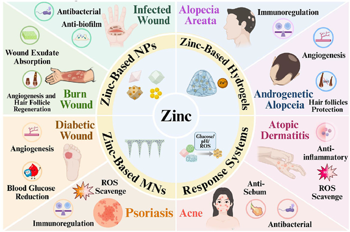
Figure 1.
Schematic illustration of the mechanisms of action of zinc-based materials in the treatment of skin diseases. Figure created with BioRender.com (BioRender, 2025).

Zinc for skin diseases treatment: Progress and prospects in advanced delivery systems
Yaling Wei , Jiaming He , Jinsong Ding , Caiyang Lu , Wenhu Zhou , Xinjie Deng


As the largest organ of the human body, the skin not only serves as a protective barrier against external insults but also plays pivotal roles in immune defense, thermoregulation, and sensory perception. The epidermis, mainly composed of keratinocytes, forms the first line of defense against external agents; the dermis, rich in collagen fibers and extracellular matrix, provides elasticity and structural support; while the subcutaneous tissue, predominantly consisting of adipose and connective tissue, cushions mechanical stress and functions as an energy reservoir [1–3]. Beyond serving as a physical shield, the skin protects against mechanical trauma, ultraviolet radiation, and pathogenic microorganisms, while also participating in immune regulation, electrolyte balance, and sensory conduction. These multifaceted roles render it indispensable for maintaining systemic homeostasis [4–8].
Due to chronic exposure to complex environmental stimuli, the skin is particularly vulnerable to disease. A wide spectrum of conditions, including inflammatory, infectious, and immune- or genetic-related disorders, affect nearly one-third of the global population, ranking skin diseases as the fourth most common cause of human illness [9,10]. Although current therapeutic strategies alleviate symptoms to some extent, limitations such as modest efficacy, adverse effects, and drug resistance highlight the urgent need for safer and more effective treatment modalities.
Zinc, the second most abundant transition metal in the human body, is integral to numerous physiological processes, including cell proliferation, differentiation, immune regulation, and wound healing [11,12]. Zinc deficiency has been closely linked to several dermatological conditions, such as acne, psoriasis, atopic dermatitis (AD), and acrodermatitis enteropathica [13–15]. For decades, both oral and topical zinc formulations have been employed to treat diverse skin diseases [16–18]. Compared with systemic administration, topical delivery offers the advantages of lower dosage and reduced risk of systemic accumulation. However, challenges such as toxicity from burst release of Zn2+ and limited mechanisms of action remain unresolved [19,20]. Historically, zinc-containing preparations, exemplified by calamine, have been used in medicine for thousands of years (Fig. S1 in Supporting information) [21–23]. Modern pharmacological studies further confirm zinc as an essential trace element with broad biological activities. With the advent of nanotechnology, zinc-based nanomaterials have emerged as promising candidates for skin disease therapy, owing to their unique physicochemical properties and multifunctional biological activities.
Zinc-based nanomaterials exert antibacterial, anti-inflammatory, and reparative effects through controlled Zn2+ release. Moreover, their assembly into metal-organic frameworks (Zn-MOFs) or nanoenzymes enables additional functions, including reactive oxygen species (ROS) generation or scavenging, as well as stimulus responsiveness. Zn-MOFs can also function as versatile drug carriers, facilitating multimodal synergistic therapies. These advances have generated significant interest in recent years. Nevertheless, clinical translation faces notable challenges: (ⅰ) The dynamic Zn2+ requirements across different disease stages complicate precise dosing; (ⅱ) the safety of certain zinc formulations in dermatological use remains controversial; (ⅲ) efficient strategies for crossing the skin barrier are still lacking; and (ⅳ) little is known about the potential of zinc-based materials for underexplored conditions such as seborrheic dermatitis and hidradenitis suppurativa.
Currently, a comprehensive overview that integrates the physiological roles of zinc, the pathophysiological mechanisms underlying zinc-related skin disorders, and the design strategies of zinc-containing materials for therapeutic use is still lacking. To address this gap, the present review highlights recent progress in zinc-based materials for the treatment of skin diseases, with particular emphasis on their mechanisms of action, functional properties, and therapeutic applications, as well as key challenges and future directions (Fig. 1). Specifically, we first discuss the physiological roles of zinc in skin biology and the impact of zinc deficiency on disease development; next, we summarize strategies for the design and application of zinc-based materials in skin disease therapy; and finally, we explore the translational challenges and future perspectives. This review aims to provide new insights and guidance for the rational design and clinical translation of zinc-based nanomaterials in dermatology.
Zinc, an essential trace element, is ubiquitously distributed across human tissues and organs, where it plays indispensable roles in maintaining structural integrity and physiological function [24,25]. Approximately 6% of the body’s total zinc content resides in the skin, with heterogeneous distribution across different layers: The epidermis contains about 60 µg/g of tissue zinc, markedly higher than the 40 µg/g detected in the dermis. This differential enrichment underpins zinc’s diverse biological functions in skin homeostasis and pathology (Fig. S2 in Supporting information) [26,27].
Physiologically, zinc is integral to keratinocyte proliferation, differentiation, and survival. In the immune system, zinc contributes to both innate and adaptive responses by modulating the maturation of T and B lymphocytes, natural killer cells, and dendritic cells. Through these effects, it enhances antibody production, antigen presentation, and phagocytosis, thereby sustaining immune homeostasis [28,29]. Also, zinc can achieve antibacterial effects through multiple pathways [30–32].
Despite these beneficial effects, excessive extracellular Zn2+ can exert cytotoxicity, triggering cell injury and apoptosis. Thus, the challenge lies in harnessing zinc’s multifaceted biological functions within a safe and effective dosage range. Therefore, developing optimized formulations that maximize therapeutic benefits while minimizing toxicity is of critical importance for clinical translation in dermatology.
There has been a lot of clinical evidence that zinc deficiency is closely related to the incidence of skin diseases. Oral or topical zinc preparations have a certain improvement effect on skin diseases. We summarized the clinical evidence between zinc deficiency and skin diseases and the clinical evidence of zinc supplementation in the treatment of skin diseases (Table S1 in Supporting information).
Acne is a chronic inflammatory disorder of the pilosebaceous unit and remains one of the most prevalent dermatological conditions worldwide, with a global prevalence of approximately 9.38% [33]. It affects up to 80% of adolescents and also a considerable proportion of adults [34]. Clinically, acne is characterized by comedones, papules, pustules, nodules, cysts, and in severe cases, permanent scarring, which can markedly impair both appearance and psychological well-being [35–37].
Accumulating clinical evidence demonstrates that serum zinc levels in acne patients are significantly lower than in healthy controls, and zinc concentration negatively correlates with disease severity [38–42]. Subsequent studies confirmed the therapeutic potential of oral zinc salts, such as zinc sulfate and zinc gluconate, in reducing acne severity [43,44]. Topical applications have also shown promise: For instance, local application of 5% zinc sulfate solution significantly reduced the acne inflammation score of patients with mild acne, and a combination of erythromycin and zinc acetate significantly reduced lesion counts compared with erythromycin alone [45,46]. Moreover, a multicenter, double-blind, randomized controlled trial involving 140 patients demonstrated that creams containing nicotinamide, antibacterial agents, and zinc pyrrolidone carboxylate effectively decreased non-inflammatory lesions with good safety profiles [47]. A systematic evaluation study that included a large amount of preclinical research evidence showed that the nanoscale size significantly enhanced the antibacterial and anti-inflammatory properties of zinc oxide (ZnO), while maintaining good acute tolerance [48]. More rigorously designed clinical trials are needed to support the clinical application of nano-zinc oxide (nZnO).
Alopecia encompasses a group of disorders characterized by progressive hair thinning or shedding, often associated with profound psychological and social consequences [49]. Androgenetic alopecia (AGA) is the most common form, caused by irreversible miniaturization of hair follicles [50,51], while alopecia areata (AA) is a chronic, relapsing autoimmune disease that affects approximately 2% of the global population [52]. Zinc plays a pivotal role in the process of hair regeneration, which is consistent with its status as the third most abundant trace element in hair.
Clinical studies have reported significantly lower serum zinc levels in patients with AGA and AA compared with healthy individuals, and in AA patients, the degree of zinc deficiency correlates with disease severity [53–55]. One research reported symptomatic relief following supplementation [56]. Topically, 1% zinc pyrithione solution can promote hair regrowth, although its efficacy is inferior to 5% minoxidil [57]. Collectively, these findings suggest that zinc may influence hair follicle regeneration and cycling, but robust evidence from large-scale clinical studies is needed.
Zinc, an essential trace element in humans, exerts its potent anti-inflammatory effects primarily by reducing the activation of nuclear factor kappa-B (NF-κB) and its target genes, including interleukin-1β (IL-1β) and tumor necrosis factor-α (TNF-α) [58]. Consequently, zinc supplementation may play a significant regulatory role in various inflammatory-related skin disorders.
AD is a chronic, relapsing inflammatory skin disorder characterized by pruritus, xerosis, and barrier dysfunction, with prevalence rates of up to 20% in children and 4.4%–16.1% in adults [59–62].
Some studies demonstrate reduced zinc levels in AD and correlations with disease severity [16,63–66]. Oral zinc administration may alleviate AD symptoms by modulating cytokine release from keratinocytes [16,17,67,68]. Topical preparations yield more consistent benefits: For example, textiles coated with nZnO effectively reduced pruritus and improved sleep quality [18].
Psoriasis is a chronic, immune-mediated inflammatory disease that affects ~125 million people worldwide [69]. It is characterized by hyperproliferation of keratinocytes, immune dysregulation, and strong involvement of the IL-23/Th17 axis, leading to erythematous plaques and scaling, frequently accompanied by psychological comorbidities [70,71].
Clinical studies consistently demonstrate that serum zinc levels in psoriasis patients are lower than in healthy individuals and negatively correlate with disease severity [42,72,73]. Therapeutically, topical 0.25% zinc pyrithione cream has been shown to reduce plaque severity and pruritus [74].
Chronic wounds represent a major global health problem, with an estimated prevalence of 2.21 per 1000 individuals, and their management is projected to reach a market value of USD 18.7 billion by 2027 [75–77].
Zinc is indispensable for wound healing due to its critical functions in cell proliferation, membrane repair, and immune maintenance, with distinct mechanistic roles in each phase [78,79].
Clinical studies report a high incidence of hypozincemia in patients with diabetic foot ulcers (DFU), suggesting zinc deficiency as a potential risk factor [80]. A randomized, double-blind controlled trial demonstrated that oral supplementation with 50 mg/day zinc sulfate in patients with grade 3 DFU significantly reduced wound area and depth [81]. In contrast, topical formulations show more encouraging results: ZnO dressings (6%–15%) improved venous ulcer healing, and ZnO paste achieved an 83% resolution rate in arterial and venous ulcers [82,83]. Overall, while preliminary evidence supports a beneficial role of zinc in wound management, high-quality, large-scale clinical trials are needed to confirm its efficacy.
Zinc has been shown to exert anti-acne effects via multiple mechanisms [44,84]: (ⅰ) Inhibiting the production of inflammatory cytokines (e.g., IL-6, TNF-α) and downregulating integrins and Toll-like receptors in keratinocytes, thereby attenuating inflammation; (ⅱ) Suppressing 5α-reductase activity, blocking the conversion of testosterone to dihydrotestosterone (DHT), and reducing sebaceous gland secretion; (ⅲ) Directly inhibiting the growth of C. acnes, thus eliminating a primary source of persistent inflammation; (ⅳ) Downregulating insulin-like growth factor-1 (IGF-1) and its receptor in keratinocytes, thereby limiting hyperproliferation and abnormal differentiation.
With advances in nanotechnology, zinc-based nanomaterials have emerged as promising strategies for acne management. For example, Xiang et al. developed a composite of zinc porphyrin MOF (ZnTCPP) and ZnO nanoparticles (NPs) (ZnTCPP@ZnO) (Fig. S3A in Supporting information) [85]. Under ultrasound activation, this system generated ROS with high efficiency, achieving bactericidal rates of 99.73% against C. acnes and 99.64% against methicillin-resistant Staphylococcus aureus (MRSA). ZNTCPP@ZnO down-regulates the release of zinc ions and promotes skin repair by up-regulating genes closely related to zinc ion absorption (Mt1, Mt2), DNA replication, and fibroblast proliferation (Rfc4, Rfc3, Lig1, Mcm6, Dut and Dctcpp1). Wen et al. constructed zeolitic imidazolate framework (ZIF-8)-ICG NPs via self-assembly of zinc and 2-methylimidazole (2-MeIM) [86]. These NPs exhibited improved photostability and enhanced ROS generation, and exerted their antibacterial effects through mechanisms including disruption of the cell membrane, disturbance of membrane potential, generation of ROS, and disruption of ATP synthesis and metabolism. When delivered transdermally via microneedles (MNs), ZIF-8-ICG NPs achieved a killing efficiency of 99.76% against C. acnes under near-infrared irradiation (Fig. S3B in Supporting information). In another study, Rihova et al. [87] employed vapor phase infiltration (VPI) technology to uniformly load ZnO nanocrystals onto polyvinyl alcohol (PVA) fibers, enabling rapid release of Zn2+ (complete release within 15 min). This system demonstrated potent antibacterial activity, with a median inhibition concentration (IC50) of 1.88 µg/mL against C. acnes, highlighting its potential as a functional anti-acne facial mask material.
Despite these advances, the clinical translation of zinc-based nanomaterials faces challenges. Their broad-spectrum antibacterial activity may disrupt commensal skin microbiota, while the acidic microenvironment of acne lesions can accelerate material degradation, leading to uncontrolled Zn2+ release, cytotoxicity, skin irritation, and even delayed wound repair [88–90]. To address these limitations, our group [91] developed hyaluronic acid (HA)-coated ZnO (HA–ZnO) NPs through a biomimetic mineralization strategy using HA as a template (Fig. S3C in Supporting information). The HA coating conferred chemotactic properties toward C. acnes, enhancing selective antibacterial effects and the adhesion and penetration of C. ances to NPs by surface charge regulation. The inhibitory activity against C. acnes was 16-fold greater than that against Staphylococcus epidermidis, a skin commensal. Additionally, HA improved ZnO stability under acidic conditions, reduced cytotoxicity, promoted fibroblast proliferation, and suppressed sebum secretion. Clinical evaluation demonstrated that HA–ZnO NPs significantly reduced acne lesions and erythema with excellent safety, thereby providing both antibacterial and reparative therapeutic benefits.
Zinc is closely associated with follicular physiology and hair health. Zn2+ participates in the metabolism of multiple vitamins and trace elements [92,93], and regulates hair follicle stem cell activity through its interaction with enzymes such as alkaline phosphatase [94], thereby facilitating follicular regeneration. In addition, Zn2+ is a critical component of the antioxidant defense system, reducing oxidative injury to DNA and proteins in follicular cells and mitigating inflammation-induced damage [95]. High-dose Zn2+ has also been shown to upregulate cell cycle–associated proteins in dermal follicular cells, thereby promoting proliferation and inhibiting apoptosis to support hair growth [96].
Building on these mechanisms, zinc-based therapeutic materials have been developed for AGA management with promising outcomes. As shown in Fig. S4 (Supporting information), Zhang et al. engineered copper/zinc co-doped mesoporous silica NPs encapsulating quercetin (ZCQ), which were further integrated into biodegradable MN patches (ZCQ/MN) (Fig. S4A) [19]. Upon MN insertion, gradual dissolution enabled sustained release of Qu, Cu2+, and Zn2+, collectively inhibiting DHT production, reducing inflammation, and promoting follicular regeneration. In animal studies, treatment yielded a 95.33% hair coverage rate within 14 days, with Zn2+ playing a pivotal role in protecting dermal follicle cells from DHT damage, suppressing macrophage cytokine secretion, and stimulating proliferation of follicular and vascular endothelial cells. Similarly, Yang et al. designed γ-polyglutamic acid (γ-PGA) MN patches (ZnMOF MNs) encapsulating a curcumin (Cur)–zinc MOF [97]. This system created transient micropores for uniform ZnMOF delivery and sustained release of Zn2+ and Cur (Fig. S4B). Zn2+ attenuated zinc deficiency-induced apoptosis of dermal papilla cells, while Cur mitigated DHT-mediated injury, together establishing a regenerative microenvironment. Compared with conventional oral zinc supplementation, ZnMOF MNs minimized Zn2+ burst toxicity and improved Cur solubility, demonstrating a safe and efficient strategy for local therapy.
Unlike AGA, AA is an autoimmune condition characterized by CD8+ and NKG2D+ T cell-mediated attack on hair follicles. Numerous studies have reported reduced serum zinc levels in AA patients [98,99]. Currently, zinc sulfate, zinc acetate, and glycyrrhizic acid zinc (GA-Zn) are commonly administered as zinc supplements. Among these, GA-Zn, composed of GA and Zn2+, not only restores zinc levels but also indirectly promotes hair regeneration by upregulating corticosteroid-metabolizing enzymes. However, oral GA-Zn is limited by poor solubility, gastrointestinal side effects, and the risk of zinc accumulation during long-term use. Transdermal delivery is therefore an attractive alternative, though the ionic nature of GA-Zn restricts skin permeability. To overcome this, Ruan et al. encapsulated GA within ZIF-8, generating GA@ZIF-8 NPs with enhanced solubility and acid-responsive release, which were administered transdermally via MNs (Fig. S4C) [20]. In AA mouse models, GA@ZIF-8 significantly elevated local Zn2+ levels, reduced CD8+NKG2D+ T cell infiltration, inhibited JAK receptor activation, and downregulated IL-15 and IFN-γ expression, ultimately restoring follicular regeneration.
The benefits of zinc supplementation in inflammatory skin diseases have been confirmed. Zinc-based materials can not only simulate the activity of various antioxidant enzymes to scavenge ROS, regulate immune responses to treat AD and contact allergic dermatitis, but also interfere with the process of psoriasis through antibacterial effects and photodynamic therapy. Recent advances in zinc-based materials for the treatment of inflammatory skin diseases such as AD, contact allergic dermatitis and psoriasis. See Section S1 in Supporting information for details.
In wound repair, zinc-based materials act through multiple pathways, including regulation of glucose metabolism and inflammatory cascades, enhancement of microcirculation and angiogenesis, activation of hair follicle stem cells, and optimization of wound moisture balance through superior exudate absorption. Moreover, their inherent antibacterial activity and ability to disrupt biofilms create a microenvironment conducive to tissue regeneration and repair. The details are in Section S2 (Supporting information).
Zinc-containing medical materials have shown distinctive therapeutic advantages in the management of diverse skin diseases (Table S2 in Supporting information). Their mechanisms of action primarily involve antibacterial, anti-inflammatory, antioxidant, immunoregulatory, and tissue-repair functions. Collectively, the application of zinc-containing medical materials highlights their multi-target and multi-pathway regulatory potential, offering not only effective strategies for the treatment of skin diseases but also new directions for the rational design of advanced biomaterials and precision therapies.
Currently, a variety of zinc-based preparations have been employed in the treatment of skin diseases, with common routes of administration including systemic delivery such as oral and injectable formulations [67,100]. Oral administration offers advantages such as diverse dosage forms, avoidance of pain, and ease of patient self-management [101]. However, its clinical utility is constrained by limited bioavailability [102]. Intravenous injection achieves high bioavailability, allows precise dose control, and circumvents first-pass hepatic metabolism. Nevertheless, as an invasive method, it is associated with pain, poor patient compliance, and medical waste disposal [103].
In comparison, local topical administration, using the skin as the delivery site, directly targets the lesion area, thereby avoiding the limitations of systemic exposure, enhancing local drug concentrations, and improving both therapeutic precision and safety. As such, topical delivery has become a central strategy to improve the therapeutic performance of zinc-based materials [104,105]. Despite these advantages, conventional topical preparations (e.g., ZnO ointments) remain limited. On the one hand, the rapid release of zinc ions may cause local irritation and even cytotoxicity, disrupting the skin microenvironment [89]. On the other hand, zinc compounds exhibit poor transdermal penetration, making it difficult for them to effectively reach deeper structures such as hair follicles, sebaceous glands, or the dermis, which limits their efficacy in treating deep-seated infections or inflammation [106,107]. Moreover, zinc ion monotherapy often fails to achieve optimal therapeutic outcomes.
Collectively, these limitations severely restrict the clinical translation and efficacy of zinc-based formulations. Consequently, the development of safe and efficient delivery systems, through the interdisciplinary integration of material science and pharmaceutical technology, has emerged as a key research focus. Such systems aim to enable controlled and sustained local release, improve skin penetration, and fully harness the multifaceted biological activities of zinc for the treatment of skin diseases.
(1) ZnO NPs. With the advancement of nanotechnology, zinc-based nanomaterials have garnered considerable attention in biomedical research due to their unique physicochemical properties and robust antibacterial activities [108]. Nanoscale particles are particularly advantageous in antibacterial applications because their large specific surface area and pronounced surface-to-volume effects facilitate enhanced adsorption onto bacterial surfaces, thereby improving bactericidal efficiency. Among these, ZnO NPs demonstrate potent inhibitory and bactericidal effects against both Gram-positive and Gram-negative bacteria, positioning them as one of the most promising metal-based antimicrobial nanomaterials [109]. Their antibacterial activity is mediated through multiple mechanisms, including direct contact and adsorption [110], Zn2+ release [111], and the generation of ROS coupled with photocatalytic activity [112,113]. The synergism of these mechanisms confers broad-spectrum and highly effective antibacterial properties.
Nonetheless, the morphology and structure of ZnO critically influence its physicochemical behavior. Conventional synthesis approaches often involve harsh conditions and afford limited control over particle morphology. Moreover, the acidic barrier of the skin exacerbates challenges, as ZnO readily dissolves under acidic conditions, releasing large amounts of Zn2+. This not only compromises antibacterial efficacy but also increases the risk of cytotoxicity and skin irritation due to burst ion release [89], thereby restricting clinical translation. As shown in Fig. S9 (Supporting information), to address these challenges, our group developed biomimetic mineralized HA–ZnO NPs using an ultrasound-assisted thermal precipitation method, employing HA as a stabilizer and Zn2+ as a precursor (Fig. S9A) [91]. The resulting spindle-shaped NPs (~228 ± 3 nm) were surface-coated with HA, which modulated ion release kinetics. Under neutral conditions (pH 6.8), both HA–ZnO and bare ZnO exhibited high stability; however, at acidic (pH 5.5), bare ZnO released ~90% of Zn2+ within 30 min, while HA–ZnO demonstrated slower release, mitigating burst ion exposure and improving biosafety. In a separate study, programmable DNA self-assembly was exploited, where C20 DNA served as a template to prepare uniform ZnO NPs (Fig. S9B) [114]. Additionally, the porous structure of graphene oxide was used as a reference to construct H–ZnO via polyvinylpyrrolidone (PVP) etching, which enhanced colloidal stability, photocatalytic activity, bacterial adhesion, and slowed Zn2+ release.
(2) Zn-MOFs. Zn-MOFs offer unique advantages in skin disease therapy owing to their ordered porous structures, high stability, and Zn2+-related biological functions. Among them, ZIF-8 is one of the most extensively studied. Its pH-sensitive properties enable responsive Zn2+ release under acidic conditions, thereby exhibiting antibacterial effects in the early stages of wound healing [115]. Nevertheless, prolonged Zn2+ release may lead to excessive ROS generation in both host and microbial cells, while its role in tissue repair remains limited [116]. Accordingly, modification strategies are being actively pursued.
For instance, He et al. [117] developed an intelligent nanoplatform (ZEM) by coating epigallocatechin gallate (EGCG)/Mg2+ complex on the surface of ZIF-8 (Fig. S9C). The modification increased particle size from 200 nm to ~250 nm and altered the morphology from rhombic dodecahedral to quasi-spherical, while preserving the crystalline framework. ZEM exhibited dual functionality: Rapid Zn2+ release under weakly acidic conditions for antibacterial activity, and H2O2-responsive Mg2+ release to promote angiogenesis during inflammation. Similarly, Wei et al. fabricated ZIF-8 hybrid materials by embedding naringin (Nar) within a bacterial cellulose matrix, conferring additional antioxidant, anti-inflammatory, and pro-angiogenic effects, thereby establishing potential as multifunctional wound dressings (Fig. S9D) [118].
Beyond ZIF-8 modification, novel Zn-MOFs have been constructed to enhance therapeutic versatility. For example, Cur, despite its potent antioxidant and anti-inflammatory effects, suffers from poor solubility and stability [119,120]. Wang et al. employed a eutectic strategy to coordinate Cur with Zn2+, forming CCM@ZIF-8, which improved Cur stability and bioavailability while enabling synergistic Zn2+/Cur release (Fig. S9E) [121]. This dual-action system enhanced anti-inflammatory, antioxidant, and pro-proliferative activities.
(3) Coordination NPs of zinc and polyphenolic compounds. Excessive Zn2+ release disrupts metal ion homeostasis and may cause toxicity, while the therapeutic efficacy of Zn2+ monotherapy is often limited. To overcome these challenges, researchers have explored coordination strategies between Zn2+ and natural polyphenolic compounds to construct multifunctional nanocomposites with enhanced efficacy and reduced toxicity.
For example, Zhang et al. developed Zn–DHM NPs through self-assembly of Zn2+ with dihydromyricetin (DHM), stabilized by PVP (Fig. S9F) [122]. These Zn–DHM NPs function as nanoenzymes with multiple therapeutic activities: Alleviating oxidative stress, maintaining metabolic and immune homeostasis, promoting cell proliferation and migration, enhancing angiogenesis, and stimulating collagen deposition and epithelialization. In diabetic mouse models, these properties collectively accelerated wound healing while maintaining favorable biosafety.
NPs have demonstrated considerable promise in drug delivery and targeted therapy. However, their application in topical formulations remains restricted by several limitations, including short retention time at the administration site, susceptibility to removal by physiological fluids, and unstable drug release profiles [123,124]. In contrast, hydrogels, owing to their high water content, excellent adhesion, controlled drug release, and biocompatibility, exhibit distinct advantages for topical applications. They can adhere to damaged skin surfaces for extended periods, provide physical protection, enable sustained release of therapeutic agents, and confer antibacterial activity, while also being easily removable to prevent secondary injury. These attributes have made hydrogels an attractive platform for skin disease management [125,126]. Nevertheless, conventional hydrogels often suffer from potential biological toxicity, limited biodegradability, or single-functionality, all of which restrict their broader clinical utility [127]. To address these challenges, functional hydrogels based on metal ion coordination have recently emerged as ideal candidates for wound-conformal treatment and multifunctional integration, benefiting from their dynamic coordination bonds, self-healing properties, and injectability [128,129]. We categorize the hydrogels into three types for discussion: Single zinc-based hydrogels, zinc-based NP-hydrogel composite systems, and zinc and other metal composite hydrogels. The details are in Section S3 (Supporting information).
The skin barrier is essential for protecting the body from external insults; however, it also restricts the effective transdermal delivery of therapeutic agents, thereby limiting clinical efficacy [130]. MNs, an emerging transdermal drug delivery platform, consist of micron-scale needle arrays mounted on a base substrate [131]. Owing to their unique physical structure, MNs can painlessly pierce the stratum corneum, creating microchannels that facilitate the efficient delivery of active compounds into the dermis [132,133]. Due to its advantages such as safety, efficiency and good patient compliance, MNs have received extensive attention in recent years and have been used to assist in the transdermal delivery of NPs. In addition, the localized micro-stimulation caused by MN insertion can enhance blood circulation, further augmenting therapeutic outcomes [134,135].
Among various soluble MNs matrices, HA has emerged as a preferred material owing to its excellent biocompatibility, biodegradability, and rapid dissolution in skin tissue. HA-based MNs dissolve within minutes of insertion, releasing encapsulated drugs in a controlled manner [134]. For instance, Ruan et al. designed HA-based soluble MNs for the delivery of ZIF-8 NPs loaded with glycyrrhetinic acid (GA@ZIF-8) (Fig. S11A in Supporting information) [20]. The MNs not only utilized micropores generated during skin penetration to facilitate GA@ZIF-8 delivery but also leveraged the physical stimulation to improve follicular blood circulation, thereby enhancing therapeutic efficacy against AA. Notably, this system incorporated a Zn2+-responsive, double-crosslinked hydrogel MN sensor unit capable of real-time Zn2+ monitoring during treatment. This dual-function patch, integrating therapeutic and diagnostic capabilities, represents a novel approach for the precise and safe application of zinc supplements. Similarly, Xiang et al. developed HA-based MNs loaded with composite NPs composed of porphyrin Zn-MOFs and ZnO (ZnTCPP@ZnO), underscoring the potential of zinc-based MNs for acne treatment [85].
Despite these advantages, polymer-based MNs often suffer from inadequate mechanical strength. Increased drug loading further compromises rigidity, leading to bending or fracture during skin penetration and subsequent delivery failure [136]. To address this limitation, photo-crosslinkable materials have been introduced to reinforce MNs mechanical integrity. Among these, photo-crosslinked methacrylated HA (MeHA) has emerged as an optimal zinc-based composite MNs matrix due to its biocompatibility, photopolymerization capability, and enhanced strength. Building upon this, Yao et al. encapsulated ZIF-8 NPs into MeHA through a molding process, yielding degradable MNs arrays with sufficient extensibility and wound-friendly properties (Fig. S11B in Supporting information) [116]. These MNs enabled sustained, controlled Zn2+ release, exhibiting potent antibacterial activity while minimizing secondary injury to the wound. Moreover, low-molecular-weight HA derived from MeHA degradation products promoted tissue regeneration and angiogenesis, further amplifying wound healing efficacy.
The multifactorial pathogenesis of skin diseases has imposed greater demands on therapeutic materials, rendering conventional passive treatments insufficient for precision medicine. In this context, zinc-based formulations with intelligent, stimuli-responsive characteristics have emerged as a research hotspot. Through rational material design, such systems enable the controlled release of Zn2+ or co-loaded therapeutic agents in response to specific microenvironmental cues, thereby enhancing therapeutic efficacy while minimizing off-target effects. This section outlines recent construction strategies according to stimulus type.
(1) Glucose-responsive systems. Diabetic wounds are typically characterized by a hyperglycemic microenvironment, which exacerbates vascular injury and promotes bacterial colonization [137]. Glucose-responsive systems generally exploit glucose oxidase (GOx) or enzyme-mimicking catalysts to modulate local glucose levels, thereby alleviating hyperglycemia and promoting wound repair. Yang et al. reported an injectable, self-healing hydrogel (DG@Gel) incorporating GOx into a zinc-based nanodrug system formed by Zn2+, organic ligands, and deferoxamine mesylate (DFO) (Fig. S12A in Supporting information) [138]. In diabetic wounds, GOx catalyzes the oxidation of glucose into gluconic acid and H2O2. While H2O2 provides antibacterial effects, gluconic acid lowers the local pH, which in turn triggers the release of Zn2+ and DFO. This cascade synergistically contributes to ROS scavenging, antibacterial activity, and angiogenesis promotion. Such glucose-responsive hydrogels highlight a promising strategy for the precise treatment of diabetic wounds.
(2) pH-responsive systems. The abnormal acidity or alkalinity of diseased tissues can be leveraged to construct pH-responsive zinc-based platforms. Acid-sensitive MOFs, such as ZIF-8, are widely used due to the facile degradation of their imidazole ligands under acidic conditions, enabling controlled release of Zn2+ and encapsulated drugs [139]. Deng et al. and Qin et al. both confirmed the therapeutic feasibility of such systems [140,141]. Similarly, ZIF-67, composed of cobalt ions and 2-methylimidazole ligands, exhibits a large surface area, tunable degradation, and excellent acid responsiveness [142,143]. Notably, low-dose cobalt ions (~0.6 ppm) can promote macrophage polarization toward an anti-inflammatory phenotype [144]. Exploiting the structural similarity between ZIF-8 and ZIF-67, researchers have developed MOF-on-MOF composites. For instance, an MZZ platform with ZIF-67 as the core, ZIF-8 as the shell, and maltodextrin as a bacterial “bait” demonstrated pH-triggered release of Zn2+ and Co2+ (Fig. S12B in Supporting information) [145]. Acidic bacterial metabolites such as acetic, lactic, and malic acids lowered the local pH, activating ion release and achieving synergistic antibacterial, immunomodulatory, and pro-angiogenic effects [145–147].
(3) ROS-responsive systems. Excessive ROS accumulation is a hallmark of multiple inflammatory skin diseases and contributes to oxidative tissue injury and sustained inflammation [148,149]. Conditions such as AD [150], psoriasis [151], and diabetic wounds [152] are strongly associated with ROS overproduction. Efficient ROS clearance is therefore essential for alleviating inflammation and accelerating healing. Compared with conventional antioxidant enzymes (e.g., superoxide dismutase (SOD), catalase (CAT)), which suffer from limited stability and high production costs, single-atom nanozymes (SANs) exhibit enhanced catalytic activity due to their atomically dispersed active sites.
A zinc-based single-atom nanozyme (Zn/C-dots), constructed by anchoring Zn atoms onto carbon dots, was shown to function through a triple-synergistic mechanism (Fig. S12C in Supporting information): (ⅰ) Direct scavenging of free radicals by the nanozyme, (ⅱ) immunoregulatory effects of Zn2+, and (ⅲ) enhanced bacterial biofilm disruption by carbon dots. To optimize delivery, Zn/C-dots were further integrated into borate ester-linked hydrogels with ROS-responsive degradation, enabling targeted release under oxidative stress while prolonging local retention and enhancing tissue penetration [153].
(4) Multi-responsive systems. Given the complexity of pathological environments, single-stimulus systems often fail to meet clinical demands. Integrating multiple responsiveness mechanisms provides a more comprehensive therapeutic approach. For example, chronic and infected wounds are typically associated with both abnormal pH and ROS accumulation. Wang et al. designed a dual-responsive MOF-PgC3Zn. This system enabled synergistic Zn2+ release under acidic and oxidative conditions (Fig. S12D in Supporting information) [154]. At acidic pH (5.5), rapid Zn2+ release reached a cumulative 75.79 µg/mL within 14 days, effectively disrupting bacterial membranes. In ROS-rich environments (200 µmol/L H2O2), Zn2+ release exhibited a biphasic profile, with an initial burst phase followed by sustained release (59.79 µg/mL over 11 days). This dual-triggered release strategy offers a promising platform for addressing delayed wound healing.
Although conventional zinc supplementation has been widely applied, its therapeutic efficacy is often constrained by poor bioavailability. In clinical contexts, repeated or high-dose administration is frequently required, which is largely attributable to the limited efficiency of Zn2+ translocation across cellular barriers [155]. To overcome this challenge, recent studies have focused on the development of zinc-based materials capable of promoting transmembrane transport through coordination with functional ligands or by leveraging specific biological pathways. These strategies have markedly enhanced intracellular Zn2+ accumulation and improved therapeutic outcomes [106,156,157].
Cellular uptake of NPs is primarily mediated through four endocytic pathways: Phagocytosis, clathrin-mediated endocytosis (CME), caveolae-mediated endocytosis (CvME), and macropinocytosis. Among these, phagocytosis is restricted to specialized phagocytic cells, whereas CME, CvME, and macropinocytosis are widely present across diverse cell types [158].
Cheng et al. synthesized nanosheets (ZnGA NSs) from Zn2+ and gallic acid (GA), which were labeled with fluorescein isothiocyanate (FITC) to investigate their uptake in L929 cells [159]. Using endocytosis-specific inhibitors and low-temperature treatment in combination with flow cytometry, the authors demonstrated distinct internalization patterns. Compared with the control (37 ℃), nystatin (CvME inhibitor) reduced intracellular fluorescence intensity by 28.2%, whereas wortmannin (macropinocytosis inhibitor) led to a reduction of 78.2%. These findings indicate that ZnGA NSs are primarily internalized through macropinocytosis, with CvME also contributing. Importantly, GA served as an effective carrier to facilitate intracellular Zn2+ delivery, thereby enhancing bioavailability.
Intracellular zinc homeostasis is tightly regulated by two major families of zinc transporters: The SLC30 (ZnT) family, which mediates Zn2+ efflux from the cytoplasm to organelles or the extracellular space, and the SLC39 (ZIP) family, which promotes Zn2+ influx from extracellular or organellar stores into the cytoplasm [160]. In parallel, metallothioneins (MTs) act as intracellular “zinc reservoirs”, buffering excess Zn2+ to prevent cytotoxicity while supporting essential processes such as cell proliferation, differentiation, and redox regulation [161].
Recent evidence indicates that Mg2+ plays a synergistic role in promoting Zn2+ transport [157]. Yang et al. designed a GelMA hydrogel incorporating zinc and magnesium particles (GelMA/Mg/Zn) and demonstrated that this composite accelerated the repair of full-thickness skin wounds in rats more effectively than single-metal hydrogels [162]. Mechanistic studies revealed that Mg2+ enhanced Zn2+ uptake in human skin fibroblasts (HSFs) by upregulating ZIP6 and ZIP10 expression while stabilizing intracellular Mg2+ levels. Elevated Zn2+ subsequently activated the phosphorylation of signal transducer and activator of transcription 3 (STAT3), which upregulated downstream targets such as α-SMA and COL1, thereby promoting fibroblast-to-myofibroblast differentiation and enhancing wound healing.
To provide a clearer overview of the structural design principles and functional advantages of the zinc delivery systems discussed above, we have summarized representative delivery platforms in Table S3 (Supporting information).
Zinc-based nanomaterials have achieved notable advances in the treatment of skin diseases, with diverse material types and functional designs continuously emerging. Despite these promising developments, several critical challenges remain in their clinical translation:
(1) Precise regulation of Zn2+ release. The therapeutic demand for Zn2+ varies across different skin diseases and their pathological stages. While various stimuli-responsive zinc-based systems have been designed for controlled release, achieving truly on-demand and spatiotemporally regulated Zn2+ delivery remains elusive. Future efforts should focus on engineering more sophisticated delivery systems capable of fine-tuned, dynamic Zn2+ release to match disease progression.
(2) Safety concerns with nZnO. nZnO has been extensively applied in sunscreens; however, its therapeutic role in different skin diseases remains controversial. For instance, some studies suggest beneficial effects in alleviating contact allergic dermatitis, whereas others report limited or even unfavorable outcomes in acne and psoriasis [90,163]. Comprehensive, systematic investigations are therefore required to clarify the safety, biodistribution, and disease-specific adaptability of nZnO in clinical dermatology.
(3) Limited efficiency of Zn2+ delivery across the skin barrier. Although Zn2+ holds considerable therapeutic potential, its clinical efficacy is frequently hampered by poor delivery efficiency due to the formidable skin barrier. Current studies primarily emphasize therapeutic verification, with relatively limited exploration of strategies to overcome barrier constraints. While preliminary concepts involving carrier-based and ion channel-mediated internalization have shown promise, these approaches lack sufficient innovation and systematic development. Future research should aim to design more efficient and intelligent transdermal delivery systems, while elucidating the molecular transport mechanisms, metabolic fates, and regulatory influences of intracellular Zn2+ on key pathological signaling pathways.
(4) Insufficient exploration of zinc in diverse skin diseases. Although zinc deficiency has been implicated in conditions such as seborrheic dermatitis, hidradenitis suppurativa, acrodermatitis enteropathica, acne rosacea, and leprosy, the application of zinc-based materials in these diseases remains largely unexplored. Expanding the research scope to include these and other skin disorders may uncover novel therapeutic opportunities and broaden the clinical applicability of zinc-based nanomaterials.
In summary, zinc-based nanomaterials represent a highly promising strategy for managing skin diseases; however, their translation into clinical practice requires addressing core challenges related to precise Zn2+ release, safety evaluation, efficient cross-barrier delivery, and expansion to additional disease indications. Through interdisciplinary integration and mechanistic research, it is anticipated that these materials will progress from preclinical exploration to practical therapeutic applications, thereby advancing precision dermatological therapy.
Zinc-based nanomaterials represent an emerging and versatile class of therapeutic platforms for the management of skin diseases. By leveraging the multifunctional biological activity of Zn2+ and the structural advantages of advanced materials, these systems demonstrate the capacity to overcome traditional therapeutic bottlenecks, including poor bioavailability, rapid clearance, and inadequate targeting. Recent studies have illustrated diverse strategies, from MN-assisted transdermal systems to intelligent multi-responsive hydrogels and zinc-MOFs, that enable precise drug delivery, enhance tissue repair, and regulate pathological microenvironments. Nevertheless, several critical issues remain to be addressed before clinical application can be realized. These include the need for precise spatiotemporal regulation of Zn2+ release, systematic evaluation of long-term safety and biosafety, development of robust cross-barrier delivery strategies, and exploration of zinc’s therapeutic potential in a wider spectrum of skin diseases. Moving forward, interdisciplinary efforts integrating materials science, dermatology, pharmacology, and bioengineering will be essential to translate zinc-based nanomaterials from bench to bedside. Furthermore, coupling these strategies with emerging approaches such as artificial intelligence–guided material design and personalized medicine holds promise to accelerate the development of next-generation zinc-based formulations for precision dermatological therapy.
Yaling Wei: Writing – original draft, Conceptualization. Jiaming He: Writing – original draft, Conceptualization. Jinsong Ding: Supervision, Conceptualization. Caiyang Lu: Conceptualization. Wenhu Zhou: Writing – review & editing, Supervision, Conceptualization. Xinjie Deng: Writing – review & editing, Supervision, Conceptualization.
The authors declare that they have no known competing financial interests or personal relationships that could have appeared to influence the work reported in this paper.
This work was supported by National Natural Science Foundation of China (No. 32571692).
Supplementary material associated with this article can be found, in the online version, at doi:
A.J. Alsaadi, F. Honarvar, M. Akrami, et al., Int. J. Pharm. 675 (2025) 125539. doi: 10.1016/j.ijpharm.2025.125539
D. Furman, J. Auwerx, A.-L. Bulteau, et al., Nat. Aging 5 (2025) 1195–1206. doi: 10.1038/s43587-025-00901-6
M. Rodrigues, N. Kosaric, C.A. Bonham, et al., Physiol. Rev. 99 (2019) 665–706. doi: 10.1152/physrev.00067.2017
K. Kabashima, T. Honda, F. Ginhoux, et al., Nat. Rev. Immunol. 19 (2019) 19–30. doi: 10.1038/s41577-018-0084-5
L.C. Biggs, C.S. Kim, Y.A. Miroshnikova, et al., J. Invest. Dermatol. 140 (2020) 284–290. doi: 10.1016/j.jid.2019.06.137
A.K. D ˛ abrowska, F. Spano, S. Derler, et al., Skin Res. Technol. 24 (2018) 165–174. doi: 10.1111/srt.12424
T. Quan, J. Dermatol. Sci. 112 (2023) 48–53. doi: 10.1016/j.jdermsci.2023.08.006
L. Hao, Y. Zhang, Y. Sun, et al., Chin. Chem. Lett. 37 (2026) 111597. doi: 10.1016/j.cclet.2025.111597
R.J. Hay, N.E. Johns, H.C. Williams, et al., J. Invest. Dermatol. 134 (2014) 1527–1534. doi: 10.1038/jid.2013.446
C. Karimkhani, R.P. Dellavalle, L.E. Coffeng, et al., JAMA Dermatol. 153 (2017) 406–412. doi: 10.1001/jamadermatol.2016.5538
Y. Dai, Q. Zhang, R. Gu, et al., Int. J. Pharm. 667 (2024) 124889. doi: 10.1016/j.ijpharm.2024.124889
G. Langer, C.S. Wan, A. Fink, et al., Cochrane Database Syst. Rev. 2 (2024) CD003216.
A. Podgórska, A. Kicman, S. Naliwajko, et al., Int. J. Mol. Sci. 25 (2024) 3823. doi: 10.3390/ijms25073823
T. Searle, F.R. Ali, F. Al-Niaimi, J. Dermatol. Treat. 33 (2022) 2455–2458. doi: 10.1080/09546634.2022.2062282
P. Zou, Y. Du, C. Yang, et al., Front. Med. 9 (2022) 1093868.
N.A. Gray, A. Dhana, D.j. Stein, et al., J. Eur. Acad. Dermatol. Venereol. 33 (2019) 1042–1050. doi: 10.1111/jdv.15524
J.E. Kim, S.R. Yoo, M.G. Jeong, et al., Acta Derm. Venereol. 94 (2014) 558–562. doi: 10.2340/00015555-1772
C. Wiegand, U.C. Hipler, S. Boldt, et al., Clin. Cosmet. Investig. Dermatol. 6 (2013) 115–121.
Z. Zhang, W. Li, D. Chang, et al., Bioact. Mater. 24 (2023) 81–95.
H. Ruan, Y. Zhong, H. Ding, et al., Chem. Eng. J. 499 (2024) 155961. doi: 10.1016/j.cej.2024.155961
A. Prasad, Am. J. Clin. Nutr. 53 (1991) 403–412. doi: 10.1093/ajcn/53.2.403
P.M. Barnes, E.J. Moynahan, Proc. R. Soc. Med. 66 (1973) 327–329. doi: 10.1177/003591577306600411
J.C. Fitzherbert, Med. J. Aust. 2 (1977) 685–686. doi: 10.5694/j.1326-5377.1977.tb99210.x
D. Xiao, Y. Huang, Z. Fang, et al., Mater. Today Bio 34 (2025) 102221. doi: 10.1016/j.mtbio.2025.102221
C. Stanescu, I. Chiscop, D. Mihalache, et al., Molecules 30 (2025) 3568. doi: 10.3390/molecules30173568
G. Michaelsson, K. Ljunghall, B.G. Danielson, Acta Derm. Venereol. 60 (1980) 295–299. doi: 10.2340/0001555560295299
E. Rallis, K. Lotsaris, V.S. Grech, et al., Nutrients 16 (2024) 4354. doi: 10.3390/nu16244354
M.I. Costa, A.B. Sarmento-Ribeiro, A.C. Gonçalves, Int. J. Mol. Sci. 24 (2023) 4822. doi: 10.3390/ijms24054822
W. Maret, Adv. Nutr. 4 (2013) 82–91. doi: 10.3945/an.112.003038
L. Foster, J.A. Foppiani, H. Xun, et al., Curr. Issues. Mol. Biol. 46 (2024) 12088–12098. doi: 10.3390/cimb46110717
Z. Al-Khafaji, S. Brito, B.H. Bin, Int. J. Mol. Sci. 23 (2022) 16165. doi: 10.3390/ijms232416165
X. Nqoro, R. Taziwa, Pharmaceutics 16 (2024) 155. doi: 10.3390/pharmaceutics16020155
J. Yang, H. Yang, A. Xu, et al., Front. Public Health 8 (2020) 450. doi: 10.3389/fpubh.2020.00450
Ö. Kutlu, A.S. Karadağ, U. Wollina, An. Bras. Dermatol. 98 (2023) 75–83. doi: 10.1016/j.abd.2022.01.006
A. Ip, I. Muller, A.W.A. Geraghty, et al., Br. J. Dermatol. 183 (2020) 349–356. doi: 10.1111/bjd.18684
A.M. Layton, J. Ravenscroft, Lancet Child Adolesc. Health 7 (2023) 136–144. doi: 10.1016/S2352-4642(22)00314-5
R.V. Reynolds, H. Yeung, C.E. Cheng, et al., J. Am. Acad. Dermatol. 90 (2024) 1006.e1–1006.e30. doi: 10.1016/j.jaad.2023.12.017
M. Kahsay, B. Assefa, F. Daba, et al., Medico Res. Chron. 4 (2017) 481–494.
S.M. Awad, H. Morsy, A.A. Sayed, et al., J. Cosmet. Dermatol. 17 (2018) 203–208. doi: 10.1111/jocd.12366
B. Mahor, B. Bhimte, H. Nasrat, et al., Int. J. Sci. Res. 03 (2023) 55–56.
B.E. Yee, P. Richards, J.Y. Sui, et al., Dermatol. Ther. 33 (2020) e14252.
C. Otami, N. Chandra, I.B.S. Sitohang, J. Gen. Proced. Dermatol. Venereol. 4 (2020) 52–57. doi: 10.19100/jdvi.v4i2.167
Y.S. Bae, N.D. Hill, Y. Bibi, et al., Dermatol. Clin. 28 (2010) 587–597. doi: 10.1016/j.det.2010.03.006
J. Cervantes, A.E. Eber, M. Perper, et al., Dermatol. Ther. 31 (2018) 12576. doi: 10.1111/dth.12576
K.E. Sharquie, A.A. Noaimi, S.A. Al-Hashimy, et al., J. Cosmet. Dermatol. Sci. Appl. 4 (2014) 60–65. doi: 10.4236/jcdsa.2014.41009
M.S. Sayyafan, M. Ramzi, R. Salmanpour, J. Dermatol. Treat. 31 (2020) 730–733. doi: 10.1080/09546634.2019.1606394
I.B.S. Sitohang, Y.F. Yahya, R. Simanungkalit, et al., J. Clin. Aesthetic Dermatol. 13 (2020) 27–31.
D. Crainic, R. Popescu, C.D. Vlad, et al., Biomedicines 13 (2025) 2156. doi: 10.3390/biomedicines13092156
C.H. Huang, Y. Fu, C.C. Chi, JAMA Dermatol. 157 (2021) 963–970. doi: 10.1001/jamadermatol.2021.2196
G. Yang, G. Chen, Z. Gu, Mol. Pharm. 18 (2021) 483–490. doi: 10.1021/acs.molpharmaceut.0c00041
W.R. Heymann, J. Am. Acad. Dermatol. 86 (2022) 301–302. doi: 10.1016/j.jaad.2021.11.013
T. Dainichi, M. Iwata, Y. Kaku, J. Dermatol. Sci. 112 (2023) 120–127. doi: 10.1016/j.jdermsci.2023.09.008
I.N. Kondrakhina, D.A. Verbenko, A.M. Zatevalov, et al., Diagnostics 10 (2020) 336. doi: 10.3390/diagnostics10050336
J. Lalosevic, M. Gajic-Veljic, J.L. Misovic, et al., Acta Derm. Venereol. 103 (2023) 13358. doi: 10.2340/actadv.v103.13358
N.S.A. Abdel Fattah, M.M. Atef, S.M.Q. Al-Qaradaghi, Int. J. Dermatol. 55 (2016) 24–29. doi: 10.1111/ijd.12769
R.D. Ead, Br. J. Dermatol. 104 (1981) 483–484. doi: 10.1111/j.1365-2133.1981.tb15323.x
R.S. Berger, J.L. Fu, K.A. Smiles, et al., Br. J. Dermatol. 149 (2003) 354–362. doi: 10.1046/j.1365-2133.2003.05435.x
M. Jarosz, M. Olbert, G. Wyszogrodzka, et al., Inflammopharmacology 25 (2017) 11–24. doi: 10.1007/s10787-017-0309-4
S.M. Langan, A.D. Irvine, S. Weidinger, Lancet Lond. Engl. 396 (2020) 345–360. doi: 10.1016/S0140-6736(20)31286-1
C. Roduit, R. Frei, M. Depner, et al., JAMA Pediatr. 171 (2017) 655–662. doi: 10.1001/jamapediatrics.2017.0556
Z.C. Chiesa Fuxench, J.K. Block, M. Boguniewicz, et al., J. Invest. Dermatol. 139 (2019) 583–590. doi: 10.1016/j.jid.2018.08.028
K. Piontek, T. Ittermann, A. Arnold, et al., Allergy 77 (2022) 1915–1917. doi: 10.1111/all.15288
M. Toyran, M. Kaymak, E. Vezir, et al., J. Investig. Allergol. Clin. Immunol. 22 (2012) 341–344.
K.L.E. Hon, S.S. Wang, E.C.W. Hung, et al., Pediatr. Allergy Immunol. 21 (2010) 831–836. doi: 10.1111/j.1399-3038.2010.01022.x
D.O.D. Abdallah, R.K. Farhan, D.W.S. Najim, Int. J. Dermatol. Sci. 6 (2024) 30–35. doi: 10.33545/26649772.2024.v6.i1a.33
O. Atay, S. Asilsoy, S. Sirin, et al., Asthma Allergy Immunol. 21 (2023) 89–94. doi: 10.21911/aai.147
Y. Wei, X. Zhu, S. Lin, et al., Biomed. Pharmacother. 177 (2024) 117129. doi: 10.1016/j.biopha.2024.117129
F.J. Bath-Hextall, C. Jenkinson, R. Humphreys, et al., Cochrane Database Syst. Rev. 2012 (2012) CD005205.
Y. Zhang, S. Dong, Y. Ma, et al., Front. Endocrinol. 15 (2024) 1308822. doi: 10.3389/fendo.2024.1308822
A.W. Armstrong, A. Blauvelt, K.C. Duffin, et al., Nat. Rev. Dis. Primer 11 (2025) 45. doi: 10.1038/s41572-025-00630-5
A.W. Armstrong, C. Read, JAMA 323 (2020) 1945–1960. doi: 10.1001/jama.2020.4006
X. Chen, W. Chen, W. Zhu, et al., J. Eur. Acad. Dermatol. Venereol. 30 (2016) 53–54.
V.M. Rao, M. Deepthi, K. Ramalingam, et al., IP Indian J. Clin. Exp. Dermatol. 5 (2025) 239–242.
G. Sadeghian, H. Ziaei, M.A. Nilforoushzadeh, Acta Dermatovenerol. Alp. Pannonica Adriat. 20 (2011) 187–190.
L. Martinengo, M. Olsson, R. Bajpai, et al., Ann. Epidemiol. 29 (2019) 8–15. doi: 10.1016/j.annepidem.2018.10.005
E. Eriksson, P.Y. Liu, G.S. Schultz, et al., Wound Repair. Regen. 30 (2022) 156–171. doi: 10.1111/wrr.12994
C.K. Sen, Adv. Wound Care 10 (2021) 281–292. doi: 10.1089/wound.2021.0026
P. Lin, M. Sermersheim, H. Li, et al., Nutrients 10 (2018) 16.
N. Mutlu, L. Liverani, F. Kurtuldu, et al., Int. J. Biol. Macromol. 213 (2022) 845–857. doi: 10.1016/j.ijbiomac.2022.05.199
J.F. Sánchez-Cruz, P. Aguilar-Alonso, H.A. Rubio-Zapata, et al., Cir. Cir. 90 (2022) 34–40.
M. Momen-Heravi, E. Barahimi, R. Razzaghi, et al., Wound Repair. Regen. 25 (2017) 512–520. doi: 10.1111/wrr.12537
E. Cereda, A. Gini, C. Pedrolli, et al., J. Am. Geriatr. Soc. 57 (2009) 1395–1402. doi: 10.1111/j.1532-5415.2009.02351.x
H.E. Strömberg, M.S. Agren, Br. J. Dermatol. 111 (1984) 461–468. doi: 10.1111/j.1365-2133.1984.tb06610.x
O. Isard, A.C. Knol, M.F. Ariès, et al., J. Invest. Dermatol. 131 (2011) 59–66. doi: 10.1038/jid.2010.281
Y. Xiang, J. Lu, C. Mao, et al., Sci. Adv. 9 (2023) eadf0854. doi: 10.1126/sciadv.adf0854
T. Wen, Z. Lin, Y. Zhao, et al., ACS Appl. Mater. Interfaces 13 (2021) 48433–48448. doi: 10.1021/acsami.1c15673
M. Rihova, S. Azpeitia, K. Cihalova, et al., J. Control. Release 383 (2025) 113777. doi: 10.1016/j.jconrel.2025.113777
A. Sirelkhatim, S. Mahmud, A. Seeni, et al., Nano-Micro Lett. 7 (2015) 219–242. doi: 10.1007/s40820-015-0040-x
J. Liu, Y. Kang, S. Yin, et al., Small 15 (2019) 1901073. doi: 10.1002/smll.201901073
F. Zhang, T. Wang, W. Wang, et al., J. Nanobiotechnology 23 (2025) 404. doi: 10.1186/s12951-025-03414-x
J. Zhu, T. Wen, Y. Ma, et al., J. Control. Release 382 (2025) 113754. doi: 10.1016/j.jconrel.2025.113754
E.R. Gatiatulina, E.A. Sheina, O.N. Nemereshina, et al., Biol. Trace Elem. Res. 197 (2020) 202–212. doi: 10.1007/s12011-019-01985-z
Y. Chen, Q. Zhu, Y. Zhou, et al., Mater. Today Bio 34 (2025) 102145. doi: 10.1016/j.mtbio.2025.102145
B.K. Handjiski, S. Eichmüller, U. Hofmann, et al., Br. J. Dermatol. 131 (1994) 303–310. doi: 10.1111/j.1365-2133.1994.tb08515.x
J. Olechnowicz, A. Tinkov, A. Skalny, et al., J. Physiol. Sci. JPS 68 (2018) 19–31. doi: 10.1007/s12576-017-0571-7
F.M. Tsai, L.K. Wang, M.L. Chen, et al., Indian J. Pharm. Sci. 81 (2019) 781–785.
Y. Yang, P. Wang, Y. Gong, et al., Theranostics 13 (2023) 3675–3688. doi: 10.7150/thno.84118
C.H. Pratt, L.E. King, A.G. Messenger, et al., Nat. Rev. Dis. Primer 3 (2017) 17011. doi: 10.1038/nrdp.2017.11
A. Alkhalifah, A. Alsantali, E. Wang, et al., J. Am. Acad. Dermatol. 62 (2010) 177–188. doi: 10.1016/j.jaad.2009.10.032
R. Kurmis, J. Greenwood, E. Aromataris, J. Burn. Care Res. 37 (2016) 143–159. doi: 10.1097/BCR.0000000000000259
J. Lou, H. Duan, Q. Qin, et al., Pharmaceutics 15 (2023) 484. doi: 10.3390/pharmaceutics15020484
M.K. Marschütz, A. Bernkop-Schnürch, Biomaterials 21 (2000) 1499–1507. doi: 10.1016/S0142-9612(00)00039-9
D. Ramadon, M.T.C. McCrudden, A.J. Courtenay, et al., Drug Deliv. Transl. Res. 12 (2022) 758–791. doi: 10.1007/s13346-021-00909-6
W.Y. Jeong, M. Kwon, H.E. Choi, et al., Biomater. Res. 25 (2021) 24. doi: 10.1186/s40824-021-00226-6
F. Sabbagh, B.S. Kim, J. Control. Release 341 (2022) 132–146. doi: 10.1016/j.jconrel.2021.11.025
C. Lu, C. Chang, Y. Zheng, et al., ACS Appl. Mater. Interfaces 16 (2024) 60027–60044. doi: 10.1021/acsami.4c15042
H. Yagai, Y. Horio, K. Wakimoto, et al., J. Funct. Foods. 116 (2024) 106176. doi: 10.1016/j.jff.2024.106176
Y. Gao, L. Wang, C. Zhou, et al., Chin. Chem. Lett. 36 (2025) 110028. doi: 10.1016/j.cclet.2024.110028
V. Deekala, B. Babu, R. Rudraraju, Indian J. Biochem. Biophys. 56 (2019) 500–505.
R. Brayner, R. Ferrari-Iliou, N. Brivois, et al., Nano Lett. 6 (2006) 866–870. doi: 10.1021/nl052326h
A. Joe, S.H. Park, K.D. Shim, et al., J. Ind. Eng. Chem. 45 (2017) 430–439. doi: 10.1016/j.jiec.2016.10.013
Y. Jiang, L. Zhang, D. Wen, et al., Mater. Sci. Eng. C 69 (2016) 1361–1366. doi: 10.1016/j.msec.2016.08.044
L. Valenzuela, A. Iglesias, M. Faraldos, et al., J. Hazard. Mater. 369 (2019) 665–673. doi: 10.1016/j.jhazmat.2019.02.073
J. Zhu, T. Wen, Y. Ma, et al., Adv. Healthc. Mater. 14 (2025) 2501009. doi: 10.1002/adhm.202501009
H. Zheng, Y. Zhang, L. Liu, et al., J. Am. Chem. Soc. 138 (2016) 962–968. doi: 10.1021/jacs.5b11720
S. Yao, J. Chi, Y. Wang, et al., Adv. Healthc. Mater. 10 (2021) 2100056. doi: 10.1002/adhm.202100056
Z. He, Y. Liu, H. Wang, et al., ACS Appl. Mater. Interfaces 14 (2022) 39172–39187. doi: 10.1021/acsami.2c07732
Z. Wei, M. Robertson, J. Qian, et al., ACS Appl. Mater. Interfaces 17 (2025) 6103–6115. doi: 10.1021/acsami.4c20399
S. Hasanzadeh, M.I. Read, A.R. Bland, et al., Pharmacol. Res. 159 (2020) 104921. doi: 10.1016/j.phrs.2020.104921
Y.S. Fu, T.H. Chen, L. Weng, et al., Biomed. Pharmacother. 141 (2021) 111888. doi: 10.1016/j.biopha.2021.111888
Y. Wang, T. Ying, J. Li, et al., Chem. Eng. J. 402 (2020) 126273. doi: 10.1016/j.cej.2020.126273
S. Zhang, X. Zhao, W. Zhang, et al., Bioact. Mater. 49 (2025) 63–84.
M.A. Shalaby, M.M. Anwar, H. Saeed, J. Polym. Res. 29 (2022) 91. doi: 10.1007/s10965-021-02870-x
D. Chenthamara, S. Subramaniam, S.G. Ramakrishnan, et al., Biomater. Res. 23 (2019) 20. doi: 10.1186/s40824-019-0166-x
X. Cui, J. Li, Y. Hartanto, et al., Adv. Healthc. Mater. 9 (2020) 1901648. doi: 10.1002/adhm.201901648
X. Liang, C. Huang, H. Liu, et al., Chin. Chem. Lett. 35 (2024) 109442. doi: 10.1016/j.cclet.2023.109442
Y. Feng, Z. Zhang, W. Tang, et al., Exploration 3 (2023) 20220173. doi: 10.1002/EXP.20220173
L. Tan, M. Li, H. Chen, et al., Nano Today 52 (2023) 101962. doi: 10.1016/j.nantod.2023.101962
Z. Liang, J. Luo, S. Liu, et al., Chem. Eng. J. 475 (2023) 146092. doi: 10.1016/j.cej.2023.146092
R. Jamaledin, C.K.Y. Yiu, E.N. Zare, et al., Adv. Mater. 32 (2020) 2002129. doi: 10.1002/adma.202002129
Y. Chen, J. Zhu, J. Ding, et al., Chin. Chem. Lett. 35 (2024) 108706. doi: 10.1016/j.cclet.2023.108706
T. Peng, Y. Chen, X. Luan, et al., Bioact. Mater. 45 (2025) 274–300.
T. Liu, J. Fu, Z. Zheng, et al., Theranostics 15 (2025) 3424–3438. doi: 10.7150/thno.103080
J. Yu, J. Wang, Y. Zhang, et al., Nat. Biomed. Eng. 4 (2020) 499–506. doi: 10.1038/s41551-019-0508-y
Y. Sun, J. Liu, H. Wang, et al., Adv. Funct. Mater. 31 (2021) 2100218. doi: 10.1002/adfm.202100218
X. Zhang, Y. Wang, J. Chi, et al., Research 2020 (2020) 7462915.
S.C.S. Hu, C.C.E. Lan, J. Dermatol. Sci. 84 (2016) 121–127. doi: 10.1016/j.jdermsci.2016.07.008
J. Yang, W. Zeng, P. Xu, et al., Acta Biomater. 140 (2022) 206–218. doi: 10.1016/j.actbio.2021.11.043
J. Dong, W. Zhou, X. Hu, et al., Biomaterials 307 (2024) 122515. doi: 10.1016/j.biomaterials.2024.122515
Z. Deng, Y. Guo, X. Wang, et al., Theranostics 15 (2025) 1439–1455. doi: 10.7150/thno.102569
K. Qin, Y. Gui, Y. Li, et al., ACS Appl. Mater. Interfaces 15 (2023) 6338–6353. doi: 10.1021/acsami.2c17328
H. Gong, G. Sun, W. Shi, et al., Carbon Energy 6 (2024) e432. doi: 10.1002/cey2.432
F. Meng, C. Qu, L. Wang, et al., J. Colloid. Interface Sci. 684 (2025) 492–502. doi: 10.1016/j.jcis.2025.01.010
J. Lin, L. Dong, Y.M. Liu, et al., Natl. Sci. Rev. 10 (2023) nwad179. doi: 10.1093/nsr/nwad179
Y. Xue, L. Zhang, J. Chen, et al., Biomaterials 321 (2025) 123293. doi: 10.1016/j.biomaterials.2025.123293
S. Lee, G. Lee, M. Oh, Acc. Chem. Res. 57 (2024) 3113–3125. doi: 10.1021/acs.accounts.4c00469
J. Li, Y. Xin, Z. Dong, et al., Chem. Eng. J. 500 (2024) 156655. doi: 10.1016/j.cej.2024.156655
T. Liu, B. Xiao, F. Xiang, et al., Nat. Commun. 11 (2020) 2788. doi: 10.1038/s41467-020-16544-7
X. Ren, Y. Hu, L. Chang, et al., Regen. Biomater. 9 (2022) rbac012. doi: 10.1093/rb/rbac012
L. Qiu, C. Ouyang, W. Zhang, et al., J. Nanobiotechnology 21 (2023) 163. doi: 10.1186/s12951-023-01924-0
J. Xu, H. Chen, H. Qian, et al., J. Nanobiotechnology 20 (2022) 448. doi: 10.1186/s12951-022-01651-y
X. Zhai, H. Hu, M. Hu, et al., Carbohydr. Polym. 334 (2024) 122064. doi: 10.1016/j.carbpol.2024.122064
S. Dai, L. Yao, L. Liu, et al., Acta Biomater. 186 (2024) 454–469. doi: 10.1016/j.actbio.2024.07.045
R. Wang, X. Li, C. Wang, et al., Biomaterials 318 (2025) 123160. doi: 10.1016/j.biomaterials.2025.123160
X. Yang, S. Wang, H. Liu, et al., J. Adv. Res. 78 (2025) 47–62. doi: 10.1016/j.jare.2025.02.005
S. Wang, X. Yang, S. Cheng, et al., Food Biosci. 58 (2024) 103722. doi: 10.1016/j.fbio.2024.103722
Y. Yu, G. Jin, Y. Xue, et al., Acta Biomater. 49 (2017) 590–603. doi: 10.1016/j.actbio.2016.11.067
H. Hillaireau, P. Couvreur, Cell. Mol. Life Sci. 66 (2009) 2873–2896. doi: 10.1007/s00018-009-0053-z
B. Cheng, J. Zhou, X. Wang, et al., Chem. Eng. J. 494 (2024) 153071. doi: 10.1016/j.cej.2024.153071
T. Kambe, T. Tsuji, A. Hashimoto, et al., Physiol. Rev. 95 (2015) 749–784. doi: 10.1152/physrev.00035.2014
S.R. Hennigar, A.M. Kelley, J.P. McClung, Adv. Nutr. 7 (2016) 735–746. doi: 10.3945/an.116.012518
F. Yang, Y. Xue, F. Wang, et al., Bioact. Mater. 26 (2023) 88–101.
X. Lai, M. Wang, Y. Zhu, et al., J. Hazard. Mater. 410 (2021) 124566. doi: 10.1016/j.jhazmat.2020.124566

 扫一扫看文章


扫一扫关注我们
 DownLoad:

 下载:
下载:
 下载:
下载: