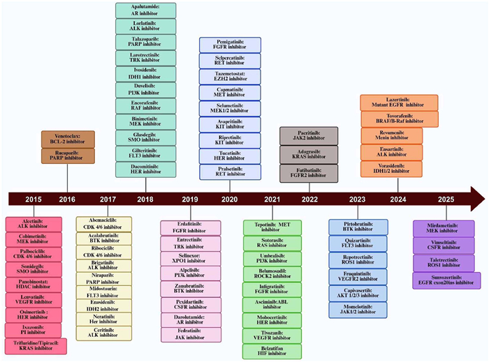
Figure 1.
FDA approval of new oral chemotherapeutics between 2015 and 2025.

Oral anticancer drug delivery: Strategies for improving bioavailability
Sinapi Umutoni , Hao Zhang , Jiayue Guo , Zehui Yu , Hao Shao , Xiaoli Yi , Chunmeng Sun , Lei Luo , Yinglan Yu


The global cancer burden has escalated significantly, with approximately 20 million new cases and 9.7 million fatalities reported in 2022, reflecting a lifetime cancer incidence rate of 20%. Projections indicate an alarming 77% increase in 2050, reaching 35 million [1,2]. While intravenous (i.v.) chemotherapy has long been the cornerstone of cancer treatment, it is associated with significant drawbacks, including patient discomfort, poor compliance, and severe local adverse reactions, such as edema, hemorrhage, and tissue necrosis. These limitations underscore the urgent need for alternative drug delivery modalities that can enhance patient outcomes and quality of life. Oral administration stands out as one of the most widely used approaches, offering distinct advantages over parenteral routes. Additionally, oral chemotherapy avoids injection-related discomfort and reduces the infection risks associated with i.v. administration. The ability to self-administer at home can reduce healthcare costs and enhance patient autonomy, and improve quality of life, particularly for patients with advanced-stage cancers who may not tolerate aggressive treatments [3].
Despite these advantages, the clinical translation of oral anticancer drugs remains hindered by low and variable bioavailability. The gastrointestinal tract (GIT) presents a complex barrier system, where dynamic pH gradients, proteolytic enzymes, mucus layers, intestinal epithelium, and the gut microbiota collectively restrict drug stability and absorption. These challenges necessitate the development of innovative strategies to enhance drug stability and permeation. Overcoming these obstacles requires innovative formulation approaches designed to enhance solubility, stability, and permeability. Recent advances, including pH modifiers, enzyme inhibitors, permeation enhancers, prodrug design, and nanoparticle (NP) engineering, have significantly enhanced the oral bioavailability of various anticancer drugs [4,5]. Notably, the U.S. Food and Drug Administration (FDA) approval pipeline for oral anticancer drugs has expanded steadily, with approximately 75 small molecules and novel agents gaining market authorization between 2015 and 2025 (compiled from Drug@FDA, Fig. 1). This review provides a critical and comprehensive analysis of the current landscape of oral anticancer drug delivery, systematically evaluating physiological and biological barriers and highlighting novel strategies to enhance bioavailability. By exploring the potential of artificial intelligence (AI) and machine learning (ML) in optimizing drug design, this work offers new insights into overcoming longstanding challenges, positioning oral anticancer therapies as a cornerstone of next-generation oncology treatments.
Despite the convenience of oral administration, the GIT imposes complex physicochemical and physiological barriers that significantly restrict the effective absorption of anticancer drugs [6]. The absorption process initiates with the drug’s release from its dosage form, a step that must be followed by maintaining chemical stability within the GIT environment to prevent degradation. This stability enables adequate dissolution in intestinal fluids, thereby ensuring sufficient solubility to drive the concentration available for uptake. The final critical phase involves permeation across the intestinal epithelium, where insufficient solubility or limited permeability markedly restricts systemic exposure.
The GIT presents a series of physiological barriers, as illustrated in Fig. 2 that limit oral absorption, encompassing variable pH conditions, digestive enzymes, the mucus layer, the intestinal epithelium, the presence of efflux pumps, and gut microbiota. Consequently, a drug must successfully navigate these obstacles to reach the systemic circulation, as they collectively determine the extent of absorption and bioavailability. Therefore, a thorough understanding of the structural and biological characteristics of the GIT is essential for developing strategies to enhance the oral bioavailability of anticancer agents. This section provides an overview of these physiological barriers and their impact on oral drug delivery.
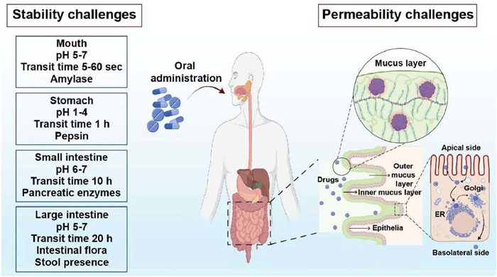
Approximately 75% of novel drug candidates, particularly anticancer agents, exhibit poor water solubility, a challenge also seen in nearly 65% of approved oral anticancer drugs, which substantially limits their therapeutic efficacy [7]. Gastric pH and buffer capacity vary depending on nutritional state, ranging from pH 1.0–2.5 under fasting conditions to pH 4.3–5.4 in the fed state, with corresponding buffer capacities of 7–19 and 14–28 mmol L−1 ΔpH−1, respectively. Moreover, physiological and pathological factors such as concomitant medications or disease states can further alter gastric pH, influencing the dissolution behavior of orally administered drugs [8]. While acidic drugs generally dissolve better at lower pH, many orally administered chemotherapeutic agents, particularly tyrosine kinase inhibitors such as erlotinib, gefitinib, lapatinib, and dasatinib, are weakly basic compounds with pH-dependent solubility. These drugs are typically more soluble in the acidic environment of the stomach, but their solubility declines significantly at higher pH levels found in the small intestine. When taken with acid-suppressing drugs, like proton pump inhibitors, histamine-2 receptor antagonists, or antacids, gastric pH increases, which can reduce the dissolution and absorption of these drugs. In healthy subjects, administering famotidine 2 h before dasatinib reduced its area under the curve (AUC) by approximately 60%. Additionally, co-administration with an aluminum/magnesium-based antacid reduced AUC by 55%-58% [9,10]. A phase I crossover trial also showed that sustained elevation of gastric pH (above 5) using ranitidine led to a 50% decrease in gefitinib’s AUC and a 71% reduction in peak blood concentration (Cmax) [11]. The simultaneous administration of omeprazole with erlotinib significantly reduced drug exposure. Specifically, omeprazole decreased erlotinib’s AUC by 46% and its Cmax by 61% [12].
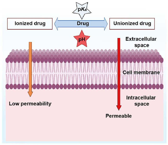
The pH of the GIT also directly affects how a drug is released from its formulated capsule or tablet. The coatings designed to control drug release may disintegrate at specific pH levels. An abnormal shift in gastric or intestinal pH may cause the drug to be released too early or too late, compromising its effectiveness. Additionally, for chemically unstable drugs prone to acid degradation, a higher stomach pH can enhance their survival and bioavailability, while a lower pH can lead to increased breakdown. Most drugs are absorbed through the intestinal membrane by passive diffusion, and the ionic state of the dissolved molecules significantly influences this process. When pH exceeds pKa, weak bases mostly exist in their unionized form, while weak acids are mostly ionized; the opposite occurs when pH is below pKa. As a result, shifts in luminal pH, whether due to physiological factors or drug effects (such as after a meal or with acid-reducing therapy), can alter the unionized fraction at the absorption site. This is explained by the pH-partition hypothesis, as illustrated in Fig. 3, which states that unionized drug species are more lipophilic and tend to penetrate membranes better [13,14]. Therefore, pH alterations can directly affect the rate and extent of drug absorption by affecting the proportion of the drug available for uptake, especially for drugs with inherently poor absorption. Consequently, the pH variations within the GIT play a key role in determining the oral bioavailability of many drugs, mainly through their effects on solubility, dissolution, stability, release from the dosage form, and final permeability across the intestinal membrane.
In addition to pH-related barriers, digestive enzymes in the GIT significantly limit the effectiveness of oral delivery of anticancer agents. Key enzymes responsible for drug degradation include proteolytic enzymes secreted into the GI lumen through gastric and pancreatic fluids, and mucosal enzymes. However, most enzymatic breakdown occurs in the lumen before the drugs can penetrate the mucus layer. The presence of a drug in the stomach may stimulate the release of pepsin from the gastric mucosa. In the upper small intestine, the pancreas secretes large amounts of trypsin, chymotrypsin, carboxypeptidase, and elastase [15]. These enzymes are particularly problematic for chemotherapeutic agents containing ester or amide bonds, which are highly susceptible to enzymatic hydrolysis. For instance, protein-based immunotherapies, including checkpoint inhibitor fragments, are easily degraded by digestive enzymes, rendering them inactive before absorption [16]. In the case of 5-fluorouracil and its prodrug tegafur (used in the S-1 regimen), enzymatic inactivation by dihydropyrimidine dehydrogenase in the gut wall significantly reduces bioavailability [17]. Additionally, etoposide shows instability in simulated gastric and intestinal fluids, likely due to enzymatic action, leading to variable and incomplete oral absorption, resulting in reduced bioavailability and inconsistent systemic exposure [18].
The intestinal mucus layer serves as the initial barrier that orally administered anticancer drugs must cross before reaching the epithelial cells involved in absorption. This viscoelastic and adhesive secretion is generated and released by specialized goblet and mucous cells lining the intestine. The mucus layer is essential for protecting epithelial cells, as it lubricates their surfaces, traps external particles, binds pathogens, and provides a physical barrier against harmful substances. This mixture comprises carbohydrates, proteins, lipids, antibodies, salts, and cellular residues. Goblet cells are the principal source of intestinal mucus secretion, and mucins are the primary protein components that fulfill various critical functions. Mucins serve as lubricants and offer protection against infection. The mucus layer also regulates the diffusion of nutrients and drugs, facilitates material transport, and maintains hydration and homeostasis in the gut environment. The mucus layer thickness varies across the GIT, increasing from ∼200 µm in the upper region to 800 µm in the lower intestine [19]. Additionally, the mucus restricts the passage of larger compounds, generally limiting diffusion to molecules with a molecular weight of 600–800 Da or less [20]. This dense and dynamic barrier poses a considerable challenge for oral anticancer drug delivery, as it restricts drug diffusion, decreases permeability, and potentially retains drug molecules before they reach the targeted sites. Anticancer drugs, especially peptide-based immunotherapies and macromolecular agents, face several challenges within the mucus layer.
(1) The mucin network comprises tightly intertwined hydrophilic and hydrophobic domains that are highly organized. These create a mesh-like structure that restricts the diffusion of hydrophobic or charged drug molecules through the intestinal mucus.
(2) Weak electrostatic or hydrophobic interactions between drugs and mucin can trap molecules on the mucus surface, further impeding transit [21]. Addressing this barrier is essential to enhancing the absorption and efficacy of oral anticancer agents.
(2) Weak electrostatic or hydrophobic interactions between drugs and mucin can trap molecules on the mucus surface, further impeding transit [21]. Addressing this barrier is essential to enhancing the absorption and efficacy of oral anticancer agents.
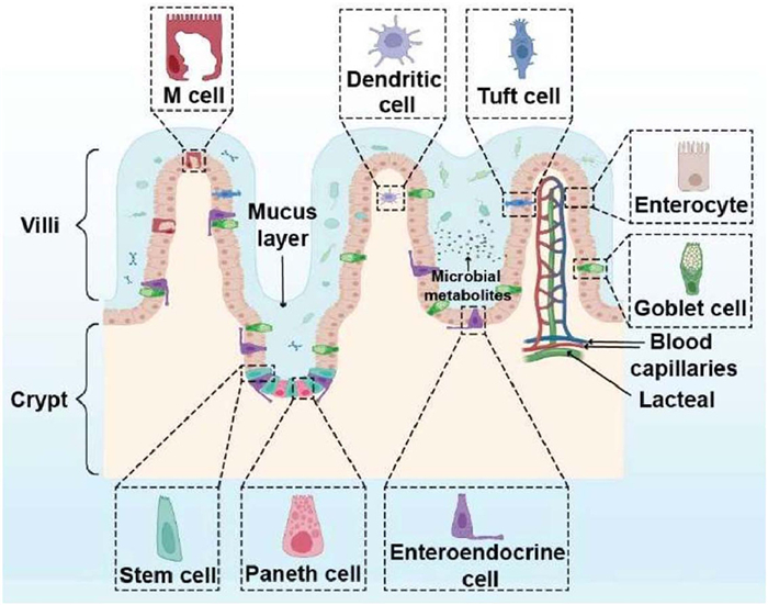
The gastrointestinal epithelium, located beneath the intestinal mucus layer, forms a continuous phospholipid bilayer that acts as a selective barrier between the external environment and the body’s internal milieu. This lining consists of a single layer of intestinal epithelial cells, as illustrated in Figs. 4 and 5, are interconnected at their apical lateral borders by tight junctions (TJs). These specialized complexes not only separate the apical and basolateral membrane domains (the fence role) but also regulate the passage of substances through the intercellular space (the gate role). As such, they serve as the first line of defense against microbial invasion, uncontrolled diffusion of luminal contents, and the entry of large hydrophilic molecules. Structurally, TJs are formed by transmembrane proteins that span the narrow space between cells, with occludin, members of the claudin family, and junctional adhesion molecules serving as the primary components. Supporting these, cytoplasmic scaffold proteins such as the zonula occludens (ZO-1, ZO-2, ZO-3) anchor the TJs complex to the actomyosin cytoskeleton, imparting both structural stability and the capacity for dynamic regulation of junctional tension and permeability. Collectively, this highly organized protein architecture ensures that the intestinal epithelium maintains barrier integrity while remaining adaptable to physiologicfal and pathological stimuli [22,23].
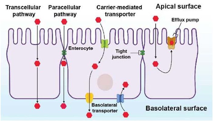
Drug permeability across this barrier is primarily dictated by its physicochemical properties, which determine the preferred route of transport. Hydrophobic molecules that can partition into the lipid-rich membrane typically cross the epithelium through the transcellular pathway, entering via the apical surface and exiting through the basolateral side into systemic circulation. However, successful transcellular passage requires a fine balance between lipophilicity and aqueous solubility; excessive hydrophobicity restricts dissolution in intestinal fluids, while excessive hydrophilicity prevents membrane penetration. This inherent trade-off presents a major challenge for many anticancer drugs and often necessitates innovative formulation strategies to optimize solubility and permeability for improved oral absorption. For hydrophilic molecules, the paracellular pathway offers an alternative route. Here, drugs move between adjacent cells through the spaces governed by tight junctions. Yet, this pathway is severely limited: Only very small molecules with a hydrodynamic radius below ∼11 Å can diffuse effectively, and the tight junctions act as dynamic gates that tightly restrict the entry of most therapeutic agents [24]. Consequently, while the transcellular route is often limited by solubility, permeability trade-offs, the paracellular route is constrained by stringent junctional control, creating a dual dilemma for oral drug delivery.
(1) Metabolic enzymes: The intestinal epithelium contains drug-metabolizing enzymes, most notably the cytochrome P450 (CYP) family, which is key to the first-pass metabolism of many orally administered anticancer agents. Among these, CYP3A4 is the most prevalent, abundantly expressed in both the small intestine and liver, which are the main sites of drug oxidation and clearance. It accounts for roughly 30% of the total hepatic P450 enzymes and about 80% of those in the small intestine. Most approved anticancer drugs, including taxanes, topoisomerase inhibitors, tyrosine kinase inhibitors, and vinca alkaloids, are CYP3A4 substrates. As a result, extensive metabolism by CYP3A4 often reduces the amount of drug that reaches the systemic circulation [25]. This enzymatic activity occurs within the enterocytes, the same absorptive cells that also express transport proteins, highlighting the coordinated role of efflux and metabolism in limiting oral drug bioavailability [26]. The interaction between CYP3A4 and P-glycoprotein (P-gp) is particularly important; this effect is largely due to their close localization within enterocytes, where P-gp is positioned on the apical membrane, and CYP3A4 resides in the endoplasmic reticulum [27]. This dual barrier can be modulated using inhibitors. Therefore, understanding and regulating both metabolic enzymes and efflux transporters is crucial for optimizing oral delivery strategies for anticancer drugs.
(2) Efflux transporters: P-gp is an ATP-dependent efflux transporter predominantly localized at the apical brush border of enterocytes lining the intestinal villus tips. Its expression increases progressively from the stomach toward the colon, and it is also present on the apical membranes of hepatocytes, renal proximal tubule cells, and at the blood-brain barrier. Structurally, P-gp is a glycosylated membrane protein of approximately 170 kDa, and it harnesses the energy released by hydrolyzing two molecules of ATP to drive the active extrusion of substrates across the cell membrane [28]. Molecular recognition by this pump is broad but commonly favors compounds that contain hydrophobic aromatic regions and tertiary amine groups. P-gp reduces intracellular exposure by expelling xenobiotics from enterocytes back into the intestinal lumen, and thereby facilitates repeated cycles of presystemic metabolism. Physiologically, this activity protects tissues from potentially harmful compounds, but clinically, it can limit oral drug absorption and promote multidrug resistance [29]. Many anticancer agents are substrates for P-gp. Examples include various antimetabolites (e.g., methotrexate, cytarabine, 5-fluorouracil), anthracyclines (doxorubicin, daunorubicin), camptothecins (topotecan, irinotecan), actinomycin D, mitomycin C, mitoxantrone, Vinca alkaloids (vinblastine, vincristine), taxanes (paclitaxel, docetaxel), selected tyrosine kinase inhibitors (imatinib, gefitinib), and certain alkylating agents (chlorambucil, cisplatin). Because of its wide substrate spectrum and regional variability in expression, P-gp represents a major determinant of intestinal drug disposition and a key barrier to effective oral delivery of many chemotherapeutics. Studies suggest that blocking or downregulating the activity of these intestinal transporters can lead to better absorption of orally administered drugs into the bloodstream. For example, a single drug can simultaneously suppress or activate multiple drug transporters. Understanding and regulating these transporters may enhance drug delivery strategies and improve therapeutic outcomes [30,31].
(2) Efflux transporters: P-gp is an ATP-dependent efflux transporter predominantly localized at the apical brush border of enterocytes lining the intestinal villus tips. Its expression increases progressively from the stomach toward the colon, and it is also present on the apical membranes of hepatocytes, renal proximal tubule cells, and at the blood-brain barrier. Structurally, P-gp is a glycosylated membrane protein of approximately 170 kDa, and it harnesses the energy released by hydrolyzing two molecules of ATP to drive the active extrusion of substrates across the cell membrane [28]. Molecular recognition by this pump is broad but commonly favors compounds that contain hydrophobic aromatic regions and tertiary amine groups. P-gp reduces intracellular exposure by expelling xenobiotics from enterocytes back into the intestinal lumen, and thereby facilitates repeated cycles of presystemic metabolism. Physiologically, this activity protects tissues from potentially harmful compounds, but clinically, it can limit oral drug absorption and promote multidrug resistance [29]. Many anticancer agents are substrates for P-gp. Examples include various antimetabolites (e.g., methotrexate, cytarabine, 5-fluorouracil), anthracyclines (doxorubicin, daunorubicin), camptothecins (topotecan, irinotecan), actinomycin D, mitomycin C, mitoxantrone, Vinca alkaloids (vinblastine, vincristine), taxanes (paclitaxel, docetaxel), selected tyrosine kinase inhibitors (imatinib, gefitinib), and certain alkylating agents (chlorambucil, cisplatin). Because of its wide substrate spectrum and regional variability in expression, P-gp represents a major determinant of intestinal drug disposition and a key barrier to effective oral delivery of many chemotherapeutics. Studies suggest that blocking or downregulating the activity of these intestinal transporters can lead to better absorption of orally administered drugs into the bloodstream. For example, a single drug can simultaneously suppress or activate multiple drug transporters. Understanding and regulating these transporters may enhance drug delivery strategies and improve therapeutic outcomes [30,31].
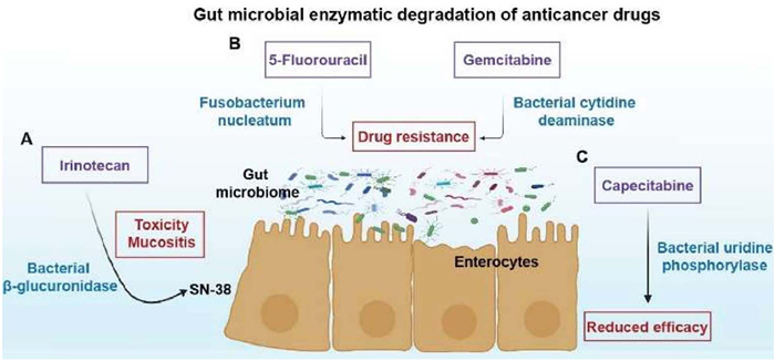
The gut microbiota, comprising bacteria, archaea, and eukaryotes, represents a complex ecosystem with an estimated population exceeding 100 trillion microorganisms. Its involvement in bacterial drug interaction has been widely acknowledged [32,33]. The primary phyla in the human gut microbiota include Proteobacteria, Bacteroidetes, Firmicutes, and Actinobacteria, with microbial density and diversity increasing distally along the GIT. In the small intestine, microbial abundance is relatively low due to factors such as short transit time, low pH, high antimicrobial concentrations, and elevated oxygen tension. By contrast, the ileum and colon harbor the most abundant and diverse microbial populations. A major challenge arises from the ability of the gut microbiota to affect drug metabolism and transport. Gut bacteria can directly or indirectly modify chemotherapeutic drugs via enzymatic degradation, biotransformation, and immune modulation. These microbial-mediated transformations may increase or reduce drug activity or generate toxic byproducts [34]. Given this substantial influence, the gut microbiota plays a pivotal role in shaping the absorption and metabolism of oral chemotherapeutics, ultimately altering both the efficacy and toxicity of these drugs [35]. This influence extends to the pharmacokinetics of anticancer agents, many of which undergo extensive first-pass metabolism. Moreover, alterations in the microbiota can affect drug transport mechanisms through substrate competition for transporters or changes in transporter expression, both of which may further decrease the absorption of anticancer drugs. Therefore, it is essential to account for microbiota dynamics when evaluating oral antineoplastic drug administration. Below, we provide specific examples of anticancer drugs whose pharmacological outcomes are strongly affected by interactions with the gut microbiota (Fig. 6).
Despite these challenges, various methods have been developed to enhance the effectiveness and minimize the toxicity of oral anticancer drugs. Recent advancements in addressing the challenges to oral drug delivery include the use of absorption enhancers, prodrugs, and nanocarriers. These novel methods facilitate the effective transport of antineoplastic agents into the bloodstream and to site-specific targets, thereby improving their bioavailability. The development of these systems requires a thorough knowledge of drug characteristics and physiological behavior. New targeting approaches have been developed to direct drugs selectively to tumor sites, thereby reducing side effects. This section provides a summary of recent progress in oral chemotherapy, highlighting major innovations that enhance absorption, bioavailability, and therapeutic efficacy.
The key obstacle to the oral delivery of anticancer agents is their pH-dependent solubility, which often limits their dissolution and absorption in the GIT. To address this issue, several formulation-based strategies have been developed to modify the local pH environment and optimize drug release. These approaches can be broadly categorized into three techniques:
(1) Microenvironmental pH modification: This strategy involves the incorporation of pH-modifying agents to alter the microenvironment around drug particles, thereby optimizing dissolution and absorption. In solid dosage forms, such as tablets or capsules, these pH-adjusting excipients are uniformly distributed within the formulation. Upon disintegration in the GIT, they rapidly dissolve and modify the pH of the diffusion layer adjacent to drug particles. For instance, organic acids such as citric, fumaric, and tartaric acids are often used to enhance the solubility of weakly basic anticancer agents, such as tyrosine kinase inhibitors. Pu et al. [36] evaluated enhanced oral formulations of a poorly water-soluble, weakly basic drug by incorporating citric acid (5%) and sodium lauryl sulfate, which significantly improved dissolution consistency across various pH conditions compared to conventional formulations. Conversely, alkalizing agents such as sodium bicarbonate are employed to facilitate the dissolution of weakly acidic drugs, including methotrexate and topotecan, which shift the microenvironment toward a more basic pH.
(2) Use of pH modifiers: In addition to traditional acids and bases, specialized pH modifiers, such as betaine chloride or fumaric acid, are incorporated into formulations to optimize drug solubility under variable gastric pH conditions. The choice of modifier depends on the physicochemical properties of the drug, including its solubility and pKa, as well as its interaction with the modifier. This precise tailoring ensures that drugs maintain favorable dissolution profiles even in altered gastric environments. Allu et al. [37] reported the use of pH modifiers such as methane sulfonic acid, benzene sulfonic acid, ethane sulfonic acid, and adipic acid to form salts or cocrystals of palbociclib in the less acidic intestinal environment (higher pH), where weakly basic drugs tend to precipitate, increased the palbociclib solubility compared to the pure drug.
(2) Use of pH modifiers: In addition to traditional acids and bases, specialized pH modifiers, such as betaine chloride or fumaric acid, are incorporated into formulations to optimize drug solubility under variable gastric pH conditions. The choice of modifier depends on the physicochemical properties of the drug, including its solubility and pKa, as well as its interaction with the modifier. This precise tailoring ensures that drugs maintain favorable dissolution profiles even in altered gastric environments. Allu et al. [37] reported the use of pH modifiers such as methane sulfonic acid, benzene sulfonic acid, ethane sulfonic acid, and adipic acid to form salts or cocrystals of palbociclib in the less acidic intestinal environment (higher pH), where weakly basic drugs tend to precipitate, increased the palbociclib solubility compared to the pure drug.
(3) pH-responsive drug delivery systems: More advanced strategies involve the use of pH-sensitive carriers or nano prodrugs that dynamically respond to pH changes in the GIT. These systems can alter their structures, such as transitioning from larger aggregates to smaller NPs, when exposed to pH variations. This enhances not only absorption in the gut but also drug accumulation in tumors, leading to improved therapeutic efficacy and reduced systemic toxicity [38]. For example, Rehman et al. [39] developed a smart pH-responsive hydroxypropyl-β-cyclodextrin (HP-β-CD)/agarose-g-poly hydrogel for oral capecitabine delivery. The system showed high drug loading (71%−90%) and strong pH responsiveness, with 95% swelling and 94% drug release at pH 7.4 compared to pH 1.2.
(3) pH-responsive drug delivery systems: More advanced strategies involve the use of pH-sensitive carriers or nano prodrugs that dynamically respond to pH changes in the GIT. These systems can alter their structures, such as transitioning from larger aggregates to smaller NPs, when exposed to pH variations. This enhances not only absorption in the gut but also drug accumulation in tumors, leading to improved therapeutic efficacy and reduced systemic toxicity [38]. For example, Rehman et al. [39] developed a smart pH-responsive hydroxypropyl-β-cyclodextrin (HP-β-CD)/agarose-g-poly hydrogel for oral capecitabine delivery. The system showed high drug loading (71%−90%) and strong pH responsiveness, with 95% swelling and 94% drug release at pH 7.4 compared to pH 1.2.
Intestinal CYP3A4 enzyme plays a crucial role in drug metabolism, influencing the bioavailability and effectiveness of many orally administered drugs. Inhibiting this enzyme can raise drug levels, leading to stronger therapeutic effects [40]. A well-known example is grapefruit juice, which selectively blocks intestinal CYP3A4 and has been key in demonstrating the significance of first-pass intestinal metabolism [41]. However, differences in CYP3A4 expression and activity across individuals cause unpredictable oral bioavailability. This issue might be addressed by selectively inhibiting intestinal CYP3A4 while sparing hepatic CYP3A4 to preserve normal drug clearance. Inhibitors such as ketoconazole, ritonavir, and cyclosporin A have been extensively studied for their ability to reduce presystemic metabolism and improve the absorption of anticancer drugs, including taxanes and tyrosine kinase inhibitors [42-44]. For instance, cyclosporin A, a potent dual inhibitor of CYP3A4 and P-gp, has been shown to increase the oral bioavailability of paclitaxel 9-fold compared to when it is administered alone [45].
Natural bioenhancers, especially phytochemicals, including curcumin, piperine, quercetin, and resveratrol, are increasingly being investigated as safer alternatives and have demonstrated the ability to suppress CYP3A4 activity, with some also targeting other metabolic pathways [46]. In rat studies, curcumin inhibited CYP3A4-mediated metabolism of docetaxel, resulting in a 1.86-fold increase in AUC and terminal half-life 1.55-fold [47]. Piperine, derived from black pepper, inhibits CYP3A4, which significantly enhances the pharmacokinetics of docetaxel, leading to increased serum concentration and prolonged half-life [48]. These dual actions position natural inhibitors as promising candidates for enhancing the efficacy of oral chemotherapy while potentially minimizing toxicity. While synthetic and natural enzyme inhibitors offer significant promise, their clinical application requires caution. Factors such as drug-drug interactions, patient variability in enzyme expression, and safety must be evaluated carefully. Ultimately, combining enzyme inhibition with complementary strategies, such as transporter modulation or advanced formulations, may provide the most effective pathway for improving oral anticancer drug delivery.
Prodrugs are inactive compounds that undergo metabolic transformation within the body to become pharmacologically active agents. Despite their minimal therapeutic effects in their original state, their structures facilitate their conversion into active drugs after administration. This transformation may occur via chemical changes or enzymatic activity. The conversion often entails the removal of a molecular component added to cover up the drug or enhance its characteristics, or it may involve inducing a structural alteration that activates the drug's therapeutic function [49], which offers a way to alter the drug's characteristics both physically and chemically to enhance their absorption, distribution, metabolism, and excretion while minimizing their toxic effects [50]. A prodrug is usually associated with a covalent bond between an active drug molecule and a chemical moiety. The immediate purpose of prodrugs is to produce novel substances with high efficacy, selectivity, and low toxicity. The timing and location of active drug release depend on its intended use, and the active compound must be released before, during, or after absorption, or in a specified target tissue or organ. The main goals of prodrug design in the context of oral administration of antineoplastics are to increase absorption in the GIT by making drugs more water-soluble and enhancing their capacity to penetrate the cell membranes. Drugs are typically designed to dissolve faster, have less polarity (by covering charged groups), and interact better with transport proteins.
A common method involves the chemical modification of active pharmaceutical compounds by adding phosphate or sulfate groups to enhance their solubility. The incorporation of ester groups may enhance membrane permeability. Phosphate-based prodrugs are generally activated in the intestinal wall by enzymes, such as alkaline phosphatases. Sulfate-based prodrugs exhibit greater resistance to degradation by sulfatases; however, they can be activated by intestinal esterase [51]. Etoposide phosphate, an etoposide prodrug, exemplifies improved solubility, exhibiting 20 mg/mL in contrast to 0.1 mg/mL of the parent compound. This formulation, presented in a gelatin capsule, demonstrated a 4-fold enhancement in oral bioavailability while minimizing the requirement for toxic co-solvents. However, clinical trials have demonstrated that it does not significantly decrease variability in patient response [52]. Several oral prodrugs of fluoropyrimidines are clinically established, such as capecitabine (Xeroda®), an oral prodrug of 5-fluorouracil that undergoes enzymatic conversion after intestinal absorption, leading to preferential activation within tumors and reduced systemic toxicity. It is FDA-approved for metastatic colorectal cancer and as monotherapy in metastatic breast cancer resistant to anthracyclines or taxanes. Beyond these indications, capecitabine has shown efficacy as both monotherapy and in combination regimens across a wide range of cancers, including gastric, pancreatic, biliary tract, renal, prostate, and head and neck cancers [53]. Tegafur, a prodrug of 5-fluorouracil, is metabolized to active 5-fluorouracil and used broadly in formulations such as S-1 (tegafur/gimeracil/oteracil) and UFT (tegafur/uracil) [54]. It is orally administered across multiple cancer types. Similarly, trifluridine/tipiracil (TAS-102; Lonsurf®), which combines the nucleoside analog trifluridine with the thymidine phosphorylase inhibitor tipiracil, significantly increases trifluridine bioavailability by inhibiting its degradation [55].
Transmembrane drug efflux driven by P-gp is a significant obstacle to oral drug absorption; however, this challenge can be addressed by co-administering P-gp modulators or inhibitors. Several mechanisms underlie P-gp inhibition, including blocking the drug-binding site (competitively, non-competitively, or allosterically), interfering with adenosine triphosphate (ATP) hydrolysis, altering membrane integrity, reducing P-gp expression, depleting ATP, and disrupting ATP binding sites [56]. By selectively blocking P-gp activity, these strategies aim to enhance oral bioavailability, improve membrane uptake, and increase the therapeutic effectiveness of drugs, particularly in cancer therapy. P-gp inhibitors are structurally diverse and have been classified into three generations [26]. First-generation inhibitors are pharmacologically active drugs, such as verapamil and cyclosporin A, that incidentally block P-gp. However, their use is limited by high serum concentrations, off-target effects, and unpredictable pharmacokinetic interactions. Second-generation inhibitors, including non-immunosuppressive cyclosporin analogs (PSC833) and dexverapamil, were designed to have stronger P-gp binding with reduced pharmacological activity. However, they interfere with CYP3A4 and other transporters, leading to complex metabolic interactions. Third-generation inhibitors, such as tariquidar, have been developed using structure activity relationship studies to achieve high specificity, minimal toxicity, and improved clinical efficacy. Collectively, these advances highlight the potential of P-gp inhibition as an approach to overcome drug efflux and enhance oral drug delivery [57].
Recent studies have demonstrated that natural compounds, such as curcumin, can act as effective P-gp inhibitors and improve the pharmacokinetic profiles of poorly absorbed anticancer drugs. In a murine model, oral paclitaxel (35 mg/kg) showed poor absorption, with a bioavailability of 2.8%. However, when pretreated with curcumin (100 mg kg−1 day−1 for 4 days), the AUC of paclitaxel nearly doubled (3.6–5.8 µg h mL−1), Cmax increased, and absolute bioavailability improved to 4.5% [46]. These findings are consistent with using synthetic inhibitors; for instance, in vivo oral coadministration of elacridar and taxanes increased their plasma concentration [58]. These studies demonstrate that modulating intestinal efflux, whether through natural bioenhancers or synthetic inhibitors, represents a promising strategy to overcome the oral absorption barriers of anticancer drugs.
Functional excipients have advanced pharmaceutical development by evolving from conventional inert roles to actively optimizing drug delivery, particularly in improving the oral bioavailability of anticancer drugs challenged by issues of solubility and permeability. Absorption enhancers, as a key category of these excipients, promote drug absorption by facilitating passage through cell membranes independent of solubility enhancement [59], and are particularly important for hydrophilic or high-molecular-weight anticancer drugs, whose polarity limits transcellular diffusion and directs them to the restricted paracellular pathway, which is constrained by the limited surface area and narrow pores of tight junctions [60]. These enhancers temporarily modify epithelial structure to increase permeability without permanent damage, either by expanding tight junctions to support paracellular transport or by enhancing transcellular transport through drug complex formation, inhibition of enzymatic degradation, and mild disruption of membrane integrity. This approach originated in a 1961 study demonstrating that EDTA improved the oral absorption of heparin in dogs, leading to extensive research that has evaluated >250 compounds in preclinical studies, including chelating agents, surfactants, fatty acids, polymers, and bacterial toxins, each operating via specific mechanisms to increase intestinal permeability [61]. Other functional excipients complement these efforts by addressing solubility limitations, such as alkalizing agents, co-solvents, and surfactants (e.g., sodium lauryl sulfate, polysorbate) [62,63].
Recent efforts emphasize co-processed or multi-component excipient blends in tablets and capsules, in which excipients work in combination to address multiple barriers including low solubility Biopharmaceutics Classification System (BCS) Class Ⅱ/Ⅳ drugs, pH-dependent degradation, and efflux-mediated poor absorption, without relying on complex delivery vehicles like NPs.
Polysorbate 80 and cyclodextrin combinations for oral paclitaxel. Shanmugam et al. conducted a study in beagle dogs, where polysorbate 80 (0.5%−2%, w/v) was paired with β-cyclodextrin in solid dispersions embedded within starch-based matrices. The surfactant reduced interfacial tension to boost wetting and micelle formation, while cyclodextrin formed inclusion complexes that amorphized paclitaxel, increasing solubility by 15–20 fold. Mechanistically, this duo inhibited P-gp efflux at the intestinal epithelium, extending absorption windows. Synergy was evident in pharmacokinetic data, with combined use yielding 2.5-fold higher AUC compared to single-excipient variants, attributed to polysorbate permeation aid enhancing cyclodextrin solubilizing effect [64]. pH-adjusting excipients in matrix systems. Dharshini et al. employed calcium carbonate (5%−10%) integrated with sodium carbonate and polymeric carriers (PVP) in controlled-release tablets for pH-sensitive dasatinib, thereby regulating the microenvironmental pH from 1.2 (gastric) to 6.8 (intestinal), preventing acid-catalyzed degradation and increasing bioavailability by 30%. The carbonates buffered dissolution, while PVP stabilized the amorphous state; safety was affirmed through in vitro cytotoxicity assays showing no excipient-induced GIT irritation. This integrative design revealed synergistic pH-triggered release, where carbonate effervescence accelerated polymer erosion for burst-then-sustained profiles [65]. Functional excipients and absorption enhancers can greatly increase oral exposure of anticancer drugs, but their safety may pose concerns. Surfactants, bile-salt mimetics, co-solvents, and permeation enhancers may cause epithelial irritation, temporary barrier disruption, or microbiota changes, particularly with chronic use or high doses. Some synthetic polymers and complexing agents can also provoke hypersensitivity or cumulative toxicity. Thus, excipients should be treated as active formulation components whose local and systemic safety, reversibility, and long-term effects on intestinal barrier integrity require rigorous evaluation in line with regulatory guidance [66,67].
ILs are organic salts that are liquid at or near room temperature and can be tailored by pairing different cations and anions. Their unique combination of structural tunability, negligible volatility, and excellent solvating power has led to increasing interest in pharmaceutical applications [68]. In oral delivery, IL can act as powerful cosolvents, permeation enhancers, or be integrated directly into the active pharmaceutical ingredient as so-called active pharmaceutical ingredient ILs (API‑IL), in which the drug itself forms one of the charged species. IL‑based strategies have been explored to overcome the low solubility and variable absorption of several anticancer agents [69]. For instance, methotrexate has been converted into a series of API‑IL by pairing the anionic drug with biocompatible cations. Among the five ILs evaluated, proline ethyl ester methotrexate exhibited a 4.6-fold increase in oral bioavailability compared with sodium methotrexate, along with significant improvements in Cmax and AUC [70]. Despite these advantages, the clinical translation of IL-based formulations faces several challenges. Conventional IL may exhibit cytotoxicity or environmental persistence, motivating the development of bio‑ILs constructed from endogenous or biodegradable ions. In addition, comprehensive toxicological evaluation, control of residual solvents, and the establishment of regulatory standards for IL‑containing products will be essential before such systems can be widely adopted.
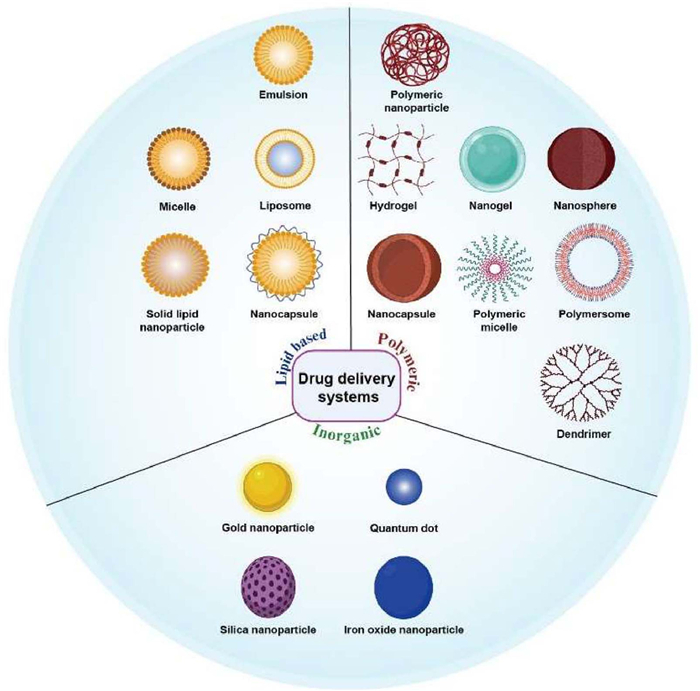
According to the National Nanotechnology Initiative, NPs are defined as structures possessing a size within the range of 1–100 nm in at least one dimension. However, in practice, the term often includes particles of up to several hundred nanometers in size. Nanomedicine was first introduced clinically in 1995 with the approval of Doxil®, a liposomal formulation of doxorubicin for the treatment of breast cancer, Kaposi’s sarcoma, and ovarian cancer, marking a substantial advancement in cancer treatment [71]. The translation of oral nanomedicine from laboratory research to clinical applications remains a considerable challenge, primarily due to formulation stability and regulatory constraints. These structures, which are generally created via top-down or bottom-up techniques, provide specific benefits owing to their small size and unique characteristics. These materials are frequently biocompatible, biodegradable, and stable in colloidal suspensions and can encapsulate water soluble or fat-soluble drugs [72]. Nanomedicine offers numerous advantages for drug delivery, including protecting drugs against premature degradation, enhancing their bioavailability, prolonging their circulation time within the body, enabling drugs to reach specific tissues or cells, and mitigate their side effects. Nanocarriers include liposomes, solid lipid NPs (SLN), micelles, dendrimers, polymers, silicon- or carbon-based particles, magnetic NPs, and exosomes (Fig. 7). The efficacy of these nanocarriers is contingent on their structural design and drug incorporation method. Anticancer drugs can be incorporated into NPs by dissolving, adsorbing, entrapping, or attaching to the carrier material [73].
The intestinal mucus layer coating the epithelial cells presents the first obstacle for nanomedicine to reach the intestinal villous cells. Scientists face challenges in developing orally delivered NPs because these particles must possess opposing surface properties to overcome different barriers. They should be hydrophilic and neutral to easily pass through mucus, but hydrophobic or positively charged for efficient absorption by intestinal cells. A promising approach involves using NPs engineered to change their surface properties after passing through the mucus layer [74]. For an orally administered anticancer nanomedicine to be effective, it must successfully overcome the GIT physiological and biochemical barriers, subsequently, during its journey through the bloodstream, to reach the tumor site. Recent research has established that for a nanomedicine to effectively penetrate the thick intestinal mucus, its surface must be nonfouling, that is, it must resist binding to biomacromolecules. A common strategy to achieve this is coating NPs with dense hydrophilic polymers, such as polyethylene glycol (PEG). These PEGylated particles can move rapidly through mucus. However, this nonfouling property also causes them to go undetected by cells. Consequently, even when they reach the intestinal villi, they are poorly internalized, presenting a significant challenge for drug delivery [75]. A recent study demonstrated that a zwitterionic betaine polymer can avoid nonspecific protein adsorption while selectively interacting with the proton-assisted amino acid transporter 1, which is highly expressed on intestinal epithelial cells. This dual behavior enables the material to move effectively through the mucus layer and achieve high transepithelial uptake for oral drug delivery. However, despite its favorable stealth and targeting characteristics, the polymer’s intricate synthesis may introduce challenges for large-scale manufacturing [76].
The physical properties of NPs significantly influence their ability to cross the mucus layer and epithelial lining during absorption. In particular, particle size is a major factor in enhancing mucus clearance. NPs smaller than the typical pore size of mucus (∼200 nm) can penetrate the mucus layer more easily than larger NPs. However, if the particles are excessively small, they may encounter difficulties in effectively entering cells owing to insufficient force to overcome the resistance of the cell membrane [77,78]. Very small particles may also have insufficient surface areas for effective binding, which may limit absorption in situations where cellular uptake is dependent on particular ligand-receptor interactions. Studies have indicated that NPs measuring 30–60 nm are typically optimal for cellular uptake, independent of their core material or surface characteristics. Particles as large as 500 nm can be absorbed, depending on their characteristics and the specific cell types involved. Particles exceeding this size are more prone to uptake by specialized immune cells in the intestine, including M cells, and are subsequently transported via the lymphatic system. Additionally, properties like shape, elasticity, surface charge, and size of NPs also influence their ability to cross the intestinal mucosa. These characteristics make nanotechnology a viable platform for advancing next-generation drug delivery systems [79,80]. Researchers have developed various NPs-based drug delivery systems to enhance the effectiveness of cancer treatment. Table 1 [81-96] lists recent nanocarrier systems and their reported improvements in oral cancer drugs.
 DownLoad:
CSV
| Drug delivery system | Anticancer drug | Outcome | Ref. |
| GSH-responsive NPs (Cyssome) | Gemcitabine | Cyssome exhibited optimal Young’s modulus for efficient transepithelial absorption, and in vivo imaging confirmed the highest accumulation of Cyssome, 17.59%, compared to the rest of the NPs | [81] |
| Solid lipid NPs (SDPN) | Luteolin (LU) and silibinin (SL) | Improved oral intestinal and Peyer’s patches absorption, inhibited OXPHOS and glycolysis in 4T1 cells, hence alleviated breast cancer lung metastasis by regulating tumor-associated macrophages (TAMs), and tumor microenvironment (TME) | [82] |
| Zwitterionic polypeptide NPs (ZIPP) | Paclitaxel | ZIPP NPs exhibited 1.5-fold lower spleen and liver uptake than the control owing to its stealth property, and the tumor accumulation was 1.6-fold higher than the rest of the NPs | [83] |
| Fluorinated chitosan (FCS@IgG) | Anti-programmed death antibody (αPD1) | Fluorocarbon-modified chitosan (FCS) enhanced the oral bioavailability of αPD1 4.73%, a 5-fold higher than the i.v. dose | [84] |
| Mesoporous hydroxyapatite NPs (Gd-MHAp/5-FU/Gef/CS/PAA NPs) | 5-Fluorouracil, gefitinib | The modification of CS and PAA enhanced NPs efficacy; the NPs significantly shrank the tumors compared to the control group | [85] |
| LHRH-modified polymeric microsphere (PLGA-PEG/PTX-LHRH) | Prodigiosin, paclitaxel | The NPs released the drug in non-Fickian diffusion, which reduced the cell viability compared to unconjugated drugs. The in vivo experiment showed nonrecurrence of TNBC post-tumor resection and treatment with the microsphere | [86] |
| Butyrate-modified NPs (Bu-NPs/Sor/Sal) | Sorafenib, salinomycin | Caco-2 and Hepa1–6 cells had high NPs uptake, and there was no significant difference in LO2 cells. The NPs exhibited a high immune response memory against the recurrence of cancer | [87] |
| Probiotic spore-based NPs (DOX/SOR/Spore-DA NPs) | Doxorubicin, sorafenib | ASBT-mediated endocytosis in Caco-2 cells by 61.4% higher than in the control group in SW260 cells, the NPs exerted a potent toxicity on tumor cells | [88] |
| Ligand-mediated active targeting liposome (GCA-NPs) | Paclitaxel | Qu-coating enhanced the P-gp inhibition, hence increased PTX absorption in the intestinal epithelium. The AUC0-∞ for PTX@GCA-NPs was greater than oral Taxol® and PTX@NPs, with a 19- and 4-fold increase, respectively | [89] |
| Prebiotic Xylan-stearic acid NPs (SCXN) | Capecitabine (Cap) | Tumor inhibition rate increased from 5.29% (free Cap) to 71.78%; median survival increased from 14 to 33.5 days; enhanced CD8+ T cell infiltration; modulated gut microbiota | [90] |
| Yeast microcapsule (YC/PreCDDP) | Cisplatin (CDDP) | AUC0-∞ of YC/PreCDDP was 480% higher than CDDP, which showed strong antitumor efficacy in the CT26 tumor model | [91] |
| Engineered exosome (FMR/Cur) | Curcumin (Cur) | Folate-modified MR (FMR) was found to localize within the endoplasmic reticulum (ER)region of HCT116 cells. The research demonstrated that FMR encapsulating Cur successfully transported Cur into the ER, leading to heightened ER stress and subsequent cell death | [92] |
| Dextran NPs (IDNPs) | Irinotecan | In the caecum tumor model with CT26luc cells, the mice treated with both A-phages and d-IDNPs demonstrated the smallest tumor volume and weakest bioluminescence intensity | [93] |
| Solid lipid NPs (DFSLNs) | Doxorubicin, Superparamagnetic iron oxide NPs (SPIONs) |
The overexpression of folate receptor (FAR)on CT26 cells mediated the uptake of DFSLNs via FAR-mediated endocytosis, which enhanced tumor accumulation by 4-fold. The tumor decreased 15-fold after dual treatment of the NPs under a high-frequency magnetic field | [94] |
| Saccharomyces cerevisiae Chitosan NPs | 5-Fluorocytosine (5-FC) | Converted 5-FC to 5-fluorouracil (5-FU)in TME, inhibited tumor growth, increased apoptosis, and prolonged survival in the CT26 cancer model with low toxicity | [95] |
| Zwitterionic and polysaccharide polymer-coated nanocomplexes (HTPBD) | CRISPR-Cas9 | The nanocomplex efficiently passed through the mucus and was transported via transporter-mediated transport, which enhanced the disruption of the TRAP1 gene in the CT26 tumor model, thus enhancing the synergy effect | [96] |
Nanocrystals have emerged as a promising drug delivery system, particularly for poorly soluble drugs, due to their ability to enhance solubility, achieve high drug loading, improve stability, and enable sustained release. These nanosized particles, ranging from 10 nm to 1000 nm, consist solely of the drug substance, forming colloidal dispersions known as nanosuspensions [97]. Unlike other NP formulations, nanocrystals are carrier-free, eliminating the need for a structural carrier and thereby minimizing toxicity while maximizing drug content. To maintain their stability and prevent aggregation, small amounts of surfactants or polymers are used as stabilizers, adsorbing onto the nanocrystal surface to form a protective layer. This stabilization ensures that the particles remain dispersed without significantly contributing to the particle’s volume or structure, thereby preserving the high drug concentration [98,99]. By addressing the challenges of low solubility and bioavailability, nanocrystals represent a significant advancement in pharmaceutical formulations, offering a robust platform for delivering oral anticancer drugs with reduced excipient-related risks. Zhu et al. [100] developed rod-shaped nanocrystals of nintedanib (BIBF), a poorly soluble BCS Class Ⅱ anticancer drug, which were shown to markedly enhance oral bioavailability. Compared with the raw drug, the nanocrystal formulation significantly improved dissolution and intestinal absorption. Pharmacokinetic analysis revealed that oral administration of BIBF nanocrystals achieved a 2.5-fold higher bioavailability, demonstrating more sustained drug exposure. Mechanistic investigations further indicated that absorption was mediated through multiple endocytic pathways, including caveolae- and clathrin-dependent uptake as well as macropinocytosis. Despite their simplicity and high drug loading, nanocrystals can suffer from physical instability ripening and aggregation and may require relatively high levels of surfactants or polymers for stabilization, which reintroduces concerns about excipient‑related toxicity and regulatory acceptance for chronic oral regimens [101].
Lipid-based nanocarriers enhance drug solubility, stability, and absorption in the GIT, overcoming the limitations of conventional oral delivery systems [102]. Lipid-based nanocarriers are versatile and can encapsulate both hydrophilic and lipophilic drugs, making them suitable for various anticancer agents. While these nanocarriers offer significant potential, ongoing research is essential to overcome challenges such as physiological barriers and scaling up production [103]. The development of advanced targeting mechanisms and responsive systems holds promise for further improvement of their clinical efficacy and safety. For instance, Liporaxel®, an oral nano-formulation of paclitaxel approved in South Korea (2018) and China (2024) for second-line treatment of advanced gastric cancer, exemplifies the clinical success of self-emulsifying drug delivery systems (SEDDS). By spontaneously forming nanoemulsions in the GIT, SEDDS like Liporaxel® bypass key absorption barriers-such as P-gp efflux and first-pass metabolism boosting the oral bioavailability of hydrophobic antineoplastics like paclitaxel to levels approaching 50%−70% of i.v. equivalents [104]. Nonetheless, lipid-based systems can be sensitive to digestive lipases, food effects, and interpatient variability in bile secretion, which may result in variable exposure. Moreover, some surfactants and oils used in SEDDS or self-microemulsifying drug delivery system (SMEDD)have dose‑dependent gastrointestinal irritation and may complicate long‑term safety assessment and scale‑up [105].
Polymer nanocarriers are highly adaptable platforms for drug delivery, composed of natural macromolecules or specially engineered synthetic polymers. They offer significant benefits, including excellent biocompatibility and resistance to the harsh GIT conditions [106]. Methacrylic copolymer coatings, such as Eudragit, are commonly used to stabilize oral dosage forms, including tablets and capsules, and can also enhance the stability of polymeric systems. As a result, these coatings are often incorporated into hybrid technologies to enhance the oral delivery [107]. Despite these advantages, the production of polymer NPs can be costly, and scaling up for large-scale manufacturing remains a significant challenge. A widely used example is poly(lactic-co-glycolic acid) (PLGA), a biodegradable copolymer approved by the FDA. PLGA can be easily modified for targeted drug delivery applications. Another common modification is PEGylation, in which PEG, a hydrophilic polymer, is used to coat NPs. This process helps increase the circulation time of NPs by reducing their interactions with biological components in the bloodstream. Chitosan, a biodegradable and FDA-approved polymer, is also frequently used in nanomedicine. Its positive charge and modifiable surface groups make it ideal for targeting specific areas [108]. For example, Liu et al. designed a versatile, flexible micelle system for paclitaxel. The flexibility of the micelles enhances their ability to penetrate the intestinal mucus layer. Furthermore, their surface was modified with a ligand (cholic acid) to actively target the apical sodium-dependent bile acid transporter in ileum enterocytes. This dual strategy of mucosal penetration and active ileum targeting led to a dramatic 8.7-fold increase in oral bioavailability [109].
Dendritic macromolecules, encompassing dendrimers, dendrons, hyperbranched polymers, and their hybrids, have emerged as versatile carriers for drug applications in both in vitro and in vivo studies. Unlike linear cationic polymers, their highly branched, globular architecture imparts unique characteristics such as enhanced solubility, low intrinsic viscosity, and nanoscale dimensions [110]. Structurally, these macromolecules radiate outward from a central core, creating a fractal-like morphology rich in terminal functional groups. The resulting dendritic structures exhibit key features including monodispersity, controlled size, and abundant surface functionalities, which position them as a prominent class of nanoscale delivery systems. Dendrimers, in particular, are categorized based on properties such as biodegradability, surface functionality, and responsiveness to stimuli, with notable examples including poly(amidoamine), poly(propyleneimine), polylysine, and poly(ester amide) [111]. Highly engineered copolymers with complex synthesis and purification steps can also be difficult to reproduce under good manufacturing practice (GMP) conditions, and batch-to-batch variability in molecular weight or end‑group density may translate into inconsistent in vivo behavior. In addition, concerns about immunogenicity (e.g., anti‑PEG antibodies) and long‑term polymer accumulation must be carefully addressed in safety evaluations [112].
Inorganic NPs are promising nanocarriers for oral delivery. Their exceptionally high surface areas allow them to be effectively loaded with therapeutic compounds. A key advantage over organic alternatives is their unique set of inherent properties, including magnetism, optics, and stability at body temperature. These features enable precise control over the carrier design, such as its size, shape, and surface charge, to optimize drug delivery, targeting, and distribution within the body; for instance, some can even be guided using an external magnetic field [113]. Recent advancements have focused on creating hybrid nanostructures that combine robust inorganic materials with biocompatible organic compounds like chitosan [114]. For example, Vong et al. [115] developed a hybrid system using silica-containing redox NPs to orally deliver a novel catalytic inhibitor of topoisomerase Ⅱ (TOP2), SP-4–84. The silica core provided stability, while the redox-active polymer shell responded to the elevated reactive oxygen species environment in colitis-associated colon cancer, triggering drug release and significantly improving survival in a murine model. These hybrid systems provide enhanced regulation of drug release rates and strengthen the overall stability of the delivery platform. On the other hand, inorganic carriers often raise additional safety questions, including long‑term retention, potential release of metal ions, and biotransformation in the dynamic GIT environment. These factors may complicate regulatory evaluation and necessitate extensive chronic toxicity and environmental fate studies compared with biodegradable organic systems. A thorough understanding of these interactions is crucial to guarantee the safety and effectiveness of oral nanomedicines [116,117].
Bioinspired nanocarriers represent an edge advancement in drug delivery. They are naturally derived or isolated, often requiring fewer complex synthetic steps than fully artificial systems, and offer substantial potential for safe and efficient drug transport. These carriers emulate natural structures such as viruses, exosomes, and cellular components to optimize biocompatibility, targeting specificity, and controlled release. By drawing on biological design principles, they can address important limitations of conventional delivery systems, including poor oral bioavailability and off-target toxicity [118]. For example, a recent work has shown that rational engineering of exosomal membranes can markedly improve the cytosolic delivery of nucleic-acid therapeutics. Zhuo et al. enriched milk-derived exosomes with cholesterol, which promoted membrane-fusion-mediated entry into cancer cells and efficient cytosolic release of siRNA. In colorectal tumor-bearing mice, siPLK1-loaded cholesterol-rich exosomes produced stronger gene silencing and antitumor effects than conventional lipid NPs after both oral and i.v. administration, highlighting the potential of bioinspired vesicles as versatile carriers for oral RNA-based anticancer therapy [119]. However, despite these promising results, bioinspired nanocarriers still face important translational challenges, including low and variable yields of natural vesicles, heterogeneous composition, difficulties in large-scale purification and sterilization, and regulatory uncertainty regarding their biologically complex surfaces [120].
The gut microbiota plays a pivotal role in shaping the efficacy of orally administered anticancer drugs, acting both as a barrier and a modulator of therapeutic response. The emerging field of pharmacomicrobiomics captures this complex interplay, highlighting how microbial activity influences pharmacokinetic processes such as absorption, distribution, metabolism, and excretion as well as pharmacodynamic factors that affect drug targets and cellular responses. Understanding these interactions is fundamental to advancing precision medicine, as it allows for the personalization of drug therapy through targeted modulation of the gut microbial environment [121]. To overcome microbiota-mediated barriers and optimize oral drug performance, several complementary strategies have been developed. Protective formulations, including enteric coatings, pH-responsive polymers, and NP encapsulation, can shield drugs from microbial degradation until they reach their intended site in the intestine. Similarly, rational prodrug design introduces linkers that resist bacterial enzymes but are cleaved by host-specific mechanisms, ensuring controlled activation within the body [122]. Localized enzyme inhibition, exemplified by selective β-glucuronidase inhibitors that reduce irinotecan toxicity, prevents harmful drug-microbe interactions without broadly disturbing microbial balance [123,124]. In parallel, carrier systems with mucus-penetrating or mucoadhesive properties can enhance epithelial absorption while minimizing microbial exposure [125,126]. Beyond formulation-based interventions, modulation of the gut microbiota through prebiotics, probiotics, or bacteriophage therapy offers additional avenues for control. Prebiotics promote the growth of beneficial microbes, whereas probiotics are live microorganisms that provide therapeutic benefits, improve drug performance, and reduce adverse effects by enhancing immune regulation and inhibiting pathogenic colonization. Because probiotic viability is often reduced in the GIT, encapsulation within environment-sensitive biomaterial coatings has emerged as an effective strategy to preserve their stability and ensure sustained colonization [127]. At the clinical and translational levels, microbiome profiling is increasingly integrated into treatment planning as a companion diagnostic tool, allowing for patient stratification based on microbial composition and functional capacity. Furthermore, advances in predictive in vitro and in silico models, such as gut-on-chip systems [128], now enable early assessment of microbiome drug interactions, guiding the rational design of formulations and excipients [129]. Ultimately, combining protective delivery systems, enzyme inhibition, microbiota modulation, predictive modeling, and patient-specific microbiome analysis provides a holistic framework for improving the oral delivery and therapeutic success of anticancer agents while maintaining host-microbe equilibrium.
AI is the ability of machines or computational models to recognize patterns and relationships from training data and apply this knowledge to generate precise predictions on new, unseen data sets. ML focuses on how these systems improve their predictive accuracy by identifying patterns within the data and continuously refining their models through iterative learning. ML approaches can be categorized as supervised learning, where outputs are predefined, or unsupervised learning, where the system detects hidden structures and relationships in the data without prior labeling or explicit guidance [130]. The integration of AI and ML into oral drug delivery for anticancer therapies is transforming pharmaceutical research by improving drug development precision and efficiency. These technologies are being applied to optimize formulations, predict drug interactions, and refine delivery systems, ultimately enhancing therapeutic effectiveness while reducing side effects. Their strength lies in the ability to analyze vast datasets, model complex biological processes, and predict clinical outcomes with high accuracy. In cancer care more broadly, AI is advancing diagnosis and treatment by improving the speed and accuracy of detection, tailoring treatment plans, and forecasting patient responses [131]. In nanomedicine, AI plays a crucial role in the design and optimization of NP-based therapies, enabling researchers to predict NP biological interactions more accurately, enhance targeting, and mitigate toxicity. ML further supports the identification of novel compounds and formulations, accelerating drug discovery and reducing the reliance on traditional, time-consuming methods. Through computer-aided drug design, AI and ML also enable the prediction of key drug properties, including toxicity and drug-drug interactions, which are vital for developing safe and effective anticancer treatments. For instance, Cheng et al. created a quantitative model utilizing deep learning along with a physiologically based pharmacokinetic model. This model predicts the efficacy of different NPs in drug delivery to tumors by considering variables such as cancer type, tumor type, and physicochemical characteristics of NPs [132]. Also, Aljarf et al. developed PiscesCSM, a ML model that uses graph-based molecular representations and physicochemical properties to predict anticancer drug synergy. The tool demonstrated high accuracy, with area under the receiver operating characteristic (AUROC) values of up to 0.89, outperforming current approaches by over 16% in blind tests. Available as a free web server, PiscesCSM provides an accessible platform for screening and prioritizing drug pairs with favorable synergistic effects [133].
While many of the strategies summarized above remain at a preclinical stage, several have already led to marketed products or advanced clinical candidates. Table 2 maps selected oral anticancer formulations from the last decade to their dominant delivery strategies. These examples also highlight common themes underlying successful translation, including reliance on well‑characterized excipients, relatively simple and scalable manufacturing processes.
DownLoad:
CSV
| Drug/formulation | Delivery strategy | Indication | Stage/trial ID |
| Capecitabine (Xeloda®) | Enzymatically activated prodrug | Widely used oral fluoropyrimidine for colorectal, breast, gastric, and other cancers | On market |
| Tegafur (s-1, Teysuno®) | Prodrug and enzyme inhibition | Gastric cancers | On market |
| Trifluridine/tipiracil (TAS-102, Lonsurf®) | Nucleoside analog and enzyme inhibitor | Metastatic colorectal cancer | On market |
| Liporaxel®/DHP107 (oral paclitaxel) | Lipid-based self-emulsifying nanocarrier SEDDS | Gastrointestinal stromal tumors | Phase Ⅱ (2024) NCT06326346 |
| MJC-001 (oral PTX softgel capsule) | Lipid-based nanoemulsion/SEDDS with permeability enhancers | Advanced solid tumors. | Upcoming Phase Ⅰ trial (approved in Korea and China) |
| NBM-BMX capsule (Temozolomide) | Small-molecule HDAC8 inhibitor | Malignant neoplasm, malignant neoplasm of the brain | Phase Ⅰb/Ⅱ (2023) NCT06012695 |
| CRLX101 | Cyclodextrin-based polymeric NP with camptothecin | Rectal cancer | Phase Ⅰ/Ⅱ (2023) NCT02010567 |
| AZD4635 | Nanosuspension capsule | Advanced solid malignancies Non-small cell lung cancer |
Phase Ⅰ (2016) NCT02740985 |
| Docetaxel-PNP | Polymeric NP | Advanced solid malignancies | Phase Ⅰ (2010) NCT01103791 |
Oral anticancer therapy offers several meaningful advantages compared with parenteral treatments, including greater patient convenience, the possibility of long-term disease management at home, and reduced overall healthcare costs. Yet, despite these benefits, the GIT presents a complex series of barriers ranging from pH variability and enzymatic digestion to mucus and epithelial defenses, efflux transporters, and microbiota driven metabolism that continue to limit the bioavailability and consistency of many oral cytotoxic and targeted agents. In this review, we outline how strategies such as pH modulation, enzyme and transporter inhibition, prodrug design, functional excipients, IL, nanocarriers, microbiota-focused approaches, and emerging AI-based tools can help overcome these challenges. Collectively, these advances highlight a rapidly expanding toolkit for improving oral exposure and therapeutic index, while also underscoring the important safety, manufacturing, and regulatory considerations that must be addressed before these innovations can be widely adopted in clinical practice.
Looking ahead, the most impactful advances are likely to arise from rational combinations of these strategies rather than from any single technology. Nanocarriers can be integrated with prodrug chemistry, enzyme or transporter modulation, and even IL based design to create multifunctional platforms that simultaneously address solubility, permeability, and tumor selectivity. Early clinical experiences with self‑emulsifying oral paclitaxel formulations and oral fluoropyrimidine combinations already illustrate how such synergies can translate into improved exposure and patient convenience. Extending these concepts to macromolecules, immune modulators, and combination regimens will be an important frontier for the next decade.
However, these opportunities come with practical challenges that cannot be overlooked. Many advanced formulations still rely on complex multi-step syntheses, all of which elevate cost and complicate large scale GMP. In parallel, regulatory frameworks for nanocarriers, API-IL systems, and microbiota-targeted technologies continue to evolve, and long-term data on safety, immunogenicity, and microbiome disruption remain limited. Moving forward, progress will depend on developing modular, platform-based technologies built from well-characterized materials, coupled with early dialogue with regulatory agencies and prospective clinical studies that integrate pharmacokinetic, pharmacodynamic, and microbiome endpoints.
At the same time, advances in AI and systems pharmacology are poised to reshape how oral anticancer therapies are designed and personalized. Machine-learning tools that combine physicochemical properties, interpatient variability, and in vitro or in silico absorption data could streamline the selection of optimal formulation strategies. In parallel, pharmacokinetic models may support individualized dosing and scheduling. When these computational approaches are paired with routine genomic and microbiome profiling, they create a pathway toward highly personalized oral chemotherapy regimens capable of maximizing efficacy, minimizing toxicity, and adapting as tumors and host physiology change over time. Achieving this vision will require sustained collaboration among formulation scientists, clinicians, computational modelers, and regulatory authorities, but will provides a clear and realistic roadmap for the next generation of oral anticancer drug delivery.
Sinapi Umutoni: Writing – original draft, Conceptualization. Hao Zhang: Writing – review & editing. Jiayue Guo: Writing – review & editing. Zehui Yu: Writing – review & editing. Hao Shao: Writing – review & editing. Xiaoli Yi: Writing – review & editing, Funding acquisition. Chunmeng Sun: Writing – review & editing. Lei Luo: Writing – review & editing, Funding acquisition. Yinglan Yu: Writing – review & editing, Supervision, Project administration, Funding acquisition, Conceptualization.
The authors declare that they have no known competing financial interests or personal relationships that could have appeared to influence the work reported in this paper.
This research was financially supported by Science and Technology Innovation Key R&D Program of Chongqing (No. CSTB2024TIAD-STX0038), the Open Research Project from NMPA Key Laboratory for Research and Evaluation of Pharmaceutical Preparation and Excipients (No. PPE2023005), National Natural Science Foundation of China (Nos. 82504694, 82574334, 82204297), Chongqing Science and Technology Commission (No. CSTB2023NSCQ-JQX0002), Chongqing Natural Science Foundation (No. CSTB2024NSCQ-MSX0547).
R.L. Siegel, T.B. Kratzer, A.N. Giaquinto, et al., CA Cancer J. Clin. 75 (2025) 10–45. doi: 10.3322/caac.21871
F. Bray, M. Laversanne, H. Sung, et al., CA Cancer J. Clin. 74 (2024) 229–263. doi: 10.3322/caac.21834
S. Quadir, V. Saharan, D. Choudhary, et al., AAPS Pharm. SciTech 23 (2022) 3. doi: 10.1208/s12249-021-02143-2
T.L. Nguyen, V.A. Duong, H.J. Maeng, et al., Pharmaceutics 13 (2021) 1103. doi: 10.3390/pharmaceutics13071103
A. Parodi, P. Buzaeva, D. Nigovora, A. Baldin, et al., J. Nanobiotechnol. 19 (2021) 354. doi: 10.1186/s12951-021-01100-2
P.S. Burton, J.T. Goodwin, T.J. Vidmar, et al., J. Pharmacol. Exp. Ther. 303 (2002) 889–895. doi: 10.1124/jpet.102.035006
A. Budiman, A.L. Handini, M.N. Muslimah, et al., Polymers 15 (2023) 3380. doi: 10.3390/polym15163380
N. Salehi, G. Kuminek, J. Al-Gousous, et al., Mol. Pharm. 18 (2021) 3326–3341. doi: 10.1021/acs.molpharmaceut.1c00262
T. Eley, F.R. Luo, S. Agrawal, et al., J. Clin. Pharmacol. 49 (2009) 700–709. doi: 10.1177/0091270009333854
A. Indini, F. Petrelli, G. Tomasello, et al., Cancers 12 (2020) 998. doi: 10.3390/cancers12040998
W. Tang, H. Tomkinson, E. Masson, et al., Clin. Pharmacol. Drug Dev. 6 (2017) 517–523. doi: 10.1002/cpdd.337
H. Kletzl, M. Giraudon, P.S. Ducray, et al., Anticancer Drugs 26 (2015) 565–572. doi: 10.1097/CAD.0000000000000212
H. He, L. Liu, E.E. Morin, et al., Acc. Chem. Res. 52 (2019) 2445–2461. doi: 10.1021/acs.accounts.9b00228
A.Y. Abuhelwa, D.B. Williams, R.N. Upton, et al., Eur. J. Pharm. Biopharm. 112 (2017) 234–248. doi: 10.1016/j.ejpb.2016.11.034
Q. Zhu, Z. Chen, P.K. Paul, et al., Acta Pharm. Sin. B 11 (2021) 2416–2448. doi: 10.1016/j.apsb.2021.04.001
O.N. Rebeck, M.J. Wallace, J. Prusa, et al., Cell Chem. Biol. 32 (2025) 98–110. doi: 10.1016/j.chembiol.2024.10.013
P.M. Parikh, T.P. Sahoo, G. Biswas, et al., South Asian J. Cancer 13 (2024) 77–82. doi: 10.1055/s-0043-1778685
H. Sadou Yayé, L. Hassani, P.H. Secrétan, et al., J. Pharm. Biomed. Anal. 178 (2020) 112896. doi: 10.1016/j.jpba.2019.112896
R. Bansil, B.S. Turner, Adv. Drug Deliv. Rev. 124 (2018) 3–15. doi: 10.1016/j.addr.2017.09.023
I. Schoultz, Å.V. Keita, Cells 9 (2020) 1909. doi: 10.3390/cells9081909
B. Ding, Z. Zhu, C. Guo, J. Li, Y. Gan, et al., Acta Pharm. Sin. B 14 (2024) 2006–2025. doi: 10.1016/j.apsb.2024.02.019
Y. Xu, N. Shrestha, V. Préat, et al., J. Control. Release 322 (2020) 486–508. doi: 10.1016/j.jconrel.2020.04.006
Y. Yu, S. Hussain, J. Qi, et al., Chin. Chem. Lett. 35 (2024) 109673. doi: 10.1016/j.cclet.2024.109673
N.A. Motlekar, K.S. Srivenugopal, M.S. Wachtel, et al., J. Pharm. Pharmacol. 58 (2006) 591–598. doi: 10.1211/jpp.58.5.0003
F. Wang, X. Zhang, Y. Wang, et al., Drug Metab. Dispos. 51 (2023) 543–559. doi: 10.1124/dmd.122.001131
K. Thanki, R.P. Gangwal, A.T. Sangamwar, et al., J. Control. Release 170 (2013) 15–40. doi: 10.1016/j.jconrel.2013.04.020
S. Urgaonkar, K. Nosol, A.M. Said, et al., J. Med. Chem. 65 (2022) 191–216. doi: 10.1021/acs.jmedchem.1c01272
H. Zhang, H. Xu, C.R. Ashby, et al., Med. Res. Rev. 41 (2021) 525–555. doi: 10.1002/med.21739
J. Dong, Z. Qin, W. Zhang, et al., Drug Resist. Updat. 49 (2020) 100681. doi: 10.1016/j.drup.2020.100681
M. Estudante, J.G. Morais, G. Soveral, et al., Adv. Drug Deliv. Rev. 65 (2013) 1340–1356. doi: 10.1016/j.addr.2012.09.042
L.M.S. Chan, S. Lowes, B.H. Hirst, et al., Eur. J. Pharm. Sci. 21 (2004) 25–51. doi: 10.1016/j.ejps.2003.07.003
W. Fusco, L. Bricca, F. Kaitsas, et al., Best Pract. Res. Clin. Gastroenterol. 72 (2024) 101941. doi: 10.1016/j.bpg.2024.101941
S.J. Sherafat, M. Alebouyeh, S. Moghim, et al., Gastroenterol. Hepatol. From Bed. Bench. 11 (2018) 101.
J. Huang, W. Liu, W. Kang, Y. He, et al., EBioMedicine 83 (2022) 104197. doi: 10.1016/j.ebiom.2022.104197
X. Zhang, Y. Han, W. Huang, M. Jin, Z. Gao, Acta Pharm. Sin. B 11 (2021) 1789–1812. doi: 10.1016/j.apsb.2020.09.013
Y. Pu, R. Menger, Z. Tong, et al., Pharm. Dev. Technol. 27 (2022) 406–413. doi: 10.1080/10837450.2022.2070206
S. Allu, J.H. An, B.J. Park, et al., J. Mol. Struct. 1305 (2024) 137756. doi: 10.1016/j.molstruc.2024.137756
X. Liu, Z. Wang, X. Ren, et al., Asian J. Pharm. Sci. 18 (2023) 100832.
U. Rehman, R.M. Sarfraz, A. Mahmood, et al., Curr. Drug Deliv. 18 (2021) 1256–1271. doi: 10.2174/1567201818666210212085912
R. Zhang, K. Dong, Z. Wang, et al., Pharmaceutics 13 (2021) 1261. doi: 10.3390/pharmaceutics13081261
M.F. Paine, A.B. Criss, P.B. Watkins, J. Pharmacol. Exp. Ther. 312 (2005) 1151–1160. doi: 10.1124/jpet.104.076836
M.V. Eijk, H. Yu, E. Sawicki, et al., Cancer Chemother. Pharmacol. 90 (2022) 71–82. doi: 10.1007/s00280-022-04445-z
D. Tu, X. Hu, J. Lei, et al., Expert Opin. Ther. Patents 35 (2025) 503–513. doi: 10.1080/13543776.2025.2470294
J. Hakkola, J. Hukkanen, M. Turpeinen, et al., Arch. Toxicol. 94 (2020) 3671-3671. doi: 10.1007/s00204-020-02936-7
J.M. Terwogt, J.H. Beijnen, W.W. Huinink, et al., Lancet 352 (1998) 285 -285.
V. Valala, M. Paka, L.K. Mende, et al., Biochem. Biophys. Res. Commun. 778 (2025) 152334. doi: 10.1016/j.bbrc.2025.152334
X. Sun, J. Li, C. Guo, et al., Drug Metab. Pharmacokinet. 31 (2016) 269–275. doi: 10.1016/j.dmpk.2016.02.005
P. Makhov, K. Golovine, D. Canter, et al., Prostate 72 (2012) 661–667. doi: 10.1002/pros.21469
J. Rautio, N.A. Meanwell, L. Di, et al., Nat. Rev. Drug Discov. 17 (2018) 559–587. doi: 10.1038/nrd.2018.46
A. Nguyen, R. Böttger, S. Li, Biomaterials 275 (2021) 120955. doi: 10.1016/j.biomaterials.2021.120955
A.K. Jain, S. Jain, Expert Opin. Drug Deliv. 13 (2016) 1759–1775. doi: 10.1080/17425247.2016.1200554
R.S. deJong, N.H. Mulder, D.R.A. Uges, et al., Br. J. Cancer. 75 (1997) 1660–1666. doi: 10.1038/bjc.1997.282
S. Mazzaferro, K. Bouchemal, G. Ponchel, Drug Discov. Today 18 (2013) 93–98. doi: 10.1016/j.drudis.2012.08.006
H. Yen, C. Chen, C. Yeh, I. Lai, World J. Surg. Oncol. 19 (2021) 124. doi: 10.1186/s12957-021-02233-2
H.J. Lenz, S. Stintzing, F. Loupakis, Cancer Treat. Rev. 41 (2015) 777–783. doi: 10.1016/j.ctrv.2015.06.001
T. Nguyen, V. Duong, H. Maeng, Pharmaceutics 13 (2021) 1103. doi: 10.3390/pharmaceutics13071103
M.L. Amin, Drug Target Insights 7 (2013) 27–34.
P. Stasiak, J. Sopel, J.M. Lipowicz, et al., Int. J. Mol. Sci. 26 (2025) 1124. doi: 10.3390/ijms26031124
S. Wadhwa, M. Gupta, Int. J. Health Sci. 6 (2022) 1854–1866. doi: 10.53730/ijhs.v6ns6.9908
I. Ramirez-Velez, B. Belardi, Adv. Drug Deliv. Rev. 199 (2023) 114905. doi: 10.1016/j.addr.2023.114905
C. Twarog, S. Fattah, J. Heade, et al., Pharmaceutics 11 (2019) 78. doi: 10.3390/pharmaceutics11020078
M.K. Vadlamudi, S. Dhanaraj, Mater. Sci. Eng. 263 (2017) 022023.
Z. Le, Y. Chen, H. Han, et al., ACS Appl. Mater. Interfaces 10 (2018) 42186–42197. doi: 10.1021/acsami.8b18979
S. Shanmugam, H.T. Im, Y.T. Sohn, et al., Drug Dev. Ind. Pharm. 41 (2015) 1864–1876. doi: 10.3109/03639045.2015.1018275
D. Mageshvaran, S. Yadav, V. Yadav, et al., Int. J. Pharm. 668 (2025) 125007. doi: 10.1016/j.ijpharm.2024.125007
D. Dahlgren, M. Sjöblom, H. Lennernäs, et al., Eur. J. Pharm. Biopharm. 142 (2019) 411–420. doi: 10.1016/j.ejpb.2019.07.006
S. Maher, C. Geoghegan, D.J. Brayden, et al., Adv. Drug Deliv. Rev. 202 (2023) 115086. doi: 10.1016/j.addr.2023.115086
M. Handa, W.H. Almalki, R. Shukla, et al., Drug Discov. Today 27 (2022) 2415–2424. doi: 10.1016/j.drudis.2022.06.003
Y. Zhuo, H. Cheng, Y. Zhao, et al., Pharmaceutics 16 (2024) 151. doi: 10.3390/pharmaceutics16010151
R.M. Moshikur, M.K. Ali, R. Wakabayashi, et al., Int. J. Pharm. 608 (2021) 121129. doi: 10.1016/j.ijpharm.2021.121129
A.N. Al-Thani, A.G. Jan, M. Abbas, et al., Life Sci. 352 (2024) 122899. doi: 10.1016/j.lfs.2024.122899
U. Hani, V.T. Choudhary, M. Ghazwani, et al., Pharmaceutics 16 (2024) 1527. doi: 10.3390/pharmaceutics16121527
U.E. Ewii, A.L. Onugwu, V.C. Nwokpor, et al., Nano TransMed 3 (2024) 100042. doi: 10.1016/j.ntm.2024.100042
Y. Gao, Y. He, H. Zhang, et al., J. Colloid. Interface Sci. 582 (2021) 364–375. doi: 10.1016/j.jcis.2020.08.010
W. Fan, Q. Wei, J. Xiang, et al., Adv. Mater. 34 (2022) e2109189. doi: 10.1002/adma.202109189
X. Han, Y. Lu, J. Xie, et al., Nat. Nanotechnol. 15 (2020) 605–614. doi: 10.1038/s41565-020-0693-6
A. Banerjee, J. Qi, R. Gogoi, et al., J. Control. Release 238 (2016) 176–185. doi: 10.1016/j.jconrel.2016.07.051
M. Xu, Y. Qi, G. Liu, et al., ACS Nano 17 (2023) 20825–20849. doi: 10.1021/acsnano.3c05853
Y. Yu, L. Xing, L. Li, et al., J. Control. Release 341 (2022) 215–226. doi: 10.1016/j.jconrel.2021.11.026
Z. Shi, Q. Li, L. Mei, Chin. Chem. Lett. 31 (2020) 1345–1356. doi: 10.1016/j.cclet.2020.03.001
X. Zhang, K. Shi, J. Mao, et al., Nano Today 54 (2024) 102048. doi: 10.1016/j.nantod.2023.102048
X. Gu, R. Zhang, Y. Sun, et al., J. Nanobiotechnol. 21 (2023) 206. doi: 10.1186/s12951-023-01949-5
S. Banskota, S. Saha, J. Bhattacharya, et al., Nano Lett. 20 (2020) 2396–2409. doi: 10.1021/acs.nanolett.9b05094
W. Zhu, Y. Chao, Q. Jin, et al., ACS Nano 17 (2023) 4373–4386. doi: 10.1021/acsnano.2c09266
Q. Song, J. Jia, X. Niu, et al., Nanoscale 11 (2019) 15958–15970. doi: 10.1039/c9nr03802g
S.M. Jusu, J.D. Obayemi, A.A. Salifu, et al., Sci. Rep. 10 (2020) 1–23.
Y. Yu, X. Shen, X. Xiao, et al., Small 19 (2023) 2301149. doi: 10.1002/smll.202301149
Q. Song, C. Zheng, J. Jia, et al., Adv. Mater. 31 (2019) 1903793. doi: 10.1002/adma.201903793
W. Liu, Y. Han, X. Xin, et al., J. Nanobiotechnol. 20 (2022) 281. doi: 10.1186/s12951-022-01460-3
T. Lang, R. Zhu, X. Zhu, et al., Nat. Commun. 14 (2023) 4746. doi: 10.1038/s41467-023-40439-y
X. Zhou, K. Ling, M. Liu, et al., Theranostics 9 (2019) 6568. doi: 10.7150/thno.35353
L. Liang, N. Wang, X. Li, Z. Zhang, et al., Nano Today 57 (2024) 102377. doi: 10.1016/j.nantod.2024.102377
D.W. Zheng, X. Dong, P. Pan, et al., Nat. Biomed. Eng. 3 (2019) 717–728. doi: 10.1038/s41551-019-0423-2
M.Y. Shen, T.I. Liu, T.W. Yu, et al., Biomaterials 197 (2019) 86–100. doi: 10.1016/j.biomaterials.2019.01.019
Y.T. Qin, X. Liu, J.X. An, et al., ACS Nano 18 (2024) 23497–23507. doi: 10.1021/acsnano.4c07115
K. Zhao, Y. Yan, X.K. Jin, et al., Nat. Nanotechnol. 20 (2025) 935–946. doi: 10.1038/s41565-025-01904-5
H.J. Patel, J.B. Sharma, D.U. Kapoor, et al., Biomed. Mater. Devices 4 (2026) 3001–3018. doi: 10.1007/s44174-025-00432-z
P. Lhaglham, L. Jiramonai, Y. Jia, et al., iScience 27 (2024) 111185. doi: 10.1016/j.isci.2024.111185
P.S. Chary, S. Shaikh, V. Bhavana, et al., Appl. Mater. Today 40 (2024) 102334. doi: 10.1016/j.apmt.2024.102334
Y. Zhu, Y. Fu, A. Zhang, et al., Eur. J. Pharm Sci. 168 (2022) 106047. doi: 10.1016/j.ejps.2021.106047
N.K. Banappa, et al., Int. J. Pharm. Sci. 3 (2025) 2200–2218.
S.S. Quadir, G. Joshi, V. Saharan, et al., Curr. Nanomed. 14 (2023) 127–142.
A. Khan, V. Pawar, R. Bagale, et al., Solid lipid nanoparticles, in: R. Srivastava, S.K. Debnath, R. Prasad (Eds.), Nanomaterials in Healthcare, CRC Press, Boca Raton, 2023, pp. 280–296.
S.B. Kim, B. Xu, T. Sun, et al., J. Clin Oncol. 43 (2025) 1007. doi: 10.1200/JCO.2025.43.16_suppl.1007
A. Salawi, Drug Deliv. 29 (2022) 1811–1823. doi: 10.1080/10717544.2022.2083724
N. Kim, S. Kwon, G. Kwon, et al., J. Control. Release 369 (2024) 351–362. doi: 10.1016/j.jconrel.2024.03.046
M.Z. Khan, H.P. Stedul, N. Kurjaković, Drug Dev. Ind. Pharm. 26 (2000) 549–554. doi: 10.1081/DDC-100101266
L.J. Kubiatowicz, N. Pourafzal, A.T. Zhu, et al., Small Methods (2025) 2500624.
C. Liu, W. Liu, Y. Liu, et al., Acta Pharm. Sin. B 13 (2023) 3425–3443. doi: 10.1016/j.apsb.2023.05.029
K. Luo, B. He, Y. Wu, et al., Biotechnol. Adv. 32 (2014) 818–830. doi: 10.1016/j.biotechadv.2013.12.008
H. Singh, S. Tarannum, R.K. Sahoo, et al., Des. Ther. Appl. (2023) 289–328.
R. Liu, C. Luo, Z. Pang, et al., Chin. Chem. Lett. 34 (2023) 107518. doi: 10.1016/j.cclet.2022.05.032
S. Asad, A. Jacobsen, A. Teleki, Curr. Opin. Chem. Eng. 38 (2022) 100869. doi: 10.1016/j.coche.2022.100869
F. Yanar, D. Carugo, X. Zhang, Molecules 28 (2023) 5694. doi: 10.3390/molecules28155694
L. Vong, S. Kimura, Y. Nagasaki, Adv. Healthc. Mater. 6 (2017) 1700428. doi: 10.1002/adhm.201700428
R. Saker, G. Regdon, T. Sovány, J. Drug Deliv. Sci. Technol. 99 (2024) 105979. doi: 10.1016/j.jddst.2024.105979
F. Wang, L. Zhou, D. Mu, et al., Front. Public Health. 12 (2024) 1390099. doi: 10.3389/fpubh.2024.1390099
F. Liu, F. Meng, Z. Yang, et al., Chin. Chem. Lett. 35 (2024) 109335. doi: 10.1016/j.cclet.2023.109335
Y. Zhuo, Z. Luo, Z. Zhu, et al., Nat. Nanotechnol. 19 (2024) 1858–1868. doi: 10.1038/s41565-024-01785-0
X. Liu, X. Zhong, C. Li, Chin. Chem. Lett. 32 (2021) 2347–2358. doi: 10.1016/j.cclet.2021.03.015
K. Pathak, M.Z. Ahmad, J.J. Sahariah, et al., Nano Express 6 (2025) 032001. doi: 10.1088/2632-959x/adff9f
M.M. de Souza, A.L.R. Gini, J.A. Moura, et al., Pharmaceuticals 18 (2025) 297. doi: 10.3390/ph18030297
H.Y. Lin, C.Y. Chen, T.C. Lin, et al., Commun. Biol. 4 (2021) 280. doi: 10.1038/s42003-021-01815-w
A.P. Bhatt, S.J. Pellock, K.A. Biernat, et al., Proc. Natl. Acad. Sci. U. S. A. 117 (2020) 7374–7381. doi: 10.1073/pnas.1918095117
K. Han, J. Xu, F. Xie, et al., J. Immunol. 212 (2024) 208. doi: 10.4049/jimmunol.2300480
S. Paul, S. Bhuyan, D.D. Balasoupramanien, et al., ACS Omega 9 (2024) 24121–24141. doi: 10.1021/acsomega.3c10305
Q. Zhao, Y. Chen, W. Huang, et al., Signal Transduct. Target Ther. 8 (2023) 386. doi: 10.1038/s41392-023-01619-w
J.M. Donkers, J.I. Vaart, E.V. Steeg, Biomimetics 8 (2023) 226. doi: 10.3390/biomimetics8020226
S. Wang, D. Ju, X. Zeng, Biomedicines 12 (2024) 194. doi: 10.3390/biomedicines12010194
E. Fountzilas, T. Pearce, M.A. Baysal, et al., npj Digit. Med. 8 (2025) 75. doi: 10.1038/s41746-025-01471-y
P. Samathoti, R.K. Kumarachari, S.P.N. Bukke, et al., Discover. Oncol. 16 (2025) 697. doi: 10.1007/s12672-025-02469-4
Y. Cheng, C. He, J.E. Riviere, et al., ACS Nano 14 (2020) 3075–3095. doi: 10.1021/acsnano.9b08142
R. AlJarf, C.H.M. Rodrigues, Y. Myung, et al., J. Cheminformmatics 16 (2024) 81. doi: 10.1186/s13321-024-00859-4

Figure 2
Physicochemical and physiological barriers to oral anticancer drugs. Orally delivered drugs traverse the mouth, stomach, small intestine, and colon, encountering varying pH levels, enzymatic activity, and transit times that challenge absorption. Upon reaching the small intestine, drugs must penetrate the mucus layer, be absorbed by intestinal epithelial cells at the apical membrane, and cross the basolateral surface to enter systemic circulation. Created with

Figure 3
Schematic representation of the pH-partition theory. The figure shows how the ionization state of a drug affects its ability to cross the cell membrane. A drug can exist in either ionized or unionized form, depending on the pH of the environment and the drug’s pKa. Ionized drugs have low permeability and cross the membrane poorly, while unionized drugs are more permeable and can pass through the lipid bilayer into the intracellular space. Created with

Figure 4
Schematic illustration of various cell types in the intestinal epithelium. The intestinal lining comprises a single layer of epithelial cells covered by a mucus layer and includes several specialized cell types: Enterocytes, goblet cells, Paneth cells, enteroendocrine cells, tuft cells, dendritic cells, and M cells. Stem cells at the base of the crypts sustain epithelial renewal, while microbial metabolites from the gut microbiota interact with these cells to modulate barrier integrity and drug absorption. Beneath the epithelial layer lies the underlying vasculature (blood capillaries and lacteals). Created with

Figure 5
Schematic illustration of drug transport pathways across the intestinal epithelium. The passive transcellular pathway involves a drug crossing the cell by partitioning into the lipid membranes at both the apical and basolateral surfaces. In contrast, the paracellular pathway allows passive diffusion of small molecules through the spaces between cells at intercellular junctions. The carrier-mediated pathway represents an active mechanism in which specific transporters recognize and shuttle drug molecules from the apical to the basolateral surface. Finally, in the efflux pathway, specialized pumps actively expel drugs back into the intestinal lumen during membrane transport, thereby restricting their overall absorption. Created with

Figure 6
Influence of gut microbiota on oral chemotherapy drugs. (A) Bacterial β-glucuronidase enzymes can reactivate the inactive metabolite of irinotecan (SN-38 G) in the gut, leading to the release of the active form (SN-38) and resulting in significant gastrointestinal toxicity. (B) Fusobacterium nucleatum inactivates 5-fluorouracil, and bacterial cytidine deaminase degrades gemcitabine, leading to drug resistance. (C) Capecitabine, an oral prodrug of 5-fluorouracil, is commonly used to treat advanced cancers, such as metastatic breast cancer and colorectal cancer is degraded by uridine phosphorylase, resulting in decreased conversion to its active form, subsequently diminishing its therapeutic efficacy. These interactions between micro-ecosystems show the complexity of drug metabolism and absorption, and the difficulty in overcoming the differences in drug bioavailability caused by the microbiota.. Created with
Table 1. Recent findings (2019–2025) in nanomedicine for improved oral bioavailability of cancer drugs.
| Drug delivery system | Anticancer drug | Outcome | Ref. |
| GSH-responsive NPs (Cyssome) | Gemcitabine | Cyssome exhibited optimal Young’s modulus for efficient transepithelial absorption, and in vivo imaging confirmed the highest accumulation of Cyssome, 17.59%, compared to the rest of the NPs | [81] |
| Solid lipid NPs (SDPN) | Luteolin (LU) and silibinin (SL) | Improved oral intestinal and Peyer’s patches absorption, inhibited OXPHOS and glycolysis in 4T1 cells, hence alleviated breast cancer lung metastasis by regulating tumor-associated macrophages (TAMs), and tumor microenvironment (TME) | [82] |
| Zwitterionic polypeptide NPs (ZIPP) | Paclitaxel | ZIPP NPs exhibited 1.5-fold lower spleen and liver uptake than the control owing to its stealth property, and the tumor accumulation was 1.6-fold higher than the rest of the NPs | [83] |
| Fluorinated chitosan (FCS@IgG) | Anti-programmed death antibody (αPD1) | Fluorocarbon-modified chitosan (FCS) enhanced the oral bioavailability of αPD1 4.73%, a 5-fold higher than the i.v. dose | [84] |
| Mesoporous hydroxyapatite NPs (Gd-MHAp/5-FU/Gef/CS/PAA NPs) | 5-Fluorouracil, gefitinib | The modification of CS and PAA enhanced NPs efficacy; the NPs significantly shrank the tumors compared to the control group | [85] |
| LHRH-modified polymeric microsphere (PLGA-PEG/PTX-LHRH) | Prodigiosin, paclitaxel | The NPs released the drug in non-Fickian diffusion, which reduced the cell viability compared to unconjugated drugs. The in vivo experiment showed nonrecurrence of TNBC post-tumor resection and treatment with the microsphere | [86] |
| Butyrate-modified NPs (Bu-NPs/Sor/Sal) | Sorafenib, salinomycin | Caco-2 and Hepa1–6 cells had high NPs uptake, and there was no significant difference in LO2 cells. The NPs exhibited a high immune response memory against the recurrence of cancer | [87] |
| Probiotic spore-based NPs (DOX/SOR/Spore-DA NPs) | Doxorubicin, sorafenib | ASBT-mediated endocytosis in Caco-2 cells by 61.4% higher than in the control group in SW260 cells, the NPs exerted a potent toxicity on tumor cells | [88] |
| Ligand-mediated active targeting liposome (GCA-NPs) | Paclitaxel | Qu-coating enhanced the P-gp inhibition, hence increased PTX absorption in the intestinal epithelium. The AUC0-∞ for PTX@GCA-NPs was greater than oral Taxol® and PTX@NPs, with a 19- and 4-fold increase, respectively | [89] |
| Prebiotic Xylan-stearic acid NPs (SCXN) | Capecitabine (Cap) | Tumor inhibition rate increased from 5.29% (free Cap) to 71.78%; median survival increased from 14 to 33.5 days; enhanced CD8+ T cell infiltration; modulated gut microbiota | [90] |
| Yeast microcapsule (YC/PreCDDP) | Cisplatin (CDDP) | AUC0-∞ of YC/PreCDDP was 480% higher than CDDP, which showed strong antitumor efficacy in the CT26 tumor model | [91] |
| Engineered exosome (FMR/Cur) | Curcumin (Cur) | Folate-modified MR (FMR) was found to localize within the endoplasmic reticulum (ER)region of HCT116 cells. The research demonstrated that FMR encapsulating Cur successfully transported Cur into the ER, leading to heightened ER stress and subsequent cell death | [92] |
| Dextran NPs (IDNPs) | Irinotecan | In the caecum tumor model with CT26luc cells, the mice treated with both A-phages and d-IDNPs demonstrated the smallest tumor volume and weakest bioluminescence intensity | [93] |
| Solid lipid NPs (DFSLNs) | Doxorubicin, Superparamagnetic iron oxide NPs (SPIONs) |
The overexpression of folate receptor (FAR)on CT26 cells mediated the uptake of DFSLNs via FAR-mediated endocytosis, which enhanced tumor accumulation by 4-fold. The tumor decreased 15-fold after dual treatment of the NPs under a high-frequency magnetic field | [94] |
| Saccharomyces cerevisiae Chitosan NPs | 5-Fluorocytosine (5-FC) | Converted 5-FC to 5-fluorouracil (5-FU)in TME, inhibited tumor growth, increased apoptosis, and prolonged survival in the CT26 cancer model with low toxicity | [95] |
| Zwitterionic and polysaccharide polymer-coated nanocomplexes (HTPBD) | CRISPR-Cas9 | The nanocomplex efficiently passed through the mucus and was transported via transporter-mediated transport, which enhanced the disruption of the TRAP1 gene in the CT26 tumor model, thus enhancing the synergy effect | [96] |
 下载: 导出CSV
Table 2. Representative marketed and pipeline oral anticancer formulations linked to delivery strategies.
| Drug/formulation | Delivery strategy | Indication | Stage/trial ID |
| Capecitabine (Xeloda®) | Enzymatically activated prodrug | Widely used oral fluoropyrimidine for colorectal, breast, gastric, and other cancers | On market |
| Tegafur (s-1, Teysuno®) | Prodrug and enzyme inhibition | Gastric cancers | On market |
| Trifluridine/tipiracil (TAS-102, Lonsurf®) | Nucleoside analog and enzyme inhibitor | Metastatic colorectal cancer | On market |
| Liporaxel®/DHP107 (oral paclitaxel) | Lipid-based self-emulsifying nanocarrier SEDDS | Gastrointestinal stromal tumors | Phase Ⅱ (2024) NCT06326346 |
| MJC-001 (oral PTX softgel capsule) | Lipid-based nanoemulsion/SEDDS with permeability enhancers | Advanced solid tumors. | Upcoming Phase Ⅰ trial (approved in Korea and China) |
| NBM-BMX capsule (Temozolomide) | Small-molecule HDAC8 inhibitor | Malignant neoplasm, malignant neoplasm of the brain | Phase Ⅰb/Ⅱ (2023) NCT06012695 |
| CRLX101 | Cyclodextrin-based polymeric NP with camptothecin | Rectal cancer | Phase Ⅰ/Ⅱ (2023) NCT02010567 |
| AZD4635 | Nanosuspension capsule | Advanced solid malignancies Non-small cell lung cancer |
Phase Ⅰ (2016) NCT02740985 |
| Docetaxel-PNP | Polymeric NP | Advanced solid malignancies | Phase Ⅰ (2010) NCT01103791 |
下载: 导出CSV

 扫一扫看文章


扫一扫关注我们


 下载: