Citation:
Ming Zhou, Xiaoqin Yin, Yi Yang, Jian Li, Peng Liu, Jinhui Yang, Pengfei Xu, Shuo Hu, Feifei An. An Evans blue-modified melanin-targeting theranostic agent for improved PET imaging and radionuclide therapy of malignant melanoma[J]. Chinese Chemical Letters,
2026, 37(12): 112361.
doi:
10.1016/j.cclet.2026.112361
An Evans blue-modified melanin-targeting theranostic agent for improved PET imaging and radionuclide therapy of malignant melanoma
English
An Evans blue-modified melanin-targeting theranostic agent for improved PET imaging and radionuclide therapy of malignant melanoma
Received Date:
18 September 2025 Accepted Date:
03 January 2026 Revised Date:
25 December 2025 Available Online:
15 December 2026
Abstract:
Melanin is the characteristic product of the vast majority of melanomas which is the most lethal and invasive type of skin cancer. We synthesized a new probe that target melanin, H-3, and its variant that connected to the Evans blue, H-3-EB, were radiolabeled with [68Ga]Ga and [177Lu]Lu, respectively. Both [68Ga]Ga-H-3 and [68Ga]Ga-H-3-EB have favorable affinity and specificity for melanin, and good diagnostic efficacy for melanoma. [177Lu]Lu-H-3-EB showed a more pronounced therapeutic effect than [177Lu]Lu-H-3 in B16 tumors since its extended effective half life in vivo by its ability binding to albumin. [68Ga]Ga-H-3-EB was well tolerated by malignant melanoma (MM) patients, and detected all primary melanoma lesions and lymph node metastases. This suggests that [177Lu]Lu-H-3-EB could become a highly promising therapeutic option for patients with advanced melanoma.
Melanoma is the most lethal and invasive type of skin cancer, affecting the pigment-producing cells in the skin [1,2]. Systemic chemotherapy with drugs like dacarbazine shows limited efficacy in metastatic melanoma patients, with median survival ranging from only 6 to 10 months [1,3]. Over the past decades, targeted therapy and immunotherapy have improved survival outcomes for some advanced melanoma patients. Nevertheless, long-term remission from targeted therapy is rare, with the majority of patients experiencing disease progression within 6–12 months after initial response and inevitably develop drug resistance [4-8]. Consequently, there is an urgent need to develop additional effective treatment strategies, among which targeted radionuclide therapy (TRNT) for melanoma shows great promise [9-13].
Radionuclide therapy with tumor-targeting agents selectively deliver radiation to tumor cells, causing cell death while maximizing the protection of normal tissues and organs [14,15]. In recent years, nuclide-labeled peptides have been employed in internal irradiation radiotherapy for melanoma [16,17], primarily targeting the α-melanocyte-stimulating hormone (α-MSH) receptor (MC1R) [17-19], which is specifically expressed in melanoma. However, due to the low expression density of MC1R on melanoma cells, the continuous reduction of cell surface receptors caused by internalization, and the competition between endogenous MC1R ligands and radiolabeled peptides, the overall uptake of radiopharmaceuticals by tumors is limited, resulting in suboptimal therapeutic efficacy [20,21].
Melanin is present in most primary and metastatic melanomas, with only 1.8%–8.1% being amelanotic melanoma [22], making melanin an ideal target for radionuclide therapy in advanced melanoma. Recently, melanoma-targeting radiotherapies with benzamide, quinoxaline, or pyridinamide analogues that bind closely to melanin have been widely developed [23,24]. However, their short serum half-life allows for quick clearance from the blood circulation, beneficial for the diagnostic imaging of melanoma but limiting the bioavailability of radiotherapeutic drugs by tumors. Therefore, improving the pharmacokinetics of radiopharmaceuticals and increasing total tumor uptake may enhance the efficacy of TRNT for melanoma.
Human serum albumin is the most abundant protein in plasma, accounting for approximately 55%–60% of serum protein in the body, with a biological half-life of 19 days [25]. When drugs bind with albumin, their blood circulation time can be extended, improving drug utilization [26]. Truncated Evans blue (tEB), a high-affinity reversible binding molecule of albumin, is currently the most widely used albumin binder, with each albumin capable of binding approximately 8–14 EB molecules [26,27]. Studies have shown that the uptake and retention of radionuclide drugs by tumors can be significantly increased through the reversible binding of EB and albumin in vivo, enhancing the therapeutic effect [27,28]. Thus, we believe that improving the pharmacokinetics of radionuclide drugs targeting melanin may effectively increase total drug uptake and achieve better therapeutic outcomes.
In this study, we designed a melanin-targeting pyridinamide analogue H-3 and combined tEB to generate H-3-EB via the macrocyclic chelator DOTA. A series of detailed comparative experiments were conducted on the two compounds after labeling with 68Ga/177Lu, demonstrating prolonged blood circulation time and tumor accumulation with EB modification in [68Ga]Ga-H-3-EB. The efficacy of [68Ga]Ga-H-3-EB in detecting primary malignant melanoma lesions and metastatic lymph nodes was evaluated in 5 malignant melanoma patients.
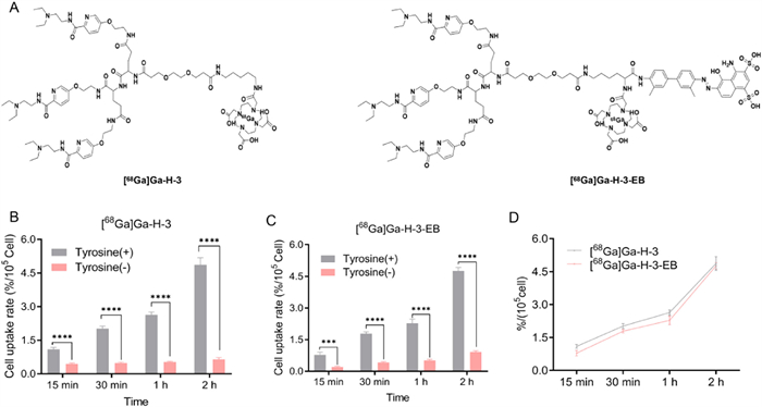
The structural integrity of [68Ga]Ga-H-3 and [68Ga]Ga-H-3-EB (Fig. 1A) was confirmed by 1H-nuclear magnetic resonance (NMR), 13C NMR, and mass spectrometry (Figs. S1–S8 in Supporting information). The radiochemical yields of [68Ga]Ga-H-3 and [68Ga]Ga-H-3-EB were 81.3% ± 5.2% and 78.5% ± 4.4%, respectively, with radiochemical purity exceeding 99%. In order to confirm whether coupling H-3 with EB reduces its biological activity, we compared the uptake of [68Ga]Ga-H-3 and [68Ga]Ga-H-3-EB in B16 melanoma cells, with and without l-tyrosine pretreatment. Melanin production was significantly higher in B16 cells pretreated with l-tyrosine than in untreated cells. Correspondingly, the uptake of [68Ga]Ga-H-3 and [68Ga]Ga-H-3-EB in l-tyrosine pretreated B16 cells was significantly higher than in untreated cells (P < 0.001, Figs. 1B and C). Uptake values increased over time, with 2-h uptake values of 4.90% ± 0.18% and 4.87% ± 0.32%, respectively, which were five times higher than those of untreated cells, confirming the specificity of the two tracers for melanin, which consistent with findings from other studies targeting melanin probes [29-31]. And uptake of both tracers in l-tyrosine pretreated B16 cells was similar at the same time points (Fig. 1D), indicating that EB coupling does not reduce the affinity and biological activity of H-3 towards melanin.
Figure 1
Figure 1.
(A) The chemical structures of [68Ga]Ga-H-3 and [68Ga]Ga-H-3-EB. (B) In vitro uptake of [68Ga]Ga-H-3 in B16 melanoma cells with (+) or without (−) tyrosine over time. (C) In vitro uptake of [68Ga]Ga-H-3-EB in B16 melanoma cells with (+) or without (−) tyrosine over time. (D) In vitro uptake of [68Ga]Ga-H-3 and [68Ga]Ga-H-3-EB in B16 melanoma cells with (+) tyrosine over time (n = 4; mean ± SD). ***P< 0.001, ****P< 0.0001.
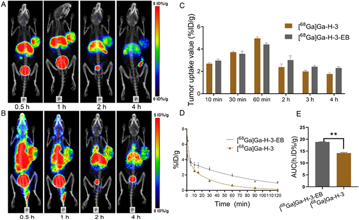
All animal experiments involved in this study were approved by the Ethics Committee of Central South University and were strictly conducted in accordance with the guidelines of the Animal Ethical and Welfare Committee (No. CSU-2022–0293). We compared the imaging of [68Ga]Ga-H-3 and [68Ga]Ga-H-3-EB in B16 tumors (Figs. 2A and B). Consistent with their biodistribution results (Fig. S9 in Supporting information), the uptake of [68Ga]Ga-H-3 and [68Ga]Ga-H-3-EB in tumors were showed in (Fig. 2C). After 1 h post-injection, the uptake peaked at 3.93 ± 0.29%ID/g and 4.58 ± 0.23%ID/g of [68Ga]Ga-H-3 and [68Ga]Ga-H-3-EB in B16 tumors. Pharmacokinetic results showed the different blood clearance rates of [68Ga]Ga-H-3 and [68Ga]Ga-H-3-EB with their effective half-life of 22.85 and 46.22 min, respectively (Fig. 2D). What is more, the area under the curve (AUC) of drug [68Ga]Ga-H-3-EB was also significantly larger than that of [68Ga]Ga-H-3 (Fig. 2E), indicating that coupling with EB may significantly enhanced tumor retention. And our results showed that [68Ga]Ga-H-3 and [68Ga]Ga-H-3-EB could accumulate rapidly at the tumor site, while the uptake of other normal tissues, especially the lung and brain, was very low, resulting in a high tumor-to-background ratios, which was significantly superior to the melanin-targeting diagnostic tracer 131I-iodofluotizamide benzamide (131I-IFNABZA), which has significant uptake in the lungs and was unfavorable for the detection of pulmonary metastases [32].
Figure 2
Figure 2.
PET/CT images of (A) [68Ga]Ga-H-3 and (B) [68Ga]Ga-H-3-EB in B16 tumor models. The uptake of [68Ga]Ga-H-3 and [68Ga]Ga-H-3-EB in tumors (C). The pharmacokinetic of [68Ga]Ga-H-3 and [68Ga]Ga-H-3-EB in B16 tumor models (D). The dosage of [68Ga]Ga-H-3 and [68Ga]Ga-H-3-EB in the blood of B16 tumor models (E). Group variation data were described as mean ± SD (n = 5). **P< 0.01.
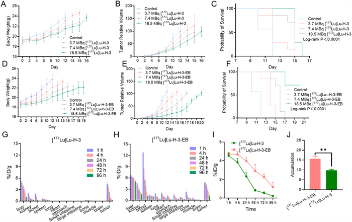
We evaluated the anti-tumor radiotherapy efficacy of [177Lu]Lu-H-3 and [177Lu]Lu-H-3-EB in B16 tumor models. Both [177Lu]Lu-H-3 (Figs. 3A–C) and [177Lu]Lu-H-3-EB (Figs. 3D–F) significantly inhibited B16 tumor growth, with [177Lu]Lu-H-3-EB showing a more pronounced therapeutic effect (P < 0.05). The average survival time of mice in the control, 18.5 MBq [177Lu]Lu-H-3 and [177Lu]Lu-H-3-EB group were 11.14 ± 1.07, 15.29 ± 0.49 and 18.57 ± 1.40 days (Figs. 3C and F). Notably, 7.4 MBq [177Lu]Lu-H-3-EB achieved a comparable therapeutic effect to 18.5 MBq [177Lu]Lu-H-3, extending the survival of B16-loaded mice by at least 8–10 days, which is significantly better than the therapeutic effect of radiotherapeutic drugs for melanoma reported by Miao and Rbah-Vidal et al. [33,34], while comparable to that of two doses of 131I-ICF01012 injection (2 × 18.5 MBq) [35], since the improved pharmacokinetics of [177Lu]Lu-H-3-EB (Figs. 3G and H) allows a single dose to achieve the therapeutic effect of two or more administrations with the total radiation dose at B16 tumor site of [177Lu]Lu-H-3-EB was significantly higher than [177Lu]Lu-H-3 (Fig. 3J) as EB delayed the washout of H-3 from the tumor (Fig. 3I), which has demonstrated in other studies combining the drug with EB [36-39] and findings showed that tumor vascularization reduction after radionuclide therapy affected the efficacy of subsequent treatments [24,40]. Ki-67 and terminal deoxynucleotidyl transferase-mediated dUTP nick end labeling (TUNEL) staining showed that tumor cell proliferation in mice treated with [177Lu]Lu-H-3-EB was significantly lower than the [177Lu]Lu-H-3 groups, while cell apoptosis was higher. And hematoxylin and eosin (H&E) staining of the tumors showed extensive necrotic areas in the [177Lu]Lu-H-3-EB treatment groups (Fig. S10 in Supporting information). 18.5 MBq [177Lu]Lu-H-3-EB had no significant systemic toxicity on mice as evidenced by the undifferentiated results of H&E staining in the heart, liver, spleen, lungs, and kidney tissues of each group (Fig. S11 in Supporting information).
Figure 3
Figure 3.
Therapy and efficacy assessment of [177Lu]Lu-H-3/H-3-EB on B16 tumor. (A–C) The body weight, tumor growth curves relative to the tumor volume on day 0 (set as 1.0) and Kaplan-Meier plot of mice in the [177Lu]Lu-H-3 treatment groups. (D–F) The body weight, tumor growth curves relative to the tumor volume at day 0 (set as 1.0) and Kaplan-Meier plot of mice in the [177Lu]Lu-H-3-EB treatment groups. Biodistribution of [177Lu]Lu-H-3 (G) and [177Lu]Lu-H-3-EB (H) in B16 models. Variation curve (I) and total dose (J) of radiation at the B16 tumor site (n = 7). Data are presented as mean ± SD. **P< 0.01.
Five pathologically confirmed malignant melanoma of the plantar skin patients were included in this study, and their clinical characteristics were summarized in Table S1 (Supporting information). The total effective radiation dose of [68Ga]Ga-H-3-EB in human, calculated by OLINDA (Version 2.2) software was 9.36 × 10⁻³ ± 6.44 × 10⁻⁴ mSv/MBq, which is lower than the radiation dose of [18F]F-FDG commonly used in clinical practice [41]. Therefore, we believe that the application of [68Ga]Ga-H-3-EB in patients is highly safe.
Owing to the low background uptake in the skin and muscles, the diagnoses of both the primary melanoma and metastatic lymph nodes were clear in [68Ga]Ga-H-3-EB PET/CT. The melanin-targeting probe [18F]F-P3BZA demonstrates high uptake in melanoma and exhibited specific imaging capabilities for both primary melanoma and its metastases. However, its significant accumulation in the stomach and moderate uptake in the eyes limit its clinical application [42]. Several other imaging agents, namely [18F]F-PEG3-FPN [43], [18F]F-DMPY2 [31], and [18F]F-PFPN [44], are currently among the better-performing agents for the clinical diagnosis of melanoma. Nonetheless, due to constraints in their molecular structures, none of these probes can be labeled with therapeutic metallic radionuclides commonly used in the clinic, such as lutetium-177 (177Lu), for the radionuclide therapy of patients with advanced melanoma. This limitation underscores the distinct advantage of H-3-EB, which can be labeled with the diagnostic radionuclide 68Ga for specific diagnosis of melanoma and also with the therapeutic radionuclide 177Lu for treating advanced melanoma, enabling a theranostic approach.
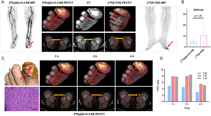
Compared with [18F]F-FDG, [68Ga]Ga-H-3-EB PET/CT detected all primary tumor lesions and metastatic lymph nodes too, like the melanin-targeting imaging agents [18F]F-P3BZA [42] and [18F]F-PFPN [44]. In some metastatic lymph nodes, the uptake of [68Ga]Ga-H-3-EB was even higher than that of [18F]F-FDG (Fig. 4A). In the primary tumor foci, standardized SUVmax of the two imaging agents were comparable (P = 1.26; calculated as the ratio of lesion SUVmax to the SUVmean of the skin in the corresponding contralateral area), indicating that melanoma exhibits relatively high uptake of [68Ga]Ga-H-3-EB (Fig. 4B). Over time, [68Ga]Ga-H-3-EB showed gradual accumulation in both primary melanoma lesions and metastatic lymph nodes, with sustained high uptake in the tumors even 4 h after injection (Figs. 4C and D). Although [68Ga]Ga-H-3-EB is not superior to [18F]F-FDG in diagnosing malignant melanoma, its greatest advantage lies in selecting candidates suitable for [177Lu]Lu-H-3-EB radionuclide therapy. These findings lay a solid foundation for the subsequent clinical translation of [177Lu]Lu-H-3-EB in treating advanced malignant melanoma.
Figure 4
Figure 4.
(A) [68Ga]Ga-H-3-EB and [18F]F-FDG PET/CT imaging of left foot cutaneous melanoma with left inguinal metastatic lymph nodes in patient |1. (B) Comparison of standardized SUVmax between [68Ga]Ga-H-3-EB and [18F]F-FDG in primary melanoma lesions (SUVmax of primary lesion divided by SUVmean of corresponding contralateral skin). (C) [68Ga]Ga-H-3-EB dynamic PET/CT imaging in patient |1 with malignant melanoma of left plantar skin, left inguinal metastatic lymph node and its pathological results of HE staining. (D) [68Ga]Ga-H-3-EB dynamic SUVmax ratio of tumor to non-tumor. Malignant melanoma of left plantar skin is indicated by the red arrow, left inguinal metastatic lymph node is indicated by the yellow arrow. T, tumor; M, muscle; B, brain; L, lymph nodes. Data are presented as mean ± SD (n = 5).
In conclusion, we synthesized a novel probe targeting melanin and enhanced its total tumor uptake by co-administering with EB. We validated its therapeutic potential against melanoma at the animal level and performed first-in-human PET/CT imaging, which demonstrated tumor uptake comparable to that of [18F]F-FDG, confirming that it is an effective and safe probe. This suggests that [177Lu]Lu-H-3-EB could become a highly promising therapeutic option for patients with advanced melanoma.
Ethical statement
The study involves human experiments with volunteers or patients has been approved by the Ethics Committee of Xiangya Hospital, Central South University (approval No. 202104001) and was conducted in accordance with the Helsinki Declaration. All participants signed informed consent forms.
The authors declare that they have no known competing financial interests or personal relationships that could have appeared to influence the work reported in this paper.
Acknowledgments
This work was supported by the Science and Technology Innovation Team Talent Project of Hunan Province (No. 2021 RC4056) and National Natural Science Foundation of China (No. 82272045).
Supplementary materials
Supplementary material associated with this article can be found, in the online version, at doi:10.1016/j.cclet.2026.112361.
Figure 1
(A) The chemical structures of [68Ga]Ga-H-3 and [68Ga]Ga-H-3-EB. (B) In vitro uptake of [68Ga]Ga-H-3 in B16 melanoma cells with (+) or without (−) tyrosine over time. (C) In vitro uptake of [68Ga]Ga-H-3-EB in B16 melanoma cells with (+) or without (−) tyrosine over time. (D) In vitro uptake of [68Ga]Ga-H-3 and [68Ga]Ga-H-3-EB in B16 melanoma cells with (+) tyrosine over time (n = 4; mean ± SD). ***P< 0.001, ****P< 0.0001.
Figure 2
PET/CT images of (A) [68Ga]Ga-H-3 and (B) [68Ga]Ga-H-3-EB in B16 tumor models. The uptake of [68Ga]Ga-H-3 and [68Ga]Ga-H-3-EB in tumors (C). The pharmacokinetic of [68Ga]Ga-H-3 and [68Ga]Ga-H-3-EB in B16 tumor models (D). The dosage of [68Ga]Ga-H-3 and [68Ga]Ga-H-3-EB in the blood of B16 tumor models (E). Group variation data were described as mean ± SD (n = 5). **P< 0.01.
Figure 3
Therapy and efficacy assessment of [177Lu]Lu-H-3/H-3-EB on B16 tumor. (A–C) The body weight, tumor growth curves relative to the tumor volume on day 0 (set as 1.0) and Kaplan-Meier plot of mice in the [177Lu]Lu-H-3 treatment groups. (D–F) The body weight, tumor growth curves relative to the tumor volume at day 0 (set as 1.0) and Kaplan-Meier plot of mice in the [177Lu]Lu-H-3-EB treatment groups. Biodistribution of [177Lu]Lu-H-3 (G) and [177Lu]Lu-H-3-EB (H) in B16 models. Variation curve (I) and total dose (J) of radiation at the B16 tumor site (n = 7). Data are presented as mean ± SD. **P< 0.01.
Figure 4
(A) [68Ga]Ga-H-3-EB and [18F]F-FDG PET/CT imaging of left foot cutaneous melanoma with left inguinal metastatic lymph nodes in patient |1. (B) Comparison of standardized SUVmax between [68Ga]Ga-H-3-EB and [18F]F-FDG in primary melanoma lesions (SUVmax of primary lesion divided by SUVmean of corresponding contralateral skin). (C) [68Ga]Ga-H-3-EB dynamic PET/CT imaging in patient |1 with malignant melanoma of left plantar skin, left inguinal metastatic lymph node and its pathological results of HE staining. (D) [68Ga]Ga-H-3-EB dynamic SUVmax ratio of tumor to non-tumor. Malignant melanoma of left plantar skin is indicated by the red arrow, left inguinal metastatic lymph node is indicated by the yellow arrow. T, tumor; M, muscle; B, brain; L, lymph nodes. Data are presented as mean ± SD (n = 5).


 DownLoad:

 下载:


 下载:

