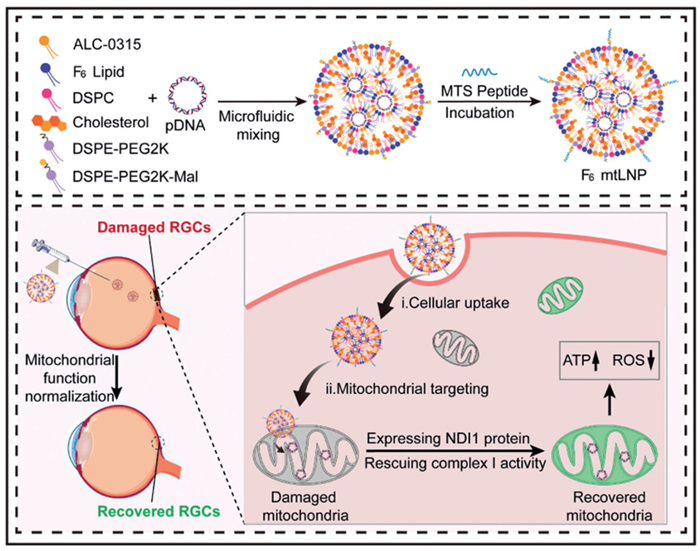
Scheme 1.
Schematic illustration of in situ mitochondrial gene delivery system (F6 mtLNP) construction and its effect mechanism for Leber's hereditary optic neuropathy therapy.

Mitochondria-targeted lipid nanoparticles to rescue mitochondrial complex Ⅰ deficiency for Leber's hereditary optic neuropathy therapy
Min Zhao , Huan-Ni Liu , Hai-Xin Xie , Hao-Yuan Yu , Jing-Song Yang , Na-Hui Liu , Jia-Qi Chen , Lu-Xin Qie , Zi-Juan Yi , Tian-Jiao Zhou , Lei Xing , Xian Wu Cheng , Yi Wang , Hu-Lin Jiang


Mitochondrial complex Ⅰ (NADH: ubiquinone oxidoreductase) serves as a cornerstone of the respiratory chain and is essential for cellular energy metabolism [1]. It couples electron transfer from NADH to ubiquinone with proton pumping across the inner mitochondrial membrane, thereby establishing the electrochemical gradient that is indispensable for adenosine triphosphate (ATP) synthesis. Mitochondrial complex Ⅰ dysfunction, primarily induced by genetic mutations or oxidative stress, results in structural disintegration and functional loss. This impairment severely compromises ATP synthesis and amplifies reactive oxygen species (ROS) production, thereby triggering a cascade of cellular damage that culminates in cell death [2]. These pathological processes underlie a spectrum of severe and often fatal mitochondrial disorders, including Leber's hereditary optic neuropathy (LHON) and Parkinson's disease [3,4]. Currently, no effective treatments exist for these mitochondrial disorders involving complex Ⅰ deficiency, highlighting the urgent need for innovative therapeutic strategies.
Gene therapy holds a distinct advantage for treating mitochondrial disorders through functional compensation of defective proteins. For complex Ⅰ deficiencies, bypass therapy has emerged as a promising strategy by supplementing alternative enzymes to circumvent the impaired complex. The single-subunit NADH dehydrogenase (NDI1) from Saccharomyces cerevisiae represents an ideal candidate, as it can replace entire mammalian complex Ⅰ by transferring electrons from NADH to ubiquinone, thereby restoring electron transport chain function [5]. Current NDI1-based gene therapies primarily rely on viral vector-mediated allotopic expression, which has demonstrated efficacy in restoring mitochondrial function in various models [6–9]. However, clinical translation faces challenges including inefficient delivery caused by twice transmissions, immunogenicity, oncogenic risks, and high manufacturing costs [10–13]. These limitations emphasize the necessity for non-viral approaches for in situ mitochondrial gene therapy. The central obstacle of such non-viral strategies, however, remains the development of delivery systems capable of traversing the mitochondrial double membrane and permitting direct introduction of genetic therapeutics into the matrix [14].
Existing mitochondria-targeted strategies primarily based on lipophilic cation and mitochondrial targeting signal (MTS) peptide [15–18], yet both have limitations. The lipophilic cation-mediated strategy, which relies on the mitochondrial membrane potential, often induces adverse effects such as obvious cytotoxicity due to high positive charge density [19–21]. Meanwhile, the MTS peptide pathway, mediated by the translocase of the outer membrane (TOM) complex, has unproven efficacy for delivering large nucleic acid cargos or even nanoparticle [14,22]. In contrast, fluorination presents a promising alternative. Our previous work identified fluorinated groups as effective membrane-penetrating motifs with intrinsic mitochondrial affinity [23]. Their unique properties, including hydrophobicity, lipophobicity, and low surface energy, contribute to enhanced membrane affinity and facilitate efficient lysosome escape [24–26]. Moreover, incorporating fluoroalkyl motifs in place of hydrocarbon chains results in a reduction of cytotoxic effects and hemolysis, thereby enhancing biocompatible properties [27]. Consequently, fluorination overcomes the key challenges of charge-associated toxicity and biological cargo constraints, suggesting a new direction for designing mitochondrial gene delivery systems.
Here, we synthesized a novel ionizable lipid featuring a fluorinated hydrophobic tail (F6 lipid) as a membrane-penetrating and mitochondrial membrane affinity component to construct fluorinated lipid nanoparticles (F6 LNP). The surface of the LNP was further modified with MTS peptides to obtain F6 mtLNP for enhancing the targeting affinity for mitochondrial membranes. Under the dual action of MTS and F6 lipid, the nanoparticles can efficiently enrich and traverse the double membrane structure of mitochondria, increasing the accumulation of functional gene therapeutic NDI1 in the mitochondrial matrix. As a result, efficient in situ mitochondrial gene delivery and functional protein expression are achieved, leading to recovery of complex Ⅰ activity and restoration of mitochondrial homeostasis (Scheme 1). Overall, this strategy may offer a new therapeutic avenue for mitochondrial disorders involving complex Ⅰ deficiency.
Firstly, we synthesized F6 lipid via esterification reaction, coupling a partially fluorinated hydrophobic tail chain with a hydrophilic amine headgroup (Fig. 1A and Fig. S1 in Supporting information). The chemical structure of F6 lipid was confirmed by 1H nuclear magnetic resonance (NMR) and 19F NMR spectra (Figs. S2 and S3 in Supporting information). For comparison, a non-fluorinated ionizable lipid analog (F0 lipid) was also prepared and verified by 1H NMR (Figs. S4 and S5 in Supporting information).
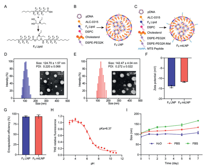
Fluorinated group tail-modified lipid nanoparticles (F6 LNP) were formulated using a microfluidic mixing device [28]. The formulation of F6 LNP included F6 lipid, ALC-0315, 1,2-distearoyl-sn-glycero-3-phosphocholine (DSPC), cholesterol, and 1,2-distearoyl-sn-glycero-3-phosphoethanolamine-N-[methoxy-(polyethylene glycerol)-2000] (DSPE-PEG2K) (Fig. 1B). The optimal molar ratio of the F6 lipid in F6 LNP was determined to be 25% based on the evaluation of cellular internalization efficiency and mitochondrial transfection performance, the latter of which was assessed using a mitochondrial green fluorescent protein (mtGFP) reporter plasmid (Fig. S6 in Supporting information). This mtGFP plasmid (Supplementary sequence 1 in Supporting information) is designed to express GFP exclusively in the mitochondrial matrix via the mitochondrial codon system, enabling specific assessment of mitochondrial transfection [29]. Two key design strategies ensure specificity: (1) The inclusion of a cytoplasmic stop codon prevents translation in the cytosol [30,31]; (2) the absence of a mitochondrial targeting sequence excludes protein import into mitochondria [32,33].
To further enhance the targeting affinity for mitochondrial membranes, 1,2-distearoyl-sn-glycero-3-phosphoethanolamine-N-[maleimide(polyethylene glycol)-2000] (DSPE-PEG2K-Mal) was added to the F6 LNP formulation in an appropriate proportion, and the terminal thiol-containing MTS peptide was grafted onto the surface of F6 LNP through michael addition reaction, thereby obtaining F6 mtLNP (Fig. 1C). Based on the cellular internalization efficiency and mtGFP transfection performance, the optimal molar ratio of DSPE-PEG2K-MTS was determined to be 0.75% (Fig. S7 in Supporting information). For the assessment the biosafety and mitochondrial targeting efficacy of F6 mtLNP, we additionally prepared three control LNPs, namely: ALC-0315 LNP (containing only the commercial ionizable lipid ALC-0315), F0 LNP (containing F0 lipid instead of F6 lipid), and F0 mtLNP (containing both F0 lipid and MTS peptide).
Following preparation, the LNPs were characterized. The average particle sizes of F6 LNP and F6 mtLNP were measured as 124.70 ± 1.57 and 142.47 ± 4.04 nm, respectively, with low polydispersity index (PDI). Transmission electron microscopy (TEM) images showed uniform spherical structures in both formulations (Figs. 1D and E). The zeta potential values of F6 LNP and F6 mtLNP were −13.87 ± 0.72 and −11.70 ± 0.26 mV, respectively (Fig. 1F). The encapsulation efficiencies (EE) of F6 LNP and F6 mtLNP were 93.13% ± 2.04% and 93.97% ± 3.57%, respectively (Fig. 1G). The pKa values of F6 LNP and F6 mtLNP were determined to be 6.46 and 6.37, respectively (Fig. 1H and Fig. S8 in Supporting information). Under physiological conditions, this confers a negative surface charge that improves biological safety, while the ionization properties enable a shift to a cationic state in acidic lysosomes to facilitate lysosomal escape [34,35]. Furthermore, both F6 LNP and F6 mtLNP demonstrated good size stability in H2O and PBS over 7 days. Although a slight increase in hydrodynamic size occurred in 10% fetal bovine serum (FBS)-Dulbecco's Modified Eagle Medium (DMEM), attributable to the formation of a protein corona [36,37], the size remained well below 160 nm (Fig. 1I and Fig. S9 in Supporting information). TEM imaging further confirmed that the spherical morphology and structural integrity of the nanoparticle cores were maintained under all tested conditions (Fig. S10 in Supporting information), collectively indicating favorable stability for subsequent biological applications. The physicochemical properties of three control LNPs were also characterized (Table S1 in Supporting information).
Furthermore, we conducted the methylthiazolyldiphenyl-tetrazolium bromide (MTT) assay and mitochondrial function evaluations to assess the biosafety profiles of different LNP formulations. The experimental results revealed that F6 mtLNP did not significantly affect cell viability and mitochondrial function adversely, particularly the ATP production (Figs. S11 and S12 in Supporting information).
To further investigate the mitochondrial targeting efficacy of F6 mtLNP, we assessed key cellular pathways, including cellular internalization and specific subcellular trafficking to mitochondria. Flow cytometry revealed that both F6 LNP and F6 mtLNP exhibited significantly higher mean fluorescence intensity (MFI) of Cy5-labeled plasmid gene (Cy5-pDNA) compared to other groups, a property attributed to the superior membrane permeability of their fluorinated moieties. Among them, F6 mtLNP achieved the most efficient cellular internalization (Fig. 2A). To elucidate the internalization mechanism, we employed pharmacological inhibitors of key endocytic pathways: Wortmannin (macropinocytosis), genistein (caveolae), and chlorpromazine (clathrin) [38,39]. The result indicated that the entry of F6 mtLNP involved multiple mechanisms, among which caveolae-mediated endocytosis was served as the predominant route (Fig. 2B). This pathway typically utilizes neutral caveosomes for cytosolic delivery, thereby minimizing lysosomal degradation, which may explain the enhanced functional delivery observed [40,41]. To further assess the ability of lysosomal escape, we tracked the co-localization of F6 mtLNP with lysosomes over time using confocal laser scanning microscopy (CLSM). The results showed that after 3 h of incubation, the Pearson's correlation coefficient (R) of F0 mtLNP with lysosomes (R = 0.80) was higher than that of F6 mtLNP (R = 0.64). After 5 h of incubation, the R value of F0 mtLNP with lysosomes remained at a relatively high level of 0.72, while the R value of F6 mtLNP further dropped to 0.55 (Fig. S13 in Supporting information). The results demonstrate the superior lysosomal escape capability of the F6 lipid, which we attribute to a dual mechanism in which the ionizable headgroup protonates to electrostatically disrupt the lysosomal membrane while the fluorinated tail enhances this effect via hydrophobic and lipophobic interactions [42,43].
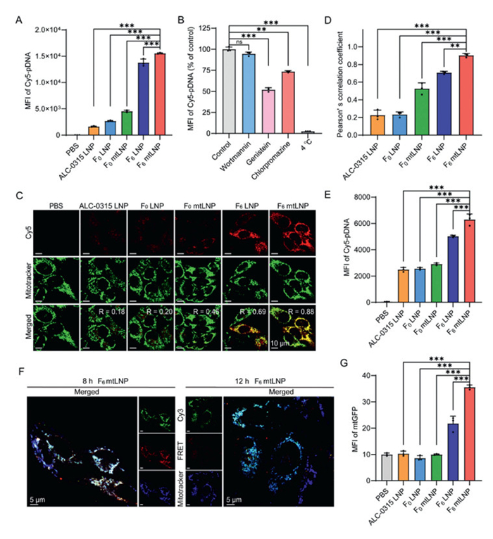
Next, we investigated the capability of F6 mtLNP for mitochondria-targeted gene delivery. As shown in Figs. 2C and D, both F6 LNP and F6 mtLNP exhibited strong mitochondrial co-localization of Cy5-pDNA, whereas other non-fluorinated formulations showed markedly weaker signals, underscoring the role of F6 lipid in mitochondrial targeting. Additionally, the Pearson's correlation coefficient of F6 mtLNP (R = 0.88) was significantly higher than that of F6 LNP (R = 0.69), highlighting the additional benefit of MTS modification for selective mitochondrial delivery. Consistent with these findings, direct measurement of Cy5-pDNA MFI in purified mitochondria, which were isolated and treated with proteinase K to remove external contamination [44], confirmed the significant enhancement of mitochondrial accumulation achieved by F6 mtLNP (Fig. 2E). These results indicate the strong mitochondria-targeted gene delivery capability of F6 mtLNP, attributed to the synergistic action of the MTS peptide and the high membrane affinity and penetration capacity of F6 lipid.
Given that efficient gene delivery to the mitochondria, specifically to the matrix, is essential for in situ mitochondrial gene therapy to rescue dysfunctional proteins, we next employed high-sensitivity structured illumination microscopy (HiS-SIM) and TEM to determine the precise sub-mitochondrial localization of F6 mtLNP. HiS-SIM images showed successful delivery of Cy5/Cy3-pDNA into mitochondria by F6 mtLNP, with specific accumulation within cristae structures, as confirmed by co-localization with the inner mitochondrial membrane (Fig. S14 in Supporting information). The outer and inner mitochondrial membranes were specifically labeled with Tom20-mEmerald and Mitotracker Green, respectively, to validate this intramitochondrial distribution. To further trace the intracellular trajectory of the nanoparticles, we prepared F6 mtLNP loaded with 10 nm gold colloids for TEM visualization. Micrographs revealed F6 mtLNP with gold colloids surrounding the mitochondria. Following a 12 h incubation, some F6 mtLNP with gold colloids were successfully found in the mitochondrial matrix (Fig. S15 in Supporting information).
After the confirmation of mitochondria-targeted gene delivery capability of F6 mtLNP, we further investigated whether the genes delivered into the mitochondrial matrix could be released from the carrier using the fluorescence resonance energy transfer (FRET) assay [45]. Cy3-pDNA and Cy5-pDNA were co-encapsulated in F6 mtLNP to monitor the FRET signals at different incubation time points. When Cy3-pDNA and Cy5-pDNA are in close proximity, both Cy3 and FRET fluorescence are detectable under Cy3 excitation; however, when they dissociate, only Cy3 fluorescence remains observable in the absence of FRET signals. CLSM images showed that at 8 h, both Cy3 and FRET fluorescence were present and co-localized with mitochondria. In contrast, after 12 h of incubation, the FRET signal decreased significantly, while Cy3 fluorescence remained associated with mitochondria (Fig. 2F). These results suggest that F6 mtLNP did not disassemble immediately upon mitochondrial entry but instead gradually release their genetic cargo within the mitochondria over time.
Owing to the superior mitochondrial delivery and release capability of F6 mtLNP, we next assessed its transfection efficiency. As shown in Fig. 2G, F6 mtLNP mediated significantly enhanced mitochondrial gene transfection efficiency, demonstrating that exogenous plasmid DNA was successfully delivered into mitochondria and translated into the target protein.
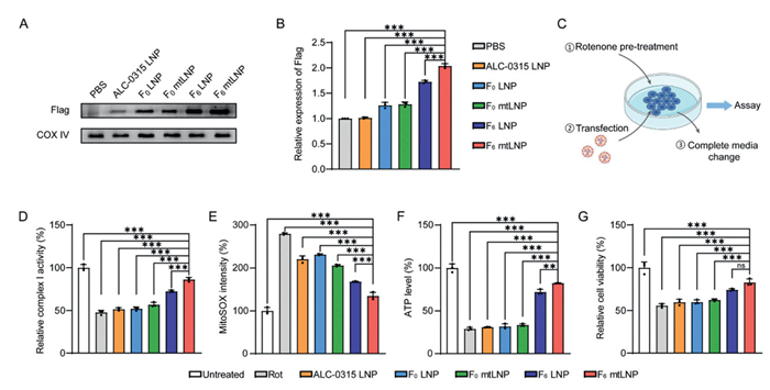
Mitochondrial complex Ⅰ dysfunction represents a major causative factor in the development of various mitochondrial disorders. Currently, no effective therapies exist for these mitochondrial disorders involving complex Ⅰ deficiency. Most reported mitochondrial gene therapy strategies focus on rectifying defects in individual subunits of the complex [46,47]. In contrast, an approach that functionally compensates for the entire complex Ⅰ could offer a broadly applicable treatment for all such deficiencies. A unique and promising therapeutic strategy involves expressing the alternative NDI1 from Saccharomyces cerevisiae. This single-subunit enzyme integrates into the mitochondrial inner membrane of mammalian cells and catalyzes the oxidation of NADH, thereby serving as a functional substitute for the endogenous 45-subunit complex Ⅰ. Therefore, we constructed a mitochondrially codon-optimized plasmid (NDI1) encoding an NDI1 protein with a C-terminal Flag tag for further investigation (Supplementary sequence 2 in Supporting information). Successful mitochondrial transfection of F6 mtLNP/NDI1 in vitro was validated by Western blot analysis, as evidenced by significantly enhanced protein expression (Figs. 3A and B), suggesting that F6 mtLNP delivering the NDI1 gene still maintained superior mitochondrial transfection over other treatments.
To investigate whether F6 mtLNP/NDI1 can restore mitochondrial function through functional supplementation of the NDI1 gene under the pathological condition of complex Ⅰ deficiency, we introduced rotenone (Rot)-induced cells as mitochondrial complex Ⅰ deficiency cells based on our previous studies and those of other researchers to assess in vitro efficacy of F6 mtLNP/NDI1 [23,48,49]. The efficacy of Rot as a classic complex Ⅰ inhibitor is attributed to its mechanism of blocking electron transfer from NADH to ubiquinone by specifically binding to the ubiquinone-binding site of complex Ⅰ [50].
We first determined the optimal Rot concentration using an MTT assay to mimic mitochondrial complex Ⅰ deficiency. The Rot concentration was selected at 0.1 µmol/L to achieve about 50% reduction in cell viability for subsequent in vitro experiments (Fig. S16A in Supporting information). We further evaluated mitochondrial membrane potential (MMP) under this condition using the mitochondrial protonophore uncoupler carbonyl cyanide m-chlorophenyl hydrazone (CCCP) as a control group. Flow cytometric analysis with JC-1 staining revealed that Rot treatment significantly decreased red fluorescence intensity, accompanied by a 29.7% increase in the depolarized cell population (Q3 quadrant)–a response pattern similar to that induced by CCCP (Fig. S16B in Supporting information). These results confirm that the selected Rot concentration effectively impairs mitochondrial function, leading to a decrease in MMP.
Following the establishment of the Rot-induced complex Ⅰ deficiency cell model, the cells were treated with F6 mtLNP/NDI1 and other formulations to evaluate their in vitro therapeutic efficacy (Fig. 3C). The key mitochondrial functions were evaluated, including complex Ⅰ activity, mitochondrial ROS level and ATP generation. As shown in Fig. 3D, the F6 mtLNP/NDI1 treatment exhibited a significant recovery of complex Ⅰ activity relative to other groups, confirming effective functional compensation. This restoration was accompanied by a pronounced reduction in mitochondrial ROS levels (Fig. 3E) and increased ATP generation (Fig. 3F), which concertedly mitigated Rot-induced cellular damage (Fig. 3G). In contrast, the F6 mtLNP/mtGFP control group showed no beneficial effects across all measured parameters (Fig. S17 in Supporting information), underscoring that the observed recovery specifically resulted from NDI1 expression rather than non-specific vector effects. Collectively, these results demonstrated that F6 mtLNP/NDI1 effectively delivered and expressed NDI1, compensating for complex Ⅰ deficiency and restoring mitochondrial homeostasis under pathological conditions.
Given the promising in vitro efficacy of F6 mtLNP/NDI1 in rescuing mitochondrial complex Ⅰ function, we transitioned to evaluating its therapeutic potential in vivo. LHON was chosen as the disease model based on the rationale that it is a classic ocular mitochondrial disorder driven primarily by monogenic mutations in complex Ⅰ subunits, offering a compelling and relevant model for evaluating the efficacy of NDI1-based functional compensation in vivo. All animal experimental procedures were approved by the Regional Ethics Committee of China Pharmaceutical University and conducted in accordance with institutional guidelines.
We first investigated whether F6 mtLNP could mediate the delivery and expression of an exogenous functional gene NDI1 in mice. Whole-eye protein extracts were collected and analyzed for Flag-tagged expression at various time points. Throughout the one-month observation period, F6 mtLNP/NDI1 enabled sustained Flag-tagged protein expression, which peaked between 3 and 7 days post-administration (Fig. S18 in Supporting information). These results demonstrate that a single F6 mtLNP/NDI1 treatment promoted persistent expression of the NDI1 in mitochondrial compartments of cells in vivo, with functional protein detectable for at least one month. The biosafety of F6 mtLNP/NDI1 at various doses was evaluated in vivo using hematoxylin and eosin (H&E) staining. Histopathological examination revealed no noticeable structural damage in corneal or retinal tissues at any tested dosage (Fig. S19 in Supporting information), supporting its suitability for subsequent in vivo efficacy studies.
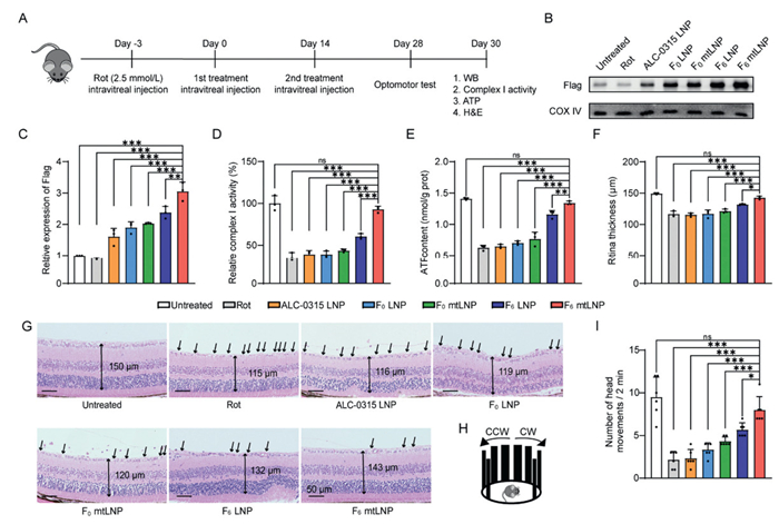
We next investigated whether mitochondrial delivery of NDI1 could rescue visual impairment in mice. Mirroring our in vitro strategy, we established a Rot-induced mouse model to evaluate the therapeutic potential of F6 mtLNP/NDI1 in vivo. The model was generated by intravitreal injection of 2.5 mmol/L Rot into C57BL/6J male mice (6–8 weeks old) [23,51]. Three days post-induction, Rot-induced mice received two treatments with respective formulations at 14-day intervals. On day 28 after the initial treatment, visual function was assessed using an optomotor response test, followed by a series of evaluations including NDI1 expression, complex Ⅰ activity, ATP production, and retinal structural changes (Fig. 4A). As shown in Figs. 4B and C, F6 mtLNP/NDI1 treatment significantly enhanced NDI1 expression compared to other formulations. Consistent with this observation, a pronounced restoration of mitochondrial complex Ⅰ activity was documented (Fig. 4D), confirming functional compensation of NDI1 to the impaired complex Ⅰ. Furthermore, the treatment facilitated the recovery of ATP generation (Fig. 4E), supporting the rescue of mitochondrial bioenergetics. Concurrent with these biochemical improvements, histological analysis revealed that F6 mtLNP/NDI1 ameliorated Rot-induced retinal damage, including retinal ganglion cells (RGCs) loss and layer thinning, restoring retinal architecture and thickness to near-normal levels (Figs. 4F and G). Moreover, in the optomotor test, F6 mtLNP/NDI1-treated mice exhibited significantly increased head tracking movements (Figs. 4H and I), confirming that mitochondrial delivery of NDI1 effectively restored visual function.
In conclusion, we have developed a novel mitochondria-targeted lipid nanoparticle (F6 mtLNP) incorporating a fluorinated ionizable lipid (F6 lipid) and surface-modified with MTS peptide. The hydrophobic and lipophobic nature of the fluorinated groups promoted lysosomal escape and enhanced cellular uptake, while the MTS peptide enabled precise mitochondrial targeting. The high mitochondrial membrane affinity and penetration capability of the fluorinated groups further allowed the system to overcome the double-membrane barrier, facilitating efficient accumulation of the therapeutic gene NDI1 within the mitochondrial matrix. As a result, this platform successfully restored mitochondrial complex Ⅰ function through functional compensation of the defective protein, leading to the rescue of visual impairment in a Rot-induced mouse model. These findings support the potential of F6 mtLNP as a promising therapeutic strategy not only for LHON but also for a broader spectrum of mitochondrial disorders involving complex Ⅰ deficiency.
Min Zhao: Writing – review & editing, Writing – original draft, Visualization, Validation, Software, Methodology, Investigation, Formal analysis, Data curation, Conceptualization. Huan-Ni Liu: Writing – review & editing, Visualization, Validation, Methodology, Investigation, Formal analysis, Data curation. Hai-Xin Xie: Writing – review & editing, Visualization, Validation, Methodology, Investigation, Formal analysis, Data curation. Hao-Yuan Yu: Writing – review & editing, Visualization, Validation, Methodology, Investigation, Formal analysis, Data curation. Jing-Song Yang: Writing – review & editing, Visualization, Validation, Methodology, Investigation, Formal analysis. Na-Hui Liu: Writing – review & editing, Visualization, Validation, Methodology, Investigation, Formal analysis. Jia-Qi Chen: Writing – review & editing, Visualization, Validation, Methodology, Investigation, Formal analysis. Lu-Xin Qie: Writing – review & editing, Visualization, Validation, Methodology, Investigation, Formal analysis. Zi-Juan Yi: Writing – review & editing, Visualization, Validation, Methodology, Formal analysis. Tian-Jiao Zhou: Writing – review & editing, Visualization, Validation, Formal analysis, Conceptualization. Lei Xing: Writing – review & editing, Visualization, Validation, Formal analysis, Conceptualization. Xian Wu Cheng: Writing – review & editing, Visualization, Validation, Resources, Funding acquisition, Conceptualization. Yi Wang: Writing – review & editing, Visualization, Validation, Supervision, Resources, Project administration, Investigation, Funding acquisition, Formal analysis, Conceptualization. Hu-Lin Jiang: Writing – review & editing, Visualization, Validation, Supervision, Resources, Project administration, Investigation, Funding acquisition, Formal analysis, Conceptualization.
The authors declare that they have no known competing financial interests or personal relationships that could have appeared to influence the work reported in this paper.
This work was financially supported by the National Natural Science Foundation of China (Nos. 82020108029, 82304416, 82473867, 82370424), Natural Science Foundation of Jiangsu Province (No. BK20231016), Jilin Provincial Foundation of Changbai Talent Outstanding Team (No. 202410006), State Key Laboratory of Natural Medicines China Pharmaceutical University (No. SKLNMZZ202021), Double First-class University Projects (No. CPU2018GY06, China), Double First-Rate construction plan of China Pharmaceutical University (No. CPU2022QZ18) and 2024 Project on Comprehensive Research of Multi-Target Natural Medicines (No. SKLNMZZ2024JS08).
Supplementary material associated with this article can be found, in the online version, at doi:
J. Zhu, K.R. Vinothkumar, J. Hirst, Nature 536 (2016) 354–358. doi: 10.1038/nature19095
L.Y. Meng, G.Z. Wu, RSC Adv. 13 (2023) 20476–20485. doi: 10.1039/d3ra03313a
P. Yu-Wai-Man, P.G. Griffiths, G. Hudson, et al., J. Med. Genet. 46 (2009) 145–158. doi: 10.1136/jmg.2007.054270
J.L. Li, T.Y. Lin, P.L. Chen, et al., Front. Mol. Neurosci. 14 (2021) 797833. doi: 10.3389/fnmol.2021.797833
B.B. Seo, T. Kitajima-Ihara, E.K. Chan, et al., Proc. Natl. Acad. Sci. U. S. A. 95 (1998) 9167–9171. doi: 10.1073/pnas.95.16.9167
N. Chadderton, A. Palfi, S. Millington-Ward, et al., Eur. J. Hum. Genet. 21 (2013) 62–68. doi: 10.1038/ejhg.2012.112
N. Chadderton, A. Palfi, D.M. Maloney, et al., Pharmaceutics 15 (2023) 322. doi: 10.3390/pharmaceutics15020322
S. Millington-Ward, A. Palfi, C. Shortall, et al., Int. J. Mol. Sci. 25 (2024) 8876. doi: 10.3390/ijms25168876
H. Li, B. Sun, Y. Huang, et al., Mol. Med. 28 (2022) 29.
H. Yin, R.L. Kanasty, A.A. Eltoukhy, et al., Nat. Rev. Genet. 15 (2014) 541. doi: 10.1038/nrg3763
G.N. Nguyen, J.K. Everett, S. Kafle, et al., Nat. Biotechnol. 39 (2021) 47. doi: 10.1038/s41587-020-0741-7
S. Guan, A. Munder, S. Hedtfeld, et al., Nat. Biotechnol. 14 (2019) 287–297. doi: 10.1038/s41565-018-0358-x
H. Yu, V. Porciatti, A. Lewin, et al., Sci. Rep. 3 (2018) 8.
Y. Wang, J.S. Yang, M. Zhao, et al., Adv. Drug Deliv. Rev. 211 (2024) 115355. doi: 10.1016/j.addr.2024.115355
P. Ning, W.J. Wang, M. Chen, et al., Chin. Chem. Lett. 28 (2017) 1943–1951. doi: 10.1016/j.cclet.2017.09.026
Z.Y. Yu, X. Luo, C. Zhang, et al., Chin. Chem. Lett. 35 (2024) 109519. doi: 10.1016/j.cclet.2024.109519
Y. Yamada, M. Hibino Satrialdi, et al., Adv. Drug Deliv. Rev. 154-155 (2020) 187–209. doi: 10.1016/j.addr.2020.09.010
F. Tong, Y. Wang, Y. Xu, et al., Nat. Commun. 15 (2024) 10382. doi: 10.1038/s41467-024-54854-2
J. Zielonka, J. Joseph, A. Sikora, et al., Chem. Rev. 117 (2017) 10043–10120. doi: 10.1021/acs.chemrev.7b00042
S.S. Liew, X. Qin, J. Zhou, et al., Angew. Chem. Int. Ed. 60 (2021) 2232–2256. doi: 10.1002/anie.201915826
L.H. Huang, Z.H. Sun, Q. Shen, et al., Chin. Chem. Lett. 33 (2022) 4146–4156. doi: 10.1016/j.cclet.2022.02.047
N. Yoshinaga, T. Miyamoto, M. Odahara, et al., Adv. Funct. Mater. 34 (2024) 2306070. doi: 10.1002/adfm.202306070
Y. Wang, L.F. Hu, P.F. Cui, et al., Adv. Mater. 33 (2021) e2103307. doi: 10.1002/adma.202103307
C. Zhang, K. Yan, C.K. Fu, et al., Chem. Rev. 122 (2022) 167–208. doi: 10.1021/acs.chemrev.1c00632
J.H. Gong, Y. Wang, L. Xing, et al., Int. J. Pharm. 535 (2018) 180–193. doi: 10.1016/j.ijpharm.2017.11.015
H. Han, J. Xing, W. Chen, et al., Nat. Commun. 14 (2023) 944. doi: 10.1038/s41467-023-36625-7
Z. Zhang, W. Shen, J. Ling, et al., Nat. Commun. 9 (2018) 1377. doi: 10.1038/s41467-018-03779-8
A. Raza, R. Zhang, R.N. Lu, et al., Nanomedicine 20 (2025) 2545–2569. doi: 10.1080/17435889.2025.2555507
D. Lyrawati, A. Trounson, D. Cram, Pharm. Res. 28 (2011) 2848–2862. doi: 10.1007/s11095-011-0544-0
Y. Yamada, T. Ishikawa, H. Harashima, Biomaterials 136 (2017) 56–66. doi: 10.1016/j.biomaterials.2017.05.016
T. Ishikawa, K. Somiya, R. Munechika, et al., J. Control. Release 274 (2018) 109–117. doi: 10.1016/j.jconrel.2018.02.005
S. Yang, S.Q. Ma, X. Wan, et al., EBioMedicine 10 (2016) 258–268. doi: 10.1016/j.ebiom.2016.07.002
Y. Zhang, X. Li, J. Yuan, et al., Clin. Exp. Ophthalmol. 47 (2019) 774–778. doi: 10.1111/ceo.13515
E. Samaridou, J. Heyes, P. Lutwyche, Adv. Drug Deliv. Rev. 154 (2020) 37–63.
J.L. Guo, M.R. Gu, Y.H. Chen, et al., Chin. Chem. Lett. 36 (2025) 110849. doi: 10.1016/j.cclet.2025.110849
E. Voke, M.L. Arral, H.J. Squire, et al., Nat. Commun. 16 (2025) 8699. doi: 10.1038/s41467-025-63726-2
R. Rampado, S. Crotti, P. Caliceti, et al., Front. Bioeng. Biotechnol. 8 (2020) 166. doi: 10.3389/fbioe.2020.00166
L.Y. Qi, Y. Wang, L.F. Hu, et al., J. Control. Release 341 (2022) 511–523. doi: 10.1016/j.jconrel.2021.11.046
M.L. Zhou, S. Liang, J. Wang, et al., Adv. Funct. Mater. 35 (2025) 2414918. doi: 10.1002/adfm.202414918
M.A. Islam, T. Park, J. Firdous, et al., Prog. Mater. Sci. 128 (2022) 100961. doi: 10.1016/j.pmatsci.2022.100961
X. Yuan, B. Qin, H. Yin, et al., ACS Cent. Sci. 6 (2020) 174–188. doi: 10.1021/acscentsci.9b01052
S. Chatterjee, E. Kon, P. Sharma, et al., Proc. Natl. Acad. Sci. U. S. A. 121 (2024) e2307800120. doi: 10.1073/pnas.2307800120
J. Lv, H. Wang, G. Rong, et al., Acc. Chem. Res. 55 (2022) 722–733. doi: 10.1021/acs.accounts.1c00766
Y.X. Zhou, J. Wei, G. Deng, et al., Nat. Cell Biol. 25 (2023) 937–949. doi: 10.1038/s41556-023-01160-6
M. Zheng, Y. Liu, Y. Wang, et al., Adv. Mater. 31 (2019) e1903277. doi: 10.1002/adma.201903277
R.D. Koilkonda, H. Yu, T.H. Chou, et al., JAMA Ophthalmol. 132 (2014) 409–420. doi: 10.1001/jamaophthalmol.2013.7630
M.A.E. van de Wal, M.J.W. Adjobo-Hermans, J. Keijer, et al., Brain 145 (2022) 45–63. doi: 10.1093/brain/awab426
Y. Wang, N.H. Liu, L.F. Hu, et al., Acta Pharm. Sin. B 14 (2024) 5435–5450. doi: 10.1016/j.apsb.2024.08.007
Y. Aoyama, S. Inagaki, K. Aoshima, et al., J. Pharmacol. Sci. 147 (2021) 200–207. doi: 10.1016/j.jphs.2021.07.003
D. Kampjut, L.A. Sazanov, Science 370 (2020) eabc4209. doi: 10.1126/science.abc4209
E.T. Tsai, S.Y. Peng, Y.R. Wu, et al., Cells 12 (2023) 2617. doi: 10.3390/cells12222617

Scheme 1 Schematic illustration of in situ mitochondrial gene delivery system (F6 mtLNP) construction and its effect mechanism for Leber's hereditary optic neuropathy therapy.

Figure 1 Preparation and characterization of F6 mtLNP. (A) The primary synthesis route of F6 lipid. (B) The formulation of F6 LNP. (C) The formulation of F6 mtLNP. (D, E) Size distribution and TEM image (inserted) of F6 LNP and F6 mtLNP. (F) Zeta potential of F6 LNP and F6 mtLNP. (G) Encapsulation efficiency of F6 LNP and F6 mtLNP. (H) The pKa determination of F6 mtLNP. (I) Stability of F6 mtLNP in H2O, PBS and 10% FBS-DMEM. Data are expressed as mean ± standard deviation (SD) (n = 3 independent samples).

Figure 2 Cellular internalization and mitochondria-targeted behavior of F6 mtLNP. (A) Cellular internalization of different formulations. The mean fluorescence intensity (MFI) of Cy5-pDNA was detected by flow cytometry. (B) Cellular internalization mechanism of F6 mtLNP. The MFI of Cy5-pDNA was detected by flow cytometry. (C) Mitochondria-targeted behavior of different formulations assessed by CLSM. Scale bar: 10 µm. (D) Pearson's correlation coefficients (R) of different formulations. (E) Mitochondria-targeted behavior of different formulations in isolated mitochondria. The MFI of Cy5-pDNA was detected by flow cytometry. (F) Intracellular DNA release detected by FRET assay. Scale bar: 5 µm. (G) In situ mitochondrial transfection efficiency of different formulations. The MFI of mtGFP was detected by flow cytometry. Data are expressed as mean ± SD (n = 3 independent samples). ns: not significant. **P < 0.01, ***P < 0.001.

Figure 3 Therapeutic effect of F6 mtLNP in a Rot-induced complex Ⅰ deficiency cell model. (A) Western blot analysis of Flag-tagged protein expression. (B) Quantitative analysis of Flag-tagged relative expression. (C) Schematic illustration of NDI1 transfection in Rot-induced cells. (D) Detection of relative complex Ⅰ activity. (E) Detection of the mitochondrial ROS level. (F) Examination of the ATP level. (G) Examination of cell viability following Rot injury measured by MTT assay. Data are expressed as mean ± SD (n = 3 independent samples). **P < 0.01, ***P < 0.001.

Figure 4 Therapeutic effect of F6 mtLNP in a Rot-induced complex Ⅰ deficiency animal model. (A) Flowchart and dosing regimen of animal pharmacodynamic studies. (B) Western blot analysis of Flag-tagged expression. (C) Quantitative analysis of Flag-tagged relative expression. (D) Examination of relative complex Ⅰ activity. (E) Assessment of ATP content in retinal tissues. (F) Calculation of retina thicknesses. (G) Representative H&E-stained retinal sections. Black arrows indicate retinal ganglion cells (RGCs) loss. Scale bar: 50 µm. (H) Illustration of optomotor test (CW: clockwise; CCW: counterclockwise). (I) Quantification of head-tracking movement. Data are expressed as mean ± SD (C-F: n = 3 independent samples; I: n = 6 eyes per group). *P < 0.05, **P < 0.01, ***P < 0.001.

 扫一扫看文章


扫一扫关注我们
 DownLoad:
 下载: