
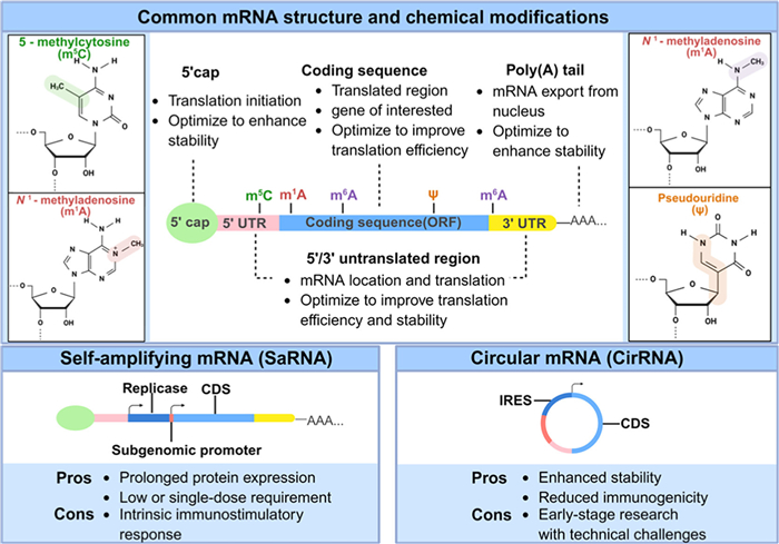
Figure 1.
Schematic representation of different types of mRNA cargos, including conventional mRNA, saRNA, and circRNA and common chemical modifications in mRNA. Created with BioRender.com .


Since its discovery in the 1960s, messenger RNA (mRNA) has been continuously studied and applied by researchers [1]. The exploration of mRNA's therapeutic potential began with the use of in vitro transcribed (IVT) mRNA in mouse models [2]. However, its early progress was slowed by problems with instability, low protein production, and poor delivery methods. In the past three decades, advances in chemical modifications and delivery technologies have greatly improved mRNA stability and reduced immune reactions [3]. Specifically, the coronavirus disease 2019 (COVID-19) mRNA-lipid nanoparticle (LNP) vaccine, as the first approved mRNA drug for human use, proved the safety and effectiveness of mRNA technology. It also showed that mRNA could be used for other medical treatments [4,5]. Chemically modified mRNA offers several advantages: It is biocompatible, allows for controlled temporary protein expression based on dosage, avoids activating the innate immune response, and can be made in large amounts.
Unlike DNA, mRNAs do not integrate into the host genome, so it is less likely to cause problems like mutations [6,7]. These properties have enabled mRNA therapeutics to show broad applications across diverse areas [8], including cancer immunotherapy [9-11], vaccine development [12-15], protein replacement therapy [16-18], cellular reprogramming [19,20] and gene editing therapies [21-23]. Compared with other RNA modalities, mRNA imposes distinct requirements on its delivery systems due to its size and mechanism of action. While small RNAs like siRNA and miRNA, primarily function through gene silencing or regulation and demand carriers that ensure cytoplasmic stability and release [24,25], mRNA is uniquely designed for protein expression. This necessitates delivery platforms that not only protect the larger, more labile mRNA molecule but also actively facilitate its endosomal escape and subsequent access to the ribosome. These requirements have positioned LNPs, extracellular vesicles (EVs), and biomaterial-based scaffolds as central to advancing mRNA therapeutics. Importantly, such delivery innovations are particularly relevant to dermatology, where unique skin barriers and regenerative demands pose additional challenges.
Thanks to a deeper understanding of gene expression during tissue repair and aging [26-29], recent studies have emphasized the promise of mRNA therapies for skin regeneration and rejuvenation. As the body's largest organ, the skin protects internal organs, bones, muscles, and other soft tissues. It comprises three main structures: The epidermis, the dermis, and the subcutaneous tissue. The skin plays a crucial role in resisting external stimuli, preventing pathogenic microorganisms and chemicals, and maintaining water and electrolyte balance as well as thermal homeostasis in the body [30,31]. When the skin encounters external injury, the temporary disruption of local function usually triggers a series of healing and repair responses while initiating complex physiological and pathological changes [32-34]. Skin repair and aging are closely linked, involving reactions to external stimuli and cellular damage. Effective regeneration and rejuvenation are, therefore, central to dermatological care. Gene-based therapies, including mRNA, offer a promising approach to delivering key endogenous factors directly to damaged skin, leading to higher levels of tissue regeneration.
This article explores the latest progress in mRNA therapy for skin regeneration and rejuvenation. We analyze the remaining challenges and discuss emerging solutions and future directions. We aim to provide a comprehensive perspective and a theoretical foundation for advancing mRNA therapy in dermatological applications by addressing these topics (Figs. S1 and S2 in Supporting information).
The clinical translation of mRNA therapeutics remains challenging, with only a limited number of drug candidates progressing to clinical trials. Key hurdles include poor stability, high immunogenicity, short half-life, limited targeting ability, and inefficient delivery to the cytoplasm [35,36].
To tackle these challenges, chemical modifications to mRNAs are critical to improve their stability and minimize immunogenicity [37]. At the same time, developing efficient and safe in vivo delivery platforms is essential, along with innovative tissue-engineered scaffolds [38]. These approaches aim to improve mRNA drug penetration, enhance transfection efficiency, and enable localized and sustained delivery to the target tissue. In this section, we focus on the basic information of the key points mentioned and recent advancements related to skin.
Due to size, synthetic mRNAs for clinical use are typically generated via IVT from DNA plasmids using phage RNA polymerase. mRNA functional domains generally consist of 5′-untranslated region (5′-UTR), the 3′-UTR, and the coding sequence (CDS) [39]. Specific nucleoside modifications such as the 5-methylcytosine (m5C), N1-methyladenosine (m1A), and N6-methyladenosine (m6A), as well as the incorporation of pseudouridine (
Researchers are also exploring new modifications, such as dual chemical modifications, demonstrating that alterations of
Computational advancements have also revolutionized mRNA design. With ∼2.4 million potential mRNA sequences for the severe acute respiratory syndrome coronavirus 2 (SARS-CoV-2) spiking protein due to synonymous codons, the LinearDesign algorithm can find the optimal mRNA design in 11 min. This approach has notably improved mRNA stability, protein expression, and antibody production, with studies showing a 128-fold increase in antibody titers in mice [45]. Codon optimization algorithms further refine mRNA sequences for enhanced translational output, offering a pathway toward unmodified therapeutic mRNAs. The UTRs are also essential in maintaining stability and controlling translation. Many mRNA therapies choose UTR sequences from highly expressed human genes like α- and β-globin. Recent research suggests that alternative UTR designs may achieve even greater protein expression levels [46]. Deep learning models are now used to optimize UTR sequences, making significant progress in genome-editing mRNA applications [47,48].
A key limitation of conventional mRNA therapies is their relatively short protein expression period, which requires repeated dosing. To overcome this challenge, researchers are developing advanced mRNA platforms, including self-amplifying mRNAs (saRNAs) and circular RNAs (circRNAs) (Fig. 2) [49]. Self-amplifying mRNAs (saRNAs) have a special design with a viral replicase-CDS and a subgenomic promoter inside the open reading frame (ORF). This design enables the synthesis of RNA-dependent RNA polymerase (RDRP), which enables mRNA to self-replicate. Consequently, saRNAs achieve sustained protein production with minimal or single dosing [50], and they can even achieve similar protein expression levels with up to a tenfold lower dose [7]. However, saRNAs have an intrinsic immunostimulatory response that may interfere with intracellular signaling during self-replication [39]. In a study on diabetic wound healing, the translation efficiency of saRNA encoding hepatocyte growth factor (HGF) was reduced by over 80%, significantly hindering therapeutic outcomes. To address this, the research team co-delivered an auxiliary mRNA encoding the vaccinia virus E3 protein with the therapeutic saRNA. The E3 protein inhibits the protein kinase R (PKR) phosphorylation pathway, effectively resolving immune interference with saRNA translation. This approach not only achieves long-term therapeutic effects but also gradually overcomes immune barriers, offering a new technical paradigm for chronic disease management and tissue regeneration [51]. circRNAs offer a promising alternative to linear mRNA treatments [52]. They are formed from precursor RNAs through an intramolecular ring-forming process. Due to their looped structure that's held together by covalent bonds, circRNAs exhibit enhanced stability for resisting degradation by exonucleases. Furthermore, they show lower immunogenicity compared to linear mRNAs. A key advantage of circRNAs is their ability to directly drive protein expression in host cells without converting into a linear form. The presence of specific elements like internal ribosome entry site (IRES) sequences and m6A modifications might enable this capability. These parts help bring ribosomes to circRNAs and start the process of translation [49,53]. For skin regeneration, circRNAs have shown remarkable promise. For example, research by Huang and colleagues shows that circCDK13 can encourage the proliferation and migration of human epidermal keratinocytes and dermal fibroblasts [54]. However, circRNA research remains in its early stages. There is an urgent need for standardized protocols to isolate, characterize, express, and delete circRNAs to realize their therapeutic potential [55].
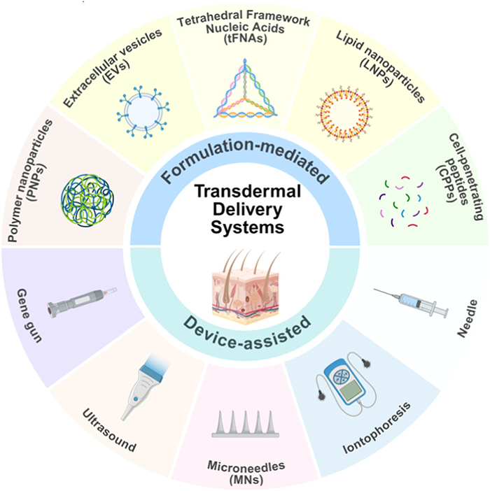
Because of its negative charge, mRNA can break down quickly by enzymes both in vivo and in vitro [56], which makes effective delivery vehicles crucial for mRNA therapy. The skin’s barrier function, especially the stratum corneum (SC), poses a major obstacle to the diffusion and penetration of drug molecules. This challenge is further compounded in damaged skin, such as in wounds, where oxidative stress and ischemia disrupt the physiological microenvironment, complicating treatment [57]. This section primarily focuses on non-viral mRNA delivery systems like LNPs, EVs, and tetrahedral framework nucleic acids (tFNAs), as well as administration methods reliant on medical devices, such as microneedles (MNs) and needle-free injections (Fig. 2). The description of tFNAs (Figs. S3, S4 and Table S1 in Supporting information) and other formulation-mediated transdermal delivery materials is in the supporting information (Tables S2 and S3 in Supporting information).
LNPs are the most commonly studied mRNA delivery system [58-60]. They comprise ionizable lipids, auxiliary phospholipids, polyethylene glycol (PEG) lipids, and cholesterol [61]. These components collectively self-assemble into stable nanoparticles that encapsulate and protect mRNA from ribonuclease (RNase)-mediated degradation, while also providing a versatile strategy to overcome multiple intracellular delivery barriers. LNPs are internalized by the cell through endocytosis, and upon reaching the acidic environment of the endosome, they become positively charged, which aids in the release of RNA into the cytoplasm [62]. Although commercial products like liposomes or TransITTM RNA have been utilized for in vitro mRNA delivery [17], the cytotoxicity and instability of LNPs limit their broader application [63]. However, chemical modification and scaling of LNPs components are promising strategies to enhance their function [64].
Topical use of LNPs for dermal delivery is challenging because the SC is difficult to penetrate. LNPs formulated for mRNA delivery often accumulate in the liver, likely because of interaction between apolipoprotein E (ApoE) on the LNPs surface and ApoE receptors on hepatocytes. Several studies have attempted to optimize LNPs delivery for skin applications. Researchers have focused on receptors expressed by skin cells. Based on the mannose receptor expressed by dermal fibroblasts and the low-density lipoprotein receptor (LDLR) expressed by epidermal keratinocytes, researchers have devised the co-delivery of LNPs using unstable PEG lipids and ApoE. The unstable PEG lipids can be rapidly dissociated after reaching the target cells, promoting cell uptake; The ApoE modification enhanced the specific binding of LNPs to LDLR. Experiments confirmed that this modification strategy significantly improved the transfection efficiency of LNPs in primary human skin cells [65]. Blakney used a design of experiments (DoE) method to refine the delivery of LNPs carrying saRNAs for luciferase into human skin. Their findings showed that skin immune cells selectively internalize and express saRNAs and highlighted the need for further exploration of how LNPs formulations can be tailored to improve uptake by epithelial cells and fibroblasts for improved protein expression [66]. They also investigated how complex lipids affect cellular uptake and mRNA expression, aiming to maximize protein expression in different human skin ex vivo models [67].
Beyond delivery efficiency, the inherent immunostimulatory nature of LNPs poses a significant hurdle for therapeutic applications, especially in inflammatory conditions. To address this, innovative LNP designs are emerging that focus on mitigating inflammatory responses. For instance, one study incorporated lipoic acid, an antioxidant molecule, into the ionizable lipid structure. The resulting LNP formulation demonstrated dual functionality: It effectively facilitated transdermal delivery of clustered regularly interspaced short palindromic repeats-CRISPR-associated protein 9 (CRISPR-Cas9) mRNA and simultaneously suppressed reactive oxygen species (ROS) and pro-inflammatory cytokines in a psoriasis model, leading to significant symptom alleviation [68]. This example illustrates that rational LNP design can concurrently overcome both physical delivery barriers and undesirable immune activation, paving the way for mRNA therapeutics in inflammatory skin diseases.
In vitro studies are also a significant challenge for mRNA therapeutics. Daniel Anderson developed a barcode-based screening system to identify novel biodegradable LNPs. They screened >400 LNPs formulations containing 384 ionizable lipids in just 8 mice [69]. Another approach, known as proteomics-associated LNP screening (PALS), involves using barcodes encoded at the 3′ end of functional mRNA molecules to evaluate the efficiency of LNPs in delivering mRNA [70]. Additionally, artificial intelligence has been used for the screening and optimization of LNPs in mRNA delivery, and has achieved certain results [71,72]. Future studies need to explore further the chemical modification of LNPs, lipid composition adjustment, and targeting of specific cells and tissues for safer and more effective mRNA therapies [73].
EVs are innate nanoscale structures made up of phospholipid bilayers, which are actively secreted by both eukaryotic and prokaryotic cells. EVs function as natural carriers that transport and deliver molecular cargo between cells via endocytosis and other cytotransport mechanisms. Among them, exosomes are the most studied and characterized EVs [74]. Compared to LNPs, EVs offer lower immunogenicity and more extraordinary targeting ability, making them better suited for crossing biological barriers [75]. In a full-thickness skin wound model, mice receiving five consecutive injections of vascular endothelial growth factor A (VEGF-A) mRNA loaded EVs exhibited markedly accelerated wound healing. This repeated dosing regimen did not elicit detectable immunogenic reactions. In contrast, both adeno-associated virus (AAV) and LNP-based VEGF-A delivery systems failed to achieve comparable therapeutic outcomes under identical repeated administration conditions [76]. These findings position EVs as a highly promising intervention tool in regenerative medicine, with broad application prospects [77,78].
EVs derived from various origins contain unique components inherited from their parent cells, endowing them with distinct functional capabilities. Take mesenchymal stem cell-derived EVs (MSC-EVs) as an instance; they are capable of transporting proteins, genetic material, and signaling factors that preserve stemness and elicit reparative behaviors. These EVs can also suppress cell death, regulate immune reactions, and facilitate the repair of damaged tissues [79]. Similarly, saliva-derived exosomes (Saliva-Exos) have been found to encapsulate mRNAs that enhance endothelial cell function and accelerate wound healing. Delivery of UBE2O mRNA via these exosomes downregulates SMAD family member 6 (SMAD6) expression, leading to subsequent activation of the bone morphogenetic protein 2 (BMP2) signaling pathway and ultimately promoting angiogenesis [80].
Other EV sources show distinct therapeutic advantages: Fibroblast-derived EVs promote collagen synthesis and re-epithelialization through Akt/β-catenin signaling, while keratinocyte-derived EVs enhance cell proliferation via mitogen-activated protein kinase (MAPK)/extracellular signal-regulated kinase (ERK) pathways. Although not yet applied in mRNA delivery, these native EVs possess inherent tropism for skin cells, positioning them as promising vehicles for future mRNA-based wound therapies.The diverse functional profiles of EVs from various sources, including macrophages, platelets, and endothelial cells, enable precise regulation of wound healing phases, highlighting their potential as targeted therapeutic platforms in regenerative medicine [81].
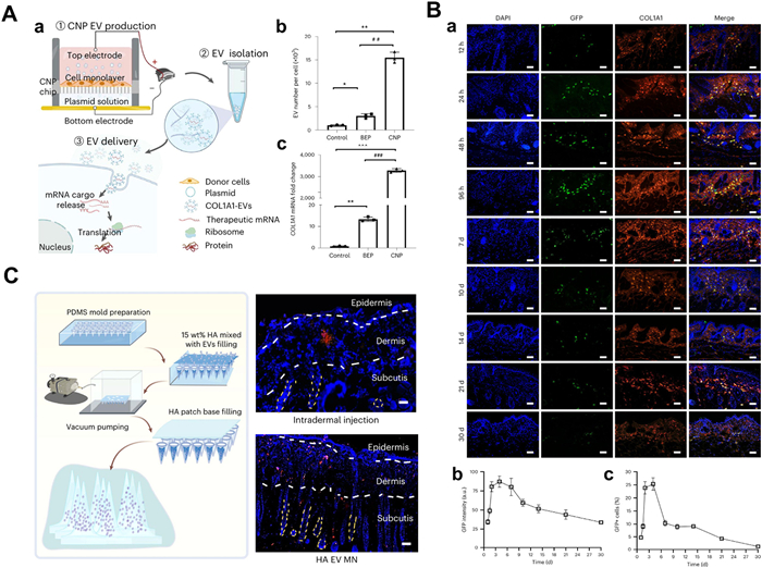
Despite their therapeutic potential, the clinical translation of EV-based mRNA delivery has been constrained by challenges in scalable production and efficient loading of exogenous mRNA. Andrew and colleagues demonstrated that cellular nanoporation (CNP) can encourage cells to generate and emit exosomes that incorporate specific nucleotide sequences. This method uses nanostructured electrodes to apply localized electrical stimulation to cells, which temporarily permeabilizes cell membranes, promoting the uptake of specific genetic material and significantly enhancing exosome secretion. Compared to other exosome production strategies, a 50-fold enhancement in exosome count was achieved through CNP, even in cells that inherently secrete exosomes at low rates. It also showed an over 1000-fold surge in mRNA content within exosomes (Fig. 3A) [82]. The CNP approach offers a new strategy for improving the production of exosomes and their use as multifunctional genetic material vectors. Andrew and colleagues combine different cellular nanoelectroporation systems with mRNA therapy to apply in diverse fields such as oncology, cancer therapy [83,84], bone regeneration [85], and collagen regeneration [86]. Therefore, EV as a carrier has excellent therapeutic potential and clinical value for skin regeneration and rejuvenation [87,88]. By properly selecting and engineering EV-derived cells, targeted delivery systems can be developed for more precise treatment strategies [89,90].
The method of administration significantly influences both treatment effectiveness and patient compliance. This is especially true for nucleic acid drugs, such as mRNAs. Direct Injection into the skin rather than systemic delivery offers benefits like stronger local treatment effects and a lower chance of quick removal. However, the skin's barrier properties present challenges for efficient gene delivery. Therefore, in addition to gene carriers such as LNPs and multilamellar bodies, physical devices are widely used to improve delivery efficiency. Traditional intradermal drug delivery is usually achieved using invasive methods, such as needle injections. These approaches cause discomfort, swelling, and risk of infection and require skilled personnel to perform the procedure. To overcome these limitations, numerous noninvasive and minimally invasive methods have been investigated [91,92], including iontophoresis (ItP) [93], ultrasound, electroporation, gene gun [94], and needleless injection [95]. Darade et al. systematically compared various transdermal strategies, demonstrating that both intradermal injection and MN administration achieved robust protein expression across epidermal and dermal layers, with significantly higher delivery efficiency than fractional laser ablation [96]. Notably, MN systems maintained comparable efficacy to conventional injections while addressing their inherent limitations, positioning them as a transformative technology for nucleic acid delivery.
MNs, measuring from 25 µm to 2000 µm in length, have shown great promise for treating various dermatological conditions, such as acne, atopic dermatitis, psoriasis, and skin cancer [97,98]. Their use promotes skin proliferation and barrier renewal, enhancing therapeutic outcomes [99]. MN platforms employ distinct mechanisms to overcome specific cutaneous barriers. Solid MNs, typically fabricated from metallic materials, create temporary microchannels through the SC, enabling subsequent permeation of topical formulations. This two-step approach effectively breaches the primary physical barrier while maintaining the structural integrity of therapeutic payloads. Hollow MNs function as direct conduits for intradermal infusion, bypassing the SC entirely through precise interstitial delivery. Remarkably, studies using porcine skin models have demonstrated that hollow MNs facilitate uniform protein expression even with unformulated mRNA, suggesting their potential for simplified delivery protocols [100].
For sustained release applications, biodegradable systems offer advanced solutions. Dissolving MNs, fabricated from polymers such as polyvinylpyrrolidone (PVP) or hyaluronic acid, release their payload gradually as the matrix degrades within the skin microenvironment. The tunable degradation kinetics of these systems enable extended therapeutic expression, as demonstrated by PVP-based RNA patches that maintained luciferase expression for up to 72 h. Hydrogel MNs represent a further refinement, utilizing swellable polymers that absorb interstitial fluid to form controlled-release networks. These systems are particularly valuable for chronic conditions requiring prolonged drug exposure, as their porous architecture modulates release kinetics while maintaining a favorable biocompatibility profile [101].
Beyond penetration enhancement, MN technologies address mRNA stability challenges through innovative formulation strategies. Incorporating mRNA in a solid form like cryomicroneedle (CryoMN) technology reduces the complexities associated with liquid mRNA formulations and maximizes mRNA stability [102]. Another promising approach is using one-shot vaccine printers (MVPs) to manufacture MNs containing mRNA vaccines encapsulated in LNPs or other payloads. The MN printing process involves doping stabilized, soluble polymers into mRNA-LNP inks. After extensive screening, researchers found that these formulations could maintain the stability of LNP-encapsulated mRNA vaccines at room temperature for a minimum of six months [103].
Building on this, researchers innovatively combined MN technology to integrate nucleic acid drug delivery with medical data management, creating a new system named on-patient medical record-keeping (OPMR) technology for storing case information. In a study, poly (methyl methacrylate) particles with near-infrared (NIR) fluorescent dye and therapeutic mRNA were incorporated into dissolving MN patches. This system provided treatment while storing and transmitting medical information in the skin. Animal experiments showed the system maintained nearly 90% fluorescence intensity after 30 days and the encoded information remained identifiable after 70 days, indicating excellent spatiotemporal stability. Histological analysis confirmed only transient mild inflammation at the implantation site, with no long-term adverse effects like hyperkeratosis or fibrosis. This platform is suitable for any mRNA-based therapeutics in transdermal delivery. It could manage chronic diseases by enabling sustained therapeutic protein expression via a single MN delivery and securely storing key patient information, offering hardware support for telemedicine and personalized treatment [104].
Among drug delivery methods, MN-based transdermal delivery excels by increasing drug bioavailability with little to no discomfort. Advances in micro- and nanofabrication, along with innovations in nanotechnology, are expected to increase payloads per patch, improve the stability of drugs, and optimize transdermal drug delivery efficiency. Unique MN designs allow for the sustained release of drugs, rendering them a promising option for chronic disease management. This approach reduces dosing frequency and enhances patient adherence. Continued research on MNs and nanocarriers holds great potential for accelerating their clinical application in transdermal drug delivery [105].
There is a structured sequence in skin wound healing, typically consisting of four phases: Hemostasis, inflammation, proliferation, and remodeling [79,106]. These interdependent phases involve dynamic interactions between cells, cytokines, signaling molecules, and other mechanisms that work together to ensure effective skin repair following injury [107]. However, poor healing due to trauma, surgery, and acute or chronic disease affects millions of people worldwide each year, reflecting impaired regulation of healthy tissue repair responses [108]. Also, it is suggested that poor wound healing is a key aspect of the onset of various diseases, especially chronic diseases [109]. Regenerative wound therapy is an emerging and rapidly growing field aimed at restoring the skin's original function and rebuilding damaged cells and tissues without scarring. Evidence supports the efficacy of using cytokines and growth factors to enhance wound healing. Growth factors like VEGF, fibroblast growth factor (FGF), keratinocyte growth factor (KGF), epidermal growth factor (EGF), or platelet-derived growth factor (PDGF) have been seen as promising targets for mRNA therapies to treat cutaneous wound healing [110].
VEGF-A is crucial for angiogenesis and has been shown to enhance wound healing through the promotion of new blood vessel formation [111]. The healing effectiveness of VEGF-A mRNA has been further enhanced by integrating advanced delivery systems like ionizable LNPs. Using microfluidic methods, Li et al. encapsulated VEGF-A mRNA within an ionizable lipid-based delivery system, demonstrating significant improvements in wound healing, particularly in a diabetic wound model. After 14 days, wounds treated with LNP/VEGF-A mRNA achieved an average wound area of only 2.47%, significantly outperforming groups treated with mRNA alone (11.93%), LNPs alone (21.55%), or phosphate-buffered saline (PBS) (22.08%) [112,113].
Furthermore, the team optimized their approach by synthesizing VEGF-A circRNA through a self-catalyzing type Ⅰ intron strategy. This method demonstrated enhanced stability and sustained protein expression, attributed to the circRNAs’ resistance to degradation and the protective effects of the LNPs. Encapsulated circRNAs exhibited exceptional stability at low temperatures. In vivo experiments revealed that by day 12, diabetic mice administered U-LNP/VEGF-A circRNA in a single dose showed nearly complete wound closure, while wounds persisted in all other groups [114].
FGF, particularly FGF-2, plays a key role in re-epithelialization, extracellular matrix (ECM) production, keratinocyte proliferation, and angiogenesis. However, the short intracellular half-life of FGF-encoding mRNA restricts local protein synthesis. To overcome this, researchers developed mineralized microparticles (MCMs) that allow proteins to be gradually released over time in diabetic mouse models, enhancing wound healing through the delivery of FGF-2-modified nucleoside mRNA. The bioactivity of mRNA-encoded basic fibroblast growth factor (bFGF) produced by cells was significantly higher than recombinant human bFGF (rhbFGF). This suggests that gene delivery can activate endogenous autocrine and paracrine signaling pathways, offering advantages over traditional protein delivery methods [115].
Recent research has also explored the role of inflammatory factors in wound healing, particularly the impact of macrophage phenotypes on the process. Persistent inflammation in wounds is often linked to the M1 macrophage phenotype, which is pro-inflammatory, whereas the M2 macrophage phenotype enhances wound healing through anti-inflammatory responses [116]. Wang et al. used interleukin-4 (IL-4) mRNA to prepare TS2 LNPs to reprogram macrophages from the M1 phenotype to the M2 phenotype. The TS2-IL4 LNP-mRNA-treated group showed faster wound healing starting on day 9, achieving about 80% wound closure by day 12. Moreover, the percentage of M2 macrophages in this group was considerably higher compared to the untreated free IL-4 mRNA and MC3-IL-4 LNP-mRNA groups, with increases of 1.8, 1.5, and 1.3 times, respectively [117]. Researchers used adipose-derived stem cells (hADSCs) loaded with IL-10 mRNA modified chemically. They found that IL-10 expression peaked at 24 h post-transfection and gradually declined over the next two days. However, early IL-10 bursts in diabetic wounds may not always be beneficial, potentially leading to less favorable wound healing outcomes. Therefore, in vivo studies are required to optimize the timing and dosing of IL-10 mRNA to ensure safe and practical application in further study in wound healing [118].
Additionally, other signaling pathways also play key roles in wound healing. For instance, the stromal cell-derived factor 1 (SDF-1)/C-X-C chemokine receptor type 4 (CXCR4) signaling pathway, which stimulates epidermal stem cell migration and proliferation, is crucial for epithelialization [119]. Luo et al. combined small skin grafts with SDF-1α modRNA to enhance skin defect repair. Immunofluorescence analysis revealed that skin grafts treated with SDF-1α modRNA demonstrated greater cell proliferation by day 21 post-operation, while the control group showed cell apoptosis. This approach demonstrated the ability to promote blood perfusion and inhibit cell apoptosis, highlighting its potential to enhance skin regeneration in diabetic full-thickness injury models [120].
Telomerase sustains telomere length and is essential for regulating cellular aging and proliferation [121]. Chang et al. used the optimized human telomerase reverse transcriptase (hTERT) mRNA LNPs system to transfect keratinocytes, fibroblasts, and donor-derived autologous human skin cell suspensions (hSCS) (Figs. S5A and B in Supporting information). This treatment boosted telomerase activity and evaluated key markers such as Lamin A, 53BP1, p21, and Ki67. The results demonstrated that the human cells in the hTERT mRNA LNPs group doubled in number, with a fivefold increase in proliferating cells compared to control groups, and markers of cellular senescence and DNA damage notably reduced (Fig. S5D in Supporting information). These findings suggest that mRNA-encoding telomerase could improve cell viability and actively promote wound healing [122].
Combination therapy using multiple mRNAs has shown an additive effect compared to monotherapy. In research by Tejedor et al., FGF was found to be a potential co-candidate with VEGF-A for wound healing. The concurrent delivery of FGF1 mRNA and VEGF-A mRNA was shown to upregulate 10 genes associated with skin growth, blood vessel formation, and epithelial cell proliferation, including matrix metalloproteinase 3 (MMP3), placental growth factor (PGF), midkine (MDK), tissue inhibitor of metalloproteinase 1 (TIMP1), fibulin-2 (FBLN2), ornithine decarboxylase 1 (ODC1), progestin and adipoQ receptor family member 3 (PAQR3), and high mobility group AT-hook 2 (HMGA2) in a diabetic mouse model. The gene upregulation was only observed with the combined mRNA therapy, suggesting that it may reduce the therapeutic agent's effective dose while maintaining or increasing the therapeutic effect [123]. Wang et al. explored the effectiveness of human adipose-derived stem cells (hADSCs) loaded with transforming growth factor beta 3 (TGF-β3) and IL-10 mRNA in promoting scarless wound repair. In a preclinical model, ADSCs loaded with both showed heightened MMP expression and reduced myofibroblast activation along with decreased collagen deposition. This combination therapy counteracted the wound healing delay observed with ADSCs-IL-10 alone, demonstrating the potential of co-delivered mRNA therapies to reduce scar formation and promote more efficient wound healing [124]. Current non-clinical/clinical approaches for skin wound healing by mRNA therapy are included in Table S4 (Supporting information).
Intrinsic aging and extrinsic aging are two mechanisms of the skin's aging process. Intrinsic aging, a natural and inevitable process, affects tissues throughout the body over time. External aging, also known as photoaging, is primarily driven by prolonged ultraviolet (UV) radiation exposure and is affected by environmental factors including lifestyle, diet, and stress response [125-127]. Studies have shown that the condition of the skin can reflect an individual's overall health, risk of mortality, and life expectancy [128]. Skin aging involves changes in gene expression that disrupt cellular homeostasis. These changes are marked by increased production of MMPs, accumulation of oxidative stress, and chronic inflammation, which serve as molecular indicators of deteriorating skin quality, leading to a decline in the function and quality of the entire tissue [129]. Aging also leads to skin atrophy, characterized by diminished skin cell proliferation and the breakdown of ECM proteins. As a result, older skin exhibits increased stiffness, reduced elasticity, and lower tensile strength compared to younger skin, making it more prone to injuries and stress-related damage [130].
Given the characteristics and molecular changes of aging skin, current mRNA therapies focus on these targets. By regulating the expression of key molecules, mRNA therapy has the potential to enhance skin cell proliferation and increase the amount of ECM proteins. This could lead to an enhancement in the skin's overall function and quality. Collagen is the richest protein in the skin, constituting 90.1% of total matrix proteins. The loss of collagen that cannot be restored is a sign of skin aging, which results in skin atrophy [131]. In response, You et al. engineered EVs carrying human collagen I alpha I (COL1A1) mRNA to assess its potential to replenish collagen in UV-irradiated mouse skin. The study found that the corresponding protein began to be translated at 12 h and peaked on day 4, with effects lasting for several weeks, depending on the dose (Fig. 3B). After COL1A1-EV treatment, the number and area of wrinkles decreased from day 7, significantly decreasing to levels observed in non-irradiated sham controls by the day 14. They also successfully applied MNs, which allowed for uniform distribution of EVs into the dermis and subcutis, resulting in long-term retention without membrane rupture (Fig. 3C). On further examination of collagen replacement and skin wrinkle improvement, the treatment of COL1A1-EV MN patches had the greatest collagen fiber deposition and dermal thickness, with these effects persisting for up to three months. This group also showed the most notable wrinkle improvement at 70 days, outlasting the effects of syringe administration by a factor of two [86].
Elastin, another vital ECM protein, is crucial in restoring skin function and elasticity [132]. Since elastin is synthesized in early development and not replaced. It is typically lost irreversibly, leading to impaired tissue function. Avci-Adali et al. addressed this by microinjecting synthetic modified tropoelastin (TE) mRNA into cells and pig skin, significantly increasing elastin synthesis, with an approximately 1.2-fold increase in elastin content in ex vivo pig skin, equivalent to a 20% increase [133]. Given the long half-life of elastin, even a few treatments or one could improve the elasticity of affected tissues. They also stressed that it could ensure the most effective protein expression by refining synthetic mRNA. After screening various codon-optimized and nucleotide-modified TE mRNAs, they found the me1
Oxidative stress is a hallmark of skin aging, with reductions in key antioxidant genes such as glutathione S-transferase M2 (GSTM2) in aged skin. As shown in Fig. S6 (Supporting information), it is demonstrated that treatment with EVs carrying GSTM2 (EVs-Gstm2) enhanced mitochondrial oxidative phosphorylation in senescent fibroblasts, improved oxidative stress, and promoted epidermal cell renewal via paracrine signaling (Fig. S6A). In aged mouse models, EVs-Gstm2 increased collagen expression by 40% and reduced MMP2 levels (Fig. S6B). In wound healing models, EVs-Gstm2 accelerated closure rates, achieving 97% wound closure by day 12 compared to 87% in controls. Histological analyses also revealed denser and more organized collagen deposition, showcasing its efficacy in improving skin homeostasis and wound healing (Fig. S6C) [135].
Stem cells are essential for maintaining tissue homeostasis and repair. Through single-cell omics analysis, recent studies have highlighted the importance of basal stem cells in skin aging, revealing their age-related changes. Activating transcription factor 3 (ATF3), a transcription factor within basal stem cells, has been identified as a pivotal regulator [136]. Treatment with mRNA encoding ATF3 in cellular experiments significantly increased the expression of critical skin proteins. This modulation also reduced fibroblast senescence and enhanced collagen synthesis, emphasizing its potential for reversing skin aging.
Also, the discovery of induced pluripotent stem cells (iPSCs) by Yamanaka et al. using four transcription factors (octamer-binding transcription factor 4 (OCT4), Krüppel-like factor 4 (KLF4), SRY-box transcription factor 2 (SOX2), cellular myelocytomatosis oncogene (c-Myc)) revolutionized regenerative medicine [137]. Browder et al. demonstrated that cyclic partial reprogramming in aging mice using these factors prevented age-related histological changes and enhanced wound healing without adverse effects, even with long-term applications [138]. mRNA therapy offers an innovative approach for transient reprogramming by avoiding issues associated with gene expression vectors, providing direct protein translation, and increasing reprogramming efficiency [139-141]. This method has been used to introduce reprogramming factors that rejuvenate skin cells and counteract the clinical symptoms of aging [142,143].
Siddharth et al. utilized LNPs to deliver a mix of mRNAs encoding OCT4, SOX2, KLF4, LIN28, C-MYC, and NANOG (OSKLMN). This transient epigenetic reprogramming increased fibroblast proliferation, reduced markers of collagen degradation (MMP1) and oxidative stress (SOD2), and slowed downstream aging processes. These effects were sustained through multiple cell passages, indicating long-lasting rejuvenation [144]. Similarly, Hsia et al. used mRNA-loaded LNPs to treat adult fibroblasts and keratinocytes, leading to enriched ECM protein deposition, reduced cytokine signaling, and increased proliferation [145]. However, these studies are currently limited to cellular models, necessitating further evaluation in animal models to confirm safety and efficacy. Although mRNA-based methods or safer genes or small molecules that act as chromatin regulators instead of OSKM genes are promising, a comprehensive assessment and mechanism study is needed to determine their safety and efficacy in humans and explore their potential for clinical treatment applications. Also, alternative strategies, such as utilizing safer genes or small-molecule chromatin regulators instead of traditional reprogramming factors, may enhance clinical viability. Comprehensive mechanism studies and clinical trials are crucial to translating these advancements into effective treatments for human skin aging. Current non-clinical approaches to mRNA therapy for skin anti-aging are included in Table S5 (Supporting information).
Although preclinical studies have fully demonstrated the potential of mRNA therapy in skin regeneration and anti-aging, its clinical translation is still in its early stages. At present, the skin therapies with mRNA as the active ingredient that have entered clinical research mainly focus on two directions: One is the tissue repair strategy centered on protein replacement, and the other is the immunotherapy for malignant skin tumors.
In the field of tissue repair, the VEGF-A mRNA candidate drug AZD8601 is the most representative. Preclinical studies have shown that in diabetic mouse models, subcutaneous injection of AZD8601 can dose-dependently improve local oxygen metabolism, promote angiogenesis and tissue reconstruction. The pharmacokinetic-pharmacodynamic model further revealed the crucial significance of cumulative administration in maintaining the continuous healing effect, providing an important basis for the design of clinical administration regimens [146]. On this basis, researchers have created models to evaluate how AZD8601 influences wound healing through its pharmacokinetic and pharmacodynamic properties. These models revealed that cumulative dosing was key to sustained wound healing acceleration [147].
To assess the safety of VEGF-A mRNA (AZD8601), Gan conducted a randomized, double-blind, placebo-controlled Phase 1 trial involving patients with type 2 diabetes mellitus. It showed good tolerability, with minor injection site reactions as the sole treatment-related adverse event, likely due to VEGF-A-induced vasodilation [148]. The development process of AZD8601 marks the successful expansion of mRNA therapy from infectious disease vaccines to the field of skin regeneration, verifying the feasibility of regulating human skin physiological processes through local expression of growth factors.
In contrast, mRNA has demonstrated more active and diversified progress in the field of skin tumor immunotherapy. Fixed-antigen vaccines, exemplified by BNT111, which deliver mRNA encoding multiple melanoma-associated antigens intravenously via LNPs, have been evaluated in Phase Ⅱ clinical trials in combination with immune checkpoint inhibitors. These vaccines have achieved significant response rates in patients with advanced melanoma that has relapsed after programmed cell death protein 1 (PD-1) blockade and have received fast track designation from the Food and Drug Administration (FDA) [149]. This success demonstrates that mRNA vaccines can elicit robust systemic T-cell immunity.
Overall, clinical research on mRNA skin therapy has shown a clear differentiation pattern: In non-neoplastic skin diseases, the number of candidate drugs is still limited, and AZD8601 is one of the few representatives that have entered the clinical stage and proven safety; In the field of skin tumors, mRNA technology has demonstrated diverse pathways and broad prospects, covering strategies such as systemic vaccines and local immune regulation. This current situation highlights the opportunities and challenges in the future, how to further apply the successful experience accumulated in tumor immunotherapy to a wider range of clinical needs such as chronic wounds, scars, hereditary skin diseases and skin rejuvenation. The progress of AZD8601 has laid a key feasibility foundation for this transformation, and the continuous innovation in formulation technology, administration strategies and combination therapy in the field of tumor immunology has also provided a strong impetus for the expansion of mRNA therapy in dermatology.
Through this review, we explored the applications of mRNA therapy in skin regeneration and rejuvenation. Delivering mRNA encoding skin repair-related key proteins induces high expression of factors that enhance cell adhesion, migration, differentiation, angiogenesis, and growth factor release, supporting its potential for promoting wound healing and mitigating aging effects.
Advances in mRNA stability, particularly via nucleotide modifications and circRNA engineering, have extended mRNA half-lives from hours to weeks while reducing innate immune activation. Concurrently, delivery systems have advanced beyond traditional LNPs. Notably, skin offers unique advantages as a target organ for mRNA therapy: Local injection or grafting enables direct delivery to the site of action, reducing systemic side effects and facilitating complication monitoring.
While mRNA therapies hold significant promise, skin-related clinical studies remain limited. The clinical translation of mRNA therapies for non-life-threatening dermatological conditions, such as diabetic ulcers or photoaging, is subject to particularly stringent safety requirements. Because repeated dosing is often needed to maintain therapeutic benefit, treatments must demonstrate no systemic exposure, no worsening of local inflammation, and no carcinogenic risk, especially in photo-damaged skin that already carries a high mutational burden.
Clinical development is further complicated by patient heterogeneity. Variables such as age, skin barrier integrity, and microbiome composition markedly influence drug penetration and immune response. As a result, dosing regimens effective in young or healthy skin may not translate to elderly or photo-damaged skin, highlighting the need for broader and more tailored preclinical validation.
Endpoint selection also remains a major hurdle. While “time to complete wound closure” is an accepted endpoint in ulcer studies, rejuvenation trials lack validated surrogate markers. Current measures, such as histological collagen density or wrinkle scoring, remain exploratory and are not qualified as primary endpoints, necessitating larger cohorts and longer follow-up in Phase Ⅱ/Ⅲ trials and thereby increasing both cost and complexity.
From a regulatory perspective, stringent requirements apply to product characterization, including accurate quantification of encapsulated versus free mRNA, precise analysis of particle size distribution, and tight control of dsRNA and other impurities. These raise the bar for both process development and quality assurance.
Manufacturing scalability presents additional barriers. Production of good manufacturing practice (GMP)-grade ionizable lipids is still a bottleneck: First-generation clinical lipids are restricted by patents and remain costly, while next-generation skin-penetrating lipids exist only at gram scale and require complex synthetic routes that are difficult to expand safely. Similarly, large-scale EV production is limited by low yields and costly purification, while terminal sterilization can damage carriers or the mRNA payload itself.
Until lipid supply chains diversify and high-throughput bioprocessing systems for vesicles or nanocarriers are developed, clinical trials will likely remain restricted to small-area indications such as scar revision or actinic keratosis. Without innovations in scalable manufacturing and cost reduction, broader applications in chronic wounds and skin rejuvenation will face significant economic and practical barriers.
Looking forward, the future of mRNA-based dermatology will likely be shaped by following trends. Next-generation mRNA medicines are expected to evolve from passive protein replacement to dynamic systems. Smart biomaterials or engineered lipid carriers may sense pathological cues, such as protease activity, pH shifts, or oxidative stress, and trigger controlled release or translational activation. Such “on-demand” delivery would maximize efficacy while minimizing off-target risks, a critical advance for chronic dermatological conditions.
Optimizing mRNA sequences, lipid chemistry, and formulation parameters is inherently multidimensional. AI-assisted platforms can accelerate this process, from designing skin-tropic lipids to refining codon usage for stability and translation. These advances dovetail with personalized medicine, where mRNA may be tailored to a patient’s genetic background or biomarker profile, echoing the precedent set by individualized cancer vaccines.
Beyond infectious disease and oncology, the modularity of mRNA technology positions it for monogenic skin disorders (e.g., epidermolysis bullosa), immune-mediated conditions such as psoriasis and vitiligo, and regenerative or aesthetic applications that fundamentally recalibrate skin biology.
In conclusion, while the path to clinical adoption is fraught with translational, regulatory, and manufacturing hurdles, the convergence of molecular biology, biomaterial engineering, and digital design holds the key to unlocking the full potential of mRNA-based dermatology. By addressing these challenges through interdisciplinary collaboration, mRNA therapies may soon transition from experimental promise to clinical reality, offering not only aesthetic enhancement but also fundamental restoration of skin health and function.
Mingxuan Zhang: Writing – review & editing, Writing – original draft, Visualization, Investigation, Conceptualization. Xingcheng Pan: Writing – review & editing, Investigation. Jiajie Li: Writing – review & editing, Supervision, Funding acquisition, Conceptualization. Hang Wang: Writing – review & editing, Supervision, Project administration, Funding acquisition, Conceptualization.
The authors declare that they have no known competing financial interests or personal relationships that could have appeared to influence the work reported in this paper.
We are grateful for the financial support from the National Key Research and Development Program of China (No. 2022YFC2409804), the Sichuan Provincial Natural Science Foundation (No. 2023NSFSC1500), the Research and Develop Program, West China Hospital of Stomatology Sichuan University (No. RD-02–202504), and Sichuan Provincial Natural Science Foundation (No. 2025ZNSFSC1577).
Supplementary material associated with this article can be found, in the online version, at doi:
S. Brenner, F. Jacob, M. Meselson, Nature 190 (1961) 576–581. doi: 10.1038/190576a0
J.A. Wolff, R.W. Malone, P. Williams, et al., Science 247 (1990) 1465–1468. doi: 10.1126/science.1690918
P. Huang, H. Deng, C. Wang, et al., Adv. Mater. 36 (2024) e2307822. doi: 10.1002/adma.202307822
F.P. Polack, S.J. Thomas, N. Kitchin, et al., N. Engl. J. Med. 383 (2020) 2603–2615. doi: 10.1056/nejmoa2034577
K.S. Corbett, D.K. Edwards, S.R. Leist, et al., Nature 586 (2020) 567–571. doi: 10.1038/s41586-020-2622-0
H. Parhiz, E.N. Atochina-Vasserman, D. Weissman, Lancet 403 (2024) 1192–1204. doi: 10.1016/S0140-6736(23)02444-3
E. Rohner, R. Yang, K.S. Foo, et al., Nat. Biotechnol. 40 (2022) 1586–1600. doi: 10.1038/s41587-022-01491-z
Y. Shi, M. Shi, Y. Wang, et al., Signal Transduct. Target. Ther. 9 (2024) 322. doi: 10.1038/s41392-024-02002-z
J.S. Weber, M.S. Carlino, A. Khattak, et al., Lancet 403 (2024) 632–644. doi: 10.1016/S0140-6736(23)02268-7
L.A. Rojas, Z. Sethna, K.C. Soares, et al., Nature 618 (2023) 144–150. doi: 10.1038/s41586-023-06063-y
J.F. Gainor, M.R. Patel, J.S. Weber, et al., Cancer Discov. 14 (2024) 2209–2223. doi: 10.1158/2159-8290.cd-24-0158
M. Alberer, U. Gnad-Vogt, H.S. Hong, et al., Lancet 390 (2017) 1511–1520. doi: 10.1016/S0140-6736(17)31665-3
C.D. Palmer, A.R. Rappaport, M.J. Davis, et al., Nat. Med. 28 (2022) 1619–1629. doi: 10.1038/s41591-022-01937-6
B. Essink, L. Chu, W. Seger, et al., Lancet Infect. Dis. 23 (2023) 621–633. doi: 10.1016/S1473-3099(22)00764-2
C.J. Wayne, A.K. Blakney, Cell 187 (2024) 1822 -1822.e1821. doi: 10.1016/j.cell.2024.03.018
D. Koeberl, A. Schulze, N. Sondheimer, et al., Nature 628 (2024) 872–877. doi: 10.1038/s41586-024-07266-7
M. Nawaz, S. Heydarkhan-Hagvall, B. Tangruksa, et al., Adv. Sci. 10 (2023) e2206187. doi: 10.1002/advs.202206187
D.W. Wolfson, N.K. Kim, K.H. Lee, et al., Nat. Biomed. Eng. 8 (2024) 1124–1141. doi: 10.1038/s41551-024-01211-9
J.G. Rurik, I. Tombácz, A. Yadegari, et al., Science 375 (2022) 91–96. doi: 10.1126/science.abm0594
S. Nie, Y. Qin, L. Ou, et al., Adv. Mater. 36 (2024) e2310168. doi: 10.1002/adma.202310168
L. Breda, T.E. Papp, M.P. Triebwasser, et al., Science 381 (2023) 436–443. doi: 10.1126/science.ade6967
J. Popovitz, R. Sharma, R. Hoshyar, et al., Adv. Drug Deliv. Rev. 200 (2023) 115026. doi: 10.1016/j.addr.2023.115026
S. Soroudi, M.R. Jaafari, L. Arabi, J. Control. Release 372 (2024) 113–140. doi: 10.1016/j.jconrel.2024.06.023
K. Li, M. Lu, X. Xia, et al., Chin. Chem. Lett. 32 (2021) 1010–1016. doi: 10.1016/j.cclet.2020.09.010
R. Li, M. Zhu, X. Hu, et al., Chin. Chem. Lett. 36 (2025) 110736. doi: 10.1016/j.cclet.2024.110736
J.A. Goldman, K.D. Poss, Nat. Rev. Genet. 21 (2020) 511–525. doi: 10.1038/s41576-020-0239-7
Q. Zhao, Y. Zheng, D. Zhao, et al., Protein Cell 14 (2023) 398–415.
H. Bao, J. Cao, M. Chen, et al., Sci. China Life Sci. 66 (2023) 893–1066. doi: 10.1007/s11427-023-2305-0
Z. Li, J. Cheng, P. Huang, et al., Chin. Chem. Lett. 35 (2024) 109153. doi: 10.1016/j.cclet.2023.109153
E.S. Chambers, M. Vukmanovic-Stejic, Immunology 160 (2020) 116–125. doi: 10.1111/imm.13152
P. Di Meglio, G.K. Perera, F.O. Nestle, Immunity 35 (2011) 857–869. doi: 10.1016/j.immuni.2011.12.003
S. Knoedler, L. Knoedler, M. Kauke-Navarro, et al., Mil. Med. Res. 10 (2023) 49.
L. Li, C. Guo, Y. Fang, et al., Chin. Chem. Lett. 36 (2025) 110839. doi: 10.1016/j.cclet.2025.110839
Y. Hong, Q. Su, X. Wu, et al., Chin. Chem. Lett. 37 (2026) 111846. doi: 10.1016/j.cclet.2025.111846
H.H. Wei, L. Zheng, Z. Wang, Fundam. Res. 3 (2023) 749–759. doi: 10.1016/j.fmre.2023.02.022
M. Qin, G. Du, X. Sun, Accounts Chem. Res. 54 (2021) 4262–4271. doi: 10.1021/acs.accounts.1c00493
Y. Shi, X. Zhen, Y. Zhang, et al., Chem. Rev. 124 (2024) 929–1033. doi: 10.1021/acs.chemrev.3c00611
A. Zielińska, J. Karczewski, P. Eder, et al., J. Control. Release 359 (2023) 207–223. doi: 10.1016/j.jconrel.2023.05.042
D.D. Kang, H. Li, Y. Dong, Adv. Drug Deliv. Rev. 199 (2023) 114961. doi: 10.1016/j.addr.2023.114961
Y. Shi, J. Mao, S. Wang, et al., Biomaterials 314 (2025) 122853. doi: 10.1016/j.biomaterials.2024.122853
K. Li, J. Peng, C. Yi, Fundam. Res. 3 (2023) 738–748. doi: 10.1016/j.fmre.2023.05.010
L. Bornewasser, C. Domnick, S. Kath-Schorr, Chem. Sci. 13 (2022) 4753–4761. doi: 10.1039/d2sc00670g
Y. Zhu, A.S. Saribas, J. Liu, et al., Mol. Ther. 31 (2023) 1136–1158. doi: 10.1016/j.ymthe.2023.02.012
S. Son, M. Park, J. Kim, et al., Adv. Sci. 11 (2024) e2307541. doi: 10.1002/advs.202307541
H. Zhang, L. Zhang, A. Lin, et al., Nature 621 (2023) 396–403. doi: 10.1038/s41586-023-06127-z
A.G. Orlandini von Niessen, M.A. Poleganov, C. Rechner, et al., Mol. Ther. 27 (2019) 824–836. doi: 10.1016/j.ymthe.2018.12.011
S. Castillo-Hair, S. Fedak, B. Wang, et al., Nat. Commun. 15 (2024) 5284. doi: 10.1038/s41467-024-49508-2
S.M. Castillo-Hair, G. Seelig, Acc. Chem. Res. 55 (2022) 24–34. doi: 10.1021/acs.accounts.1c00621
C. Liu, Q. Shi, X. Huang, et al., Nat. Rev. Cancer 23 (2023) 526–543. doi: 10.1038/s41568-023-00586-2
A.K. Minnaert, H. Vanluchene, R. Verbeke, et al., Adv. Drug Deliv. Rev. 176 (2021) 113900. doi: 10.1016/j.addr.2021.113900
Y. Xue, Y. Zhang, Y. Zhong, et al., Nat. Commun. 15 (2024) 739. doi: 10.1038/s41467-024-45094-5
H. Shi, S. Peng, M. Yang, et al., Chin. Chem. Lett. 36 (2025) 111160. doi: 10.1016/j.cclet.2025.111160
R. Chen, S.K. Wang, J.A. Belk, et al., Nat. Biotechnol. 41 (2022) 262–272.
Q. Huang, Z. Chu, Z. Wang, et al., Nat. Commun. 15 (2024) 3904. doi: 10.1038/s41467-024-48284-3
A.F. Nielsen, A. Bindereif, I. Bozzoni, et al., Nat. Methods 19 (2022) 1208–1220. doi: 10.1038/s41592-022-01487-2
S. Pawar, P. Pingale, A. Garkal, et al., Int. J. Biol. Macromol. 267 (2024) 131139. doi: 10.1016/j.ijbiomac.2024.131139
C. Hemrajani, P. Negi, A. Parashar, et al., Biomed. Pharmacother. 147 (2022) 112633. doi: 10.1016/j.biopha.2022.112633
B. Sun, W. Wu, E.A. Narasipura, et al., Adv. Drug Deliv. Rev. 200 (2023) 115042. doi: 10.1016/j.addr.2023.115042
Y. Tian, M. Li, Y. Yang, et al., Chin. Chem. Lett. 35 (2024) 109270. doi: 10.1016/j.cclet.2023.109270
H. Xu, J. Fan, J. Liu, et al., Chin. Chem. Lett. 36 (2025) 110833. doi: 10.1016/j.cclet.2025.110833
J. Guo, M. Gu, Y. Chen, et al., Chin. Chem. Lett. 36 (2025) 110849. doi: 10.1016/j.cclet.2025.110849
S. Chen, X. Huang, Y. Xue, et al., Nat. Rev. Method. Prim. 3 (2023) 63. doi: 10.1038/s43586-023-00246-7
C. Wang, Y. Zhang, Y. Dong, Acc. Chem. Res. 54 (2021) 4283–4293. doi: 10.1021/acs.accounts.1c00550
W. Xiao, F. Wang, Y. Gu, et al., Chin. Chem. Lett. 35 (2024) 108755. doi: 10.1016/j.cclet.2023.108755
C.H. Gregersen, R. Mearraoui, P.P. Søgaard, et al., Eur. J. Pharm. Biopharm. 197 (2024) 114219. doi: 10.1016/j.ejpb.2024.114219
H.H. Ly, S. Daniel, S.K.V. Soriano, et al., Mol. Pharm. 19 (2022) 1892–1905. doi: 10.1021/acs.molpharmaceut.2c00032
A.K. Blakney, P. Deletic, P.F. McKay, et al., J. Control. Release 330 (2021) 1250–1261. doi: 10.1016/j.jconrel.2020.11.033
C. Wang, Q. Pan, S. Chen, et al., J. Am. Chem. Soc. 147 (2025) 34449–34465. doi: 10.1021/jacs.5c08376
L.H. Rhym, R.S. Manan, A. Koller, et al., Nat. Biomed. Eng. 7 (2023) 901–910. doi: 10.1038/s41551-023-01030-4
U. Odunze, N. Rustogi, P. Devine, et al., Nucleic Acids Res. 52 (2024) 9384–9396. doi: 10.1093/nar/gkae648
J. Witten, I. Raji, R.S. Manan, et al., Nat. Biotechnol. 43 (2025) 1790–1799. doi: 10.1038/s41587-024-02490-y
Y. Xu, S. Ma, H. Cui, et al., Nat. Commun. 15 (2024) 6305. doi: 10.1038/s41467-024-52940-z
B.B. Mendes, J. Conniot, A. Avital, et al., Nat. Rev. Methods Primers. 2 (2022) 24. doi: 10.1038/s43586-022-00104-y
M. Muskan, P. Abeysinghe, R. Cecchin, et al., Mol. Ther. 32 (2024) 2939–2949. doi: 10.1016/j.ymthe.2024.02.025
S. Liu, X. Wu, S. Chandra, et al., Acta Pharm. Sin. B 12 (2022) 3822–3842. doi: 10.1016/j.apsb.2022.05.002
Y. You, Y. Tian, R. Guo, et al., Eur. Heart J. 46 (2025) 1662–1676. doi: 10.1093/eurheartj/ehae883
M.E. Al-Masawa, M.A. Alshawsh, C.Y. Ng, et al., Theranostics 12 (2022) 6455–6508. doi: 10.7150/thno.73436
Y. Xu, L. Ma, Y. Wang, et al., Chin. Chem. Lett. 36 (2025) 109766. doi: 10.1016/j.cclet.2024.109766
C. Zhou, B. Zhang, Y. Yang, et al., Stem Cell Res. Ther. 14 (2023) 107. doi: 10.1186/s13287-023-03345-0
B. Mi, L. Chen, Y. Xiong, et al., J. Nanobiotechnol. 18 (2020) 68. doi: 10.1186/s12951-020-00624-3
Y. Meng, Z. Yao, X. Ke, et al., J. Control. Release 378 (2025) 438–459. doi: 10.1016/j.jconrel.2024.12.010
Z. Yang, J. Shi, J. Xie, et al., Nat. Biomed. Eng. 4 (2020) 69–83.
C.L. Chiang, Y. Ma, Y.C. Hou, et al., Nat. Commun. 14 (2023) 6692. doi: 10.1038/s41467-023-42402-3
S. Dong, X. Liu, Y. Bi, et al., Nat. Commun. 14 (2023) 6610. doi: 10.1038/s41467-023-42365-5
Y. Ma, L. Sun, J. Zhang, et al., Adv. Sci. 10 (2023) e2302622. doi: 10.1002/advs.202302622
Y. You, Y. Tian, Z. Yang, et al., Nat. Biomed. Eng. 7 (2023) 887–900. doi: 10.1038/s41551-022-00989-w
D. Bian, Y. Wu, G. Song, et al., Stem Cell Res. Ther. 13 (2022) 24. doi: 10.1186/s13287-021-02697-9
H. Wu, J. Wang, Y. Zhao, et al., Chem. Eng. J. 452 (2023) 139553. doi: 10.1016/j.cej.2022.139553
A.H. Mohammadi, Z. Ghazvinian, F. Bagheri, et al., Biodrugs 37 (2023) 353–374. doi: 10.1007/s40259-023-00595-5
C. Ming-Kun, C. Zi-Xian, C. Mao-Ping, et al., Cancer Commun. 44 (2024) 205–225. doi: 10.1002/cac2.12518
P. Singh, I. Muhammad, N.E. Nelson, et al., Drug Deliv. Transl. Res. 12 (2022) 2613–2633. doi: 10.1007/s13346-022-01138-1
W. Zhang, Y. Jiao, Z. Zhang, et al., J. Control. Release 371 (2024) 516–529. doi: 10.1016/j.jconrel.2024.06.013
M. Hasan, A. Khatun, K. Kogure, Pharmaceutics 14 (2022) 525. doi: 10.3390/pharmaceutics14030525
P. Peking, U. Koller, S. Hainzl, et al., Mol. Ther. Nucleic Acids 5 (2016) e287. doi: 10.1038/mtna.2016.3
S. Hu, Z. Li, J. Cores, et al., ACS Nano 13 (2019) 11273–11282. doi: 10.1021/acsnano.9b04384
A. Darade, M. Lapteva, T. Hoffmann, et al., Pharmaceutics 14 (2022) 151. doi: 10.3390/pharmaceutics14010151
F. Qu, R. Geng, Y. Liu, et al., Theranostics 12 (2022) 3372–3406. doi: 10.7150/thno.69999
M. Zheng, T. Sheng, J. Yu, et al., Nat. Rev. Bioeng. 2 (2024) 324–342.
X. Liu, R. Barresi, M. Kaminer, et al., Sci. Rep. 12 (2022) 18115. doi: 10.1038/s41598-022-22481-w
S. Golombek, M. Pilz, H. Steinle, et al., Mol. Ther. Nucleic Acids 11 (2018) 382–392. doi: 10.1016/j.omtn.2018.03.005
K.J. Koh, Y. Liu, S.H. Lim, et al., Sci. Rep. 8 (2018) 11842. doi: 10.1038/s41598-018-30290-3
J. Yu, C. Kuwentrai, H.R. Gong, et al., Acta Biomater. 148 (2022) 133–141. doi: 10.1016/j.actbio.2022.06.015
A. Vander Straeten, M. Sarmadi, J.L. Daristotle, et al., Nat. Biotechnol. 42 (2024) 510–517. doi: 10.1038/s41587-023-01774-z
J. Han, M. Kanelli, Y. Liu, et al., Nat. Mater. 24 (2025) 794–803. doi: 10.1038/s41563-024-02115-4
G. Subramanian, K. Kalidasan, S. Quah, et al., Int. J. Pharm. 661 (2024) 124435. doi: 10.1016/j.ijpharm.2024.124435
G.C. Gurtner, S. Werner, Y. Barrandon, et al., Nature 453 (2008) 314–321. doi: 10.1038/nature07039
O.A. Peña, P. Martin, Nat. Rev. Mol. Cell Biol. 25 (2024) 599–616. doi: 10.1038/s41580-024-00715-1
S.A. Eming, P. Martin, M. Tomic-Canic, Sci. Transl. Med. 6 (2014) 265 sr266.
P. Martin, C. Pardo-Pastor, R.G. Jenkins, et al., Science 386 (2024) eadp2974. doi: 10.1126/science.adp2974
Y. Firmansyah, V.M. Sidharta, L. Wijaya, et al., Asian J. Med. Health 22 (2024) 49–61. doi: 10.9734/ajmah/2024/v22i3992
M. Rodrigues, N. Kosaric, C.A. Bonham, et al., Physiol. Rev. 99 (2019) 665–706. doi: 10.1152/physrev.00067.2017
W. Zha, J. Wang, Z. Guo, et al., Int. J. Pharm. 632 (2023) 122565. doi: 10.1016/j.ijpharm.2022.122565
S. Dong, J. Wang, Z. Guo, et al., Bioorg. Med. Chem. 78 (2023) 117135. doi: 10.1016/j.bmc.2022.117135
J. Liu, Y. Zhang, C. Liu, et al., J. Control. Release 373 (2024) 319–335. doi: 10.1016/j.jconrel.2024.07.018
A.S. Khalil, X. Yu, J.M. Umhoefer, et al., Sci. Adv. 6 (2020) eaba2422. doi: 10.1126/sciadv.aba2422
A.E. Louiselle, S.M. Niemiec, C. Zgheib, et al., Transl. Res. 236 (2021) 109–116. doi: 10.1016/j.trsl.2021.05.006
S. Wang, Y. Zhang, Y. Zhong, et al., Proc. Natl. Acad. Sci. U. S. A. 121 (2024) e2322935121. doi: 10.1073/pnas.2322935121
Y. Zhang, W. Wang, L. Chen, et al., Bioeng. Translat. Med. 10 (2025) e10711. doi: 10.1002/btm2.10711
H. Chen, G. Li, Y. Liu, et al., Front. Immunol. 12 (2021) 668758. doi: 10.3389/fimmu.2021.668758
Z. Luo, Y. Bian, R. Zheng, et al., Cell Prolif. 55 (2022) e13318. doi: 10.1111/cpr.13318
J. Ramunas, E. Yakubov, J.J. Brady, et al., FASEB J. 29 (2015) 1930–1939. doi: 10.1096/fj.14-259531
D.F. Chang, K.A. Court, R. Holgate, et al., Adv. Healthc. Mater. 13 (2024) e2302029. doi: 10.1002/adhm.202302029
S. Tejedor, M. Wågberg, C. Correia, et al., Cells 13 (2024) 414. doi: 10.3390/cells13050414
W. Wang, L. Chen, Y. Zhang, et al., Bioeng. Translat. Med. 9 (2024) e10620. doi: 10.1002/btm2.10620
M.C. McCabe, R.C. Hill, K. Calderone, et al., Matrix Biol. Plus 8 (2020) 100041. doi: 10.1016/j.mbplus.2020.100041
F. Papaccio, A. D′Arino, S. Caputo, et al., Antioxidants 11 (2022) 1121. doi: 10.3390/antiox11061121
A.C. Franco, C. Aveleira, C. Cavadas, Trends Mol. Med. 28 (2022) 97–109.
H. Lee, Y. Hong, M. Kim, Int. J. Mol. Sci. 22 (2021) 12489. doi: 10.3390/ijms222212489
R. Wahhab, M. Sanders, N. Kokikian, et al., J. Am. Acad. Dermatol. 93 (2025) 905–914. doi: 10.1016/j.jaad.2024.08.044
M.J. Blair, J.D. Jones, A.E. Woessner, et al., Adv. Wound Care 9 (2020) 127–143. doi: 10.1089/wound.2019.1021
Y. Yan, H. Quan, C. Guo, et al., Nat. Biomed. Eng. 14 (2024) 900.
J. Halper, Adv. Exp. Med. Biol. 1348 (2021) 105–126. doi: 10.1007/978-3-030-80614-9_4
M. Lescan, R.M. Perl, S. Golombek, et al., Mol. Ther. Nucleic Acids 11 (2018) 475–484. doi: 10.1016/j.omtn.2018.03.013
S. Golombek, T. Hoffmann, L. Hann, et al., Mol. Ther. Nucleic Acids 33 (2023) 642–654. doi: 10.1016/j.omtn.2023.07.035
H. Wu, Z. Yao, H. Li, et al., J. Nanobiotechnology 22 (2024) 307. doi: 10.1186/s12951-024-02541-1
L. Li, Z. Tang, X. Portillo, et al., bioRxiv (2024), doi: 10.1101/2024.11.12.623261.
K. Takahashi, S. Yamanaka, Cell 126 (2006) 663–676. doi: 10.1016/j.cell.2006.07.024
K.C. Browder, P. Reddy, M. Yamamoto, et al., Nat. Aging 2 (2022) 243–253. doi: 10.1038/s43587-022-00183-2
L. Warren, C. Lin, Mol. Ther. 27 (2019) 729–734. doi: 10.1016/j.ymthe.2018.12.009
P.K. Mandal, D.J. Rossi, Nat. Protoc. 8 (2013) 568–582. doi: 10.1038/nprot.2013.019
L. Warren, P.D. Manos, T. Ahfeldt, et al., Cell Stem Cell 7 (2010) 618–630. doi: 10.1016/j.stem.2010.08.012
T.J. Sarkar, M. Quarta, S. Mukherjee, et al., Nat. Commun. 11 (2020) 1545. doi: 10.1038/s41467-020-15174-3
E. Beyret, P.M. Redondo, A. Platero Luengo, et al., Circ. Res. 122 (2018) 128–141. doi: 10.1161/CIRCRESAHA.117.311866
S. Menon, C. Monteleon, A.S.J. Rhodes, et al., Dermatol. Surg. 50 (2024) S145–S148. doi: 10.1097/dss.0000000000004433
E. Hsia, C. Monteleon, S. Menon, et al., J. Invest. Dermatol. 144 (2024) S182.
N. Sun, B. Ning, K.M. Hansson, et al., Sci. Rep. 8 (2018) 17509. doi: 10.1038/s41598-018-35570-6
J. Almquist, S.M. Rikard, M. Wågberg, et al., CPT Pharm. Syst. Pharmacol. 9 (2020) 384–394. doi: 10.1002/psp4.12516
L.M. Gan, M. Lagerström-Fermér, L.G. Carlsson, et al., Nat. Commun. 10 (2019) 871. doi: 10.1038/s41467-019-08852-4
S. Zoroddu, L. Bagella, Cells 14 (2025) 1476. doi: 10.3390/cells14181476

Figure 1
Schematic representation of different types of mRNA cargos, including conventional mRNA, saRNA, and circRNA and common chemical modifications in mRNA. Created with

Figure 2
Scheme illustrating different types of mRNA transdermal delivery systems, including formulation-mediated transdermal delivery and device-assisted transdermal delivery systems. Created with

Figure 3 Applications of COL1A1 mRNA in photoaged skin. (A) CNP generates large quantities of EVs loaded with COL1A1 mRNA. (B) In vivo kinetics of COL1A1-EV mRNA delivery and protein formation in murine skin. (C) A MN delivery system for improved EV distribution in tissue. Copied with permission [86]. Copyright 2023, Springer Nature Publishing Group.

 扫一扫看文章


扫一扫关注我们
 DownLoad:
 下载:
 下载: