Department of Biotherapy, Cancer Center and State Key Laboratory of Biotherapy, West China Hospital, Sichuan University, Chengdu 610041, China
b.
Department of Pharmacy, The Third Affiliated Hospital of Nanchang University (The First Hospital of Nanchang), Nanchang 330008, China
c.
School of Life Science and Engineering, Southwest Jiaotong University, Chengdu 610031, China
* Corresponding author. E-mail address: anderson-qian@163.com (Z. Qian). 1 These authors contributed equally to this work.
Received Date:
28 September 2025 Accepted Date:
28 December 2025 Revised Date:
24 December 2025 Available Online:
15 December 2026
Abstract:
Proteolysis-targeting chimeras (PROTACs) represent a transformative therapeutic approach but face clinical translation hurdles due to high molecular weight, poor physicochemical properties, and off-target risks. We present a comprehensive review and analysis of various current PROTAC delivery strategies. These strategies fall into three main categories: Nanocarrier-based systems utilizing polymers, liposomes, and other materials, often with stimuli-responsive control; biomacromolecular platforms based on antibodies, peptides, or nucleic acids; and novel chemical methods such as prodrug modifications through esterification or photocaging. All three approaches have been developed to enhance delivery efficiency and specificity. Finally, this review outlines prospective future directions toward intelligent and precise delivery systems. These systems hold promise for expanding the spectrum of degradable undruggable targets and facilitate novel combination therapies. Collectively, these advancements provide valuable theoretical insights and practical guidance for the clinical translation of PROTACs.
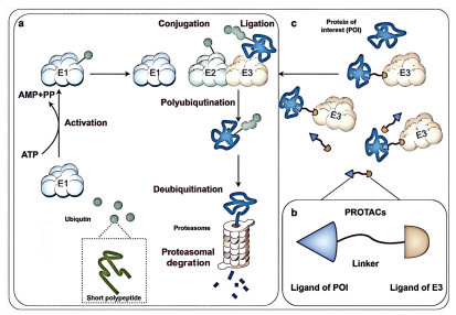
Proteolysis-targeting chimeras (PROTACs) technology has emerged as a revolutionary therapeutic strategy within the biopharmaceutical landscape, attracting intense research focus due to its unique mechanism of action and potential to address longstanding challenges in drug development [1,2]. Unlike conventional small-molecule inhibitors that typically function by binding to an active site to inhibit protein function, PROTACs are heterobifunctional molecules designed to harness the cell’s intrinsic protein degradation pathways (Fig. 1a). They consist of three core elements (Fig. 1b): A ligand that binds with high affinity to a protein of interest (POI), a ligand that recruits an E3 ubiquitin ligase, and a linker connecting these two moieties. By simultaneously binding both the POI and the E3 ligase, the PROTAC facilitates the formation of a ternary complex. This proximity induces the transfer of ubiquitin molecules from the E3 ligase complex onto the POI. The polyubiquitinated POI is then recognized and irreversibly degraded by the proteasome, the cell’s primary protein degradation apparatus (Fig. 1c) [3-5]. This catalytic, event-driven mode of action offers several compelling advantages [6]. Most significantly, PROTACs can target proteins traditionally considered “undruggable” with inhibitors, such as transcription factors [7], scaffolding proteins [8], or proteins lacking well-defined functional pockets [9]. Furthermore, their catalytic nature allows for sub-stoichiometric activity, potentially enabling efficacy at lower concentrations. Importantly, PROTACs have the potential to overcome certain forms of acquired drug resistance, such as mutations in the target protein’s active site that prevent inhibitor binding [10], by engaging alternative binding sites to facilitate the protein’s ubiquitination and subsequent degradation. However, significant challenges impede the clinical translation of PROTACs. Their inherently large molecular weight and complex structure typically result in poor physicochemical properties [11], including low aqueous solubility, high lipophilicity, and limited cell membrane permeability, culminating in inadequate oral bioavailability and poor pharmacokinetic (PK) profiles [12-15]. Achieving tissue-specific targeting and selectivity remains difficult, raising concerns about uncontrolled “on-target” degradation in non-diseased tissues and potential “off-target” degradation due to unintended ternary complex formation. These limitations are reflected in the current clinical landscape: While over 30 PROTAC molecules have entered various stages of clinical development, targeting proteins like the androgen receptor (AR), estrogen receptor (ER), Bruton’s tyrosine kinase (BTK), and signal transducer and activator of transcription 3 (STAT3), none have yet achieved approval. Consequently, addressing these delivery and selectivity constraints has emerged as a non-negotiable prerequisite for clinical viability. This imperative necessitates the strategic engineering of customized drug delivery systems (DDS) specifically designed to overcome PROTACs’ bioavailability and spatial control limitations.
Figure 1
Figure 1.
PROTACs selectively degrade POI via the ubiquitin-proteasome system (UPS). (a) UPS selectively degraded proteins. E1: ubiquitin-activating enzyme; E2s: ubiquitin-conjugating enzymes; E3s: ubiquitin ligase. The target protein is labeled with a polyubiquitin tag through the cascade reactions of E1, E2 and E3. The proteasome recognizes these ubiquitin-tagged protein substrate, unfolds the tagged protein substrate, and subsequently translocates it into the proteolytic core, where the unfolded protein is degraded into small peptides. (b) Three-component structure of PROTACs. (c) PROTACs simultaneously bind to a target POI and an E3 ubiquitin ligase, thereby bringing the POI into proximity with the E3 ligase. This proximity induces the ubiquitination of the POI and results in the degradation of POI via the proteasome. This figure was drawn by Figdraw.
2.
Delivery system design principles-addressing PROTACs-specific challenges
The clinical translation of PROTACs depends on overcoming their unique pharmacological challenges, which requires a paradigm shift from conventional drug delivery approaches. To address these constraints, PROTACs delivery systems require core design principles focused on PK improvement, targeting selectivity, and precise spatiotemporal regulation.
A primary challenge is the lack of intrinsic tissue selectivity, compounded by the characteristically high molecular weight and poor membrane permeability of PROTACs, which severely limit their passive diffusion and cellular uptake. One key strategy is the incorporation of active targeting ligands. DDS can be functionalized with ligands (e.g., monoclonal antibodies [16], peptides [17,18], aptamers [19,20]) that recognize disease-specific biomarkers. This approach helps concentrate PROTACs within pathological microenvironments, promotes receptor-mediated cellular internalization to bypass the permeability limitation, increases intracellular PROTAC concentrations to therapeutic levels, and reduces off-target degradation and systemic toxicity.
Moreover, PROTACs operate catalytically, meaning a single molecule can degrade multiple target proteins. Because of this amplifying effect, even minimal off-target activation has the potential to cause significant unintended protein loss. Therefore, it is essential to integrate stimuli-responsive release mechanisms to prevent uncontrolled degradation. DDS platforms can be designed to respond to endogenous pathophysiological cues such as tumor-associated acidosis (pH 6.5–7.0) [21,22], dysregulated protease expression (e.g., matrix metallopeptidase 2 (MMP-2) [23,24], cathepsins [25]), elevated glutathione (GSH) [25] or reactive oxygen species (ROS). Alternatively, exogenous triggers (e.g., near-infrared (NIR) light [26] and ultrasound [27]) can be used to ensure selective PROTAC release within target cells or subcellular compartments.
Finally, to overcome the poor PKs of PROTACs, which are exacerbated by their high molecular weight and unfavorable physicochemical properties, their formulation can be optimized by engineering nanocarriers (e.g., polymeric, lipid, or inorganic systems) or conjugates. These systems are primarily designed to achieve passive targeting, most notably through the enhanced permeability and retention (EPR) effect in tumor tissues. To this end, they are designed to extend plasma circulation [28], incorporate biomimetic surfaces [21], resist degradation, cross barriers like the blood-brain barrier (BBB) [29], and modulate release kinetics to sustain therapeutic exposure.
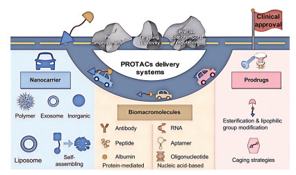
Precision targeting, trigger-dependent activation, and PK refinement are integrated strategies designed to confine target protein degradation to diseased tissues, thereby translating PROTACs’ in vitro potency into viable in vivo therapies (Fig. 2). However, their application involves inherent compromises. For instance, high-density ligand conjugation may accelerate plasma clearance, and incorporating stimuli-responsive elements can complicate manufacturing. Consequently, an optimal design requires balance the specific properties of the PROTAC molecule, the pathogenesis of the target disease, and the intended route of administration, rather than adopting a one-size-fits-all approach.
Figure 2
Figure 2.
PROTACs delivery systems promote the clinical translation of PROTACs by focusing on PK optimization, targeting selectivity and spatiotemporal control. This figure was drawn by Figdraw.
Nanocarrier-based delivery systems have emerged as a pivotal strategy to overcome the delivery challenges of PROTACs. These systems including polymer nanoparticles (PNPs), exosomes, liposomes, inorganic nanoparticles (INPs), and self-assembled nanoplatforms, enhance the delivery of PROTACs through encapsulation or chemical conjugation, significantly improving their aqueous solubility and protecting them from rapid degradation and clearance in vivo. By functionalizing the nanocarrier surface with targeting ligands such as peptides, antibodies, or aptamers, PROTACs can be selectively delivered and enriched at the disease site. Furthermore, smart PROTAC nanosystems that can respond to specific triggers have been designed [30]. These triggers can be endogenous stimuli within the tumor microenvironment (TME), such as pH, enzymes, GSH, or ROS, or external ones like light or ultrasound. This responsiveness enables the spatiotemporally controlled release of PROTACs and maximizes target degradation efficiency while minimizing off-target effects. This section systematically reviews recent advances of various nanocarriers for PROTACs delivery.
3.1.1
PNPs
PNPs, typically composed of natural or synthetic polymers (such as poly(lactic acid) (PLA), poly(lactic-co-glycolic acid) (PLGA)), exhibit favorable biocompatibility and biodegradability [31]. Their particle size generally ranges from tens to hundreds of nanometers, and surface properties such as hydrophilicity and charge can be modulated through chemical modification [32-34]. Owing to the tunability, biocompatibility, and multifunctionality, PNPs have emerged as a promising delivery platform for PROTACs. This delivery system effectively addresses key pharmacological delivery challenges associated with PROTAC administration in oncology therapeutics.
The PEG-PLGA copolymer and its derivatives represent a cornerstone in PROTACs delivery due to their excellent biocompatibility and modifiability. Saraswat et al. formulated PLGA-PEG nanoparticles (ARV-NP) to encapsulate the bromodomain protein 4 (BRD4) degrader ARV-825 [35]. The resulting NPs exhibited uniform size, high encapsulation efficiency, and sustained release (<9% over 48 h), which collectively extended the drug’s plasma half-life (t1/2) from 19 min to 177 min. In pancreatic cancer models, ARV-NP effectively degraded BRD4 and indirectly suppressed the MYC proto-oncogene (also known as c-MYC). Advancing this platform, Cimas et al. engineered the first antibody-conjugated PROTAC NPs (MZ1-ACNPs) by encapsulating MZ1 within a PLA core and covalently conjugating trastuzumab to the surface [36]. This design enabled active targeting of HER2-positive breast cancer cells, demonstrating pH-responsive release and enhanced lysosomal escape to overcome drug resistance. Further refinement was achieved by Zou et al., who utilized PEG-β-PLGA to load PROTAC prodrugs (ARV771 or D43). By incorporating reduction-sensitive disulfide bonds and the targeting peptide CRGDK, they created a “smart” nanoplatform activated by the high intracellular GSH concentration in tumor cells, leading to significantly improved tumor accumulation by 4.5 times compared to free ARV771, degradation efficiency, and synergistic tumor suppression when co-delivered with doxorubicin (DOX) [37]. Similarly, two independent studies by He et al. employed cRGD peptide-modified polymers (PEG-PLA or PEG-PCL) for the co-delivery of DOX and the BRD4 degrader ARV-825 [28,38], demonstrating the potent synergy between chemotherapy and targeted protein degradation to overcome the BBB in glioma and combat chemoresistance in colorectal cancer.
Beyond classical polyesters, several innovative polymer and complexation systems offer unique functionalities. Sun et al. developed a hybrid polyplex system based on the self-assembly of a cationic conjugated polyelectrolyte (CPE) and single-stranded DNA (ssDNA) [39]. This platform efficiently delivered a nucleic acid-based PROTAC (P9TC) without the need for transfection reagents, achieving robust degradation of the estrogen receptor ERα in breast cancer cells and presenting a novel paradigm for nucleic acid delivery. Koshkina et al. introduced a biodegradable polyphosphonate (PPE) micelle system. The unique P-C bonds in the polymer backbone shifted the 31P nuclear magnetic resonance (NMR) signal away from endogenous phosphate backgrounds, enabling the first demonstration of background-free 31P magnetic resonance imaging (MRI) tracking. Simultaneously, these micelles can encapsulate hydrophobic PROTACs, achieving true theranostic integration [40].
Semiconducting polymer nano-PROTACs developed by Zhang and colleagues further expand the functional repertoire of these delivery systems. The SPNpro platform achieves TME-specific activation through cathepsin B cleavage, resulting in over 90% suppression of tumor growth and lung metastasis in 4T1 models, as well as associated off-target toxicity [41]. Similarly, the SPNCOX system combines enzyme-triggered PROTACs release with phototherapeutic capabilities, enabling simultaneous degradation of immunosuppressive proteins and immunogenic cell death induction for enhanced antitumor immunity [42]. The dual-programmable semiconductor polymer nano-PROTACs (SPNFeP) developed by Wang et al. employs an ROS-cleavable amphiphilic polymer (DSPE-TK-PEG) to achieve simultaneous responsiveness to endogenous H2O2 and external ultrasound irradiation [27]. The on-demand release and spatiotemporal controllable activation of PROTACs have demonstrated precise synergistic advantages in the treatment of deep tissue tumors.
The innovative application of bioorthogonal POLY-PROTAC nanoplatforms introduces a multi-stage targeting approach. Gao et al. demonstrated that covalent grafting of PROTACs onto diblock polymer backbones creates NPs capable of sequential response to extracellular MMP-2, acidic endosomal pH, and intracellular GSH, resulting in a 3.9-fold increase in tumor accumulation and prolonged intratumoral retention [43]. This sophisticated design effectively addresses the critical challenges of insufficient tumor distribution and off-target effects.
Significant formulation innovations are addressing the hurdles of oral bioavailability and localized delivery of PROTACs. Pöstges et al. tackled the poor solubility of the PROTAC ARCC-4 by fabricating amorphous solid dispersions (ASDs) using enteric polymers (HPMCAS and Eudragit L 100–55). These ASDs provided gastric protection and released the drug in the intestinal environment, stabilizing a supersaturated state to extend the absorption window and paving the way for oral PROTACs development [44]. As shown in Fig. S1 (Supporting information), for localized therapy, Cheng et al. has further designed pH-responsive microneedle patches that co-load the ER degrader ERD308 and the cyclin-dependent kinase 4/6 (CDK4/6) inhibitor palbociclib using MPEG-PAE micelles (Fig. S1a). In the TME (pH 6–7), these micelles rapidly dissociate, leading to an explosive release of the drug (Figs. S1b-e). Local administration via microneedle patches achieves an impressive tumor accumulation rate of 87.7% (Fig. S1f) and maintains sustained release for four days (Fig. S1g), significantly enhancing targeting and safety [45].
The efficacy of PROTACs therapy has been significantly advanced through the rational design of stimuli-responsive polymeric nanocarriers that exploit pathological gradients within the TME for spatially and temporally controlled drug release. Among these, redox-responsive systems based on disulfide bonds have shown particular promise. For instance, Ma et al. employed a disulfide-linked FA-PEG-PROTAC conjugate [46], while Liu et al. utilized GSH-responsive polydisulfide amide (PDSA) NPs [47]. Both systems undergo reductive cleavage in the high-GSH TME to selectively release their PROTAC payloads, demonstrating markedly enhanced tumor accumulation and remarkable tumor regression in vivo. Beyond single-mechanism responsiveness, more sophisticated dual-targeting and multi-responsive platforms are emerging to further improve specificity and therapeutic outcomes. For example, Guan and colleagues developed a dual-responsive polymer, designated as DS-PLGA (disulfide bond-linked PLGA), which was utilized to encapsulate dBET6, resulting in the formation of DPB NPs. These NPs employed a dual-targeting strategy using a Lewis lung cancer cell membrane (LLCM) coating to prolong blood circulation and a conjugated cRGD peptide (RLDBP) to target αvβ3 integrin on tumor tissues. This system effectively controlled the release of dBET6 within tumor cells, achieving a circulation t1/2 of 48 h and enhanced active targeting [48]. These results collectively illustrate how the successful and precise application of PROTACs in lung cancer treatment has been demonstrated, offering significant insights for addressing the challenges associated with epigenetic PROTACs delivery. In addition to GSH-sensitive systems, acid-responsive platforms have also been widely explored. Representative work includes that of Li et al., whose indocyanine green functionalized polyethylene glycol (PEG-ICG)-based nano-PROTAC dissociates in the acidic TME to release ARV-825 and initiate potent photo-immunotherapy [49]. To achieve even higher targeting precision, multi-stimuli-responsive nano-PROTACs have recently been developed. For instance, Yang et al. used pH/enzyme-dual responsive polymers to create NPs that shrink for better penetration and then release a CDK4/6 PROTACs upon enzymatic activation [50]. Similarly, Chen et al. fabricated a polyphenol-vanadium oxide-mPEG-NH2 composite nano-PROTAC responsive to pH, H2O2, and GSH for synergistic the enhancer of zeste homolog 2 (EZH2) degradation, and photothermal therapy (PTT) [51]. A particularly promising direction is the integration of PROTAC technology with immunotherapy, as exemplified by Chen et al., whose block copolymer-based nanochaperone enables pH-dependent sequential release of the chemokine (C-X-C motif) ligand 9 (CXCL9) and a BRD4 degrader, boosting CD8+ T cell infiltration to 32.1% [52]. Further expanding this concept, Wang et al. demonstrated a self-assembly strategy using DSPE-mPEG2000 to co-deliver a PROTAC and photosensitizer Ce6, achieving acidic pH-enhanced release and synergistic photo-PROTAC therapy [53]. Culminating in system-level complexity, researchers have even developed localized depot formulations for sustained multi-modal therapy. Wu et al. engineered an injectable nanocomposite hydrogel based on thermosensitive PLGA-PEG-PLGA, which acts as a depot for spatiotemporally controlled multi-agent delivery, effectively combining PROTAC-based proteolysis with chemotherapy and immunotherapy [54].
Although PNPs offer benefits in targeting, stability, and multifunctional integration for PROTACs delivery (Table S1 in Supporting information), they also encounter significant limitations. The size of these NPs affects their interaction with the TME, which can restrict deep tissue penetration and hinder uniform spatial distribution [55]. Additionally, while sustained-release formulations extend therapeutic effects, they often do not achieve the rapid degradation required, leading to less optimal release profiles and reduced therapeutic efficacy [56,57]. Other ongoing challenges include complex manufacturing processes, potential toxicity, and constraints in biodistribution [58]. To advance next-generation intelligent nano-PROTACs toward clinical application, a collaborative approach that integrates materials engineering with biological insights is essential.
3.1.2
Exosome
Exosomes, which are extracellular vesicles (EVs) ranging from 30 nm to 150 nm in size [59], represent promising PROTACs delivery vectors due to their natural biocompatibility, low immunogenicity, and targeting capabilities [60-62]. Camel milk-derived exosomes (CMEs) demonstrate efficient encapsulation of ARV-825 (42.75% loading efficiency) via ultrasonication-assisted emulsification, enhancing gastrointestinal stability and facilitating ileum-specific absorption to achieve a 5.56-fold increase in the area under curve (AUC) in SD rats [21]. Concurrently, microfluidic droplet-electroporation (μDES) enables high-throughput, low-damage loading of YX968 into cell-derived EVs, preserving vesicle integrity and leveraging homotypic tumor targeting. This strategy promotes sustained HDAC3/8 degradation within triple-negative breast cancer (TNBC) xenografts, significantly enhancing tumor growth suppression compared to free YX968 while mitigating hepatorenal toxicity [63] Both strategies leverage the endogenous biocompatibility and targeting properties of exosomes, synergizing PROTAC-mediated target degradation with vesicular cargo to potentiate anticancer efficacy.
However, several challenges remain in exosome-mediated PROTACs delivery, including inherently low delivery efficiency [64] and complex preparation process [65]. Current loading methods rely on passive diffusion (e.g., co-incubation) or active techniques such as electroporation, with the later may damage the exosomes and affect their targeting function [66]. Additionally, the high molecular weight and hydrophobicity of PROTACs typically result in limited loading efficiency [67]. Furthermore, exosomes may indirectly inhibit the anti-tumor immune response by carrying immune checkpoint ligands derived from mother cells (such as the programmed cell death ligand 1 (PD-L1)) or immunosuppressive molecules (such as the transforming growth factor-β (TGF-β) and interleukin-6 (IL-6)) [68]. Therefore, the impact of long-term use of exosome delivery of PROTACs on immune response still needs to be further evaluated.
3.1.3
Liposome
Liposomes consist of phospholipids that spontaneously assemble into closed lipid bilayers in aqueous environments, resulting in the formation of NPs at the nanoscale [69]. The application of liposomes for the delivery of PROTACs presents significant advantages in improving PK properties, addressing challenges such as poor solubility, rapid clearance associated with high lipophilicity, and the large molecular weight. Notable PROTAC molecules, including BRD4-targeting agents (ARV-825, ARV-825 derivatives, dBET57, dBET6), DT2216 (targeting the B-cell lymphoma extra-large (BCL-xL)), LC-2 (targeting the Kirsten rat sarcoma viral oncogene homolog (KRAS)), DQ (targeting the NAD(P)H quinone dehydrogenase 1 (NQO1)), and bifunctional degraders like GNE-01/02 (targeting the BRM (Brahma homolog; also known as SWI/SNF-related, matrix-associated, actin-dependent regulator of chromatin subfamily A member 2 or SMARCA2)), EPRO/BPRO (targeting the epidermal growth factor receptor (EGFR)/BRD4), dGPX4 (targeting the glutathione peroxidase 4 (GPX4)), demonstrate improved performance when encapsulated within specifically engineered liposomal formulations.
These nanocarrier systems significantly enhance the solubility and PK profiles of PROTACs. For instance, cyclodextrin-incorporated liposomes encapsulating bivalent PROTACs (GNE-01/02) within their hydrophilic cores demonstrated a remarkable 80- to 380-fold increase in AUC in Sprague-Dawley rats [70]. Similarly, liposomes co-loaded with agents such as docetaxel and ARV-825 (DTX/ARV-lip) elevated PROTACs solubility to 1 mg/mL [71]. Furthermore, modifications involving cell-penetrating peptides, exemplified by R8-functionalized liposomes delivering the KRAS-targeting PROTAC LC-2, prolonged the t1/2 from 1.47 h to 25.7 h and enhanced the AUC by 39-fold [72].
Liposomes also facilitate advanced targeting and synergistic therapy. Galactose-functionalized nanoliposomes (GALARV) were employed to achieve active targeting of ARV-825 to hepatocellular carcinoma cells via asialoglycoprotein receptor (ASGPR) mediation, resulting in a 4.5-fold increase in tumor accumulation relative to the free drug [73]. Cationic liposomes co-delivering DT2216 with siRNA, incorporating fluorinated polyethyleneimine (F-PEI) for endosomal escape, achieved triple the BCL-xL degradation efficiency compared to the PROTAC alone and increased apoptosis rates in resistant cells to 45.7% [74]. Similarly, Zhang et al.’s co-delivery platform (PN liposomes) simultaneously delivered PROTAC DQ (targeting NQO1) and ROS inducer withaferin A (WA), effectively disrupting the ROS negative feedback loop to amplify apoptosis in tumor cells [75].
Therapeutic resistance was consistently overcome through co-delivery strategies demonstrating notable effectiveness. For example, co-delivering EGFR and BRD4 PROTACs (EPRO/BPRO) via histidine-nickel coordination induced 45% apoptosis in resistant cells [76]. Additionally, oral lipid nanocomplexes (NANOVB) co-encapsulating ARV-825 with vemurafenib (VEM) successfully reversed BRAF inhibitor resistance, reducing tumor growth by 79.5% in resistant melanoma models by targeting the c-Myc pathway [77].
Furthermore, liposomal systems overcome biological barriers. Neurotransmitter-derived lipidoid-PROTAC-DNA nanocomplexes (NPD) efficiently crossed the BBB, achieving over 90% degradation of pathological tau protein in the hippocampus of Alzheimer’s disease models and demonstrating cognitive improvement [29]. Innovative designs like “pre-fused PROTAC” lipid NPs (LNP), where PROTACs are complexed with E3 ligases (e.g., VHL) prior to encapsulation, simplified ternary complex formation and accelerated BRD4 degradation (50% within 6 h) [78]. Protein kinase C inhibitor (PC)-anchored PEGylated nanoliposomes (LARPC) conferred a strong positive charge (+26.6 mV), enhancing electrostatic adsorption to tumor vasculature, inhibiting vasculogenic mimicry, and reducing the half maximal inhibitory concentration (IC50) of ARV-825 to 0.14 µmol/L [79].
Stimulus-responsive liposomal systems represent a pivotal advancement in the targeted delivery of PROTACs. Among various stimuli, the elevated ROS levels in tumor cells have been widely exploited as a prominent trigger. For instance, Wang et al. engineered a ROS-responsive liposomal prodrug delivering a STAT3-targeting PROTAC. This system, functionalized with cRGD for targeting integrin αvβ3, undergoes cleavage of a thioketal linker in the high-ROS TME, releasing the active PROTAC. Concurrently, the cationic lipids facilitate endosomal escape via the proton sponge effect, enabling efficient cytosolic STAT3 degradation and offering a novel paradigm to overcome cancer stem cell resistance [80]. Similarly, Luo et al. developed a ROS-degradable LNP encapsulating dGPX4, a GPX4-targeting PROTAC. This formulation achieved over 60% drug release within 12 h under high ROS conditions, selectively inducing ferroptosis in cancer cells and demonstrating a remarkable 80% of tumor growth inhibition (TGI) in vivo with minimal systemic toxicity [81]. Beyond ROS, other microenvironmental cues are effectively harnessed. Chan et al. utilized a pH-responsive LNP platform for the intracellular delivery of recombinant bioPROTACs. This system relies on pH-dependent endosomal escape to release the bioPROTACs into the cytoplasm. This process results in the rapid and broad-spectrum degradation of otherwise “undruggable” targets, with an efficiency reaching 95% within 8 h. Consequently, it inhibits the growth of KRAS-mutant pancreatic cancer cells by 46% [82]. Similarly, the high GSH concentration in tumor cells provides another important trigger for selective activation. In this context, Xu et al. designed a biomimetic hybrid nanovesicle co-delivering temozolomide (TMZ) and a BRD4-targeting PROTAC prodrug (PC-ss-Pro). The disulfide bond in the prodrug core is cleaved in the high-GSH intracellular environment, releasing the active PROTAC to degrade BRD4. This action blocks multiple DNA repair pathways activated in TMZ-resistant glioblastoma, presenting an intelligent delivery strategy to reverse chemoresistance [83]. Furthermore, to achieve even greater specificity and therapeutic efficacy, recent efforts have focused on integrating multiple stimuli-responsive mechanisms into a single platform. A notable example comes from Zhang et al., who developed a dual-responsive system using disulfide-bonded PLGA to encapsulate dBET6, a BRD4-degrading PROTAC. The NPs disintegrate in the acidic TME (pH 5.0–6.5), with the high intracellular GSH concentration further promoting drug release. This strategy not only directly induced lung cancer cell apoptosis but also remodeled the immunosuppressive TME by reducing tumor-associated macrophages via the colony-stimulating factor 1 receptor (CSF1R) pathway inhibition [25]. These stimulus-responsive liposomal platforms, by integrating the potent protein degradation capability of PROTACs with the spatiotemporally controlled release properties of smart nanomaterials, provide a powerful and versatile technological foundation. They directly address critical challenges in oncology drug delivery, including tumor-specific targeting, overcoming therapy resistance, and minimizing off-target toxicity, thereby significantly advancing the therapeutic potential of PROTACs.
While liposomes can target tumors via the EPR effect, their accumulation efficiency remains suboptimal. In comparison to exosomes, liposomes’ complex composition makes optimizing component ratios more challenging [84]. Additionally, the inclusion of certain materials, such as F-PEI, may increase cytotoxicity [74]. Future research should focus on integrating targeted ligands with stimuli-responsive release strategies to optimize therapeutic efficacy.
3.1.4
INPs
INPs, comprising inorganic particles and biodegradable polycations [85], have garnered significant interest as delivery carriers for PROTACs. This attention stems from their unique physicochemical properties and controllable surface functionalization characteristics. Various inorganic materials, including gold, silica, selenium, and metal-organic frameworks (MOFs), have been explored for PROTACs delivery.
Gold-based NPs demonstrate notable success in improving the tumor-specific accumulation of PROTACs. Ma et al. [86] utilized ultrasmall gold NPs (AuNPs, ≈5 nm) deliver a peptide-based PROTAC targeting AR-V7, which extended its t1/2 from 1.9 h to 25.1 h and achieved potent degradation of AR/AR-V7 (the half-maximal degradation concentration (DC50) = 48.8/79.2 nmol/L), leading to significant tumor regression in enzalutamide-resistant prostate cancer models. Similarly, Wang et al. [87] employed smaller gold nanoclusters (GNCs, ≈2.7 nm) to construct hybrid nanoPROTACs (GNCTACs). GNCTACs stably conjugated a HER2-targeting peptide and a cereblon (CRBN) E3 ligase ligand (thalidomide derivative) via Au-S bonds. Combining GNCTACs with a fasting-mimicking diet (FMD) strategy yielded synergistic effects: FMD enhanced tumor cell endocytosis, deep tumor spheroid penetration, proteasome activity, and autophagy. This combination boosted HER2 degradation efficiency to 97% (DC50 reduced from 1.8 µmol/L to 0.3 µmol/L) and achieved 85% tumor suppression in SKBR3 xenograft models. In a similar approach focused on kinase degradation, Wang et al. developed multi-headed PROTACs on gold NPs (Cer/Pom-PEG@GNPs) that co-delivered ceritinib (an anaplastic lymphoma kinase (ALK) inhibitor) and pomalidomide (a CRBN ligand), enabling GSH-responsive release and effective degradation of ALK protein [88]. Expanding the application of gold nanomaterials further, Li et al. designed a photothermal-activated system using gold nanocages (AuNCs) to co-deliver a PROTAC prodrug (proPROTAC, targeting BRD4) and a palladium catalyst. Upon NIR irradiation, phase-change materials melted, triggering bioorthogonal activation of dBET1 and resulting in substantial TGI (tumor volume reduced to 217 mm3) in a breast cancer model [89]. Finally, demonstrating another functional adaptation, Niu et al. conjugated a GPX4-targeting peptide PROTAC (PGPD) to gold NPs via disulfide bonds, enabling reducible release and effective induction of ferroptosis in acute lymphoid leukemia [90].
Silica-based and MOF carriers enable precise delivery. Fang et al. [91] designed silica NP-based EGFR degraders (SIPROTACs, ≈ 50 nm). By separately conjugating the CRBN ligand and a gefitinib-derived EGFR ligand onto the silica surface, SIPROTACs formed a multivalent ligand display platform that enhanced ternary complex formation and subsequent degradation potency. A lipid coating (DOPC/DSPE-PEG2000/cholesterol) was applied to improve solubility and stability. SIPROTACs efficiently degraded EGFR at concentrations as low as 50 nmol/L in NSCLC cells, outperforming the free PROTAC (PROC8), and demonstrated comparable TGI in HCC-827 xenografts models while avoiding the nephrotoxicity associated with the free agent. Following a different strategy for controlled release, He et al. developed an upconversion NP-modified mesoporous silica nanocage system (UMSNs@phoBET1) loaded with a photo-caged PROTAC prodrug [92]. Upon 980 nm NIR irradiation, the core converted NIR light to UV emission, triggering photolysis to release active dBET1. This spatiotemporally controlled activation effectively degraded BRD4 (achieving 58.7% tumor suppression) and circumvented off-target toxicity. Another innovative silica-based platform was constructed by Zhan et al., who developed a NIR-regulated system (USDPR) comprising upconversion NPs coated with mesoporous silica and loaded with dBET6. Under 980 nm light, rose bengal-generated ROS promoted endo/lysosomal escape and cytoplasmic release of the PROTAC [93]. Beyond pure silica systems, Tan et al. engineered a cancer-cell-membrane-coated silica-iron oxide core-shell NP (NP@M) for homologous targeting and pH-responsive co-delivery of FAK- and BRD4-targeting PROTACs [94]. Shifting to MOF-based carriers, Zhu et al. constructed a hyaluronic acid (HA)-modified ZIF-8 MOF platform (ZMCH) for co-delivery of the BRD4 degrader MZ1 and photosensitizer Ce6 [95]. HA conferred CD44-mediated active targeting and prolonged circulation. ZMCH exhibited an acid-responsive release profile (60% release at pH 5.0 within 2 h vs. 8% leakage at physiological pH), synchronously activating photodynamic therapy (PDT) and proteolysis. This synergy induced significant apoptosis, reshaped the tumor immune microenvironment (3-fold increase in cytotoxic CD8+ granzyme B+ T cell infiltration, reduced T-regs), and suppressed 71% of lung metastases.
Selenium-based NPs offer intrinsic antioxidant activity and biocompatibility, supporting redox-responsive drug release and synergistic therapy. As a representative example, Xu et al. [96] covalently conjugated a p21-activated kinase 4 (PAK4)-targeting peptide PROTAC (PpD) to biocompatible nano-selenium particles via Se-S bonds. This conferred enhanced cellular penetration and in vivo stability (t1/2 ~ 9.6 h). GSH-responsive release in the TME enabled selective PAK4 degradation (DC50 ≈ 200 nmol/L, with no off-target effect on PAK1–3), suppressed oncogenic pathways, activated immune-related pathways, and enhanced CD8+ T cell infiltration, demonstrating potent targeted-immunotherapy synergy in renal carcinoma models. Building upon this platform while demonstrating its therapeutic versatility, Ma et al. utilized an artificial intelligence (AI)-designed AR-targeting peptide PROTAC conjugated to nano-selenium (Se-AR PROTAC), which was further integrated into a HA-based microneedle patch for transdermal delivery. This construct enabled ROS-responsive release and local accumulation at androgenetic alopecia sites, effectively degrading AR while mitigating oxidative stress [18].
In addition, stimuli-responsive composite systems represent a sophisticated approach to PROTACs delivery. Huang et al. developed a pH-responsive platform featuring magnetic nanoclusters coated with calcium-doped manganese carbonate (MNC@Ca/MnCO3), co-loaded with the BRD4-targeting PROTAC ARV-825 and an anti-PD1 antibody. Upon exposure to the acidic TME, the inorganic shell dissociates, releasing the therapeutic cargo. This process concurrently induces ferroptosis, facilitates tumor antigen capture, and promotes dendritic cell activation, collectively establishing a highly effective in situ tumor vaccine and durable anti-tumor immunity [22].
INPs present benefits including high stability, targeted delivery, and multivalent effects for PROTACs delivery. Nevertheless, challenges such as potential toxicity, complex synthesis procedures, and uncertain metabolic pathways warrant comprehensive investigation [97-99]. Moving forward, it is essential to integrate materials science with PKs, refine nanocarrier designs and perform long-term toxicological assessments to facilitate clinical translation.
3.1.5
Self-assembling NPs
Self-assembling nanoplatforms offer promising solutions for PROTACs delivery, effectively addressing challenges such as hydrophobicity, off-target toxicity, and suboptimal tumor targeting [100]. Inspiring by this approach, Gao et al. developed an amphiphilic affibody-PROTAC conjugate ZEGFR:1907 linked cyclin D1 degrader MS28 via a disulfide bond. It self-assembles into core-shell NPs (ZEGFR:1907−MS28 ADCN, ~96 nm) for EGFR-targeted tumor delivery. Intracellular GSH triggers PROTAC release, degrading Cyclin D1/CDK4/6 to induce G0/G1 arrest and apoptosis, significantly suppressing EGFR+ tumor growth. Enhanced PKs (t1/2 ≈ 1.84 h) and favorable biosafety profiles were also demonstrated [101]. Similarly, Li et al. created HER2-targeted system that self-assembled into stable NPs (ZHER2:342−MZ1 APCN, ~74.8 nm). This system enables HER2-mediated uptake and GSH-responsive release of the BRD4 degrader MZ1, showing enhanced cytotoxicity (IC50 = 194.2 nmol/L in SKOV-3) and superior TGI (65.1%) over free MZ1 [102].
Expanding the functional capabilities, Wang et al. designed a carrier-free co-delivery system where a CDK4/6-targeting PROTAC and photosensitizer Ce6 self-assemble via hydrophobic/π-π interactions (~140 nm). This enhances solubility and tumor accumulation. Crucially, PROTAC-induced G1 arrest sensitizes cells to Ce6-PDT, causing synergistic mitochondrial damage, apoptosis, amplified immunogenic cell death (ICD), and tumor immune microenvironment remodeling [103].
To combat multi-drug resistance and immune evasion in metastatic disease, Zhang et al. implemented a “minimalist self-assembly” strategy, co-assembling irinotecan (ICD inducer), BRD4 degrader MZ1, and PD-L1 siRNA into NPs (~68 nm) via non-covalent interactions. Encapsulation in RGD-liposomes (~111 nm) further enhanced liver metastasis targeting to combat chemotherapy resistance and immune evasion [104].
Of particular significance are stimulus-responsive self-assembled nanoplatforms that enable precise spatiotemporal control over PROTACs activation within the TME. These systems can be categorized based on their activation mechanisms: Wang et al. developed an exogenous light-triggered system where the BRD4 degrader ARV771 conjugates with a photosensitizer through a singlet oxygen-cleavable linker, enabling optically controlled drug release and synergistic photodynamic therapy [26]. For endogenous stimuli-responsive systems, multiple approaches have emerged. Lin et al. designed a hypoxia-activated supramolecular system (NaC4A-PROTACs) that encapsulates PROTACs within a calixarene host, achieving rapid release (>75% within 15 min) under hypoxic conditions [105]. Li et al. created a redox-sensitive theranostic platform through disulfide linkage between a BRD4-targeting PROTAC and a NIR fluorophore, allowing simultaneous GSH-triggered degradation and fluorescence imaging [30]. Enzyme-responsive strategies are represented by Tong’s MMP-2-activated system (dBET6@CFMPD) that undergoes morphological transformation from NPs to tumor-penetrating nanofibers [106], and Ninglin Chen’s sulfatase-triggered Supra-PROTACs that self-assemble into nanofibers within tumor lysosomes [107]. Additionally, Lin et al. developed a unique GSH-activated click chemistry system where bifunctional peptides self-assemble into nanofibers in the high-GSH TME [108]. Beyond these chemical stimuli, Fan et al. employed a biological targeting strategy using homologous cancer cell membranes to create biomimetic NPs (CM8988-PIPD) that achieve enhanced tumor-specific delivery through homotypic recognition [109]. Wang et al. optimized a PEGylated amphiphilic PROTAC (B1-PEG) that leverages both self-assembly properties and improved PKs for enhanced tumor accumulation [110].
Collectively, these diverse stimulus-responsive strategies demonstrate sophisticated approaches to achieve tumor-specific PROTACs delivery while minimizing off-target effects. Notwithstanding their remarkable potential, self-assembling nanoplatforms for PROTACs delivery face considerable challenges that must be systematically addressed, primarily concerning the complexity and reproducibility of their manufacturing processes and the insufficient reliability of their stimulus-responsive release mechanisms under biologically heterogeneous conditions. The precise supramolecular interactions that drive NP assembly are highly sensitive to even slight changes in preparation parameters. These interactions include hydrophobic forces [104], π-π stacking [103], and host-guest complexation [105]. Because of this sensitivity, it is difficult to produce consistent NPs across different batches. Key characteristics that vary include size distribution, drug loading efficiency, and stability. This inconsistency ultimately hinders scalable manufacturing and clinical translation. Furthermore, while many systems are engineered to release PROTACs in response to specific TME cues such as hypoxia, elevated GSH, or enzyme overexpression, the high degree of inter- and intra-tumor heterogeneity often leads to incomplete or unpredictable drug release in less responsive regions, compromising therapeutic efficacy, while also raising the risk of off-target activation in normal tissues that may exhibit similar microenvironmental features. Consequently, advancing these promising platforms toward clinical application will require not only more robust and reproducible assembly techniques but also the rational design of multi-stimuli responsive or logic-gated systems capable of operating reliably within the complex and variable landscape of human tumors.
Based on the above discussion, Table S2 (Supporting information) summarizes the key research cases of PROTAC delivery by nanocarriers, highlighting the PROTACs delivered, carrier types, and the key in vivo and/or in vitro efficacy.
3.2
Biomacromolecule-conjugated delivery systems
Further details are provided in Supporting information.
3.3
Prodrug strategies for PROTACs
To address the low oral bioavailability and off-target toxicity of conventional PROTACs, researchers have developed the pro-PROTAC strategy. This approach is fundamentally a form of stimuli-responsive delivery, which enables selective activation of active PROTACs in specific microenvironments through chemical modification or targeted conjugation, significantly enhancing tumor targeting while reducing extra-tumoral toxicity and improving oral bioavailability [111]. The core principle involves masking the active PROTACs with a cleavable moiety that responds to specific biological or external stimuli, such as enzymes, redox potential, pH, or light.
3.3.1
Esterification and lipophilic group modification
For example, Wei et al. pioneered the first orally bioavailable PROTAC prodrug by conjugating the lipophilic pivaloyloxymethyl (POM) group to the glutarimide moiety of a CDK2/4/6-targeting PROTAC. This design achieved enzyme-triggered hydrolysis and release of the active PROTAC, resulting in marked CDK2/4/6 degradation and significant inhibition of B16F10 tumor growth in vivo [112]. Additionally, Wang et al. established a chemical endocytic medicinal chemistry strategy by esterification-mediated conjugation of lipophilic-ionizable groups (e.g., anionic carboxylates, cationic ammoniums, or neutral tert–butoxycarbonyl moieties) to the VHL ligand of PROTACs, enabling targeted structural modification for enhanced membrane receptor engagement. As shown in Fig. S2 (Supporting information), this design substantially improved CD36 receptor binding affinity, as demonstrated by a 2.8-fold reduction in dissociation constant (Kd = 3.4 µmol/L for MZ1-C14-Na vs. 9.6 µmol/L for parental MZ1), validated via surface plasmon resonance (SPR) analysis (Fig. S2a). The augmented CD36 binding affinity triggered receptor-mediated endocytosis, leading to a marked 22.3-fold increase in intracellular PROTACs accumulation compared to the unmodified parent compound, as quantified by UPLC-MS metabolic profiling (Fig. S2b). Concurrently, the strategy overcame bioavailability limitations through dual optimization: >1300-fold solubility enhancement (e.g., MZ1-C14-Na at 61.5 mg/mL vs. MZ1 at 0.045 mg/mL) and improved lipophilicity-LogD balance, enabling efficient cellular penetration (Fig. S2c). Collectively, these modifications significantly potentiated target degradation efficacy (e.g., accelerated BRD4 degradation kinetics) and validated the approach as a versatile prodrug platform for optimizing macromolecular protein degraders beyond the Rule-of-5 constraints (Fig. S2d), with in vivo antitumor activity further confirming its translational potential (Fig. S2e) [14]. While the primary focus was on enhancing binding and uptake, the ester linkages also represent a potential platform for enzyme-responsive activation.
3.3.2
Caging strategies
The light-controlled “caging” strategy prodrug strategies effectively mitigate off-target effects and cytotoxicity associated with protein degraders [113]. As demonstrated by Xue et al. and Li et al., appending photolabile groups like 4,5-dimethoxy-2-nitrobenzyl (DMNB) to the E3 ligase ligand of PROTACs (e.g., dBET1) generates inactive precursors. Upon irradiation with 365 nm light, these photo-caged PROTACs (pc-PROTACs) rapidly release the active degrader. This approach yielded potent and light-dependent degradation of BRD4 (Dmax ~93%) and inhibition of cancer cell proliferation, with the IC50 improving from a micromolar range in the dark (e.g., 3.1 µmol/L) to a potent nanomolar range upon irradiation (e.g., 0.4 µmol/L), with efficacy validated in zebrafish models [114,115].
Receptor-mediated targeting and activation leverages overexpressed cell-surface receptors for selective delivery. Liu et al. [116] and Chen et al. [117] pioneered the use of folate as both a targeting moiety and a caging group, connected via enzymatically cleavable (e.g., ester) or GSH-responsive (e.g., disulfide) linkers. These folate-caged PROTACs are selectively internalized into folate receptor-alpha (FOLR1)-positive cancer cells. Subsequent intracellular hydrolysis or GSH-mediated reduction releases the active PROTAC, enabling potent degradation of targets like BRD4 and ALK, and robust anti-proliferative effects in FOLR1-high cancer cells (e.g., HeLa, OVCAR-8) while sparing FOLR1-low normal cells (e.g., HFF-1, HK-2), thereby achieving a high therapeutic index.
Bioorthogonal click-and-release strategies provide an exogenous activation mechanism with exceptional specificity, independent of endogenous biological cues. Huang et al. developed a PROTAC prodrug caged with a 4-(vinyloxy)benzyl carbonate group, which is inert until it undergoes a bond-cleavage bioorthogonal reaction with an exogenously supplied tetrazine (Tz), triggering the release of the active PROTAC MZ1 [118]. Similarly, Chang et al. employed an inverse-electron-demand Diels-Alder (IEDDA) reaction between a trans-cyclooctene (TCO)-caged ARV-771 and a Tz molecule released specifically in senescent cells by senescence-associated β-galactosidase (SA-β-gal) [119]. This two-step strategy enabled the selective degradation of BRD4 in senescent cells, showcasing a novel senolytic application.
Multi-responsive caging platforms demonstrate the versatility of this approach. An et al. created a universal multi-stimuli-responsive caging strategy for PROTACs that can be activated by enzymatic activity, pH, or light within the TME. This approach facilitates selective protein degradation and significantly suppresses tumor growth both in vitro and in vivo. By masking the active site of PROTACs with cleavable linkers (e.g., photolabile groups or enzyme-sensitive sequences), off-target hepatotoxicity was reduced by 50%, while tumor drug concentration increased 8-fold compared to conventional PROTACs [120].
Overall, these advances underscore that prodrug strategies are intrinsically linked to the broader field of stimuli-responsive delivery. These strategies leverage diverse triggers. These include intrinsic biological signals, such as enzymes, pH, and redox potential. They also include externally applied physical stimuli, such as light. Through these mechanisms, PROTAC prodrugs achieve a paramount goal. They enable precise spatial and temporal control over protein degradation. This control ultimately maximizes therapeutic efficacy and safety.
As summarized in Table S3 (Supporting information), key case studies of various prodrug strategies are presented, highlighting the specific PROTACs delivered, the types of prodrugs employed, and their main in vitro and in vivo efficacy. Despite these promising proofs-of-concept, the application of prodrug strategies to PROTACs presents notable challenges. Their effectiveness relies on precise and efficient activation at the intended site, a mechanism that frequently exhibits unpredictability and suboptimal efficiency, thereby restricting target specificity and increasing the risk of off-target toxicity. Additionally, chemical modifications such as caging may adversely affect the inherent activity of the PROTAC molecule, resulting in diminished therapeutic efficacy even following activation.
3.4
Others
In addition to the previously discussed delivery strategies, advancements in delivery technologies have greatly broadened the potential applications of PROTACs through innovative dosage form engineering and the design of biological carriers. Nicole Hofmann and her team developed ASD utilizing spray drying technology. By optimizing the carrier with miniaturized solvent screening (Solupius/Eudragit E PO), they successfully loaded 10% of MS4078, enhancing its solubility and oral bioavailability [121]. This work provides essential technical support for the advancement of oral PROTACs formulations. Meloni’s team created an oral dispersive membrane (ODF) containing ARV-110, incorporating excipients like triglyceride triacetate and polyvinyl alcohol to address the challenge of extremely low water solubility [122], thereby offering a novel approach to circumvent the first-pass effect. Wang’s group developed a soluble microneedle system (PROTAC-MNs) that delivers hydrophobic AR-PROTAC (TJA-107) directly to the dermal papilla cells of hair follicles by penetrating the stratum corneum. A single application can effectively degrade the AR protein and significantly reduce the recurrence of androgenetic alopecia, demonstrating superior efficacy compared to minoxidil [123].
4.
Conclusion and outlook
PROTAC technology, distinguished by its unique mechanism of inducing targeted protein degradation, has demonstrated considerable potential in overcoming drug resistance and broadening the spectrum of therapeutic targets previously deemed “undruggable”. This approach has become a prominent area of investigation within biomedical research. However, several challenges persist that hinder its clinical application, including the relatively large molecular size of PROTACs, unfavorable physicochemical properties such as poor solubility and limited bioavailability, the risk of off-target effects, and restricted tissue specificity. The resolution of these issues largely depends on the development of efficient and precise DDS. Current research is actively advancing innovative and diverse delivery strategies. These strategies encompass the engineering of nanocarriers, such as PNPs, liposomes, INPs, and self-assembled systems, to improve delivery efficiency and stability. Additionally, novel delivery methods leveraging biological macromolecules like antibodies, peptides, and nucleic acids, as well as prodrug approaches involving esterification modifications and photocaging technologies, are being explored. The primary goal of these delivery systems is to enhance PROTAC targeting, regulate release kinetics, improve in vivo PKs, and ultimately increase therapeutic efficacy and safety.
Despite these promising technical advances, several practical challenges must be addressed to fully realize the clinical potential of PROTAC delivery systems. Key concerns include the scalability and manufacturability of complex nanocarriers and stimuli-responsive formulations, which often involve multi-step synthesis and stringent quality control. Furthermore, regulatory pathways for such innovative delivery platforms remain undefined, necessitating clear guidelines for characterization, stability, and batch-to-batch consistency. Long-term safety and immunogenicity profiles, especially for repeated administration of bio-derived carriers (e.g., exosomes, antibodies) or INPs, also require thorough and systematic evaluation. Overcoming these practical and regulatory hurdles through interdisciplinary collaboration will be essential for translating laboratory innovations into clinically viable therapies.
Looking forward, the evolution of PROTAC-based delivery systems will continue to emphasize intelligence and precision, aiming to achieve effective degradation of “undruggable” targets and facilitate combination therapies, thereby laying a strong foundation for overcoming current clinical challenges and developing transformative treatments. First, AI guides the design and conjugation of PROTAC molecules by accelerating virtual screening and enabling the de novo design of ligands. Furthermore, AI can guide the optimization of nanocarrier compositions, paving the way for data-driven, intelligent material design. Second, advanced stimuli-responsive systems will evolve beyond single triggers toward multi-stage, closed-loop, and bioorthogonal mechanisms. These systems may sequentially respond to multiple TME cues or utilize exogenous triggers (e.g., bioorthogonal chemistry) for precise spatial-temporal activation, significantly enhancing specificity and safety. By more precise targeting and intelligent delivery to achieve the maximize on-target degradation while minimizing off-towards non-oncological applications, such as neurodegenerative and autoimmune diseases, will require the development of delivery systems capable of crossing specialized biological barriers (e.g., the BBB) and responding to disease-specific physiological signals.
The authors declare that they have no known competing financial interests or personal relationships that could have appeared to influence the work reported in this paper.
Acknowledgments
This research was supported by the National Natural Science Foundation of China (Nos. 32530059, U21A20417, 82303606), the Natural Science Foundation of Sichuan Province (Nos. 2024NSFSC0046, 2025ZNSFSC1720), “1·3·5” Project for Disciplines of Excellence, West China Hospital, Sichuan University (No. ZYGD24003), and the Postdoctor Research Fund of West China Hospital, Sichuan University (No. 2024HXBH047).
Supplementary materials
Supplementary material associated with this article can be found, in the online version, at doi:10.1016/j.cclet.2025.112338.
[1]
W. Zhao, Y. Jiang, X. Li, et al., Wires Nanomed. Nanobiotechnol. 16 (2024) e2020. doi: 10.1002/wnan.2020
R. Wang, T. Zhong, Q. Bian, et al., Small Methods 7 (2023) e2201293. doi: 10.1002/smtd.202201293
Figure 1
PROTACs selectively degrade POI via the ubiquitin-proteasome system (UPS). (a) UPS selectively degraded proteins. E1: ubiquitin-activating enzyme; E2s: ubiquitin-conjugating enzymes; E3s: ubiquitin ligase. The target protein is labeled with a polyubiquitin tag through the cascade reactions of E1, E2 and E3. The proteasome recognizes these ubiquitin-tagged protein substrate, unfolds the tagged protein substrate, and subsequently translocates it into the proteolytic core, where the unfolded protein is degraded into small peptides. (b) Three-component structure of PROTACs. (c) PROTACs simultaneously bind to a target POI and an E3 ubiquitin ligase, thereby bringing the POI into proximity with the E3 ligase. This proximity induces the ubiquitination of the POI and results in the degradation of POI via the proteasome. This figure was drawn by Figdraw.
Figure 2
PROTACs delivery systems promote the clinical translation of PROTACs by focusing on PK optimization, targeting selectivity and spatiotemporal control. This figure was drawn by Figdraw.


 DownLoad:

 下载:


 下载:

