Department of Neurosurgery and Institute of Neurosurgery, State Key Laboratory of Biotherapy, West China Hospital, West China Medical School, Sichuan University, Chengdu 610041, China
b.
West China School of Pharmacy, West China Hospital, Sichuan University, Chengdu 610041, China
c.
Department of Neurosurgery and Neurosurgical Disease Research Centre, the Second Affiliated Hospital of Guangzhou Medical University, Guangzhou Medical University, Guangzhou 510120, China
d.
School of Medical Technology, Xuzhou Medical University, Xuzhou 221004, China
Received Date:
25 November 2025 Accepted Date:
28 December 2025 Revised Date:
25 December 2025 Available Online:
15 December 2026
Abstract:
Intracerebral hemorrhage (ICH) is a severe condition caused by the rupture of cerebral blood vessels and subsequent blood entry into the brain parenchyma, characterized by high mortality and poor prognosis. The formation of hematoma and the release of erythrocyte lysis products following ICH are critical factors contributing to brain injury. Rosiglitazone (RSG), a thiazolidinedione drug, has demonstrated therapeutic potential in neurological disorders. However, the clinical translation of RSG for ICH is limited by its poor water solubility, low blood-brain barrier (BBB) permeability, and off-target organ toxicity such as hepatotoxicity and nephrotoxicity. In this study, we synthesized an amphiphilic ternary copolymer by covalently conjugating poly(ethylene glycol)-poly(lactic acid) (PEG-PLA) with chitosan (CS). This polymer, designated as PEG-PLA-CS, was utilized to encapsulate RSG, forming positively charged nanoparticles (RSG@CS-NPs) to enhance mucoadhesion and transepithelial permeability. In two distinct mouse models of ICH, intranasal administration of RSG@CS-NPs enhanced erythrocyte phagocytosis by microglia and macrophages, which in turn reduced the release of cytotoxic lysis products and promoted hematoma resolution. Consequently, this treatment attenuated hematoma volume, BBB damage, and neuronal injury, leading to the amelioration of neurological deficits. This study provides a novel perspective for ICH treatment.
Intracerebral hemorrhage (ICH) is a severe cerebrovascular event resulting from the rupture of cerebral blood vessels and subsequent extravasation of blood into the brain parenchyma. Along with ischemic stroke and subarachnoid hemorrhage, it constitutes one of the three major pathological types of stroke [1,2]. According to the 2021 Global Burden of Disease (GBD) study, the global annual age-standardized incidence rate of stroke was 141.55 per 100000 population. The incidence of ICH was 40.83 per 100000, accounting for 28.8% of all stroke cases. The incidence of ischemic stroke was more than twice that of ICH, representing 65.3% of stroke cases. However, ICH exhibits a significantly higher post-onset mortality rate compared to ischemic stroke due to its distinct pathogenesis. Studies indicate that the case fatality rate within the first month after ICH can be as high as 27%−50%, with the majority of survivors experiencing severe disability [3,4]. Given the correlation between hematoma volume and mortality/poor prognosis, surgical intervention is a crucial method for hematoma evacuation [5]. However, procedures such as craniotomy and intracranial catheterization carry significant risks of infection and additional tissue damage [6]. Therefore, there is an urgent need for feasible pharmacological strategies to accelerate the endogenous clearance of hematoma.
The onset of ICH begins with vessel rupture and blood extravasation. The rapidly forming hematoma exerts a mass effect, causing mechanical deformation and compression of brain tissue. Larger hematomas lead to increased intracranial pressure, resulting in primary injury. Within subsequent hours, factors released from the primary physical damage [7], components of the extravasated serum, and lysis of erythrocytes trigger neuroinflammation, blood-related toxicity, and oxidative stress, leading to secondary brain injury over days to weeks [8]. Erythrocyte lysis, commencing around 24 h post-ICH, releases substantial amounts of harmful products such as hemoglobin, heme, and iron, which induce iron deposition and neuronal damage [2]. Factors released from damaged tissue and extravasated blood are detected by pattern recognition receptors on microglia. Activated microglia are insufficient to fully resolve the hematoma. Subsequently, they signal endothelial cells to recruit peripheral macrophages [9]. Studies have demonstrated that accelerating the phagocytosis of erythrocytes by microglia/macrophages improves neurological deficits in mouse models of ICH [10-12]. Therefore, enhancing the phagocytic clearance of erythrocytes by microglia and macrophages is essential for hematoma resolution and recovery after ICH [13,14].
Rosiglitazone (RSG) is a thiazolidinedione class drug, clinically used primarily for diabetes treatment, exerting its hypoglycemic effects by increasing cellular sensitivity to insulin [15,16]. However, a growing body of research reports that RSG can ameliorate neurological disorders. Research by Cui et al. indicated that RSG significantly restored sphingolipid reductions and behavioral deficits caused by defects in lipid metabolism [17]. Studies by Zhou et al. demonstrated that RSG pretreatment inhibits ferroptosis, significantly reducing cerebral infarct volume and improving neurological function after stroke in mice [18]. However, the therapeutic application of RSG for brain diseases is limited by its poor water solubility, inability to permeate the blood-brain barrier (BBB), and various adverse effects [19]. Therefore, improving the water solubility of RSG, enhancing brain targeting, and reducing toxicity to other organs would facilitate its application in treating neurological diseases.
Intranasal administration offers a direct and non-invasive route for drug delivery to the central nervous system (CNS) [11,20], bypassing the BBB. It provides numerous advantages, including rapid onset of action, enhanced drug bioavailability, reduced systemic side effects, and improved brain targeting. Polymer nanoparticles (NPs) have been extensively investigated for intranasal delivery, improving the water solubility of hydrophobic drugs and enhancing brain targeting [21]. Chitosan (CS) is a biodegradable and biocompatible polymer that carries a positive charge at physiological pH, possessing mucoadhesive and permeation-enhancing properties [22,23], making it an ideal candidate for enhancing nose-to-brain transport [24].
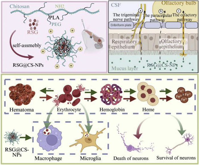
In this study, we conjugated CS to the hydrophobic terminus of poly(ethylene glycol)-poly(lactic acid) (PEG-PLA) to synthesize the amphiphilic ternary copolymer, PEG-PLA-CS. This polymer self-assembles to encapsulate RSG, forming positively charged NPs (RSG@CS-NPs), which improve the water solubility and bioavailability of RSG. Following intranasal administration, the positively charged RSG@CS-NPs adhere to the nasal mucosa and open tight junctions between epithelial cells, facilitating rapid transport from the nasal cavity to the ICH site. Upon reaching the hematoma site, RSG@CS-NPs enhance the phagocytosis of erythrocytes by microglia and macrophages, promoting hematoma absorption and thereby alleviating brain injury (Scheme 1). Administration of RSG@CS-NPs via the intranasal route resulted in favorable therapeutic outcomes in two distinct mouse models of ICH.
Scheme 1
Scheme 1.
In two mouse models of intracerebral hemorrhage, intranasal administration of RSG@CS-NPs enhances erythrocyte phagocytosis by microglia and macrophages, promotes hematoma resolution, thereby reducing hematoma volume, BBB damage, and neuronal injury, and ultimately improving neurological deficits.
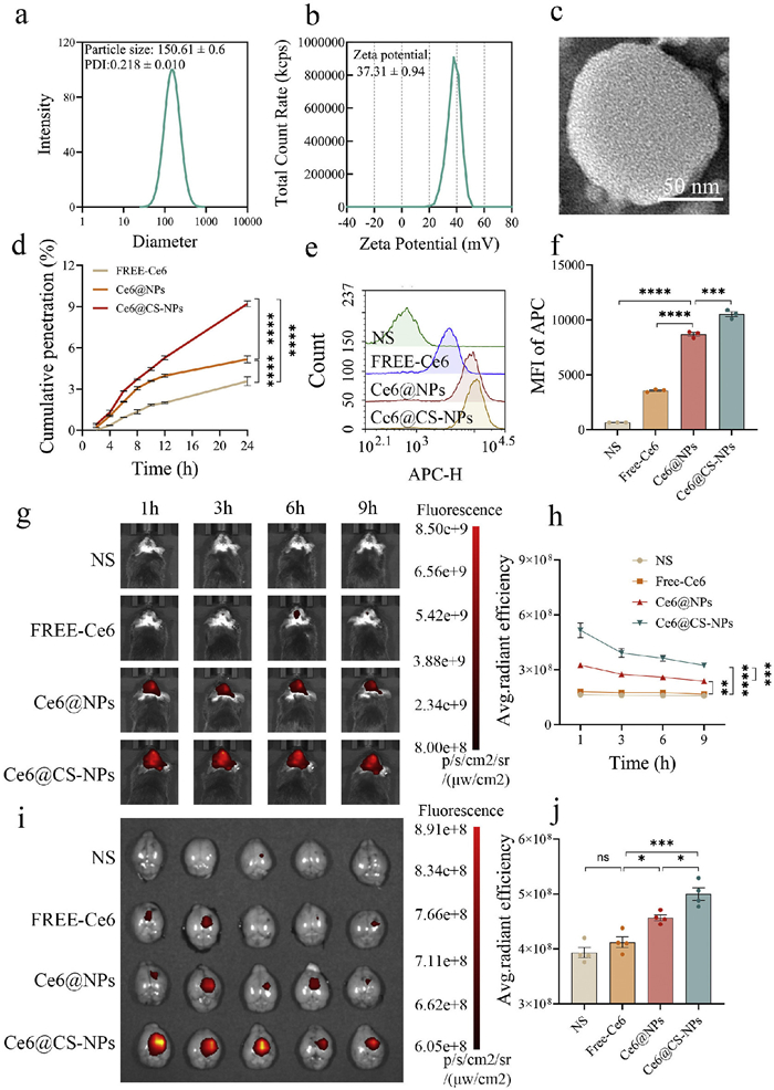
To address the issues of poor water solubility and low bioavailability of RSG, we first synthesized PEG-PLA via ring-opening reaction (Fig. S1a in Supporting information). PEG-PLA was then reacted with succinic anhydride to generate PEG-PLA-COOH (Fig. S1b in Supporting information). Subsequently, catalyzed by 1-ethyl-3-(3-dimethylaminopropyl)carbodiimide (EDC) and N-hydroxysuccinimide (NHS), the amino groups of CS reacted with the carboxyl groups of PEG-PLA-COOH, synthesizing the amphiphilic ternary polymer PEG-PLA-CS (Fig. S1c in Supporting information). Proton nuclear magnetic resonance (1H NMR) characterization confirmed the structures of the compounds. As shown in Fig. S2 (in Supporting information), the synthesized ternary copolymer exhibited proton signals not only from the CS chain (4, 6–9, 5, 10) but also from PEG-PLA (1–2, n, m). Furthermore, proton signals from succinic anhydride (3), used to link PEG-PLA and CS, were clearly observed. These results collectively confirmed the successful synthesis of PEG-PLA-CS. To prepare RSG@CS-NPs, PEG-PLA-CS was dissolved in a 1:1 mixture of 2-(N-morpholino)ethanesulfonic acid (MES) buffer (pH 6.0) and acetone. Acetone-dissolved RSG was then added under water bath sonication. After evaporating the organic solvent, the solution was concentrated using an ultrafiltration tube. Dynamic light scattering (DLS) analysis showed that RSG@CS-NPs had a particle size of 150.61 ± 0.6 nm and a polydispersity index (PDI) of 0.218 ± 0.010 (Fig. 1a). The zeta potential was 37.31 ± 0.94 mV (Fig. 1b). Transmission electron microscopy (TEM) imaging confirmed the spherical morphology of RSG@CS-NPs (Fig. 1c). The encapsulation efficiency and drug loading capacity of RSG@CS-NPs were 84.33% ± 3.79% and 7.78% ± 0.32%, respectively. Based on previous studies, NPs for intranasal delivery should ideally have a size <200 nm, PDI <0.3 [25], and encapsulation efficiency >80% [26]. Given these parameters, our RSG@CS-NPs are well-suited for intranasal administration.
Figure 1
Figure 1.
Characterization of RSG@CS-NPs, permeability across the nasal epithelial barrier, and brain targeting in vitro and in vivo. (a) Size distribution of RSG@CS-NPs. (b) Zeta potential of RSG@CS-NPs. (c) TEM image of RSG@CS-NPs. Scale bar: 50 nm. (d) Cumulative permeability of Free-Ce6, Ce6@NPs, and Ce6@CS-NPs across the RPMI 2650 nasal epithelial barrier model at 2, 4, 6, 8, 12, and 24 h (n = 3). (e, f) Uptake of Free-Ce6, Ce6@NPs, and Ce6@CS-NPs by BV2 cells in a co-culture model with the nasal epithelial barrier (n = 3). (g, h) Fluorescence intensity changes in the brains of ICH mice at 1, 3, 6, and 9 h after intranasal administration of Free-Ce6, Ce6@NPs, and Ce6@CS-NPs (n = 4). (i, j) Ex vivo fluorescence intensity of brain tissues from ICH mice at 9 h after intranasal administration of Free-Ce6, Ce6@NPs, and Ce6@CS-NPs (n = 4). ns, not significant. *P < 0.05, **P < 0.01, ***P < 0.001, ****P < 0.0001. Data are presented as mean ± SEM.
An intact BBB impedes the entry of most blood-borne substances, including 98% of small-molecule drugs and all large-molecule drugs, into the brain, which limits the efficacy of intravenous or oral administration for treating CNS diseases [27]. In contrast, owing to the unique anatomical connections between the nasal cavity and the brain, drugs administered intranasally can bypass the BBB via several pathways, enabling direct delivery to the olfactory bulb, cerebrospinal fluid (CSF), and brain parenchyma target sites. This facilitates localized treatment in brain tissue and improves drug bioavailability in the CNS [28]. These pathways primarily include: the olfactory pathway, the trigeminal nerve pathway, and the paracellular pathway [29,30].
During intranasal delivery, mucus and the nasal epithelium represent major barriers [31]. Therefore, we utilized human nasal epithelial cells (RPMI 2650) cultured in Transwell inserts under air-liquid interface (ALI) conditions to establish an in vitro model of the nasal epithelial barrier [32]. This model was used to evaluate the permeability of RSG@CS-NPs. First, we assessed the cytotoxicity of PEG-PLA-CS on RPMI 2650 cells. As shown in Fig. S3 (Supporting information), cell viability remained unchanged with increasing concentrations of PEG-PLA-CS compared to the control group, confirming the safety of our nanomaterial. Next, we encapsulated the fluorescent tracer chlorin e6 (Ce6) into PEG-PLA or PEG-PLA-CS to prepare Ce6@NPs or Ce6@CS-NPs, respectively. The medium in the upper chamber was replaced with 500 µL of solution containing Free-Ce6, Ce6@NPs, or Ce6@CS-NPs, ensuring an equal total amount of Ce6 (50 µg) per well. Samples were taken from the lower chamber medium at different time points, with an equal volume of fresh medium replenished each time. The amount of Ce6 permeating the nasal epithelial barrier model was measured and calculated. Compared to Free-Ce6, experimental results (Fig. 1d) revealed that encapsulation in PEG-PLA NPs (Ce6@NPs) enhanced the transepithelial permeability of Ce6 [33], and this effect was significantly strengthened by CS conjugation, with the positively charged Ce6@CS-NPs showing superior penetration compared to their unmodified counterparts. This aligns with previous literature reports that positively charged drugs exhibit enhanced transmembrane transport across nasal epithelial cells [24]. Furthermore, we established a co-culture model of the RPMI 2650 nasal epithelial barrier and BV2 microglial cells (BV2 cells) to assess drug uptake by microglia after penetrating the nasal barrier and entering the brain. Results (Figs. 1e and f) showed that BV2 cells internalized significantly more Ce6@CS-NPs than Ce6@NPs. These findings indicate that encapsulating hydrophobic drugs in PEG-PLA NPs enhances permeability across the nasal mucosal barrier and promotes uptake by microglia, while positively charged PEG-PLA-CS NPs enable even more efficient permeation and cellular uptake.
The collagenase-induced ICH mouse model was used to study the ability of PEG-PLA-CS to deliver drugs from the nose to the brain in vivo. All experimental procedures adhered to the Guide for the Care and Use of Laboratory Animals and received approval from the Animal Ethics Committee of the State Key Laboratory of Biotherapy, Sichuan University (Chengdu, China). Three days post-model induction, mice were randomly divided into four groups: Normal saline (NS) group, Free-Ce6 group, Ce6@NPs group, and Ce6@CS-NPs group. In vivo imaging system (IVIS) fluorescence imaging was performed at 1, 3, 6, and 9 h post-intranasal administration. Results (Figs. 1g and h) showed that only very weak fluorescence signals, statistically insignificant, were detected after intranasal administration of Free-Ce6, indicating the difficulty for hydrophobic drugs to reach the brain via direct intranasal delivery [34]. In contrast, mice in the Ce6@NPs and Ce6@CS-NPs groups exhibited significant fluorescence signals, demonstrating the effective delivery capability of NP-based intranasal administration. Among the three nose-to-brain pathways, the paracellular route is the fastest, allowing drugs to rapidly cross epithelial tight junctions and reach the brain within 1 h, while the other pathways deliver drugs more slowly, taking about 3 h [29,35]. Notably, at 1 h post-administration, the fluorescence signal in the brains of the Ce6@CS-NPs group was significantly higher than that in the Ce6@NPs group, suggesting CS's role in opening epithelial tight junctions, enabling rapid and substantial Ce6 delivery to the brain within 1 h. Furthermore, the fluorescence signal in the Ce6@CS-NPs group remained higher and relatively stable compared to the Ce6@NPs group during the subsequent 3–6 h, indicating good retention properties of Ce6@CS-NPs for sustained and efficient delivery of hydrophobic drugs to brain tissue. Ex vivo fluorescence analysis at 9 h post-administration (Figs. 1i and j) showed persistently high fluorescence intensity in the brain tissue of the Ce6@CS-NPs group, consistent with the overall trend of in vivo imaging results. Organ fluorescence analysis revealed only weak signals detectable in the liver (Fig. S4 in Supporting information). In summary, these findings indicate that using PEG-PLA-CS as a carrier increases the epithelial permeability of hydrophobic drugs, facilitates rapid brain targeting, and enables sustained accumulation and release.
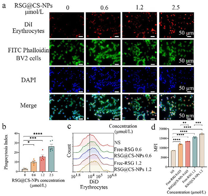
The mass effect of the hematoma and the erythrocyte lysis products are major contributors to brain injury after ICH. Microglia in the brain and peripheral macrophages recruited following ICH can phagocytose erythrocytes, thereby reducing hematoma volume and damage from erythrocyte breakdown by-products [12]. To verify whether RSG@CS-NPs promote erythrocyte phagocytosis by microglia and macrophages in vitro, we isolated erythrocytes from mouse whole blood and labeled them with the plasma membrane fluorescent probes DiI (1,1′-dioctadecyl-3,3,3′,3′-tetramethylindocarbocyanine perchlorate) or DiD (1,1′-dioctadecyl-3,3,3′,3′-tetramethylindodicarbocyanine, 4-chlorobenzenesulfonate salt) to generate fluorescent erythrocytes. BV2 cells pretreated with different concentrations of Free-RSG and RSG@CS-NPs were co-incubated with a five-fold excess of fluorescently labeled erythrocytes. After a specified time, non-phagocytosed erythrocytes were washed away, and phagocytosis was assessed using flow cytometry to detect fluorescence signals or observed via fluorescence microscopy after paraformaldehyde fixation [36]. Fluorescence microscopy revealed that pretreatment with RSG@CS-NPs concentration-dependently enhanced erythrocyte phagocytosis by both BV2 cells (Figs. 2a and b) and bone marrow-derived macrophages (BMDMs) (Figs. S5a and b in Supporting information). As the concentration of RSG@CS-NPs increased, the phagocytic activity of both BV2 cells and BMDMs was progressively enhanced. Flow cytometric analysis yielded consistent results, demonstrating that RSG@CS-NPs promoted erythrocyte phagocytosis by BV2 cells (Figs. 2c and d) and BMDMs (Figs. S5c and d in Supporting information). RSG is a potent agonist of the peroxisome proliferator-activated receptor γ (PPARγ) [37]. Studies have shown that PPARγ activation can upregulate the expression of the scavenger receptor CD36 on the surface of microglia, thereby enhancing their phagocytic clearance of erythrocytes [38]. This is consistent with our experimental observation that RSG@CS-NPs promote erythrocyte phagocytosis by microglia/macrophages. Furthermore, flow cytometric analysis of BV2 cells indicated that at the same dosage, RSG@CS-NPs induced a stronger pro-phagocytic effect than Free-RSG, which is likely attributable to the enhanced cellular uptake of RSG facilitated by the PEG-PLA-CS NP delivery system. In summary, our results demonstrate that RSG@CS-NPs promote erythrocyte phagocytosis by microglia and macrophages.
Figure 2
Figure 2.
RSG@CS-NPs enhance erythrocyte phagocytosis by BV2 cells in vitro. (a, b) Representative fluorescence images of BV2 cells phagocytosing fluorescently labeled erythrocytes. BV2 cells were pretreated with different concentrations of RSG@CS-NPs for 24 h, then co-incubated with DiI-labeled erythrocytes (red) for 3 h; BV2 cells were labeled with fluorescein isothiocyanate (FITC) (green) (n = 5). Scale bar: 50 µm. DAPI, 4′,6-diamidino-2′-phenylindole. (c, d) Flow cytometric analysis of DiD-labeled erythrocyte phagocytosis by BV2 cells. BV2 cells were pretreated with different concentrations of Free-RSG or RSG@CS-NPs for 24 h, then co-incubated with DiD-labeled erythrocytes for 0.5 h (n = 3). *P < 0.05, **P < 0.01, ***P < 0.001, ****P < 0.0001. Data are presented as mean ± SEM.
In the study by Chang et al. [39], using a mouse model of collagenase-induced ICH, it was shown that intraperitoneal injection of 0.5 mg/kg RSG could reduce hematoma volume and improve neurological deficits. Jiao et al. employed both collagenase-induced and autologous blood-induced ICH models in rats [40]. After model establishment, a total of 0.1 mg/kg RSG was administered via local injection at four points around the hematoma, and the results demonstrated that RSG effectively lowered neurological deficit scores, brain edema content, and hematoma volume. Based on these previous studies, we selected 0.5 and 1 mg/kg as the experimental doses for the intranasal administration of RSG@CS-NPs.
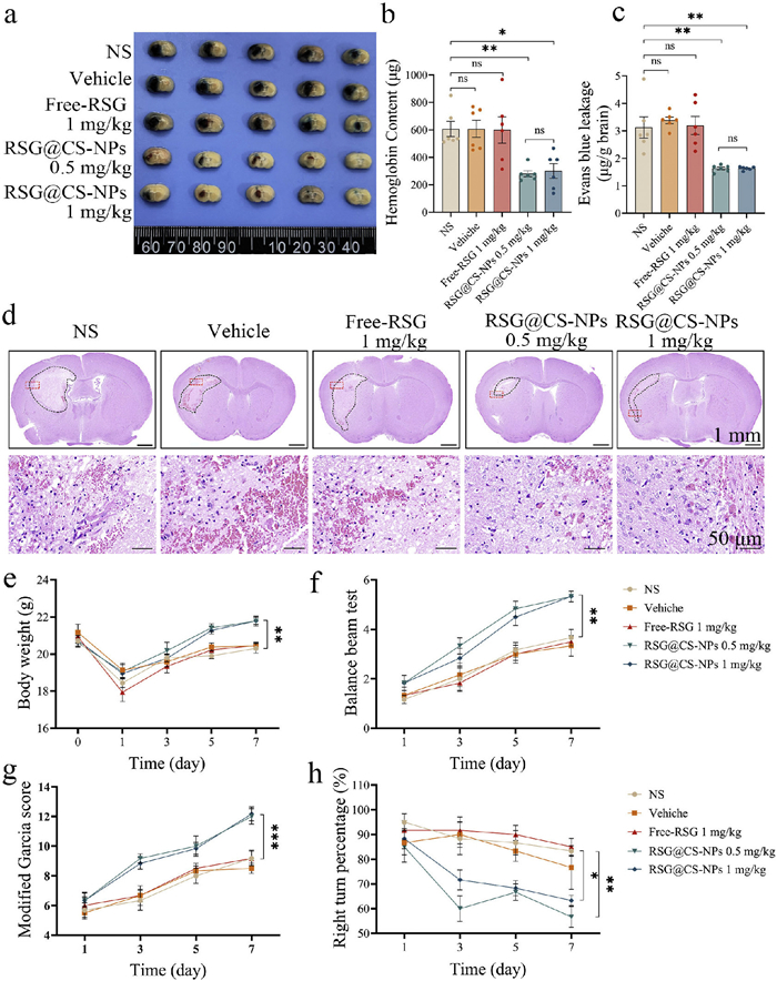
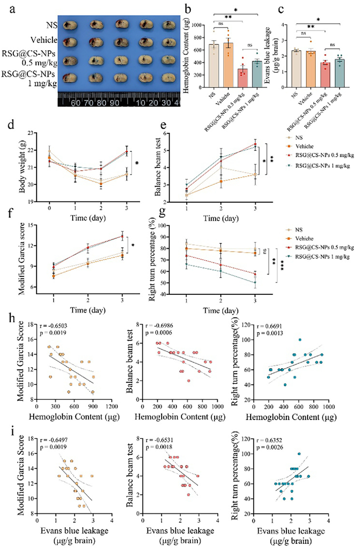
Following collagenase injection, mice were randomly divided into five groups: NS group, vehicle group, Free-RSG (1 mg/kg) group, RSG@CS-NPs (0.5 mg/kg) group, and RSG@CS-NPs (1 mg/kg) group. Intranasal administration treatment commenced 1 h after collagenase injection and continued once daily for seven days. Three days post-treatment initiation, mice were intravenously injected with 4% (w/v) Evans blue (EB) in saline (4 mL/kg). After phosphate buffer saline (PBS) perfusion, brain tissues were harvested, sectioned for hematoma size observation, and subsequently assessed for hemoglobin levels in brain tissue. BBB integrity was evaluated based on EB extravasation. Results (Figs. 3a-c) showed that intranasal administration of Free-RSG at 1 mg/kg failed to reduce hematoma volume or BBB disruption, whereas both 0.5 and 1 mg/kg RSG@CS-NPs significantly reduced ICH-induced hematoma volume and BBB damage. Hematoxylin and eosin (H&E)-stained brain sections from mice treated for three days (Fig. 3d) also demonstrated that RSG@CS-NPs administration reduced hematoma volume. Furthermore, high-magnification observation around the hematoma revealed that RSG@CS-NPs promoted phagocyte erythrophagocytosis. Neurological deficits were assessed using body weight change, balance beam test (which evaluates the walking ability of mice on the beam, with scores ranging from 1 to 7, where lower scores indicate more severe neurological deficits), modified Garcia score (a composite neurological assessment with scores ranging from 4 to 18, in which higher scores reflect milder impairment), and right turn test (where mice are placed at the entrance of a 30° angle formed by two boards and the percentage of right turns is calculated, with a higher percentage indicating more severe neurological impairment). Results (Figs. 3e-h) indicated that compared to other groups, RSG@CS-NPs administration significantly attenuated ICH-induced body weight loss, improved performance on the balance beam test, resulted in higher modified Garcia scores, and significantly reduced the percentage of right turns. Correlation analysis (Fig. S6 in Supporting information) showed that hemoglobin level and BBB leakage were negatively correlated with balance beam test scores and modified Garcia scores, but positively correlated with the percentage of right turns. Brain sections from treated mice were subjected to Nissl staining, Prussian blue staining, and Luxol fast blue (LFB) staining, followed by microscopic examination. Experimental results demonstrated that intranasal administration of RSG@CS-NPs increased neuronal survival around the hematoma (Figs. S7a and b in Supporting information), protected myelin integrity (Figs. S8b and d in Supporting information), and reduced iron deposition (Figs. S8a and c in Supporting information) compared to other groups. Analysis confirmed a significant correlation between neuronal density and the recovery of neurological function (Figs. S7c-e in Supporting information). In the autologous blood injection ICH mouse model, similar therapeutic effects were observed, namely that intranasal RSG@CS-NPs reduced hemoglobin level and BBB disruption (Figs. 4a-c) and improved neurological impairments post-ICH (Figs. 4d-g). The correlation between hematoma volume/BBB leakage and behavioral test results was consistent with the results from the collagenase injection model (Figs. 4h and i).
Figure 3
Figure 3.
Therapeutic effects of intranasally administered RSG@CS-NPs in the collagenase-induced mouse ICH model. (a) Stereoscopic images of brain tissues from ICH mice after three days of intranasal treatment with different drug solutions. (b) Hemoglobin content in the ipsilateral hemisphere of ICH mice after three days of intranasal treatment. (c) Evans blue leakage in the ipsilateral hemisphere of ICH mice after three days of intranasal treatment. (d) Representative brain sections from ICH mice after three days of intranasal treatment, showing enhanced erythrocyte phagocytosis by microglia/macrophages in the RSG@CS-NPs group. Scale bar: 1 mm (upper), 50 µm (lower). (e-h) Body weight, balance beam test scores, modified Garcia scores, and percentage of right turns in mice on days 1, 3, 5, and 7 after intranasal administration of different drug solutions (n = 6). *P < 0.05, **P < 0.01, ***P < 0.001, ****P < 0.0001. Data are presented as mean ± SEM.
Figure 4.
Therapeutic effects of intranasally administered RSG@CS-NPs in the autologous blood-induced mouse ICH model. (a) Stereoscopic images of brain tissues from ICH mice after three days of intranasal treatment with different drug solutions. (b) Hemoglobin content in the ipsilateral hemisphere of ICH mice after three days of intranasal treatment. (c) Evans blue leakage in the ipsilateral hemisphere of ICH mice after three days of intranasal treatment. (d-g) Body weight, balance beam test scores, modified Garcia scores, and percentage of right turns in mice on days 1, 2, and 3 after intranasal administration of different drug solutions (n = 5). (h, i) Correlation analysis between hemoglobin content/BBB disruption and various neurological assessments in the autologous blood-induced ICH model. *P < 0.05, **P < 0.01, ***P < 0.001, ****P < 0.0001. Data are presented as mean ± SEM.
We conducted a comprehensive study on the systemic toxicity of RSG@CS-NPs. Blood biochemical parameters on day 7 post-administration showed no significant deviations compared to control mice (Fig. S9 in Supporting information), indicating negligible toxicity of RSG@CS-NPs. H&E staining of the heart, liver, spleen, lungs, and kidneys revealed no obvious abnormalities (Fig. S10 in Supporting information), suggesting that RSG@CS-NPs did not cause significant toxicity in major organs. Additionally, blood glucose tests indicated that intranasal administration of RSG@CS-NPs at 1 mg/kg did not induce hypoglycemia (Fig. S11 in Supporting information), thereby avoiding associated side effects. Collectively, these data provide strong support for the safety of intranasally administered RSG@CS-NPs in treating ICH.
In conclusion, we successfully developed a nanodrug delivery system capable of encapsulating the hydrophobic drug RSG within a flexible PLA hydrophobic core, surrounded by hydrophilic PEG and CS. RSG@CS-NPs exhibit a positive surface charge under physiological conditions, aiding their retention in the nasal cavity and facilitating the opening of epithelial tight junctions. This enables rapid and sustained drug delivery from the nasal cavity to the brain via multiple pathways, while also promoting drug uptake by microglia and macrophages. Following internalization by microglia or peripheral macrophages, the drug enhances the phagocytic capacity of these cells towards erythrocytes, thereby promoting hematoma resolution and reducing brain tissue damage caused by erythrocyte lysis products. In two mouse models of ICH, intranasal administration of RSG@CS-NPs demonstrated promising therapeutic efficacy, reducing hematoma volume and BBB damage, and significantly improving neurological function in ICH mice. This positively charged NP delivery system, coupled with the therapeutic strategy of enhancing erythrocyte phagocytosis to accelerate hematoma resolution, provides a novel perspective for ICH treatment.
The authors declare no known competing interests or personal relationships that could have influenced the work reported in this paper.
Acknowledgments
This work was supported by the National Key Research and Development Program of China (No. 2024YFA1210202), the National Natural Science Foundation of China (Nos. 32222046 and 82172630), the Sichuan Science and Technology Program (No. 2023NSFSC1931, China), and 1·3·5 Project for Disciplines of Excellent, West China Hospital, Sichuan University (No. ZYYC25008, China). We would also like to thank BioRender (www.biorender.com) for assisting us with drawing Scheme 1.
Supplementary materials
Supplementary material associated with this article can be found, in the online version, at doi:10.1016/j.cclet.2025.112336.
[1]
T. Liu, W. Jiang, M. Zhang, et al., MedComm 6 (2025) e70436. doi: 10.1002/mco2.70436
[2]
J. Magid-Bernstein, R. Girard, S. Polster, et al., Circ. Res. 130 (2022) 1204–1229. doi: 10.1161/circresaha.121.319949
Scheme 1
In two mouse models of intracerebral hemorrhage, intranasal administration of RSG@CS-NPs enhances erythrocyte phagocytosis by microglia and macrophages, promotes hematoma resolution, thereby reducing hematoma volume, BBB damage, and neuronal injury, and ultimately improving neurological deficits.
Figure 1
Characterization of RSG@CS-NPs, permeability across the nasal epithelial barrier, and brain targeting in vitro and in vivo. (a) Size distribution of RSG@CS-NPs. (b) Zeta potential of RSG@CS-NPs. (c) TEM image of RSG@CS-NPs. Scale bar: 50 nm. (d) Cumulative permeability of Free-Ce6, Ce6@NPs, and Ce6@CS-NPs across the RPMI 2650 nasal epithelial barrier model at 2, 4, 6, 8, 12, and 24 h (n = 3). (e, f) Uptake of Free-Ce6, Ce6@NPs, and Ce6@CS-NPs by BV2 cells in a co-culture model with the nasal epithelial barrier (n = 3). (g, h) Fluorescence intensity changes in the brains of ICH mice at 1, 3, 6, and 9 h after intranasal administration of Free-Ce6, Ce6@NPs, and Ce6@CS-NPs (n = 4). (i, j) Ex vivo fluorescence intensity of brain tissues from ICH mice at 9 h after intranasal administration of Free-Ce6, Ce6@NPs, and Ce6@CS-NPs (n = 4). ns, not significant. *P < 0.05, **P < 0.01, ***P < 0.001, ****P < 0.0001. Data are presented as mean ± SEM.
Figure 2
RSG@CS-NPs enhance erythrocyte phagocytosis by BV2 cells in vitro. (a, b) Representative fluorescence images of BV2 cells phagocytosing fluorescently labeled erythrocytes. BV2 cells were pretreated with different concentrations of RSG@CS-NPs for 24 h, then co-incubated with DiI-labeled erythrocytes (red) for 3 h; BV2 cells were labeled with fluorescein isothiocyanate (FITC) (green) (n = 5). Scale bar: 50 µm. DAPI, 4′,6-diamidino-2′-phenylindole. (c, d) Flow cytometric analysis of DiD-labeled erythrocyte phagocytosis by BV2 cells. BV2 cells were pretreated with different concentrations of Free-RSG or RSG@CS-NPs for 24 h, then co-incubated with DiD-labeled erythrocytes for 0.5 h (n = 3). *P < 0.05, **P < 0.01, ***P < 0.001, ****P < 0.0001. Data are presented as mean ± SEM.
Figure 3
Therapeutic effects of intranasally administered RSG@CS-NPs in the collagenase-induced mouse ICH model. (a) Stereoscopic images of brain tissues from ICH mice after three days of intranasal treatment with different drug solutions. (b) Hemoglobin content in the ipsilateral hemisphere of ICH mice after three days of intranasal treatment. (c) Evans blue leakage in the ipsilateral hemisphere of ICH mice after three days of intranasal treatment. (d) Representative brain sections from ICH mice after three days of intranasal treatment, showing enhanced erythrocyte phagocytosis by microglia/macrophages in the RSG@CS-NPs group. Scale bar: 1 mm (upper), 50 µm (lower). (e-h) Body weight, balance beam test scores, modified Garcia scores, and percentage of right turns in mice on days 1, 3, 5, and 7 after intranasal administration of different drug solutions (n = 6). *P < 0.05, **P < 0.01, ***P < 0.001, ****P < 0.0001. Data are presented as mean ± SEM.
Figure 4
Therapeutic effects of intranasally administered RSG@CS-NPs in the autologous blood-induced mouse ICH model. (a) Stereoscopic images of brain tissues from ICH mice after three days of intranasal treatment with different drug solutions. (b) Hemoglobin content in the ipsilateral hemisphere of ICH mice after three days of intranasal treatment. (c) Evans blue leakage in the ipsilateral hemisphere of ICH mice after three days of intranasal treatment. (d-g) Body weight, balance beam test scores, modified Garcia scores, and percentage of right turns in mice on days 1, 2, and 3 after intranasal administration of different drug solutions (n = 5). (h, i) Correlation analysis between hemoglobin content/BBB disruption and various neurological assessments in the autologous blood-induced ICH model. *P < 0.05, **P < 0.01, ***P < 0.001, ****P < 0.0001. Data are presented as mean ± SEM.


 DownLoad:

 下载:


 下载:

