o-Phenanthrenequinone-mediated guanidinium nucleophilic attack of tanshinone IIA to Arg145 on Annexin A2 inhibits endothelial-monocyte adhesion and contributes to relieve atherosclerosis
Citation:
Wenjuan Liu, Yixu Wang, Sihan Xu, Jiawei Li, Mengting Song, Qinghe Du, Zhenzhen Wang, Qianqian Wang, Min Jiang, Gang Bai. o-Phenanthrenequinone-mediated guanidinium nucleophilic attack of tanshinone IIA to Arg145 on Annexin A2 inhibits endothelial-monocyte adhesion and contributes to relieve atherosclerosis[J]. Chinese Chemical Letters,
2026, 37(12): 112320.
doi:
10.1016/j.cclet.2025.112320
o-Phenanthrenequinone-mediated guanidinium nucleophilic attack of tanshinone IIA to Arg145 on Annexin A2 inhibits endothelial-monocyte adhesion and contributes to relieve atherosclerosis
English
o-Phenanthrenequinone-mediated guanidinium nucleophilic attack of tanshinone IIA to Arg145 on Annexin A2 inhibits endothelial-monocyte adhesion and contributes to relieve atherosclerosis
State Key Laboratory of Medicinal Chemical Biology, College of Pharmacy and Tianjin Key Laboratory of Molecular Drug Research, Nankai University, Tianjin 300350, China
Received Date:
28 September 2025 Accepted Date:
23 December 2025 Revised Date:
23 December 2025 Available Online:
15 December 2026
Abstract:
Natural products have long served as invaluable resources for drug discovery, providing unique scaffolds to investigate disease-related targets. Here, we report a novel mechanism by which tanshinone IIA (TIIA), a bioactive constituent of Salvia miltiorrhiza Bge., exerts vascular protective effects through covalent modification of Annexin A2 (ANXA2). By employing an alkynylated TIIA probe (A-TIIA) in combination with chemical proteomics, we identified ANXA2 as a direct binding target of TIIA, wherein the 11,12-ortho-phenanthrenequinone moiety of TIIA forms an amine-quinone adduct with the guanidinium group of Arg145 in ANXA2. This interaction inhibits protein tyrosine phosphatase 1B (PTP1B)-mediated dephosphorylation of ANXA2 at Tyr24, locking the protein in an inactive conformation and disrupting the ANXA2-integrin α5-focal adhesion kinase (FAK)/intercellular adhesion molecule 1 (ICAM-1) signaling axis. This study not only uncovers a previously unknown mechanism underlying the therapeutic action of TIIA but also underscores the potential of natural product-inspired covalent modifiers in modulating protein-protein interactions. The evolutionarily conserved Arg145 residue in ANXA2 emerges as a promising target site for future drug development.
Natural products, distinguished by their unique chemical structures and diverse biological activities, have long served as a critical foundation for innovative drug discovery [1-3]. As valuable precursors in the design of chemical probes, natural products offer a unique opportunity to identify target proteins associated with bioactive compounds whose mechanisms of action remain poorly understood [4,5]. Elucidating these binding mechanisms provides not only novel structural insights but also facilitates the advancement of clinical drug development, thereby accelerating the trajectory of innovative drug discovery [6,7]. Chemical biology leverages methodologies including chemical synthesis and rational molecular design to facilitate precise structural modifications of natural products. By harnessing advanced analytical technologies, it allows for precise elucidation of the interaction mechanisms between chemical probes and target proteins across multiple levels, ranging from molecular and cellular systems to in vivo environments [8-11]. This approach enhances the clarity of target information and enables more efficient screening strategies in drug development, thereby accelerating the translation of basic discoveries into clinical applications and significantly advancing the field of pharmaceutical research and development [12-14].
Tanshinone IIA (TIIA), the core bioactive lipophilic component of the traditional Chinese medicinal herb Salvia miltiorrhiza Bge. (Danshen), has been widely recognized to possess significant anti-inflammatory and cardiovascular protective effects [15,16]. The unique value of this molecule originates from its o-phenanthrenequinone moiety, a quinone substructure characterized by high electrophilic reactivity. The α,β-unsaturated carbonyl group within the ortho-quinone can act as a covalent warhead, facilitating covalent modification of target proteins through Michael addition reactions with nucleophilic residues such as Cys, His, Lys, and Arg [17-19]. Nevertheless, no studies have yet reported covalent modulation of proteins by TIIA, and the role of this functional group in target recognition and functional regulation remains unclear. Identifying TIIA’s target proteins and its covalent modification mechanisms is essential for understanding its pharmacological effects and provides a chemical basis for developing new covalent drugs based on the o-phenanthrenequinone scaffold.
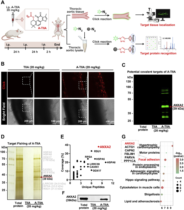
In this study, an alkynylated derivative of TIIA (A-TIIA) was synthesized by introducing an alkyne group at the C1 position of TIIA (Figs. S1–S4 in Supporting information). This derivative was designed to identify potential covalent binding targets of TIIA in vivo through click chemistry-mediated fluorescence labeling of target organs or proteins combined with proteomic profiling (Fig. 1A). Initial validation at the cellular level confirmed that alkynylation at the C1 position did not compromise TIIA's anti-inflammatory and calcium antagonistic activities (Fig. S5 in Supporting information), indicating that A-TIIA retains the pharmacological properties of TIIA and can serve as a suitable probe for target protein recognition and analysis. The animal experiments were approved by the Animal Experimental Ethics Committee of Nankai University (No. 2024-SYDWLL-000153). Mice were intraperitoneally injected with A-TIIA (20 mg/kg), fluorescent localization analysis was performed to assess its distribution in the thoracic aorta. Compared with the control group, A-TIIA exhibited specific accumulation in the thoracic aorta (Fig. 1B), suggesting a potential interaction between A-TIIA and vascular proteins. Notably, gel imaging analysis demonstrated that the fluorescent signal of A-TIIA remained specifically associated with certain protein bands even after rigorous denaturation with SDS and electrophoretic separation, in contrast to the negative control (Fig. 1C). This observation suggested that A-TIIA may form covalent bonds with specific target proteins. Subsequently, functional azide-modified magnetic microspheres were utilized to isolate potential target proteins of A-TIIA from decellularized blood vessels. Following the target fishing process, sodium dodecyl sulfate-polyacrylamide gel electrophoresis (SDS-PAGE) combined with silver staining demonstrated that some potential target proteins were specifically enriched (Fig. 1D).
Figure 1
Figure 1.
ANXA2 is identified as a potential target in the thoracic aorta. (A) Schematic representation of the experimental workflow used by the A-TIIA probe to capture and identify its binding targets. (B) The metabolic label imaging of intraperitoneal administration of the A-TIIA probe (20 mg/kg) on mice vascular tissue. Scale bar: 100 or 200 µm. (C) Fluorescence gel imaging was performed to analyze thoracic aortic lysates obtained from mice intraperitoneally administered with A-TIIA or TIIA. Total protein: Total vascular protein + BDP; TIIA: TIIA+ BDP; A-TIIA: A-TIIA probe + BDP. (D) SDS-PAGE and silver staining of proteins captured by A-TIIA probe-target fishing from blood vessels. Total protein: Total vascular protein; TIIA: TIIA + magnetic microspheres; A-TIIA: A-TIIA probe + magnetic microspheres. (E) The potential targets ranking of overlapping proteins was illustrated by a scatter plot. (F) WB was performed to verify the presence of ANXA2 among the proteins captured by the A-TIIA probe. (G) KEGG analyses for potential target pathways and functions.
To investigate TIIA's covalent binding mechanism, mass spectrometry (MS)-based protein profiling of A-TIIA-captured proteins was conducted. A total of 222 proteins were identified, and among them, 77 were initially selected as candidate proteins based on gel imaging within the molecular weight ranges of 70–100 and 25–45 kDa. Incorporating protein sequence coverage and scoring criteria, Annexin A2 (ANXA2), a critical member of the calcium-dependent phospholipid-binding protein family, was identified as the top candidate (Fig. 1E), which serve as a novel therapeutic target for hemodynamically induced vascular endothelial dysfunction and atherosclerosis [20]. Western blot (WB) analysis further validated that A-TIIA specifically captures ANXA2 (Fig. 1F). Kyoto encyclopedia of genes and genomes (KEGG) pathway analyses of these 77 proteins showed they are mainly involved in signaling pathways including cell adhesion, among which the adhesion-related genes in the potential targets include ANXA2, filamin binding LIM protein 1 (FBLIM1), parvin alpha (PARVA), etc. (Fig. 1G). Based on the above experimental evidence, we preliminarily speculate that ANXA2 may be one of the potential covalent binding target proteins through which TIIA exerts its therapeutic effects on cardiovascular diseases.
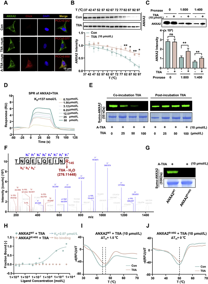
To validate the interaction between ANXA2 and TIIA, a series of in vitro protein–small molecule binding assays were conducted. Cellular immunofluorescence co-localization analysis demonstrated that A-TIIA specifically labeled EA.hy926 cells, generating pseudo-red fluorescence via a click reaction with the azide-conjugated tracer BODIPY FL-Azide (BDP) (Fig. 2A). This fluorescence signal exhibited precise co-localization with ANXA2 protein (pseudo-green), resulting in merged yellow. The high Pearson's correlation coefficient (PCC = 0.85) suggested a robust intracellular interaction between A-TIIA and ANXA2. Furthermore, the cellular thermal shift assay (CETSA) revealed that pre-treatment with 10 µmol/L TIIA significantly attenuated the thermal degradation of ANXA2 in EA.hy926 cells (Fig. 2B). In parallel, drug affinity responsive target stability (DARTS) analysis corroborated this interaction by showing that TIIA binding enhanced the resistance of ANXA2 to proteolytic digestion in cell lysates (Fig. 2C). Additionally, surface plasmon resonance (SPR) assay confirmed the direct binding affinity between TIIA and recombinant ANXA2 protein, yielding a dissociation constant (KD) of approximately 137 nmol/L (Fig. 2D).
Figure 2
Figure 2.
TIIA covalent binds to ANXA2 at Arg145. (A) Co-localization assay between ANXA2 and TIIA in EA.hy926 cells. Scale bar: 15 µm. (B) The impact of TIIA treatment enhances the thermal stability of ANXA2 in CETSA analysis. (C) The DARTS assay confirms that pre-incubation with TIIA enhances the proteolytic stability of ANXA2. (D) SPR assays demonstrate that TIIA interacts with ANXA2 with high affinity. (E) In-gel imaging combined with co- or post-incubation TIIA competition assays indicates that TIIA binds to ANXA2 irreversibly. (F) LC-MS/MS identification reveals TIIA covalently binds to ANXA2 at Arg145. (G) In-gel imaging contrastive analysis of TIIA binding to ANXA2WT and its mutant protein ANXA2R145G. (H) MST analysis of TIIA binding to ANXA2WT and ANXA2R145G. (I, J) FTS analysis of TIIA binding to ANXA2WT and ANXA2R145G. Data are expressed as mean ± SEM (n = 3). Statistical analysis was performed using analysis of variance (ANOVA). *P < 0.05, **P < 0.01 vs. Con. ns, no statistical difference.
To clarify the binding mode, a competitive in-gel imaging assay was performed. It showed that co-incubation of recombinant Sumo-ANXA2 with TIIA (25, 50 and 100 µmol/L) and 10 µmol/L A-TIIA reduced A-TIIA’s fluorescence signal on Sumo-ANXA2. However, pre-incubating Sumo-ANXA2 with A-TIIA before adding TIIA did not change the fluorescence, indicating a covalent bond between A-TIIA and Sumo-ANXA2 (Fig. 2E). We also confirmed that A-TIIA did not combine with the Sumo tag (Fig. S6 in Supporting information). Subsequently, mass spectrometry analysis of the TIIA-ANXA2 complex revealed a covalent modification occurring specifically at the Arg145 residue (R145) of ANXA2. As shown in Fig. 2F, the molecular weight of the modified Arg145 peptide is 276.11448 Da higher than that of the unmodified peptide, which is highly consistent with the theoretical value (276.11448 Da) of “TIIA molecule (molecular weight 294.1258 Da) minus 1 molecule of water (18 Da)”, indicating that dehydration occurs during the reaction. To confirm the specificity of the covalent interaction between TIIA and ANXA2 at residue R145, a recombinant mutant protein, ANXA2R145G, was constructed by substituting the arginine residue at position 145 with glycine (Arg145 → Gly145). As anticipated, in-gel fluorescence imaging demonstrated robust A-TIIA labeling of wild-type (WT) ANXA2WT, whereas the ANXA2R145G exhibited minimal signal (Fig. 2G). The binding affinity was further evaluated using microscale thermophoresis (MST), which revealed that TIIA bound to ANXA2WT with a KD of 2.07 µmol/L, while no significant binding was observed for the ANXA2R145G (Fig. 2H). Additionally, fluorescence thermal shift (FTS) assays indicated that TIIA enhanced the thermal stability of ANXA2WT by 1.5 ℃ (ΔTm = +1.5 ℃), but had no measurable effect on the thermal stability of the ANXA2R145G (ΔTm = 0 ℃) (Figs. 2I and J). Collectively, these results conclusively demonstrated that TIIA directly interacts with ANXA2 and specifically forms a covalent bond with Arg145 residue.
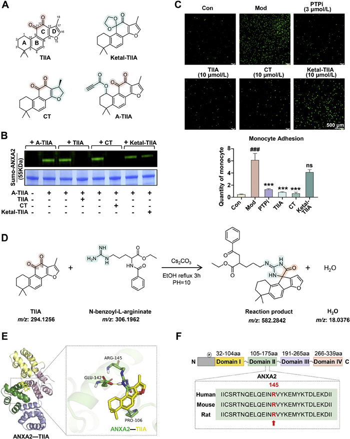
To elucidate the pharmacophore and reaction mechanism underlying TIIA binding to ANXA2, we performed acid-catalyzed ketal protection on the 11-keto group of TIIA, which disrupted the o-phenanthrenequinone structure and yielded compound Ketal-TIIA (Figs. S7–S10 in Supporting information). Furthermore, cryptotanshinone (CT), a natural derivative of TIIA that features a single bond between carbons 17 and 18, was also used to contrast the contribution of double bond in TIIA (Fig. 3A). First, in-gel imaging was employed to assess whether TIIA analogues could competitively inhibit the binding of A-TIIA to ANXA2. As shown in Fig. 3B, incubation of A-TIIA with ANXA2 for 6 h followed by click reaction with azide-BDP produced a clear fluorescent signal. However, when TIIA or CT was pre-incubated with ANXA2, no fluorescence was detected. In contrast, the Ketal-TIIA group showed fluorescence similar to the control. The results suggest that the intact o-phenanthrenequinone moiety present in TIIA and CT is likely capable of binding to the Arg145 residue of ANXA2. In contrast, Ketal-TIIA, which features a disrupted ortho-quinone structure, demonstrates significantly reduced capacity to compete for this binding site.
Figure 3
Figure 3.
TIIA covalently binds to the guanidino group of the Arg145 residue in ANXA2 through a cycloaddition reaction. (A) Structure of TIIA, Ketal-TIIA, CT and A-TIIA. (B) Competitive in-gel imaging analysis of TIIA and its derivatives in binding to ANXA2 with A-TIIA. (C) Inhibition effects of TIIA and its derivatives on blocking monocyte adhesion to vascular endothelial EA.hy926 cells. Scale bar: 500 µm. Data are expressed as mean ± SEM (n = 3). Statistical analysis was conducted by ANOVA. ###P < 0.001 vs. Con group; ***P < 0.001 and ns vs. Mod group. (D) The reaction schematic diagram of TIIA with arginine derivatives (N-benzoyl-L-argininate). (E) Covalent molecular docking of TIIA with ANXA2. (F) Comparative analysis of ANXA2 Domain Ⅱ core sequences across species.
Recent studies have demonstrated that ANXA2 activates the downstream focal adhesion kinase (FAK)/intercellular adhesion molecule-1 (ICAM-1) signaling pathway following phosphorylation. This activation markedly enhances monocyte adhesion to endothelial cells and contributes to the progression of cardiovascular diseases, such as atherosclerosis [20,21]. Based on its biological function, the effects of TIIA and its derivatives on ANXA2-regulated monocyte adhesion were further validated using a co-culture system of vascular endothelial cells and monocytes. Stimulation with Yoda1 (1 µmol/L), an ANXA2 phosphorylation inducer, significantly enhanced the adhesion of calcein AM-labeled THP-1 monocytes to vascular endothelial EA.hy926 cells. Both TIIA and CT (10 µmol/L) markedly inhibited this adhesion, showing effects comparable to those of the ANXA2 phosphorylation inhibitor PTPi (3 µmol/L) (Fig. 3C). In contrast, Ketal-TIIA failed to inhibit monocyte adhesion. These results indicate that the 11,12-o-phenanthrenequinone pharmacophore may directly interact with the Arg145 guanidinium residue on ANXA2, thereby modulating its biological function.
To elucidate the binding mechanism, a model reaction was conducted using an arginine derivative, ethyl N-benzoyl-L-argininate, with TIIA. As shown in Fig. 3D, a condensation reaction occurred between the o-phenanthrenequinone moiety of TIIA and the guanidino group of the arginine derivative (product molecular weight: 582.2842 Da). The mechanism may be that under alkaline conditions, the guanidino group in the arginine side chain remains protonated and positively charged, with its three nitrogen atoms participating in the conjugated system; the terminal nitrogen atom acts as a nucleophile and attacks the electrophilic ring of the orthoquinone (Fig. S11 in Supporting information). Further analysis by HRMS and NMR that the molecular weight of the product was 18 Da lower than the theoretical value of “TIIA + arginine derivative” (Figs. S12–S15 in Supporting information). This observation highly aligns with the characteristic molecular weight change “loss of one water molecule (molecular weight 18 Da)” detected for the covalent modification reaction between TIIA and the ANXA2-Arg145 in Fig. 2F, thereby strongly corroborating the likelihood of a dehydration reaction during the binding process between TIIA and the arginine residue. Molecular docking simulations involving the o-benzoquinone structure of TIIA and residue Arg145 of ANXA2 further indicated that Glu142 and Pro106 on ANXA2 can form hydrogen bonds with TIIA, potentially enhancing its binding affinity (Fig. 3E). Furthermore, the residue Arg145 is located in Domain Ⅱ of the ANXA2, which is one of the four highly homologous Annexin repeats in the core domain, contains five α-helices, and can bind to negatively charged phospholipid membranes in a calcium-dependent manner to mediate membrane aggregation, membrane bridging, and vesicle transport [22-24]. Sequence alignment analysis revealed that Arg145 is conserved across human, mouse, and rat species (Fig. 3F), suggesting that TIIA may interact with ANXA2 through a similar mechanism in these species.
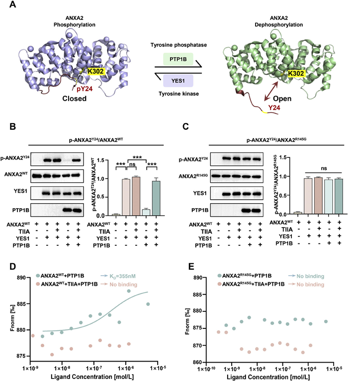
Recent studies demonstrated that the activity of ANXA2 is directly regulated by the phosphorylation status of its tyrosine residue at position 24 (Tyr24). The phosphorylation is mediated by non-receptor protein tyrosine kinases, such as Yes proto-oncogene 1 (YES1), which specifically recognize and phosphorylate Tyr24 within the N-terminus of ANXA2. The phosphorylation of the N-terminal Tyr24 enables the formation of a salt bridge with the C-terminal residue Lys302, thereby inducing a “closed” or inactive conformation of ANXA2 [25,26]. Conversely, protein tyrosine phosphatase 1B (PTP1B) catalyzes the dephosphorylation of ANXA2 at Tyr24. The disruption of salt bridge formation leads to a conformational change in ANXA2, shifting it into an “open” or active state (Fig. 4A) [20,27,28]. To investigate whether the covalent binding of TIIA to Arg145 on ANXA2 affects the ability of YES1 to phosphorylate or of PTP1B to dephosphorylate ANXA2 at Tyr24, an in vitro assay was performed. The results demonstrated that active YES1-mediated phosphorylation of ANXA2 at Tyr24 remained unaffected by TIIA treatment. In contrast, TIIA administration significantly inhibited PTP1B-mediated dephosphorylation of ANXA2 at Tyr24 (Fig. 4B).
Figure 4
Figure 4.
Covalent binding of TIIA to ANXA2 disrupts its dephosphorylation and affects the interaction between ANXA2 and PTP1B. (A) Schematic representation of ANXA2 conformational regulation in closed and states. (B, C) The impacts of TIIA on YES1 or PTP1B in regulating Tyr24 phosphorylation or dephosphorylation of ANXA2WT and ANXA2R145G proteins. (D, E) MST assay of the interaction between ANXA2WT or ANXA2R145G and PTP1B, in the presence or absence of TIIA. Data are expressed as mean ± SEM (n = 4). ***P < 0.001 (t-test).
Parallel assays conducted using the ANXA2R145G mutant produced consistent results. Active PTP1B failed to dephosphorylate Tyr24 on the ANXA2R145G mutant, and TIIA similarly showed no effect on the phosphorylation status of this mutant (Fig. 4C). These findings suggested that TIIA binding to Arg145 may interfere with the interaction between PTP1B and ANXA2, thereby attenuating PTP1B's ability to dephosphorylate ANXA2 at Tyr24. Subsequently, the MST assay was employed to validate this hypothesis. As shown in Fig. 4D, the KD between ANXA2 and PTP1B was 355 nmol/L. However, pre-incubation with TIIA completely disrupted this interaction. Moreover, the ANXA2R145G mutant failed to bind to PTP1B, indicating that the Arg145 residue is critical for the ANXA2-PTP1B interaction, and TIIA exerted no further inhibitory effect in this case.
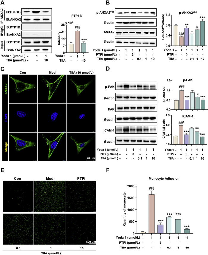
To validate the impact of TIIA's covalent targeting of ANXA2 on the ANXA2 signaling axis, co-immunoprecipitation assays were performed to evaluate how TIIA affects the interaction between ANXA2 and PTP1B in EA.hy926 cells. The results demonstrated that 1 µmol/L Yoda1 treatment significantly enhanced the interaction between ANXA2 and PTP1B, an effect that was markedly inhibited by the administration of 10 µmol/L TIIA (Fig. 5A). Under the same conditions, the ANXA2–PTP1B interaction-induced dephosphorylation of ANXA2 at Tyr24 was also attenuated (Fig. 5B).
Figure 5
Figure 5.
TIIA modulates the ANXA2-integrin α5-FAK/ICAM-1 signaling pathway in EA.hy926 cells. (A) The effects of Yoda1 and TIIA on the ANXA2-PTP1B interaction were verified by co-immunoprecipitation using EA.hy926 cell lysates. (B, C) The impacts of Yoda1 and TIIA on ANXA2 protein phosphorylation and on the membrane translocation of the ANXA2 protein. Scale bar: 20 µm. (D) WB analysis of FAK phosphorylation and ICAM-1 expression in TIIA treated EA.hy926 cells. (E, F) The inhibitory effect of TIIA treatment on monocyte adhesion to EA.hy926 cells. Scale bar: 500 µm. Data are expressed as mean ± SEM (n = 4). ###P < 0.001 vs. Con group; *P < 0.05, **P < 0.01; ***P < 0.001 vs. Mod group.
In general, calcium-induced dephosphorylation of ANXA2 promotes its binding to integrin α5, facilitating the translocation of integrin α5 to lipid rafts on the cell membrane [20]. This interaction activates the downstream FAK/ICAM-1 signaling pathway, which enhances monocyte recruitment to endothelial cells and accelerates cardiovascular disease progression [20,21]. These mechanistic insights are consistent with our experimental findings regarding ANXA2 membrane translocation, FAK phosphorylation, ICAM-1 expression, and monocyte adhesion. Specifically, the dephosphorylation of ANXA2 in EA.hy926 cells promotes its translocation to the cell membrane (Fig. 5C). The translocation induced a marked increase in both FAK phosphorylation levels and ICAM-1 protein expression (Fig. 5D), thereby promoting significant monocyte recruitment to the endothelial cells (Figs. 5E and F). Notably, all these effects were effectively attenuated by TIIA treatment.
Previous studies show orthoquinone has high electrophilicity, mainly due to its 1,2-dicarbonyl structure, highly reactive to nucleophilic attack [29]. Under physiological states, arginine’s side-chain guanidine group (pKa: 12.5) stays protonated and positively charged, its three nitrogen atoms involved in a conjugated system [30,31]. As a nucleophile, arginine’s terminal nitrogen mainly undergoes Michael addition by attacking orthoquinone’s electrophilic ring, forming a single-substituted carbon-nitrogen adduct. Subsequently, the guanidine group’s second nitrogen may attack another electrophilic site on the same orthoquinone. This causes an intramolecular double nucleophilic interaction, forming a stable fused heterocyclic structure between arginine and orthoquinone [32,33].
Both endogenously generated quinones (e.g., dopaquinone, estrogen quinone, and oxidized mitochondrial ubiquinone) and exogenously introduced quinones (including environmental pollutants and certain therapeutic compounds) can covalently altering arginine residues in proteins via this pathway. These alterations can alter protein conformation, functionality, and enzymatic activity, potentially contributing to oxidative stress-associated damage, impaired signal transduction, or enzyme malfunction [34,35]. However, this covalent modification mechanism which leverages the electrophilic properties of o-phenanthrenequinone to specifically target arginine residues is exceptionally uncommon in natural product pharmacology.
This work establishes the natural diterpenoid TIIA as a novel covalent inhibitor of ANXA2 by revealing a unique mechanism: Its o-phenanthrenequinone moiety may covalently bind to the Arg145 site of ANXA2; this interaction can inhibit PTP1B-mediated dephosphorylation of ANXA2-Tyr24, block the ANXA2-integrin α5-FAK/ICAM-1 pathway, and thereby exert vascular protective effects. However, the specific binding mechanism between ANXA2 and TIIA in vivo requires further experimental validation. Beyond partly elucidating the mechanistic basis for the cardiovascular benefits of Salvia miltiorrhiza Bge., this study identifies o-phenanthrenequinone as a privileged pharmacophore for the development of precise covalent therapeutics, presenting a novel chemical biological strategy that transcends conventional kinase-targeting paradigms.
All authors declare that they have no known competing financial interests or personal relationships that could have appeared to influence the work reported in this paper.
Acknowledgment
This research was funded by the National Natural Science Foundation of China (No. 82474058).
Supplementary materials
Supplementary material associated with this article can be found, in the online version, at doi:10.1016/j.cclet.2025.112320.
[1]
T. Rodrigues, D. Reker, P. Schneider, et al., Nat. Chem. 8 (2016) 531–541. doi: 10.1038/nchem.2479
[2]
A.G. Atanasov, S.B. Zotchev, V.M. Dirsch, et al., Nat. Rev. Drug Discov. 20 (2021) 200–216. doi: 10.1038/s41573-020-00114-z
Figure 1
ANXA2 is identified as a potential target in the thoracic aorta. (A) Schematic representation of the experimental workflow used by the A-TIIA probe to capture and identify its binding targets. (B) The metabolic label imaging of intraperitoneal administration of the A-TIIA probe (20 mg/kg) on mice vascular tissue. Scale bar: 100 or 200 µm. (C) Fluorescence gel imaging was performed to analyze thoracic aortic lysates obtained from mice intraperitoneally administered with A-TIIA or TIIA. Total protein: Total vascular protein + BDP; TIIA: TIIA+ BDP; A-TIIA: A-TIIA probe + BDP. (D) SDS-PAGE and silver staining of proteins captured by A-TIIA probe-target fishing from blood vessels. Total protein: Total vascular protein; TIIA: TIIA + magnetic microspheres; A-TIIA: A-TIIA probe + magnetic microspheres. (E) The potential targets ranking of overlapping proteins was illustrated by a scatter plot. (F) WB was performed to verify the presence of ANXA2 among the proteins captured by the A-TIIA probe. (G) KEGG analyses for potential target pathways and functions.
Figure 2
TIIA covalent binds to ANXA2 at Arg145. (A) Co-localization assay between ANXA2 and TIIA in EA.hy926 cells. Scale bar: 15 µm. (B) The impact of TIIA treatment enhances the thermal stability of ANXA2 in CETSA analysis. (C) The DARTS assay confirms that pre-incubation with TIIA enhances the proteolytic stability of ANXA2. (D) SPR assays demonstrate that TIIA interacts with ANXA2 with high affinity. (E) In-gel imaging combined with co- or post-incubation TIIA competition assays indicates that TIIA binds to ANXA2 irreversibly. (F) LC-MS/MS identification reveals TIIA covalently binds to ANXA2 at Arg145. (G) In-gel imaging contrastive analysis of TIIA binding to ANXA2WT and its mutant protein ANXA2R145G. (H) MST analysis of TIIA binding to ANXA2WT and ANXA2R145G. (I, J) FTS analysis of TIIA binding to ANXA2WT and ANXA2R145G. Data are expressed as mean ± SEM (n = 3). Statistical analysis was performed using analysis of variance (ANOVA). *P < 0.05, **P < 0.01 vs. Con. ns, no statistical difference.
Figure 3
TIIA covalently binds to the guanidino group of the Arg145 residue in ANXA2 through a cycloaddition reaction. (A) Structure of TIIA, Ketal-TIIA, CT and A-TIIA. (B) Competitive in-gel imaging analysis of TIIA and its derivatives in binding to ANXA2 with A-TIIA. (C) Inhibition effects of TIIA and its derivatives on blocking monocyte adhesion to vascular endothelial EA.hy926 cells. Scale bar: 500 µm. Data are expressed as mean ± SEM (n = 3). Statistical analysis was conducted by ANOVA. ###P < 0.001 vs. Con group; ***P < 0.001 and ns vs. Mod group. (D) The reaction schematic diagram of TIIA with arginine derivatives (N-benzoyl-L-argininate). (E) Covalent molecular docking of TIIA with ANXA2. (F) Comparative analysis of ANXA2 Domain Ⅱ core sequences across species.
Figure 4
Covalent binding of TIIA to ANXA2 disrupts its dephosphorylation and affects the interaction between ANXA2 and PTP1B. (A) Schematic representation of ANXA2 conformational regulation in closed and states. (B, C) The impacts of TIIA on YES1 or PTP1B in regulating Tyr24 phosphorylation or dephosphorylation of ANXA2WT and ANXA2R145G proteins. (D, E) MST assay of the interaction between ANXA2WT or ANXA2R145G and PTP1B, in the presence or absence of TIIA. Data are expressed as mean ± SEM (n = 4). ***P < 0.001 (t-test).
Figure 5
TIIA modulates the ANXA2-integrin α5-FAK/ICAM-1 signaling pathway in EA.hy926 cells. (A) The effects of Yoda1 and TIIA on the ANXA2-PTP1B interaction were verified by co-immunoprecipitation using EA.hy926 cell lysates. (B, C) The impacts of Yoda1 and TIIA on ANXA2 protein phosphorylation and on the membrane translocation of the ANXA2 protein. Scale bar: 20 µm. (D) WB analysis of FAK phosphorylation and ICAM-1 expression in TIIA treated EA.hy926 cells. (E, F) The inhibitory effect of TIIA treatment on monocyte adhesion to EA.hy926 cells. Scale bar: 500 µm. Data are expressed as mean ± SEM (n = 4). ###P < 0.001 vs. Con group; *P < 0.05, **P < 0.01; ***P < 0.001 vs. Mod group.


 DownLoad:

 下载:


 下载:

