shichao@ytu.edu.cn (C. Shi). 1 These authors contributed equally to this work.
Received Date:
21 October 2025 Accepted Date:
23 December 2025 Revised Date:
23 December 2025 Available Online:
15 December 2026
Abstract:
The advancement of intelligent packaging systems that integrate real-time freshness monitoring with active preservation capabilities is critically important for enhancing food safety. Beyond traditional single-function packaging materials, we synthesized sulfur/selenium-substituted Nile blue (NBS/NBSe) dyes for fabricating multifunctional films (SA/NBS, SA/NBSe) using sodium alginate (SA) via an extended-flow casting method. The obtained films exhibited dual functionality of freshness monitoring and antibacterial preservation, with excellent mechanical properties and stability. Compared with SA/NBS, SA/NBSe films demonstrated high sensitivity in monitoring shrimp freshness, which aligned with the RGB analysis results obtained via a smartphone application in total volatile base nitrogen (TVB-N) determination. Theoretical analysis revealed that NBSe was more sensitive to pH changes because its acidic type possessed a larger surface distribution in the high electrostatic potential region for promoting H+ ionization. Furthermore, SA/NBSe film contributed superior preservation efficacy for fresh beef cuts (6-day extension) and strong bacteriostatic ability attributed to 1O2 and O2•− generation of photodynamic antimicrobial therapy. Overall, these findings highlight the pH-responsiveness and antibacterial potency of Nile blue–based packaging films, making them promising for the qualitative freshness monitoring and preservation of protein-rich foods.
With increasing economic development and improved living standards, clean and fresh high-quality foods have attracted considerable attention [1]. Traditional petroleum-based food packaging materials are characterized by passive barrier properties, offering physical support and protection from external environments and stimuli [2,3]. In commercial practice, the long-term storage of perishable, protein-rich products are achieved through vacuum packaging in food-grade bags labeled with static product details, including production and expiration dates [4]. Nevertheless, fluctuations in storage temperature and humidity can accelerate or decelerate product spoilage. This discrepancy between the actual shelf life and the printed expiration date can lead to consumer misinterpretation, ultimately contributing to food waste and potential health risks [5,6]. Consequently, intelligent packaging that enables the real-time monitoring of food freshness based on environmental factors has garnered significant research attention [7,8].
Intelligent packaging films are thin, flexible materials integrated with active components (such as sensors, indicators, and antimicrobial agents) that detect environmental or product changes (like spoilage gases or pH shifts) and provide a visible and real-time response to indicate quality or safety status, extending product shelf life and enhancing consumer safety [9,10]. During the spoilage of protein-rich products, such as alkaline volatile compounds containing total volatile basic nitrogen (TVB-N), dimethylamine (DMA), and trimethylamine (TMA) are produced, increasing the pH within the packaging [11,12]. Thus, the freshness and spoilage of packaged foods can be effectively tracked via the real-time monitoring of pH variations in food items throughout the supply chain [13]. Natural pH-responsive pigments, including curcumin and anthocyanins, have been widely used for intelligent films owing to their visible color changes and negligible toxicity [14]. Thereinto, anthocyanins derived from the flower petals of Clitoria ternatea, purple sweet potatoes, blueberries, red cabbage, grapes, and Jaman have been extensively reported [15-20]. Furthermore, Monvisade et al. synthesized phenol red–grafted chitosan to monitor shrimp freshness, achieving considerable color transformation upon spoilage [21]. To further improve anthocyanin stability, Zhu et al. developed a “tannin–anthocyanin conjugated” trifunctional intelligent film through chelating bilberry anthocyanins and tannin acids with SA. The film exhibited excellent stability and achieved the real-time monitoring of beef freshness by transforming its color from red to dark yellow [22]. Although natural pigments are sensitive to pH, their lack of antibacterial and preservative functionalities limits food preservation performance. Hence, developing intelligent packaging films that combine dynamic visual freshness monitoring with food preservation capabilities would enhance consumer safety and reliability.
Nile blue, a benzophenoxazine dye widely employed for staining RNA, lysosomes, and lipids, as well as for in vitro tissue imaging, has garnered considerable attention in recent years owing to its exceptional photophysical properties, stability, and biosafety [23-25]. Photodynamic antimicrobial therapy (PDAT) using Nile blue has been extensively studied. In PDAT, the photosensitizer undergoes intramolecular transitions from the ground state (S0) to the singlet excited state (S1) under specific-wavelength light irradiation, and from S1 to the triple excited state (T1) through intersystem crossing (ISC). Reactive oxygen species (ROS) are then generated when oxygen receives energy derived from T1. These ROS interact with microbial cells, causing content leakage from the cytoplasm, eventually leading to microbial death. Thereinto, Peng et al. synthesized a Nile blue derivative with a thioether substituent, which effectively increased the derivative’s triplet-state yield and enhanced ROS generation capability. Subsequently, a multifunctional antibacterial system named APNB was constructed based on the ABP bacteriophage and cationic Nile blue. Notably, APNB represented the first application of such a system for biofilm ablation, demonstrating high efficacy against multidrug-resistant Acinetobacter baumannii [26]. Afterwards, Yoon et al. designed two compounds of sulfur-replaced phenoxazinium (NBS-N) and selenium-substituted phenoxazinium (NBSe-N) for exploring differential ROS activity, enabling the selective eradication of Gram-positive bacteria. NBS-N primarily produced superoxide anion radicals (O2•−), enabling selective killing of Gram-positive bacteria. In contrast, NBSe-N possessed a high singlet oxygen (1O2) yield and effectiveness against Gram-positive and Gram-negative bacteria. This ROS-dependent selectivity may provide a general strategy for designing targeted photodynamic agents against Gram-positive infections [27]. To further improve antibacterial efficiency, alkylated Nile blue derivatives were synthesized and incorporated into hydrogels, improving PDAT efficacy and accelerating the healing of biofilm-infected wounds [28]. Although Nile blue dyes have achieved remarkable achievements in bacterial theranostics, only a few studies have explored their application in food monitoring fields.
SA has been extensively used in food packaging owing to its excellent gas permeability, water retention capability, biocompatibility, and degradability [29]. Herein, an intelligent packaging film was first proposed based on hydrogen bonding interactions between SA and Nile blue. In this system, SA served as the film matrix, and synthetic NBS or NBSe functioned as antibacterial agents and pH-responsive indicators, yielding films for dynamic visual freshness monitoring and the antibacterial preservation of food. Theoretical analysis revealed that SA/NBSe films exhibited high sensitivity and universality to various volatile amines within 2 min. In addition, these films indicated shrimp freshness through visual color changes (from blue to pink), enabling the real-time monitoring of shrimp freshness. Moreover, shrimp freshness was assessed via a smartphone application assisted with RGB values, providing a convenient and rapid visualization detection method. Furthermore, SA/NBSe films exhibited great oxygen and water vapor retention capacities and excellent antibacterial properties, which prolonged the shelf life of fresh beef cuts by approximately 6 days. In summary, the proposed intelligent packaging film can expand technological advancements in developing highly sensitive, portable, and visual detection methods for the freshness monitoring of protein-rich products.
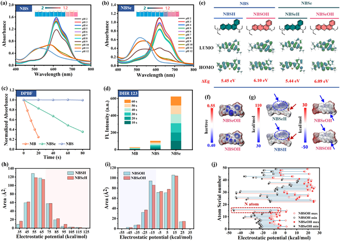
NBS dye was obtained by previous work [24]. To obtain an exceptional photosensitizer, NBSe dye was synthesized according to Scheme S1 (Supporting information). The structures of NBSe and the intermediate compounds were characterized using 1H and 13C nuclear magnetic resonance (NMR) spectroscopy, and electrospray ionization mass spectroscopy (ESI-MS) (Figs. S1–S9 in Supporting information). Subsequently, their photophysical properties were examined using ultraviolet–visible (UV–vis) and fluorescence spectra testing. As depicted in Figs. S10a and b (Supporting information), the maximum fluorescence emission peak was nearly 700 nm under 660 nm excitation, which corresponded to the maximum absorbance value of free NBSe in different solvents. Ulteriorly, the color of NBS and NBSe solutions changed from blue to pink with increasing pH, followed by a hypsochromic shift of the maximum absorption wavelength (Figs. 1a and b). Discriminatively, the NBSe solution exhibited a color change to purple at pH 9, while the color of the NBS solution changed at pH 10, indicating that NBSe was more sensitive to pH changes. The pKa values of NBS and NBSe were quantified as 9.33 and 8.63, respectively, by non-linear regression of the sigmoidal (S-shaped) titration data (Fig. S11 in Supporting information).
Figure 1
Figure 1.
UV–vis spectra of (a) NBS and (b) NBSe in solutions with varying pH (50 μmol/L). (c) Absorbance of DPBF and (d) DHR123 fluorescence for testing 1O2 and O2•− after being incubated with various dyes in dichloromethane (DCM) under irradiation (1 μmol/L, 650 nm, 1.0 mW/cm2). (e) Protonated and deprotonated forms of various dyes and the corresponding HOMO, LUMO, and energy gap (ΔEg). (f) Average local ionization energy (ALIE) and (g) ESP distribution and statistics of different dyes. ESP of the (h) acid type and (i) alkaline type, and (j) their atom serial numbers.
A previous study demonstrated that NBS was capable of simultaneously generating two ROS of O2•− and 1O2 [24]. To further compare the ROS production capacity, dihydrorhodamine 123 (DHR123) and 1,3-diphenylisobenzofuran (DPBF) were utilized for detecting O2•− and 1O2, respectively. As shown in Fig. 1c, the DPBF absorbance for NBSe decreased rapidly from 0 to 80 s (Fig. S12 in Supporting information), but remained stable with NBS under 650 nm irradiation at 1 mW/cm2. Analogously, the fluorescence intensity of DHR123 linearly increased after doping with NBSe, while that of NBS gradually increased (Fig. 1d). These results demonstrated that NBSe exhibited a stronger ability to produce 1O2 and O2•− compared with NBS, providing the basis for excellent photodynamic antibacterial efficiency.
To elucidate reasons underlying the color transformation of the two dye solutions, density functional theory (DFT) and time-dependent DFT (TD-DFT) calculations were performed to investigate changes in the photophysical properties of the solutions. In this work, the acidic and alkaline types of the NBS and NBSe dyes were designated as NBSH/NBSOH and NBSeH/NBSeOH, respectively. The frontier molecular orbitals (FMO) of NBS(e)H/NBS(e)OH in the ground state were shown in Fig. 1e. The increased energy gap between the highest occupied molecular orbital (HOMO) and lowest unoccupied molecular orbital (LUMO) in the alkaline type of NBSOH and NBSeOH resulted in considerable energy being required for electron transition, leading to a hypsochromic shift in the absorption wavelength. This result also aligned with that obtained by analyzing the electron–hole distribution of the S0–S1 transition process using the transition density matrix (Fig. S13 in Supporting information) [30]. Subsequently, the underlying mechanism of the higher pH sensitivity of NBSe compared to that of NBS was systematically explored. As shown in Fig. 1f, basic NBSOH and NBSeOH exhibited multiple electrophilic reaction sites (blue sites), which were susceptible to hydrogen ion attack [31]. Electrostatic potential (ESP) analysis [32,33] revealed that the region with a large positive ESP was concentrated at the cationic secondary amine sites of acidic NBSH and NBSeH, which promoted H+ ionization (indicated by the red arrow in Fig. 1g). After quantification, NBSeH exhibited a larger surface distribution than NBSH in the high-ESP region (94.2911 vs. 86.7789; Fig. 1h), indicating that NBSeH was more likely to ionize H+. NBSOH exhibited a larger surface distribution than NBSeOH in the low-ESP region (208.1324 vs. 206.9489; Fig. 1i), which was more susceptible to H+ binding. Furthermore, the larger ESP range in N atom of NBSeOH (–45.5817 to –6.38956 vs. –46.165 to –6.5928; Fig. 1j) and weaker N–H bonds of NBSeH (0.86 vs. 0.90; Fig. S14 in Supporting information), indicated that NBSe readily converted into NBSeOH under mildly alkaline conditions compared with NBS. Furthermore, NBSOH exhibited a higher proton affinity (294.324) than NBSeOH (293.563), indicating a stronger proton-binding capacity, which is consistent with the higher pKa value of NBS. Thus, NBSe exhibited a higher pH-responsiveness than NBS.
As discussed earlier, intelligent packaging films were prepared via extended-flow casting using Nile blue and SA. The samples with a dye content of 25 μmol/L were denoted as SA/NBS-25 and SA/NBSe-25, and those with a dye content of 50 μmol/L were denoted as SA/NBS-50 and SA/NBSe-50. As shown in Fig. S15 (Supporting information), although the pure SA film was transparent, incorporating the dye produced blue-colored films with intensity increasing with concentration, all films maintained high translucency. The mechanical performance of the films was evaluated to determine their strength and flexibility. As shown in Fig. S16 (Supporting information), the thickness of the films gradually increased with increasing dye dosage (Fig. S16a), likely owing to the increased substance content in film-forming solutions. Figs. S16b and c showed that the tensile stress (TS) of the pure SA film was 41.29 MPa, and those of the SA/NBS-50 and SA/NBSe-50 films were 63.16 and 68.40 MPa, respectively, indicating that the TS increased with the addition of dye. In addition, the elongation at break (EB) of the pure SA film was 29.40%, which was considerably lower than SA/NBS-50 and SA/NBSe-50 (Fig. S16c). This increase in TS and EB was attributed to hydrogen bond interactions between the dyes and the SA matrix, which formed a tightly crosslinked network structure [34,35]. The functional groups in the films and their intermolecular interactions were examined via Fourier transform infrared (FT-IR) spectroscopy. As is known from the chemical structure, SA contains abundant carboxyl (–COOH) and hydroxyl (–OH) groups. As shown in Fig. S16d, the peaks near 1420 and 1640 cm−1 were attributed to the symmetrical and antisymmetric stretching vibrations of the carboxylate radical (COO−) in SA, respectively. The peak near 1490 cm−1 was attributed to the stretching vibration of the C–N aromatic ring in the prepared dye. The peak at 1910 cm−1 was ascribed to C=O absorption of the –COOH group, which moved toward lower wavenumbers owing to the influence of hydrogen bonding. Furthermore, a characteristic broad band of SA at 3300–3500 cm−1 (O–H stretching) was observed. The minor peak at approximately 3350 cm−1, which emerged after dye incorporation, was assigned to the N–H stretching vibration of the secondary amine group in the dye. Notably, the peak at the 3200–3500 cm−1 regions broadened, which might be caused by hydrogen bonding interactions between the SA and dye, indicating that the prepared dye was successfully immobilized into the SA matrix [22,36]. The X-ray diffraction (XRD) pattern of the pure SA film showed four distinct peaks at 13.8°, 22.2°, 29.8°, and 41.5°, which are the characteristic peaks of amorphous SA with some overlaps, confirming the successful preparation of the composite-film substrate (Fig. S16e). The dye-containing composite films exhibited diffraction peaks at similar angles, indicating their predominantly amorphous nature [37,38]. These results demonstrated that the prepared composite films exhibited satisfactory homogeneity and compatibility between the dye and the SA matrix.
Prolonged UV exposure not only degrades the taste, nutritional content, and natural pigments of food but also promotes carcinogen formation, posing a potential threat to human health, which makes UV–vis light resistance a critical parameter for evaluating intelligent packaging films. Fig. S16f showed that the pure SA film exhibited significant visible-light transmittance (T660 = 88.5%), whereas the transmittance of the SA/NBS-50 and SA/NBSe-50 films was markedly lower (T660 = 39.5% and 44.1%, respectively). This decrease in transmittance could be attributed to the absorption peaks of NBS and NBSe near 660 nm (Fig. S10a in Supporting information). Notably, the SA/NBS-50 and SA/NBSe-50 films effectively blocked UV radiation (transmittance < 40%) in the wavelength range of 200–320 nm; thus, they can protect food against some parts of the UVA and UVB ranges and the entire UVC range. In contrast, the pure SA film exhibited higher transmittance at 320 nm (T320 = 70.4%). Furthermore, the UV–vis light resistance of the films significantly improved with increasing dye concentration, indicating that NBS and NBSe synergistically enhanced the UV–vis light resistance of the pure SA film.
Low-water vapor permeability (WVP) intelligent packaging films are essential to inhibit microbial-induced food spoilage caused by excessive moisture. As shown in Fig. S16g, the WVP of the SA/NBS and SA/NBSe films decreased significantly with increasing dye concentration. Observably, the SA/NBSe-50 film exhibited the lowest WVP value (3.76 × 10⁻¹¹ g m−1 s−1 Pa−1), representing a 21% decrease compared with that of the pure SA film (4.76 × 10⁻¹¹ g m−1 s−1 Pa−1). The low WVP of the SA/NBS and SA/NBSe films was attributed to the dense network structure formed by hydrogen bonding interactions between the SA matrix and NBS or NBSe, which effectively restricted water vapor penetration while enhancing the water vapor barrier properties [39]. Surface wettability serves as a pivotal indicator for assessing the water resistance of intelligent packaging films, directly influencing their functional efficacy. As shown in Fig. S16h, the SA, SA/NBS-50, and SA/NBSe-50 films exhibited WCAs of 53°, 86°, and 100°, respectively. The superior water resistance of the SA/NBSe films makes them more suitable for volatile amine sensing applications. Given that elevated oxygen levels accelerate the proliferation of spoilage microorganisms, oxygen permeability (OP) is a critical parameter for evaluating intelligent packaging film performance. As shown in Fig. S16i, the OP of the SA film was 1.50 × 10⁻⁷ g m−1 s−1, whereas those of the SA/NBS-50 and SA/NBSe-50 films were 1.27 × 10⁻⁷ and 1.19 × 10⁻⁷ g m−1 s−1, respectively. This result showed that the addition of dye to SA significantly improved its resistance to oxygen permeation; thus, the films are promising for practical applications in food packaging.
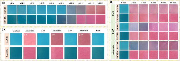
For convenience, SA/NBS-50 and SA/NBSe-50 are referred to as SA/NBS and SA/NBSe, respectively, in subsequent discussions. The SA/NBS and SA/NBSe films were soaked in buffer solutions with pH values ranging from 4 to 13 for 5 min, and their color changes were systematically observed. As shown in Fig. 2a, the SA/NBS film was blue when the solution pH was between 4 and 9. At pH 10–11, the color changed from grayish blue to purple and finally to pink at pH 12. For the SA/NBSe film, the color remained blue within the pH range of 4–8, then changed from grayish blue to light purple in the pH range of 9–10, and finally turned pink with the color gradually intensifying at pH values above 11. Notably, the SA/NBSe film exhibited superior pH sensitivity and underwent color changes at pH 9. Therefore, these intelligent packaging films are promising for monitoring protein-rich foods.
Figure 2
Figure 2.
Color changes of films after (a) immersion in solutions with various pH levels, and (b) exposure to different ammoniated atmospheres. (c) Color reversibility testing results in ammonia and acid solutions.
Total volatile basic nitrogen (TVB-N) is generated through the microbial spoilage of protein-rich animal products, yielding volatile alkaline nitrogenous compounds such as ammonia and amines. Among these, representative volatile amines, DMA and TMA are routinely used as key components to mimic TVB-N formation during the deterioration of seafood and meat products. As shown in Fig. 2b, the SA/NBSe film changed from blue to pink within 2 min upon exposure to ammonia, DMA, and TMA, demonstrating high responsiveness. Discriminatively, the SA/NBS film exhibited a more varied response: It changed directly to pink within 2 min upon exposure to TMA, underwent multiple color transitions (blue → light purple → dark purple → pink) over 6 min upon exposure to DMA, and transformed gradually from blue to purple–pink within 8 min upon exposure to ammonia. Given that the composition of gas generated during shrimp spoilage is complex, the universal and highly sensitive color change of the SA/NBSe film makes it particularly promising for practical freshness monitoring. Given the rapid response of both films to TMA, the relationship between concentration and RGB values was linearly fitted. Fig. S17 (Supporting information) showed a strong linear dependence of the R value, allowing the limit of detection (LOD) to be determined as 0.0242 mol/L for SA/NBS and 0.0171 mol/L for SA/NBSe. This direct comparison of the LODs confirmed the higher sensitivity of the SA/NBSe film. To further investigate the reversibility of color changes in response to pH variations, cyclic tests were conducted in acidic and alkaline environments. As shown in Fig. 2c, the color changes of the SA/NBS and SA/NBSe films correlated well with corresponding pH changes after three cycles of exposure to ammonia and acetic acid. These results confirmed the reversibility of color changes in the films, making them promising for various applications.
To understand the color stability of the films after exposure to light and high temperatures, we simulated refrigerated and ambient storage conditions at 0 and 25 ℃, respectively, and investigated the influence of light exposure by exposing the films to natural light and dark conditions. As shown in Figs. S18a–c (Supporting information), the color retention rate of the SA/NBS and SA/NBSe films was 96% under dark conditions with 0 ℃ and slightly lower under dark conditions with 25 ℃, indicating excellent stability. Under illumination at 25 ℃, the SA/NBS and SA/NBSe films exhibited irreversible discoloration by rapidly changing from blue to light blue, and their color retention rates were 88.88% and 57.91%, respectively, indicating that excessive ROS production not only destroyed the molecular structure of the films but also caused color fading. These results showed that the SA/NBSe film was more sensitive to light than the SA/NBS film, consistent with the ROS generation yield (Figs. 1c and d). RGB is a widely used color system and a critical indicator used for evaluating color stability. By analyzing changes in the red (R), green (G), and blue (B) channels, all colors perceivable by human vision can be comprehensively assessed. Herein, SRGB was used to evaluate the stability of the SA/NBS and SA/NBSe films. As shown in Figs. S18d and e (Supporting information), the SRGB stability of the SA/NBS film was poor (~28.43%) under ambient conditions (such as natural light exposure and storage at 25 ℃), whereas the SA/NBSe film exhibited an SRGB value of 50.00%, which was attributed to its higher light sensitivity. Under dark conditions with 0 ℃, the SA/NBS and SA/NBSe films exhibited minimal color changes (5.92% and 5.77%, respectively). Thus, the prepared films are suitable for low-temperature and light-shielded environments, which correspond to the storage environments of seafood and protein-rich products.
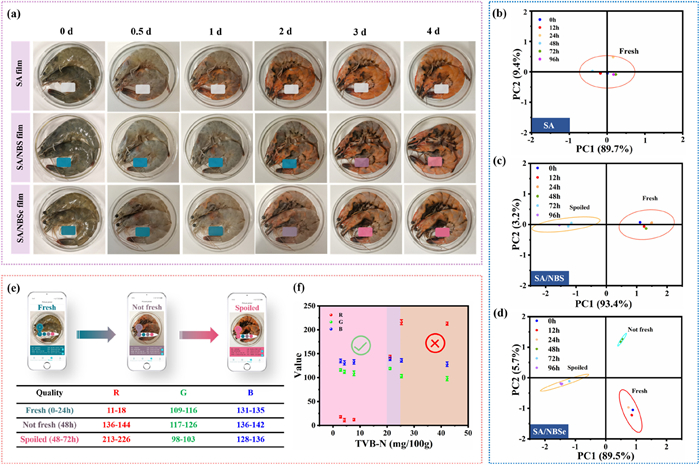
To further investigate the effectiveness of the proposed intelligent packaging films in monitoring food spoilage, live purchased nutrient-rich shrimp were purchased from a seafood market in Yantai, a coastal city known for its seafood production in the Bohai Bay region. As shown in Fig. 3a, the pure SA film exhibited minimal color change in response to variations in shrimp freshness, while the SA/NBS film exhibited a clear color transition from blue to pink. Notably, the SA/NBSe film demonstrated a more pronounced and sequential color shift from blue to purple and finally to pink, indicating superior detection sensitivity. This enhanced sensitivity was further evidenced by the SA/NBSe film’s earlier color transition (turning purple on day 2) compared to the SA/NBS film only showed a light purple hue on day 3. The comparative analysis demonstrated a clear advantage of the NBSe film over the NBS film for real-time shrimp freshness monitoring.
Figure 3
Figure 3.
(a) Photographs of shrimp freshness detection using various developed films for 96 h. PCA of the (b) SA, (c) SA/NBS, and (d) SA/NBSe films. (e) Color analysis of photographs assisted by a smartphone application. (f) Correspondence between RGB and TVB-N level of SA/NBSe films.
To quantitatively assess spoilage progression, TVB-N was measured in accordance with the national standard (GB 5009.228–2016). Based on the TVB-N levels, shrimp freshness was categorized into three stages: Fresh (TVB-N < 15 mg/100 g), not-fresh (15–25 mg/100 g), and spoiled (TVB-N ≥ 25 mg/100 g) [40]. As shown in Fig. 3f, the TVB-N content increased continuously during storage after coating with SA/NBSe films, reaching 21.084 mg/100 g after 48 h. This indicated that the shelf life of shrimp under room temperature (~25 ℃) and light-shielded conditions does not exceed 24 h. After 72 h, the TVB-N value rose to 25.300 mg/100 g, confirming that the shrimp were fully spoiled.
To further validate the film’s capability to differentiate the freshness levels of shrimp, qualitative monitoring was conducted using principal component analysis (PCA). This method classified freshness into distinct categories based on color parameters (RGB) and chemical indicators (TVB-N). As shown in Figs. 3b–d, the SA film failed to effectively distinguish between the three stages of freshness (fresh, not-fresh, and spoiled) owing to its minimal color variations. Although the SA/NBS film could differentiate between the fresh and spoiled stages, it failed to identify the “not-fresh” stage after 48 h. In contrast, the SA/NBSe film demonstrated superior discriminatory ability, clearly identifying “fresh” (0–24 h), “not-fresh” (36–48 h), and “spoiled” (48–96 h) stages of shrimp freshness. Upon shrimp spoilage (TVB-N > 20 mg/100 g at 48 h), the SA/NBSe film showed a decisive increase in R-value, in stark contrast to the stable SA/NBS film (Figs. S19a and b in Supporting information). The signal-to-noise ratios (SNR) data in Fig. S19c (Supporting information) provided a quantitative basis for this disparity: The SA/NBSe film’s SNR reached 7.5 at 48 h, far exceeding the SA/NBS film’s 5.43. The delayed response of the SA/NBS film, which only achieved a comparable SNR of 7.94 at 72 h, highlighted the superior detection capability of the SA/NBSe film. Collective evidence from both R-value and SNR measurements confirmed the superior sensitivity of the SA/NBSe film.
Recently, smartphone-based image analysis has emerged as an innovative technique for monitoring food freshness. Films that generated distinct color variations corresponding to different shrimp freshness stages—fresh, not-fresh, spoiled—were selected to evaluate their RGB values. This method established a correlation between the RGB values and TVB-N levels, enabling freshness assessment based on color changes. To enhance monitoring accuracy, data from three color channels (R, G, and B) were collected to evaluate RGB variations over time and obtain their maximum and minimum values. Specifically, when the shrimp was in the “fresh” stage, the RGB values were as follows: R = 11–18, G = 109–116, and B = 131–135 (Fig. 3e). When TVB-N levels reached 21.084–25.300 mg/100 g, indicating the “not-fresh” stage, the RGB values shifted to R = 136–144, G = 117–126, and B = 136–142. Significant changes in the RGB values were observed when the shrimp was in the “spoiled” stage (TVB-N content reaching 25.300–42.168 mg/100 g), with R values increasing sharply to 213–226, while G and B values settled at 98–103 and 128–136, respectively. Thus, RGB values obtained from the SA/NBSe film accurately reflected variations in TVB-N levels, enabling the real-time monitoring of shrimp freshness through visual signals. In addition, smartphone image-processing software was used to obtain RGB values from film images, enabling the rapid measurement of shrimp freshness. The widespread availability of smartphones makes this method highly convenient and efficient. Users can simply capture an image and analyze RGB values via a dedicated application to assess food freshness. This user-friendly and cost-effective strategy shows great potential for household and small-scale commercial applications.
Furthermore, the antibacterial performance of the SA/NBS and SA/NBSe films was evaluated. Thereinto, SA-L, SA/NBS-L, and SA/NBSe-L referred to the corresponding films subjected to 650 nm light irradiation (20 mW/cm2, 10 min). As shown in Figs. S20a and b (Supporting information), SA, SA-L, SA/NBS, and SA/NBSe films exhibited negligible antibacterial activity, whereas the SA/NBS-L and SA/NBSe-L groups demonstrated significant antibacterial activity under light irradiation, particularly against S. aureus, with survival rates as low as 2.89% and 1.52%, respectively. These results demonstrated that the observed photodynamic antibacterial activity under light irradiation was exclusive to the SA/NBS and SA/NBSe composite films, as neither light exposure alone nor the pure SA matrix induced any notable antibacterial effects. The SA/NBSe-L group demonstrated markedly superior antibacterial performance over the SA/NBS-L group. This superiority was evidenced by its significantly higher antibacterial activities (approximately 13.06-fold against Escherichia coli and 15.25-fold against S. aureus) relative to the pure SA film (Fig. S20c in Supporting information). The enhanced efficacy was further corroborated by the substantial reduction in S. aureus colonies following SA/NBSe-L treatment (22 CFU), starkly contrasting with the count after SA/NBS-L treatment (261 CFU) (Fig. S21 in Supporting information). Overall, the prepared intelligent films not only enable the dynamic monitoring of shrimp freshness but also possess excellent light-activated antibacterial properties owing to ROS generation under light conditions.
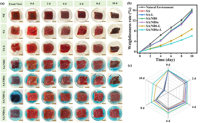
Fresh foods are typically characterized by natural aroma; absence of off-odors or spoilage indicators; and retention of desirable appearance, flavor, and color. The aforementioned results showed that the prepared films exhibited excellent antibacterial efficacy. To further investigate their preservation ability for protein-rich products, highly perishable fresh beef cuts (standard dimensions: 15 mm × 15 mm × 6 mm) were used as test samples. The fresh beef cuts were randomly divided into seven groups for parallel experiments. As illustrated in Fig. 4a, individual fresh beef cuboid samples were aseptically transferred to separate 30 mm-diameter Petri dishes, each hermetically sealed with the prepared packaging films (front view). Slight dehydration and shrinkage were observed in the SA, SA-L, SA/NBS, and SA/NBSe groups starting from day 4, with noticeable decay and dehydration observed by day 10. In comparison, fresh beef cuts in the natural environment group showed dehydration and shrinkage as early as day 2, followed by progressive moisture loss and color darkening during extended storage. These findings indicated that the fabricated films possessed good freshness retention properties, attributable to their excellent oxygen and moisture barrier performances. Notably, under light exposure, beef cuts packaged using the SA/NBS maintained freshness until day 6, after which a lusterless and loosely textured surface developed by day 8. In contrast, the SA/NBSe film prolonged the preservation period to 8 days, accompanied by minimal weight loss (4.52%) and a superior sensory evaluation score of 24.8 (GB/T 17238–2022) (Figs. 4b and c). These results demonstrated that the SA/NBS and SA/NBSe films provided effective preservation under light conditions, with the SA/NBSe film offering longer preservation duration than SA/NBS due to its superior antibacterial activity and lower water vapor and oxygen permeability.
Figure 4
Figure 4.
(a) Appearance of fresh beef cuts wrapped with the different developed films. NE indicated the sample was preserved in the natural environment. Scale bar: 0.8 cm. (b) Weight loss rates and (c) sensory evaluation of fresh beef cuts wrapped in the different developed films (SA-L, SA/NBS-L, and SA/NBSe-L were subjected to 650 nm light irradiation at 10 mW/cm2 for 5 min).
The safety and reliability of food packaging are paramount for ensuring and sustaining consumer confidence during both the purchase and consumption stages. Cytotoxicity was evaluated using the COS-7 cell line, an African green monkey kidney epithelial cell model, via standard cell viability assays. As depicted in Fig. S22a (Supporting information), the cell survival rate was maintained above 80% even at a high dye concentration of 50 μmol/L, demonstrating excellent biocompatibility. Furthermore, hemolysis assessment using healthy porcine blood revealed no significant hemolytic activity for any NBS/NBSe-incorporated film (Fig. S22b in Supporting information). The hemolysis rates for all samples were quantified to be below the 5% threshold, which strongly attested to their excellent biosafety.
In this study, intelligent films were fabricated by incorporating NBS or NBSe into an SA matrix, designated as SA/NBS and SA/NBSe films, respectively. These films exhibited a crosslinked network structure, leading to significantly enhanced mechanical strength and oxygen barrier performance. Compared to the SA/NBS film, the SA/NBSe film demonstrated superior responsiveness to various volatile nitrogenous compounds. In the actual sample testing, the SA/NBSe film exhibited excellent sensitivity for the real-time visual monitoring of shrimp freshness, transitioning from blue (fresh) to purple (not-fresh) and finally to pink (spoiled). These colorimetric responses were consistent with RGB analysis performed using a smartphone application, which was validated against the national standard method for TVB-N determination. Furthermore, the SA/NBSe film exhibited strong antibacterial activity against E. coli and S. aureus, exceeding those of the pure SA film by 13.06- and 15.25-fold, respectively. The SA/NBSe film also extended the shelf life of fresh beef cuts to 8 days. Overall, the developed films offer the dual functionality for dynamic food freshness detection and preservation, offering a promising platform for food monitoring technology to enhance consumer safety and reliability in food experiences.
Notwithstanding these promising results, the transition from laboratory proof-of-concept to practical application necessitates addressing several key challenges. While the film’s performance was validated under controlled conditions, its efficacy in real-world scenarios remains to be established. Critical questions regarding its long-term photostability in transparent packaging, mechanical and functional resilience throughout cold chain logistics, and the paramount issue of dye migration require thorough investigation. Furthermore, a comprehensive food safety assessment is imperative for regulatory approval. Consequently, future work will prioritize enhancing the film’s environmental robustness and conducting rigorous migration and toxicological studies to pave the way for its commercial viability.
The authors declare that they have no known competing financial interests or personal relationships that could have appeared to influence the work reported in this paper.
Acknowledgments
We acknowledge support from the National Natural Science Foundation of China (Nos. 22408307, 22308220), China National Postdoctoral Program for Innovative Talents (No. BX20240052), the Taishan Scholar Project of Shandong Province (No. tsqn202306160), the Natural Science Foundation of Shandong Province (No. ZR2024QB096), and State Key Laboratory of Fine Chemicals, Dalian University of Technology (No. KF2405). Shenzhen University Third-Phase Project of Constructing High-Level University (No. 000001032104), and Research Team Cultivation Program of Shenzhen University (No. 2023QNT005). We thank our department for this generous support.
Supplementary materials
Supplementary material associated with this article can be found, in the online version, at doi:10.1016/j.cclet.2025.112319.
Figure 1
UV–vis spectra of (a) NBS and (b) NBSe in solutions with varying pH (50 μmol/L). (c) Absorbance of DPBF and (d) DHR123 fluorescence for testing 1O2 and O2•− after being incubated with various dyes in dichloromethane (DCM) under irradiation (1 μmol/L, 650 nm, 1.0 mW/cm2). (e) Protonated and deprotonated forms of various dyes and the corresponding HOMO, LUMO, and energy gap (ΔEg). (f) Average local ionization energy (ALIE) and (g) ESP distribution and statistics of different dyes. ESP of the (h) acid type and (i) alkaline type, and (j) their atom serial numbers.
Figure 2
Color changes of films after (a) immersion in solutions with various pH levels, and (b) exposure to different ammoniated atmospheres. (c) Color reversibility testing results in ammonia and acid solutions.
Figure 3
(a) Photographs of shrimp freshness detection using various developed films for 96 h. PCA of the (b) SA, (c) SA/NBS, and (d) SA/NBSe films. (e) Color analysis of photographs assisted by a smartphone application. (f) Correspondence between RGB and TVB-N level of SA/NBSe films.
Figure 4
(a) Appearance of fresh beef cuts wrapped with the different developed films. NE indicated the sample was preserved in the natural environment. Scale bar: 0.8 cm. (b) Weight loss rates and (c) sensory evaluation of fresh beef cuts wrapped in the different developed films (SA-L, SA/NBS-L, and SA/NBSe-L were subjected to 650 nm light irradiation at 10 mW/cm2 for 5 min).


 DownLoad:

 下载:


 下载:

