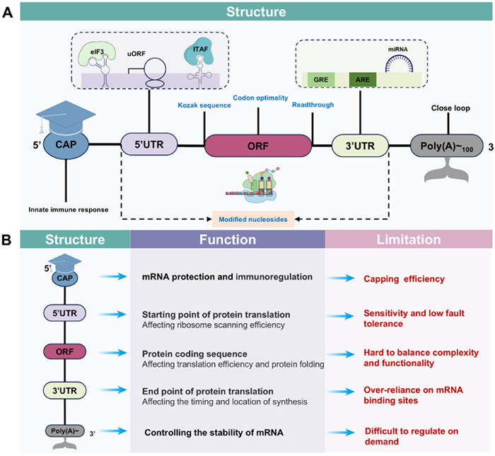
Figure 1.
Schematic diagram of linear mRNA structure (A) and the corresponding function and limitation (B).

mRNA-based vaccines: Rational antigen design, delivery optimization, and immune modulation
Shengbin Liu , Haixing Shi , Danni Dai , Tingchen Tian , Xing Duan , Ju Sun , Weitong Xu , Xiaoling Yin , Yongmei Xie , Xiangrong Song


Vaccination, pioneered by Edward Jenner in 1796, remains one of the oldest and most cost-effective medical interventions [1,2]. Vaccines have dramatically reduced morbidity and mortality from numerous diseases [3]. However, many licensed vaccines fail to elicit broad, durable immunity, are slow and costly to produce, or carry safety concerns [4]. Critically, effective human vaccines are lacking for numerous challenging pathogens, such as human immunodeficiency virus 1 (HIV-1), hepatitis C virus (HCV), and many other viruses, bacteria, fungi, and parasites responsible for millions of deaths annually. mRNA vaccines have emerged as a versatile platform in vaccinology. Over the past two decades, systematic improvements in RNA sequence engineering, chemical modification, and delivery vehicles have resolved key challenges in stability, cellular uptake, and controlled protein expression. These developments have enabled rapid vaccine design and scalable production, establishing mRNA technology as a leading approach in nanomedicine and driving extensive translational research and commercial development [5-9]. mRNA nanomedicines instruct host cells to produce defined proteins, enabling targeted immune responses for disease treatment [10-13]. Notably, the adaptable nature of mRNA synthesis permits swift modification and optimization to address diverse therapeutic indications [14]. Key advantages include: (1) Rapid production facilitating iterative screening for optimized immunogens; (2) capacity to target multiple antigens of a single pathogen [15-19] (3) scalability for pandemic response (e.g., corona virus disease 2019 (COVID-19), influenza) [20]; (4) ideal platform for designing vaccines against complex proteins (e.g., multi-transmembrane domains). Leveraging these features, the first mRNA vaccines were rapidly developed to combat COVID-19. The mRNA-lipid nanoparticle (LNP) vaccines from Pfizer-BioNTech (Comirnaty, BNT162b2) and Moderna (Spikevax, mRNA-1273) received U.S. Food and Drug Administration (FDA) Emergency Use Authorization in 2020 and 2021, and full approval in subsequent years, respectively [21,22]. Furthermore, Moderna's mRNA-LNP RSV vaccine (mRNA-1345) received approved in May 2024 [23], underscoring mRNA's potential against epidemics and pandemics. Despite a plethora of preclinical studies conducted, the clinical translation of mRNA therapeutics beyond vaccines remains limited, probably due to suboptimal design and administration for specific physiological and pathological conditions. While enabling “lightspeed” vaccine development [24], mRNA platforms demand rigorous, specialized expertise to address remaining hurdles. In addition, this review systematically evaluated the potential of artificial intelligence (AI)-assisted mRNA vaccine design in addressing the core challenges of mRNA clinical transformation.
Considering the swift advancement of mRNA nanomedicines, this review seeks to offer a thorough overview of mRNA-based therapeutic platforms, encompassing mRNA design principles, delivery technologies, current understanding of action mechanisms (focus on the latest progress), challenges in transitioning from laboratory research to clinical practice, and insights into future applications.
Structurally, mRNA molecules function as carriers of genetic information, rely on the precise collaboration of five core elements: The 5′ cap, 5′ untranslated region (5′ UTR), open reading frame (ORF), 3′ UTR, and poly(A) tail (Fig. 1A) [25]. (i) 5′ cap: Composed of 7-methylguanosine (m7G) linked via a triphosphate bridge (PPP) to the transcription start nucleotide (TSN), it serves multiple functions: (1) Protects mRNA from 5′ → 3′ exonuclease degradation, enhance stability and half-life; (2) promotes binding of translation initiation factors (eIFs), increase translation efficiency; (3) masks mRNA from innate sensors; uncapped mRNA or cap 0 structures trigger immune recognition. (ⅱ) 5′ UTR: Located downstream of the 5′ cap, modulates ribosome scanning efficiency during translation initiation. Its sequence, length, secondary structure, GC content, and presence of upstream open reading frames (uORFs) significantly influence translation efficiency. (ⅲ) ORF: The core region of mRNA that encodes proteins. Codon composition directly influences translation efficiency and protein folding. Codon optimization can improve translation rates and mRNA stability. Additionally, incorporating functional sequences within the ORF can endow proteins with specialized properties, such as transmembrane transport, secretion, or polymerization, thereby enhancing their functionality and therapeutic potential. (ⅳ) 3′ UTR: Responsible for translation termination and critically influences mRNA stability, localization, and decay. It contains regulatory elements (e.g., AU-rich elements (AREs), GU-rich elements (GREs)) that bind specific proteins. (ⅴ) Poly(A) tail: Plays a crucial role in mRNA stability and translation efficiency, synergizing with the 5′ cap. The length of the poly(A) tail significantly impacts mRNA performance, with longer tails generally correlate higher translation efficiency and stability (e.g., extending to 100 nt in dendritic cells (DCs) increased protein expression 35-fold). Interaction with poly(A)-binding protein (PABP) promotes mRNA circularization, facilitating translation initiation complex formation and boosting translation efficiency 5-10 folds [26].
Despite their indispensability, these elements present challenges limiting large-scale production and clinical application (Fig. 1B) [27]: (ⅰ) 5′ cap: Concerns include suboptimal capping efficiency and potential immune reactions. While enzymatically added caps are highly efficient but operationally complex, co-transcriptional capping is simpler yet less efficient. Uncapped mRNAs and cap 0 structures trigger immune responses and reduce translation, whereas cap 1 structures support sustained translation post-recognition. Research on type Ⅱ cap structures remains limited. (ⅱ) 5′ UTR: Function is influenced by multiple factors, including length, secondary structure, GC content, and uORFs. For instance, specific secondary structures near the start codon can facilitate or impede translation. Comprehensive understanding and precise design remain challenging [28]. (ⅲ) 3′ UTR: Regulatory function depends on the diversity and specificity of mRNA binding sites. The intricate interplay between 3′ UTR and 5′ UTR on mRNA characteristics is not fully elucidated. Designing stable 3′ UTRs often utilizes sequences from highly stable mRNAs like alpha- and beta-globin. (ⅳ) ORF: Codon optimization is complex; it is not merely replacing rare codons but involves considering the spatiotemporal coupling of ribosome elongation rate and protein folding. Incorporating functional sequences (e.g., secretion signals, transmembrane domains) within the ORF can confer desired properties but may concurrently impede overall translation efficiency or compromise protein stability. Balancing translation efficiency, folding, and function are critical. (ⅴ) Poly(A) tail: Its regulation is multifaceted and varies across biological systems (e.g., shorter tails extend half-life in plants [29], contrasting with animals). Dynamic regulation of tail length in different cell types/states requires precise control, which current technologies struggle to achieve [30]. The complex synergistic interaction between the poly(A) tail and other mRNA elements (such as 5′ cap, UTRs) is incompletely understood, including the dynamics of PABP interaction in different cellular environments [31].
Therefore, summarizing the latest mRNA design optimization strategies and analyzing the biological mechanisms behind optimizing different elements based on clinical scenarios is crucial for advancing mRNA drug application and design. The following sections elaborate on these optimizations.
mRNA therapeutics are classified into three main types and the ways in which they exert effects in cytoplasm are illustrated in Fig. 2. (ⅰ) Conventional non-replicating mRNA, which contains the target gene’s coding sequence flanked by 5′ and 3′ UTRs. This is the most established form with well-characterized structure and function [32]. (ⅱ) Self-amplifying mRNA (saRNA), builds upon non-replicating mRNA by incorporating viral replication machinery (often derived from alphaviruses, flaviviruses, or rhabdoviruses), enabling robust intracellular amplification and high antigen protein production from a small initial mRNA dose [33]. The replicase can be encoded within the same ORF or on a separate co-delivered mRNA. While offering advantages like reduced dose requirements and expedited immune activation, saRNA presents potential drawbacks including inflammation from self-amplification, delivery challenges due to increased length, and potential immunogenicity/risks from RNA replicase-host factor interactions [33]. (ⅲ) Circular RNA (circRNA), distinguished by its exceptional inherent stability due to resistance to exonuclease degradation, significantly extending intracellular half-life [34,35]. In contrast to linear mRNA, circRNA does not necessitate the costly 5′ cap or poly(A) tail. Instead, it utilizes internal ribosome entry site (IRES) sequences or N6-methyladenosine (m6A) motifs for translation initiation, simplifying manufacturing process, reduces production costs. Studies suggest circRNA can markedly reduce immunogenicity without chemical modification, although its complex topology might elicit undesirable immune responses and cell entry poses greater challenges [36].
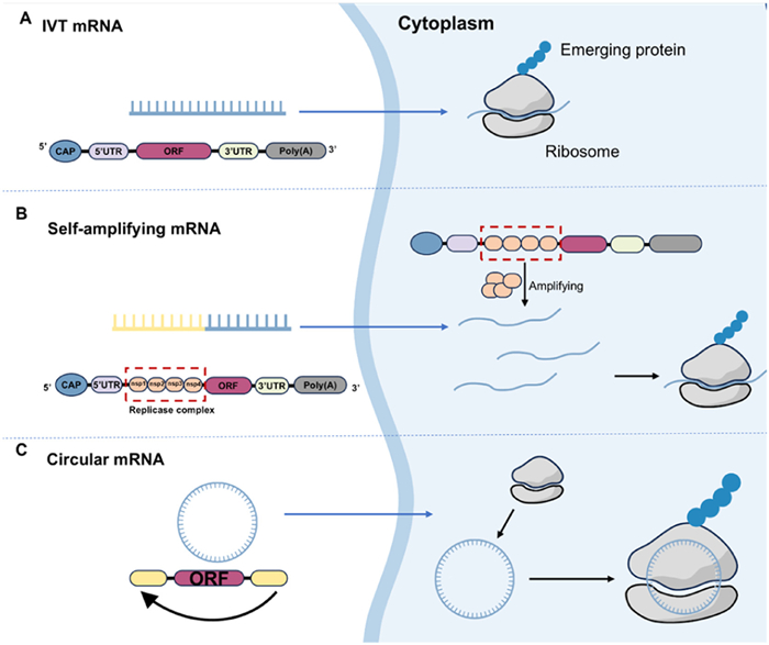
The structural fragility of mRNA renders it particularly unstable within the complex in vivo environment. Furthermore, the repulsive forces between the negatively charged mRNA and the cell membrane impede the mRNA's ability to traverse the membrane and enter the cell. Despite the advances in in vitro transcription (IVT) technology, which have enhanced the purity and quality of mRNA, there remains a discrepancy in comparison to natural mRNA, thereby underscoring the necessity for further modifications to mRNA structure and nucleotides [37].
AI and machine learning (ML) are being integrated into mRNA design, enabling more efficient optimization of sequence and structure (Table S1 in Supporting information). This section highlights strategies for mRNA structural refinement and the application of AI to enhance therapeutic performance.
The mRNA preparation technology is central in biomedicine, encompassing IVT systems, in vivo transcription mechanisms, and post-transcriptional modifications. Recent optimizations in template design (e.g., linearized plasmids, T7 promoter regulation) [38], process improvements (e.g., high-temperature transcription by thermostable RNA polymerase, reverse-phase high-performance liquid chromatography (RP-HPLC) purification) [39], and molecular modifications (e.g.,
In the IVT mRNA system, in addition to the target single-stranded mRNA (ssRNA), various enzymes, cofactors, unreacted ribonucleotides, DNA templates, and by-products are present. Among these, the by-product dsRNA has been observed to trigger innate immune responses, affecting the efficacy of mRNA drugs. According to the findings of Mu et al. [41], the optimization of RNA polymerase, nucleoside triphosphates (NTPs), reaction conditions, and template sequences can mitigate the immunogenicity of mRNA products. Regarding post-transcriptional purification, chromatography-based methods have been shown to be particularly effective [42]. Wu et al. found that using thermostable T7 RNA polymerase for transcription at higher temperatures (51-55 ℃) results in mRNA with reduced immunogenicity even without purification, and the formation of dsRNA at the 3′-end extension in the product is decreased [43]. In a separate study, Karikó et al. demonstrated that the purification of IVT products using RP-HPLC effectively reduced dsRNA impurities, lowered interferon-α (IFN-α) responses, and increased mRNA protein translation levels [44]. Nelson et al. combined m1ψ modification with process optimization (including specific NTP ratios, affinity chromatography, and RP-HPLC) to further reduce the innate immune response of mRNA [45].
The field of post-transcriptional gene regulation, particularly reversible mRNA methylation, has surged. m6A methylation, the most prevalent modification, guides mRNA processing, translation, and degradation, crucial in cell differentiation, development, and stress responses [46]. Hundreds of RNA modification types exist. Beyond m6A, others (m1A, m5C, hm5C,
mRNA serves as the cell’s “protein-synthesis blueprint” transcribed precisely from DNA and contains essential genetic instructions for the production of particular proteins [51]. In cellular settings, this mRNA functions as a molecular translator, converting genetic information into functional protein products. However, its intrinsic instability, susceptibility to RNases, and inability to cross membranes unaided demand reliable delivery vehicles for therapeutic use. Thus, mRNA delivery systems have emerged as key technologies for transporting mRNA into specific cells, enabling traversal of cellular barriers and subsequent protein expression. The principle involves encapsulating mRNA molecules within a carrier system that acts as a “protective cloak”, shielding them from enzymatic degradation, promoting cellular uptake, and facilitating endosomal escape. This strategy underlies the clinical success of mRNA vaccines and provides the foundation for expanding into gene therapy and protein replacement applications.
Contemporary delivery systems (Fig. 3a) demonstrate innovative design strategies that effectively address these challenges. The repertoire includes viral vectors and a rapidly growing range of synthetic nanomaterials, such as LNPs, polymeric nanoparticles, lipid-polymer hybrids, extracellular vesicles (EVs), and peptide-based carriers [52-57]. These platforms differ in their mechanisms of cellular entry, stability profiles, immune interactions, and translational feasibility. Collectively, they expand the therapeutic arsenal for spatially controlled mRNA expression (Fig. 3b).
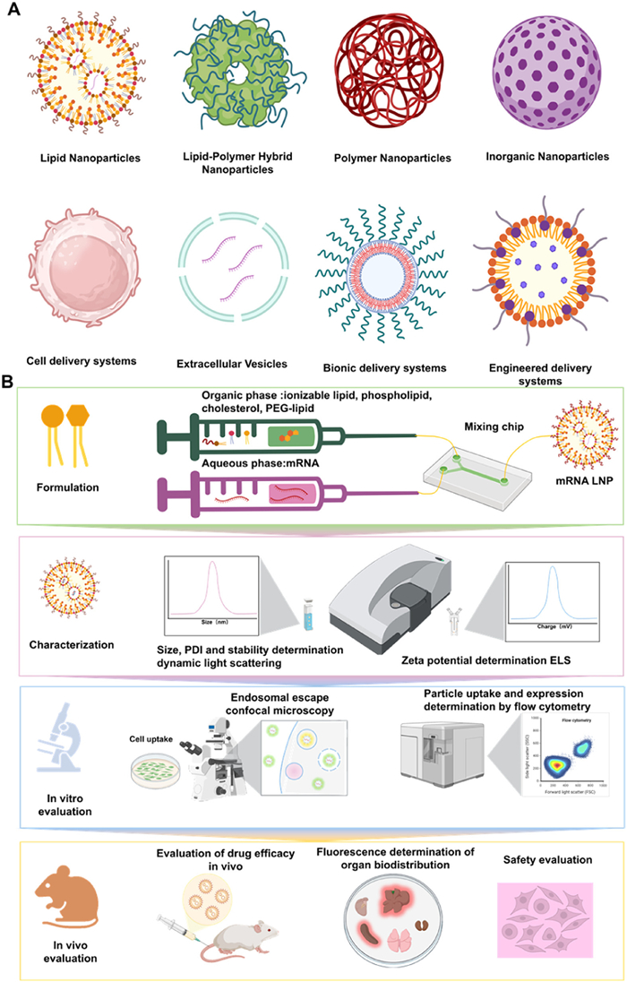
Among them, LNPs have become the most successful clinical-transformation system. Their dominance can be attributed to: (ⅰ) High encapsulation efficiency and protection of mRNA cargo; (ⅱ) pH-sensitive ionizable lipids that ensure efficient endosomal escape; (ⅲ) scalable and reproducible good manufacturing practice (GMP) manufacturing via microfluidic mixing; and (ⅳ) extensive clinical validation during the COVID-19 pandemic, which established regulatory precedence and streamlined approval pathways.
By contrast, other carriers face specific challenges before achieving clinical translation. Polymeric nanoparticles are often associated with cytotoxicity and batch variability due to permanent cationic charges. Exosomes demonstrate excellent biocompatibility and natural tropism, but standardized large-scale production remains unresolved. Peptide-based systems enable tissue-specific targeting but usually suffer from limited encapsulation efficiency and serum instability. Hybrid and inorganic nanocarriers remain largely preclinical, with concerns about complexity, safety, and long-term toxicity.
To provide a systematic overview, we summarize the mechanisms, advantages, and challenges of different delivery systems in Table S2 (Supporting information). This structured comparison highlights how LNPs currently outperform alternative carriers, while also underscoring the innovation potential of emerging platforms.
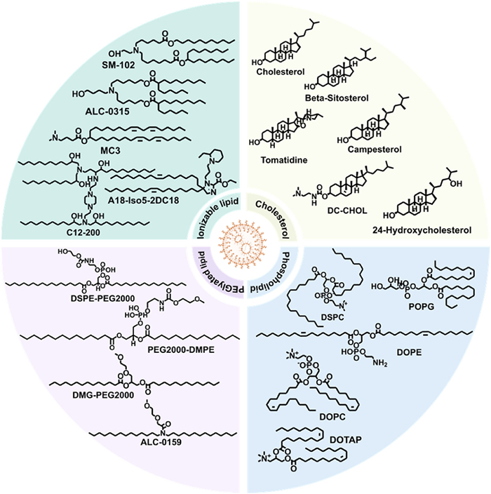
In the field of mRNA delivery systems, LNPs have become a leading platform, particularly in the context of vaccine development against the novel coronaviruses. The adoption of LNP technology by both Pfizer and Moderna highlights its effectiveness in enabling efficient mRNA vaccine delivery [58]. The unique physicochemical properties of LNPs, including their delicate structure, enable them to envelop and safeguard mRNAs, ensuring their accurate delivery into target cells and subsequent activation of the intracellular translation process. The composition of LNP comprises ionizable lipids, structured lipids, helper lipids, and PEGylated lipids (Fig. 4), meticulously engineered to safeguard the mRNA, ensure its precise delivery into the target cell, and enhance the intracellular translation process [59,60].
LPHNs represent a novel class of nanocarriers that ingeniously integrate the benefits of lipids and polymers, with the objective of significantly enhancing targeting capability, stability, and transport efficiency. This innovative design is progressively demonstrating its wide range of potential applications in several cutting-edge domains, including vaccine development, medication delivery, gene delivery, and cancer treatment [61,62]. The superior controllability and structural stability of polymers, in conjunction with the exceptional biocompatibility and membrane fusion potential of lipids, have led to a substantial enhancement in the delivery capabilities and overall functionality of LPHNs when compared to traditional LNPs [53].
LPHNs' subtlety stems from the perfect blend of lipids and polymers. Lipids, including cholesterol and phospholipids like 1,2-distearoyl-sn-glycero-3-phosphorylcholine (DSPC), provide membrane fluidity and stability. Cationic lipids (e.g., 1,2-dioleoyl-sn-glycero-3-phosphoethanolamine (DOPE), 1,2-dioleoyl-3-trimethylammonium-propane (DOTAP) encapsulate nucleic acids via electrostatic interactions, aiding cell membrane binding and endocytosis. The polymer component, polyethyleneimine (PEI), with strong positive charges, forms stable complexes with nucleic acids, boosting delivery efficiency. By tailoring polymer properties (molecular weight, structure, hydrophilicity), LPHNs enhance particle stability, regulate drug release, or maximize cell uptake. Notably, Chen's group created PLG-PPs, blending polymer and phospholipid benefits, which enhances mRNA expression and targets spleens via intravenous delivery [63]. Taratula's group studied mRNA-loaded polymer nanoparticles (PNPs) based on diethylenetriamine-substituted poly(aspartic acid), finding they induced protein expression in lungs and muscles after intravenous and IM injections, with PEG modification amount significantly affecting mRNA delivery and protein translation efficiency [64,65]. Additionally, Levy's team developed CART-FTY720, a modified CART using polymer chemistry, enhancing mRNA transport and expression in lymphocytes through ligand-receptor interactions [66].
PNPs are a type of very small delivery vehicle that is used in many ways, such as making vaccines, delivering medicines, delivering genes, and treating cancer. PNPs can be customized and controlled more easily than LNPs, making them more versatile for different medical uses. To successfully deliver drugs and ensure they are targeted correctly, it is essential to carefully control the physical and chemical properties of PNPs. This includes things like particle size, surface charge, and shape [67]. This control is achieved through careful design and synthesis.
PNPs are made from different materials. These particles can be classified as either natural or synthetic PNPs, depending on the type of polymer used. Natural PNPs, such as polysaccharides (e.g., chitosan, hyaluronic acid), proteins (e.g., gelatin, albumin), and lipid molecules (e.g., phospholipids), have shown advantages in the field of drug delivery due to their favorable compatibility with and biodegradability in living organisms. PEI and poly(β-amino ester) (PBAE) are two examples of synthetic PNPs that researchers are studying a lot right now. This is because they can be produced in large amounts, are easy to control, and can be adjusted to have certain physical and chemical properties. Researchers can precisely control how much of the drug is released and how well it targets a specific area. They can do this by changing the polymer's molecular weight, chemical structure, and hydrophilicity.
PEI, a traditional polymer for mRNA distribution, offers a high density of positive charges for mRNA complexing and endosomal escape due to its abundance of amino groups. However, the high affinity between mRNA and PEI also exerts a degree of influence on mRNA transfection efficiency [54]. Studies have shown that low molar mass PEI is less toxic but has a lower delivery effectiveness, while high molecular weight PEI exhibits exceptional nucleic acid transport efficiency but also significant toxicity [68]. Through the implementation of chemical modifications, researchers have effectively augmented PEI's transfection effectiveness and concurrently diminished its toxicity, thereby addressing this concern. For instance, the creation of PEI2k-stearic acid (PSA)/mRNA nanoparticles involved the mixing of mRNA with a specific weight ratio of PSA. This approach facilitated the efficient delivery of mRNA to DCs, subsequently inducing immune responses specific to antigens [69]. Furthermore, the alteration of polyethylene glycol (PEG) and cyclodextrin (CD) decreased PEI's charge density without compromising its capacity to condense nucleic acids. Li et al. employed CD-PEI conjugates to facilitate the transport of mRNA, leading to its migration to the lymph nodes and the subsequent triggering of immunological reactions, while preserving the intrinsic mucosal adjuvant function of mRNA [70]. Subsequent systemic delivery of PEI-g-PEG/mRNA nanoparticles bearing amino or amino acid terminal groups (PEG grafting rate of 0.5%) resulted in the most efficacious gene transfection in the lungs, as evidenced by the aforementioned hybrid production approach. These PEI-based polymer nanocarriers exhibit reduced toxicity in addition to increased transfection efficiency [71].
The hydrolysis of ester linkages endows polyester polymers, such as PBAE and polyamine coester (PACE), with high biodegradability, thereby facilitating significant support for therapeutic transformation [72,73]. Through screening of the PBAE polymer library, Kim's team was able to identify certain PNPs with effective mRNA expression capabilities. The mRNA expression exhibited a marked augmentation in T cell immune activation, yielding favorable outcomes in plaque reduction neutralization testing, and demonstrated prolonged persistence, up to two weeks, without inducing hepatic injury [74]. In a parallel study, Anderson's team produced hyperbranched PBAE (hPBAEs) to create stable and concentrated multi-chain nanoformulations suitable for inhalation. This approach facilitated the uniform distribution and effective production of luciferase mRNA in the lungs [75]. The research led by W. Mark Saltzman discovered that while in vitro transfection is somewhat limited, the stability of the multi-chain may be enhanced by blending PACE and PEG-coupled PACE in varying proportions, even if the PACE-PEG concentration is as low as 0.25% by weight. The impact of PACE-PEG incorporation on mRNA transfection in vivo varied based on the delivery method; PEGylation exhibited no influence on intravenous systemic administration, while PACE-PEG addition augmented local delivery in the lungs [76].
Despite the extensive utilization of LNPs and polymers in the domain of gene delivery, formulation stability, manufacturing scalability, and functional diversity persist as inherent challenges. In contrast to natural sources or organic materials, inorganic nanoparticles have exhibited considerable potential as gene carriers, offering a range of advantages, including straightforward controllable physical and chemical properties, relatively high stability, and frequently reduced fabrication costs [77]. The "DegradaBALL" drug delivery system (DDS), developed by Won's group, utilizes porous nanoparticles with the objective of achieving permanent gene silencing of the CTGF gene. The "DegradaBALL" DDS, developed by the research group led by Won, exhibits several advantageous properties, including effective intracellular absorption, ease of drug loading, high biocompatibility, a mild continuous release profile, and biodegradability. This modular DDS is particularly well-suited for RNA delivery [78]. However, the team led by Leeuwenburgh incorporated mRNA in lipid-coated calcium phosphate nanoparticles (LCP), which are made of fusible ionizable lipids in their outer lipid shell. They created nanocomposites with varying proportions of hydroxyapatite and gelatin, and discovered that adding more hydroxyapatite nanoparticles might improve the viscoelasticity of the nanocomposites without compromising their capacity to mend themselves. The local mRNA release was achieved by combining these nanocomposites with peptides, lipids, and LCP mRNA preparations [79]. Horcajada et al. proposed an efficient technique for embedding nucleic acids into the mesopores of biocompatible nanoscale metal-organic frameworks. These frameworks demonstrated rapid cellular absorption, RNA protection, and release capacities, which enhanced the activity of associated genes in vitro [80].
In addition to the utilization of polymers and LNPs for the transportation of mRNA, scientific research has explored methodologies that employ cells as delivery systems. This approach utilizes the paracrine capacity of cells to distribute proteins made from mRNA after it has been delivered into cells in vitro, rather than delivering mRNA cargo directly to target cells. This method offers several advantages, including the use of natural intracellular and intercellular communication, prolonged circulation life, and superior biocompatibility when compared to manufactured LNPs [81]. In 2019, Rohner's team successfully delivered vascular endothelial growth factor (VEGF) protein intramuscularly by injecting mice with skin fibroblasts that had been primed with a modified mRNA encoding VEGF [82]. In mice with ischemic limbs, this therapy dramatically decreased tissue necrosis and enhanced vascular density. In a subsequent study, the therapeutic potential of this approach was further confirmed by administering rat bone marrow-derived mesenchymal stem cells (MSCs) preloaded with modified mRNA expressing VEGF and human bone morphogenetic protein 2 (BMP-2) in a mouse model with skull defects. The treatment led to enhanced osteogenesis and angiogenesis in the treated animals, thereby aiding in the repair of the skull [83]. In order to expedite the targeting of inflammatory tissues and the expression of the potent immunosuppressive cytokine interleukin-10 (IL-10), which is not produced by MSCs themselves, Karp's team successfully infected MSCs to express functional rolling mechanisms (P-selectin glycoprotein ligand-1 (PSGL-1) and Sialyl-Lewisx (SLeX)) [84].
EV delivery has recently received a great deal of attention in the fields of vaccine development, drug delivery, and gene delivery. Cells actively produce nanoscale vesicles called EVs. They can successfully transport mRNA or other therapeutic compounds to target cells because of their excellent membrane fusion ability, minimal immunogenicity, and inherent biocompatibility [85,86]. EVs are considered the best delivery vehicles because of their natural origin and biocompatibility advantages over more conventional delivery techniques (such as LNPs, viral vectors). Based on their size, origin and biological properties, EVs can be classified as exosomes, microvesicles or apoptotic bodies.
Exosomes are often used in mRNA delivery systems because of their small size, strong membrane fusion ability, and ability to transport a variety of biomolecules [87,88]. Endosomes generate exosomes, which are then released into the extracellular space. They are essential for intercellular communication and are capable of transporting lipids, proteins, nucleic acids and RNA [89,90]. An important advantage of using exosomes for mRNA delivery is their ability to contain RNA. Their lipid bilayer can protect mRNA from extracellular enzymatic degradation, thereby maintaining its stability and integrity while being transported to the target cells [55,91]. In addition, exosomes can carry mRNA over long distances in both healthy and diseased conditions and exhibit exceptional stability in the circulation. In addition, exosomes can interact with cargo to facilitate loading and with receptor cells to promote uptake. Exosomes can efficiently target receptor cells within the same tissue due to their surface proteins and receptors [92]. As mRNA and protein drug carriers, Cheng's group created room-temperature stable, inhalable lung-derived EVs, also known as exosomes (Lung-Exos). Lung-Exos can be delivered to the lungs of rats and non-human primates via dry powder inhalation, and they have better distribution in the bronchioles and parenchyma than conventional synthetic nanoparticle liposomes (lipos). In vaccination applications, immunoglobulin G (IgG) and secretory IgA (SIgA) responses were higher in pulmonary exosomes (S-Exos) expressing the severe acute respiratory syndrome coronavirus 2 (SARS-CoV-2) spike (S) protein than in their liposomal carriers (S-Lipo) [93]. erythrocyte-derived exosomes from type O donors were loaded with anti-miR-125 and Cas9 mRNA. According to Usma et al. [94], the engineered erythrocyte-derived exosomes successfully delivered miRNA and mRNA, demonstrating effective miRNA-125 knockdown and gene knockout using clustered regularly interspaced short palindromic repeats (CRISPR)-CRISPR-associated protein 9 (Cas9) genome editing in acute myeloid leukemia and breast cancer mouse models.
Exosome-based mRNA treatments have shown promise in preclinical research and are progressively making their way to clinical trials [95]. Exosomes are gaining substantial attention as clinically relevant mRNA delivery systems, currently being explored for therapeutic use in gene regulation, oncology, regenerative medicine, and vaccination [96]. Although exosomes have numerous benefits, their clinical use is still in its infancy because of several issues that restrict its use.
In recent years, vaccine, gene therapy, and cancer immunotherapy have embraced biomimetic mRNA delivery. This system mimics the body's natural delivery mechanisms (e.g., cell membrane similarity, receptor recognition, endocytosis) using biocompatible, targeted, transmembrane, and low-immunogenicity carriers (exosomes, liposomes, nanoparticles) to enhance mRNA delivery efficiency, safety, and therapeutic effects. Inspired by natural delivery methods like liposomes and EVs (exosomes), which transfer mRNA via endocytosis or membrane fusion with low immunological response and high biocompatibility, biomimetic delivery aims to create effective mRNA vehicles. Akiyoshi's team successfully created exosome-mRNA hybrid particles, combining EV's biocompatibility/targeting with LNP's stable storage/controlled production. Despite promising evidence, this strategy is nascent and requires further investigation into specific processes [55]. Matyjaszewski's group reported exosome-polymer hybrids (EPHs) with improved stability in the presence of proteolytic enzymes and under a variety of storage conditions. Exosome surface contacts, cellular uptake, and residual biological activity can be precisely regulated by varying polymer length and surface loading. Without altering tissue distribution, the blood circulation time of EPH was increased fourfold [97]. In another study, liposomes containing chemotherapeutic drugs and genetically modified exosomes, which promote the recruitment of immune cells, formed hybrid particles that successfully prevented tumor growth in animal cancer models [98]. We can expect the rapid development of these novel delivery systems considering the recent milestone of mRNA vaccines, which highlights the enormous potential and existing limitations of LNPs.
The development of systems for delivering mRNA tailored to organs or cells is a critical component of the broader strategy for vaccine and gene therapy development. As the utilization of mRNA treatment continues to expand, there is an imperative for significant advancements in its safety, specificity, and delivery efficiency. The precise delivery of mRNA to the target organ or specific cell type is paramount to ensure the optimal therapeutic outcome. The employment of a targeted delivery system, which involves the modification of the delivery carrier's properties to ensure precise entry of mRNA into the target tissue or cell, constitutes an effective methodology for achieving this objective.
Although LNPs dominate clinical use, non-LNP delivery systems face unique translational hurdles: (1) Manufacturing and scale-up: Lack of robust, GMP-standardized protocols compared with established LNP microfluidics. (2) Safety profiles: Toxicity of cationic polymers, immunogenicity of cell-penetrating peptides (CPPs), and biodistribution uncertainty of EVs require deeper toxicological evaluation. (3) Stability and storage: Maintaining mRNA integrity in circulation and during long-term storage is unresolved. (4) Regulatory frameworks: Absence of clear approval precedents slows progress compared with LNPs. (5) Efficacy gap: Many systems show promising preclinical data but fail to achieve comparable in vivo transfection efficiency.
In summary, LNPs became the first clinically validated mRNA delivery system due to their balance of efficacy, scalability, and regulatory maturity, whereas non-LNP platforms, though innovative, must overcome challenges in safety, manufacturability, and regulatory standardization before successful clinical adoption.
mRNA therapy involves delivering functional mRNA into the body to drive therapeutic protein expression [99]. The administration route critically influences both transfection efficacy and safety. Current strategies include intramuscular (I.M.), intradermal (I.D.), subcutaneous (S.C.), intravenous (I.V.), intratumoral (I.T.), oral, and nebulized inhalation [57,100]. Each route faces distinct physiological barriers, but also provides unique advantages in terms of immune cell access, biodistribution, or patient compliance. A comprehensive evaluation of both benefits and limitations is necessary to optimize clinical translation (Fig. 5).
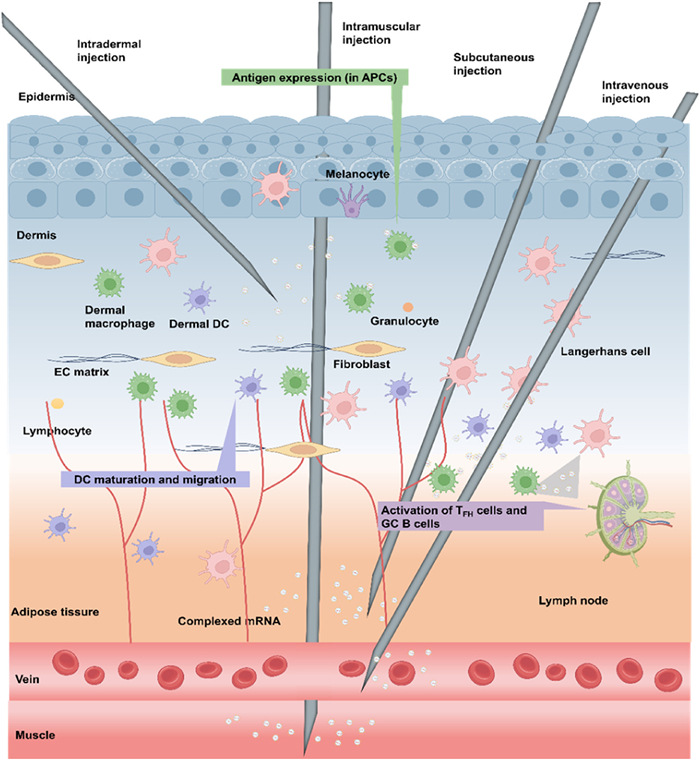
I.M. injection is the most widely adopted route, including for mRNA vaccines [101,102]. Its advantages include ease of administration, robust uptake by muscle-resident DCs, and relatively prolonged antigen expression due to slower clearance [103]. However, challenges include localized inflammation (pain, erythema, nodules) [104,105], and limited biodistribution due to restricted vascular perfusion [106].
I.T. injection provides the advantage of directly exposing tumor cells to mRNA payloads, thereby enhancing local expression while reducing systemic toxicity [107-110]. This is particularly attractive for immunotherapy and tumor vaccine strategies. However, barriers include the heterogeneous tumor microenvironment (hypoxia, dense stroma), which impedes uniform distribution, and the requirement of imaging-guided injection for precise tumor localization [111,112]. In summary: I.M. routes are suitable for prophylactic vaccines requiring systemic immune priming, while I.T. offers superior targeting but is technically demanding and tumor specific.
I.D. injection exploits the abundance of antigen-presenting cells (APCs) in dermis and epidermis [113]. This route enables efficient immune priming even at low doses, making it attractive for vaccination [104]. Its primary obstacle is the skin barrier (stratum corneum), which restricts penetration and may limit distribution beyond local tissue [102]. S.C. injection provides a depot effect, gradually releasing mRNA formulations into circulation, prolonging expression [114]. However, its drawbacks include slower absorption kinetics, restricted systemic distribution, and injection-site inflammation [101,102]. In summary: I.D. administration uniquely leverages APC-rich niches for vaccines, while S.C. offers pharmacokinetic advantages for sustained therapies, but both face limited biodistribution compared to systemic routes.
I.V. delivery enables rapid systemic biodistribution, making it ideal for indications requiring immediate therapeutic protein expression (e.g., acute infection, sepsis) [115,116]. It also bypasses digestive degradation and achieves high circulating levels. However, challenges include nonspecific organ accumulation (notably in liver and spleen), which may induce off-target protein expression [117-121], and systemic immune activation (e.g., cytokine release, fever, allergic reactions) [122-125]. Moreover, I.V. injection requires skilled personnel and carries risks of infection or infusion reactions [126,127]. In summary, I.V. delivery achieves systemic exposure but at the cost of safety risks and limited tissue selectivity.
Oral delivery is the most patient-friendly approach, with superior compliance and no injection-related trauma [115,128-131]. However, major challenges include enzymatic degradation by nucleases and gastric acid, and poor absorption due to intestinal barriers [132,133]. Nanocarrier encapsulation (liposomes, LNPs, polymeric systems) has shown promise in improving stability and absorption [115,128,129]. Yet, variability introduced by diet and gastrointestinal conditions limits reproducibility. As a result, oral administration offers unmatched patient convenience, but requires significant formulation innovation to overcome digestive degradation and bioavailability barriers.
Nebulized inhalation of mRNA has become a research focus for mucosal vaccines and lung disorder treatment. It encapsulates mRNA in nanocarriers, aerosolizing it to target lung tissue and respiratory mucosa directly, reducing systemic toxicity, bypassing first-pass metabolism, and enabling effective local delivery. Advancements in carrier design and stability are crucial. Molecularly designed ionizable lipids (e.g., MC3 derivatives) in LNPs have significantly improved mRNA stability during aerosolization and lung tissue permeability. Daniel Anderson's group at MIT says that the LNP-mRNA vaccine that's sprayed into the air can withstand three times more tearing force than regular vaccine carriers. It can also successfully make the vaccine in mouse lung cells [134]. In a recent study, Huang et al. reported that LNPs derived from the lead glycerolipid TG4C achieved highly efficient systemic in vivo mRNA delivery, resulting in ∼6-fold higher luciferase protein expression compared to commercial LNPs (SM102/MC3) [135]. Aerosolized mRNA vaccines show promise in infectious disease prevention by eliciting mucosal immunity. Preclinical studies show inhaled SARS-CoV-2 Spike mRNA vaccine reduces viral load and generates 10x higher respiratory IgA antibody titers in rhesus monkeys than IM vaccination. Phase Ⅱ clinical trials indicate aerosolized mRNA-CFTR therapy improves lung function (FEV1 by 8.2%) in cystic fibrosis (CF) patients while reducing adverse reactions compared to I.V. injection [136]. mRNA airway retention can be extended from 2 h to 6 h by coating carriers with mucus-penetrating peptides (e.g., KAL, GE11). Lv Xueguang's team developed charge-assisted stabilization (CAS), leveraging classical colloidal interface theory to stabilize LNPs during aerosolization. CAS-LNPs incorporate a biosafe negatively charged polypeptide-lipid conjugate into clinical LNP formulations, using electrostatic repulsion to prevent aggregation/fragmentation during aerosolization. In mouse, miniature pig, and beagle dog models, CAS-LNP effectively delivered mRNA to lung tissue for protein translation, achieving ∼20x higher mRNA expression efficiency than clinical LNPs [137]. Despite rapid aerosol inhalation mRNA tech progress, industrialization faces hurdles like inhalation efficiency variability, long-term toxicity evaluation, and large-scale manufacturing standards. Future research may explore CRISPR/exosome synergies for precision pulmonary delivery and AI integration to optimize carrier-aerosol parameter matching.
Repeated dosing is required for chronic conditions (e.g., cancer, persistent infections). Clinical studies of COVID-19 vaccines demonstrate that repeated boosters sustain antibody titers, but T cell responses plateau [138,139]. Long-term dosing risks include immune tolerance, autoimmune complications, and cumulative off-target distribution [140,141]. Strategies under development include adaptive dosing schedules, immune monitoring, and co-delivery of immunomodulators [142-145]. In summary: While repeated administration ensures durable efficacy, it must balance immune exhaustion and safety risks through personalized regimens.
Each administration route offers unique strengths but faces distinct physiological barriers (Table S3 in Supporting information): (1) I.D./S.C. leverage APC-rich niches but face diffusion limits. (2) I.M. is practical and effective but localized. (3) I.V. ensures systemic exposure but risks immunotoxicity. (4) Oral offers compliance but suffers from digestive degradation. (5) Inhalation provides mucosal immunity but manufacturing remains a bottleneck. (6) Long-term dosing ensures durability but raises tolerance risks. Future optimization will require matching therapeutic indication with route selection, integrating carrier engineering to overcome route-specific barriers.
mRNA-LNP enters cells by either pinocytosis, which is widespread in eukaryotic cells, or phagocytosis, which is mostly carried out by immune cells. Caveolin-mediated endocytosis, clathrin-mediated endocytosis, macropinocytosis, and other receptor-mediated endocytosis are subtypes of pinocytosis [146]. The effectiveness of mRNA-LNP transfection in 30 cancer cells was investigated by Sayers et al. [147]. They discovered that effective transfection is dependent on the early endosomal escape window prior to lysosomal fusion or exocytosis. The majority of LNPs that are swallowed by pinocytosis will go through both early and late endosomes. They will eventually reach the lysosome and undergo degradation if endosomal escape is not accomplished [148]. This is the primary challenge that the mRNA-LNP delivery systems must overcome. The success rate of MC3 LNPs escaping endosomes is about 1% to 2% of those authorized by the FDA [149]. Patel et al.'s research [150] has demonstrated that leukotriene antagonists can increase LNPs' lysosomal escape, which enhances mRNA transfection efficiency both in vitro and in vivo. Over time, the pH level rises from early endosomes to late endosomes and finally to lysosomes. Ionizable lipids are an essential part of the endosomal escape process and may adjust to pH variations [151]. Only a very tiny percentage of mRNA-LNPs can transfer IVT mRNA to the cytoplasm via the proton sponge effect or membrane phase transition mechanism [152], ionizable lipids (such as MC3, ALC-0315, SM-102) are the core functional components of LNP, and their key feature lies in the presence of one or more protonated tertiary amine head groups. The pKa of these head groups is usually designed within the range of 6.0-6.5, which enables them to undergo reversible protonation and deprotonation in different pH environments, and the charge state changes accordingly. At physiological pH (∼7.4), the tertiary amine head groups are mainly electrically neutral. At this point, the overall surface charge of LNP is close to neutral, which helps to reduce non-specific interactions with negatively charged proteins in the blood (such as serum albumin) and non-target cell membranes, thereby reducing toxicity and prolongating the circulation time in the body. Under acidic endosome pH (∼5.5-6.5), the tertiary amine head matrix is neutralized and becomes positively charged (-NH+-). This transformation is the key switch that triggers a subsequent series of structural changes and membrane interactions. SAXS research shows that at acidic pH, the protonation of ionizable lipids leads to an increase in their molecular shape parameters (P), tending to form non-layered structures such as antihexagonal phase (HII). For instance, lipids such as MC3 and KC2 are in the layered phase (Lα) at neutral pH, transform into the cubic phase (Fd3m) when pH∼6.5, and further form the HII phase at even lower pH (∼5.0-6.0). The formation of these non-layered intermediate phases provides thermodynamic driving forces for membrane fusion or pore formation [153]. Molecular dynamics simulations show that after protonation, the head base area of the lipid increases, while the hydrophobic volume of the tail hydrophobic chain relatively decreases, and the overall molecular shape is closer to an "inverted cone". Lipid molecules of this shape tend to induce negative curvature, that is, to promote the membrane to protrude outward, thereby destroying the flat structure of the lipid bilayer [154]. Studies have shown that ionizable lipids with a strong negative curvature tendency (such as DOPE) or protonated ionizable lipids, when inserted into the endosomal membrane, will "transfer" their inherent shape characteristics to the endosomal membrane, disrupting the regular arrangement of lipid molecules on the endosomal membrane. To alleviate the resulting membrane tension, the membrane may undergo local bending and form non-layered structures (such as semi-fused intermediate states or pores), ultimately leading to increased membrane permeability or even rupture. For example, after protonation of SM-102, the membrane thickness can be reduced by approximately 30% (from ∼3.8 nm to ∼2.5 nm), significantly increasing the membrane permeability [155] some mRNA-LNPs can transfer IVT mRNA to the cytoplasm via the proton sponge effect or membrane phase transition mechanism [152], after which it may translate functional proteins. Additionally, various LNPs have varying capacities to fuse with the endosomal membrane [156]. Cryo-electron microscopy studies have found that acidic pH can drive lipid infiltration into the conjunctin -46/50 pore, causing it to close, which indicates the reversibility and criticality of lipid-membrane interactions [157].
Nevertheless, endogenous mRNA that enters the cytoplasm and IVT mRNA vary in several ways. Following splicing, endogenous mRNA is transferred from DNA in the nucleus to the cytoplasm. mRNA is not "naked"; rather, it forms messenger RNA protein complexes (mRNPs) with many RNA binding proteins (RBPs) prior to exiting the nucleus. IVT mRNA may combine with LNP components to create a complex, but it does not generate mRNPs. Furthermore, to start translation, IVT mRNA needs 5′ cap and eIF4E to form a correct complex. However, the way that IVT mRNA attaches to eIF4E is yet unknown, while mRNPs around the nucleus catalyze the binding of endogenous mRNA to eIF4E in the cytoplasm [158]. The delivery vector will aid in raising the target protein's expression level if it can facilitate the creation of the translation initiation complex, translation elongation, and translation termination [159].
Delivery systems (e.g., lipid-based nanoparticles) activate immune responses through dual receptor engagement and intracellular signaling (Fig. 6). Surface components like cyclic amino head lipid A2/SAL12 bind Toll-like receptor (TLR)1/2 or TLR4, triggering nuclear factor kappa-B (NF-κB) translocation to the nucleus and inducing pro-inflammatory cytokines (e.g., IL-6, tumor necrosis factor-α (TNF-α)). Concurrently, endocytosed components (e.g., R848-C12) activate endosomal TLRs, while cytosolic nucleic acids engage the stimulator of interferon genes (STING) pathway, promoting interferon regulatory factor 3 (IRF3)/IRF7 activation and IFN-α/β production.
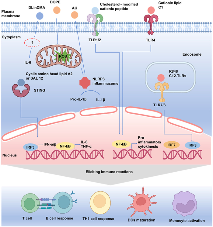
These signals converge to drive NOD-like receptor family pyrin domain-containing 3 (NLRP3) inflammasome assembly, cleaving pro-IL-1β into active IL-1β. The resulting cytokine storm (IL-6, TNF-α, IFN-α/β, IL-1β) and transcriptional reprogramming orchestrate broad immune reactions: Monocyte activation, DC maturation, Th1 cell polarization, and B cell/T cell responses. This cascade highlights how delivery systems deliberately harness innate sensors (TLRs/STING/inflammasomes) to bridge adaptive immunity, critical for vaccine efficacy but requiring careful control to avoid pathological inflammation.
LNPs are widely recognized not only as highly efficient delivery systems for nucleic acids but also as adjuvant-like modulators of the host immune response. Increasing evidence suggests that the immunogenicity of LNPs is not incidental but is determined by the chemical nature and structural properties of their lipid constituents, including ionizable lipids, PEG-lipids, and helper lipids. These components interact with innate immune receptors and downstream signaling pathways such as TLR4, STING, and the NLRP3 inflammasome, ultimately shaping the magnitude and quality of immune responses.
Adjuvants are a crucial part of vaccinations and are frequently added to them to improve the strength, scope, and longevity of the immune response. This chapter examines the most recent developments in the use of adjuvants in mRNA vaccine preclinical and clinical trials. Three broad categories may be used to classify the adjuvants in mRNA vaccines: In order to serve as a guide for the creation of novel and varied mRNA vaccination adjuvants. These include (1) mRNA-self-adjuvants, (2) exogenously added adjuvants, and (3) co-delivery adjuvants.
Polymer-based vaccine delivery systems typically enable gradual antigen release. Poly(lactic-co-glycolic acid) (PLGA), the most widely utilized synthetic polymer for nucleic acid delivery [160], effectively targets DCs in lymph nodes. For instance, Kim et al. demonstrated that encapsulating vaccine components (antigens/immunostimulants) in PLGA nanoparticles enhances DC uptake and antigen presentation while preventing systemic dispersion [161]. Similarly, virus-like particles (VLPs), self-assembled protein nanostructures derived from viral capsid proteins [162], leverage their uniform size (20–100 nm) and morphology [163] to efficiently drain via lymphatic vessels for targeted uptake by APCs in lymph nodes [164]. Gold nanoparticles further optimize antigen delivery through dual mechanisms: (1) Protecting antigens from degradation and (2) facilitating lymph node trafficking via size-controlled nanovaccine design [165]. These particles also enhance humoral immunity by activating the NLRP3 inflammasome and promoting Th2-type cytokine production, as evidenced by Zhu et al.'s work on antigen-specific antibody generation [166]. However, when employed as adjuvants in SARS-CoV-2 protein vaccines, gold nanoparticles induced robust antigen-specific IgG responses but failed to elicit sufficient cellular immunity for viral clearance [167].
As a new therapeutic approach, mRNA's clinical use requires rigorous evaluation of its immunogenicity. A key challenge is that mRNA molecules can activate the innate immune system through various pathways, acting as the first line of defense by rapidly recognizing foreign nucleic acids (Fig. 6). Immune sensors can be activated by mRNA structure, modifications, and delivery vectors, triggering cytokine production and inflammation. While potentially beneficial for vaccines (enhancing antigen presentation/immune memory), this needs careful control in gene therapy to avoid detrimental immune reactions impacting efficacy.
mRNA vaccines deliver nucleoside sequences encoding viral antigens into host cells. Following translation, these antigens elicit adaptive immunity comprising both humoral and cellular responses [168,169]. Humoral immunity involves CD4+ T cell-mediated B cell activation and subsequent antibody production by plasma cells, while cytotoxic CD8+ T lymphocytes eliminate infected cells through major histocompatibility complex class Ⅰ (MHC-Ⅰ) recognition (Fig. 7) [170,171]. Although antibody responses remain primary evaluation metrics, robust T cell activation, essential for CTL effector functions and B cell coordination, significantly enhances protective efficacy. Crucially, exogenous mRNA intrinsically activates innate immunity via pattern recognition receptors (PRRs): (1) Endosomal TLRs (TLR3/7/8) detect RNA structures, inducing NF-κB/IRF3-mediated cytokine/interferon production; (2) cytosolic RLRs (RIG-I/MDA5) trigger mitochondrial antiviral signaling protein (MAVS)-dependent antiviral responses. While evolutionarily protective, such PRR engagement risks excessive inflammation, reduced translational efficiency, and adverse effects. Mitigation strategies therefore focus on: (1) Nucleoside modifications (e.g., pseudouridine substitution) to minimize PRR recognition while enhancing protein expression; (2) advanced delivery systems (e.g., LNPs) that shield mRNA from premature immune detection and facilitate targeted uptake. These optimizations expand mRNA's therapeutic applicability across diverse diseases, with ongoing research refining immunogenicity profiles to advance personalized medicine platforms.
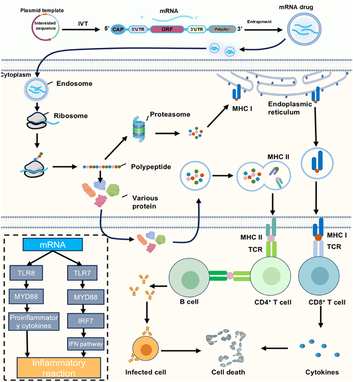
The mRNA that codes for antigen-specific proteins, antibodies, and cytokines is characterized by pathogen-associated molecular patterns and possesses intrinsic immune activation capabilities. APCs can identify it. In order to further activate immune cells and create a robust and long-lasting adaptive immune response, APCs subsequently activate pattern recognition receptors such TLR3, TLR7, and TLR8, which in turn trigger type Ⅰ interferons and proinflammatory cytokines [172]. The self-adjuvant effect of mRNA was discovered in 2004 when it was demonstrated that in vitro transcribed mRNA had an impact akin to that of vaccination adjuvants [173,174]. Compared to peptide/protein vaccines requiring exogenous adjuvants, this dual functionality, simultaneous antigen production and immune stimulation, confers significant advantages for adaptive immunity development [175]. However, the interferon-mediated cascade activates protein kinase R and 2′-5′-oligoadenylate synthetase, suppressing translation and accelerating RNA degradation [176-178]. Thus, balancing adjuvant potency with translational efficiency remains a critical challenge for vaccine efficacy.
By tailoring the nucleotide and codon sequences to control the self-adjuvant characteristics of mRNA, the German biotechnology company CureVac has created a novel technique to increase stability against degradation [179,180]. Furthermore, they have created RNActive® vaccines with self-adjuvant activity, which employ unaltered and sequence-optimized naked mRNA as antigens and co-deliver them alongside protamine. This combines strong adjuvant activity with high antigen expression to trigger the TLR7/8 signaling pathway and elicit effective immune responses [181,182]. Using mRNA to encode adjuvant immunomodulatory proteins, like TriMix, is another successful adjuvant technique. It is made up of three mRNAs that encode several immunostimulatory proteins: TLR4, CD40 ligand (CD40L), and CD70 [183]. TriMix and mRNA vaccines that encode antigens linked to cancer have been shown in several clinical trials to encourage tumor regression in advanced melanoma [184]. Two-component mRNA vaccines can also have their anti-tumor efficacy enhanced by the mRNA-encoded supplementary adjuvant molecule CD40 ligand [185]. In general, mRNA's self-adjuvant qualities offer benefits and drawbacks for the creation of mRNA vaccines. The immunological impact of mRNA vaccines can be enhanced by making appropriate use of the "self-adjuvant" function of mRNA, which can aid in the generation of vaccine-stimulated particular immune responses.
LNP-mRNA therapy holds substantial promise but faces safety challenges including toxicity, immunogenicity, and off-target effects [106,186,187]. Several strategies have been proposed to address these risks. First, the rational design and screening of ionizable lipids are critical. Optimizing lipid chemical structures enhances stability and biocompatibility, thereby reducing toxicity and immune activation [188-191]. Second, combinatorial therapeutic approaches improve efficacy while mitigating monotherapy risks through synergistic effects with adjuvants or other drugs [192]. Additionally, minimizing off-target effects requires precise control of LNP biodistribution and targeting [193]. Implementing these strategies comprehensively ensures the safety and efficacy of LNP-mRNA therapeutics.
Ionizable lipids are central to LNP-mRNA formulations, directly influencing delivery efficiency and safety [194]. Their design focuses on achieving pH-dependent behavior: Neutral at physiological pH for circulation stability and protonated in acidic endosomes to enable mRNA release [59,188]. Hydrophobic tail architecture further modulates in vivo stability and cellular uptake efficiency [189,195-197]. Combinatorial chemistry enables rapid synthesis of diverse lipid libraries, while high-throughput screening evaluates transfection efficacy and cytotoxicity [119]. Incorporating biodegradable linkers reduces long-term accumulation risks [198]. Furthermore, big data models and algorithms enhance screening precision [199,200], accelerating the development of safer LNP-mRNA systems.
Combining LNP-mRNA with other therapeutics enhances efficacy and safety. Synergistic effects allow reduced monotherapy doses, diminishing adverse events [192,201-203]. For example, co-delivering LNP-mRNA encoding immunomodulators with immune checkpoint inhibitors amplifies anti-tumor responses [110,192,204]. In vaccinology, adjuvant co-administration boosts immunogenicity and prolongs immune protection [205]. However, drug-drug interactions and patient variability necessitate rigorous preclinical evaluation and personalized regimen design to ensure clinical safety and effectiveness.
The term "off-target effects" describes how mRNA treatment affects tissues or cells that are not its intended target, leading to needless damage or adverse consequences [193]. A number of tactics have been used by researchers to reduce off-target effects. First, LNP's targeting may be enhanced to more efficiently transport it to the target tissue by improving its composition and structure [206]. For instance, precise distribution to particular cell types can be achieved by introducing lipids with particular targeting ligands [207]. Second, mRNA translation can be selectively suppressed at the cellular level by using the regulatory function of endogenous microRNA (miRNA) [208]. For instance, mRNA production in non-target cells can be decreased by including miRNA binding sites into the 3′ UTR of mRNA [209,210]. Furthermore, the distribution and release properties of LNP in various tissues may be regulated by varying the pKa value of lipids [211]. The combined use of these techniques not only increases mRNA therapy's accuracy but also offers a crucial scientific foundation for lowering the possibility of off-target effects.
mRNA technology has emerged as a landmark in biomedical science, driving advances in genetic diseases treatment, vaccine development, and cancer immunotherapy. The core strengths of this therapy lie in its high programmability and flexibility, enabling scientists to rapidly design and synthesize corresponding mRNA molecules tailored to different disease needs, thereby effectively advancing the progress of personalized treatment and large-scale vaccine research and development. The “design–delivery–expression” paradigm underpins all mRNA therapies: Sequences are generated by in vitro transcription, packaged into LNPs or other vectors for protection and targeted delivery, and translated into proteins upon cellular entry.
Clinically, mRNA vaccines demonstrated unprecedented speed and scale during the COVID-19 pandemic by designing mRNA sequences targeting the virus's spike protein, vaccines can rapidly stimulate a robust immune response in the human body. This success has inspired the application of mRNA technology in the development of vaccines against other infectious diseases such as influenza and RSV, the potential of mRNA therapies has gained widespread recognition globally. In cancer immunotherapy, personalized mRNA vaccines delivering tumor neoantigens, exemplified by Moderna/Merck’s V940 (mRNA-4157) in melanoma trials, are inducing potent anti-tumor responses. Likewise, modified mRNAs offer a promising route for replacing defective proteins in genetic disorders such as CF and muscular dystrophy.
Despite the promising prospects of mRNA therapy, it still faces some technical challenges. Firstly, the stability of mRNA remains a key factor limiting its clinical efficacy. Although the introduction of modified nucleotides and optimization of mRNA sequences can extend their half-life, their rapid degradation in vivo still impacts efficacy. Furthermore, the inherent immunogenicity of mRNA poses a risk of excessive immune responses, necessitating further optimization of mRNA design to reduce unnecessary immune reactions. Another significant challenge lies in the industrial production of mRNA drugs. Although the production cycle of mRNA is relatively short, maintaining product consistency and quality in large-scale production remains an unresolved issue. With continuous technological advancements, the development of new delivery vectors and mRNA modification technologies will help overcome these bottlenecks and expand the clinical application scope of mRNA therapy.
Looking ahead, the application range of mRNA technology will become even broader. In addition to existing vaccine development and genetic disease treatment, mRNA therapy also shows potential in cardiovascular diseases, neurodegenerative diseases, and other fields. Scientists are exploring the regulation of in vivo signaling pathways by encoding specific proteins to achieve the management and treatment of chronic diseases. The integration of AI and ML technologies will further drive the development of mRNA therapy. These emerging technologies can aid in optimizing mRNA sequence design, enhancing the efficiency of delivery systems, and providing robust data analysis support for personalized medicine. For example, AI can help predict the immunogenicity and stability of mRNA sequences, thereby designing more effective and safe therapeutic regimens.
In summary, the rapid progress of mRNA therapy not only signifies technological innovation in the field of biomedical science but also offers new treatment hopes for many intractable diseases. With continuous optimization of delivery technologies, production processes, and mRNA design, mRNA therapy will occupy an increasingly important position in the future medical field and bring new opportunities for personalized medicine and precision treatment.
Shengbin Liu: Writing – review & editing, Writing – original draft, Methodology, Investigation, Conceptualization. Haixing Shi: Writing – review & editing, Writing – original draft, Conceptualization. Danni Dai: Writing – review & editing, Writing – original draft, Conceptualization. Tingchen Tian: Investigation, Formal analysis, Data curation. Xing Duan: Data curation. Ju Sun: Writing – review & editing, Supervision. Weitong Xu: Visualization, Validation. Xiaoling Yin: Investigation, Formal analysis. Yongmei Xie: Writing – review & editing. Xiangrong Song: Writing – review & editing, Writing – original draft, Supervision, Resources, Project administration, Funding acquisition.
The authors declare that they have no known competing financial interests or personal relationships that could have appeared to influence the work reported in this paper.
This study was supported by the National Key Research and Development Program of China (No. 2023YFC3403200) and 1·3·5 Project for Disciplines of Excellence from West China Hospital, Sichuan University (No. ZYYC24009) and a key research project grant from the Chengdu Technology Bureau (No. 2024-YF05-00875-SN).
Supplementary material associated with this article can be found, in the online version, at doi:
A.J. Pollard, E.M. Bijker, Nat. Rev. Immunol. 21 (2021) 83–100. doi: 10.1038/s41577-020-00479-7
R. Rappuoli, G. Alter, B. Pulendran, Cell 187 (2024) 5171–5194. doi: 10.1016/j.cell.2024.07.021
A.J. Shattock, H.C. Johnson, S.Y. Sim, et al., Lancet 403 (2024) 2307–2316. doi: 10.1016/S0140-6736(24)00850-X
A.J. Pollard, E.M. Bijker, Nat. Rev. Immunol. 21 (2021) 129. doi: 10.1038/s41577-020-00497-5
H. Zhang, L. Zhang, A. Lin, et al., Nature 621 (2023) 396–403. doi: 10.1038/s41586-023-06127-z
M. Metkar, C.S. Pepin, M.J. Moore, Nat. Rev. Drug Discov. 23 (2024) 67–83. doi: 10.1038/s41573-023-00827-x
P.A. Mudd, A.A. Minervina, M.V. Pogorelyy, et al., Cell 185 (2022) 603–613 e615. doi: 10.1016/j.cell.2021.12.026
L. Qu, Z. Yi, Y. Shen, et al., Cell 185 (2022) 1728–1744 e1716. doi: 10.1016/j.cell.2022.03.044
J. Conde, R. Langer, J. Rueff, Nat. Nanotechnol. 18 (2023) 537–540. doi: 10.1038/s41565-023-01347-w
B. Ying, B. Whitener, L.A. VanBlargan, et al., Sci. Transl. Med. 14 (2022) eabm3302. doi: 10.1101/2021.08.25.457693
L.H. Rhym, R.S. Manan, A. Koller, G. Stephanie, D.G. Anderson, Nat. Biomed. Eng. 7 (2023) 901–910. doi: 10.1038/s41551-023-01030-4
Z. Xie, Y.C. Lin, J.M. Steichen, et al., Science 384 (2024) eadk0582. doi: 10.1126/science.adk0582
S. Ahmed, A. Herschhorn, Clin. Microbiol. Rev. 37 (2024) e0004124. doi: 10.1128/cmr.00041-24
E. Rohner, R. Yang, K.S. Foo, A. Goedel, K.R. Chien, Nat. Biotechnol. 40 (2022) 1586–1600. doi: 10.1038/s41587-022-01491-z
A.W. Freyn, J.Ramos da Silva, V.C. Rosado, et al., Mol. Ther. 28 (2020) 1569–1584. doi: 10.1016/j.ymthe.2020.04.018
S. John, O. Yuzhakov, A. Woods, et al., Vaccine 36 (2018) 1689–1699. doi: 10.1016/j.vaccine.2018.01.029
M. McMahon, G. O’Dell, J. Tan, et al., Proc. Natl. Acad. Sci. U. S. A. 119 (2022) e2206333119. doi: 10.1073/pnas.2206333119
N. Pardi, J.M. Carreño, G. O’Dell, et al., Nat. Commun. 13 (2022) 4677. doi: 10.1038/s41467-022-32149-8
A. Sajid, J. Matias, G. Arora, et al., Sci. Transl. Med. 13 (2021) eabj9827. doi: 10.1126/scitranslmed.abj9827
S. Daniel, Z. Kis, C. Kontoravdi, N. Shah, Trends. Biotechnol. 40 (2022) 1213–1228. doi: 10.1016/j.tibtech.2022.03.012
A. Mullard, Nat. Rev. Drug Discov. 20 (2021) 728.
B. Leav, W. Straus, P. White, et al., Vaccine 40 (2022) 5275–5293. doi: 10.1016/j.vaccine.2022.06.005
A. Mullard, Nat. Rev. Drug Discov. 23 (2024) 487.
K. Fernando, S. Menon, K. Jansen, et al., Drug Discov. Today 27 (2022) 697–704. doi: 10.1016/j.drudis.2021.12.010
M. Warminski, A. Mamot, A. Depaix, J. Kowalska, J. Jemielity, Acc. Chem. Res. 56 (2023) 2814–2826. doi: 10.1021/acs.accounts.3c00442
X. Xia, Vaccines 9 (2021) 734. doi: 10.3390/vaccines9070734
R.C. Gentry, N.A. Ide, V.M. Comunale, et al., Nature 637 (2025) 736–743. doi: 10.1038/s41586-024-08304-0
Y. Li, Q. Wang, Y. Xu, Z. Li, Nat. Commun. 15 (2024) 4622. doi: 10.1038/s41467-024-48963-1
J. Jia, W. Lu, B. Liu, et al., Nat. Plants 8 (2022) 1118–1126. doi: 10.1038/s41477-022-01224-9
H. Chen, D. Liu, A. Aditham, et al., Nat. Biotechnol. 43 (2025) 1128–1143. doi: 10.1038/s41587-024-02393-y
L.A. Passmore, J. Coller, Nat. Rev. Mol. Cell Biol. 23 (2022) 93–106. doi: 10.1038/s41580-021-00417-y
N. Silva-Pilipich, U. Beloki, L. Salaberry, C. Smerdou, Vaccines 12 (2024) 318. doi: 10.3390/vaccines12030318
A.K. Minnaert, H. Vanluchene, R. Verbeke, et al., Adv. Drug Deliv. Rev. 176 (2021) 113900. doi: 10.1016/j.addr.2021.113900
W.Y. Zhou, Z.R. Cai, J. Liu, et al., Mol. Cancer 19 (2020) 172. doi: 10.1186/s12943-020-01286-3
Y. Du, P.K. Zuber, H. Xiao, et al., Nat. Biomed. Eng. 9 (2025) 1062–1074.
L. Yan, Y.G. Chen, Trends Biochem. Sci. 45 (2020) 1022–1034. doi: 10.1016/j.tibs.2020.08.006
Z. Zhou, X. Li, J.Zhejiang Univ, Med. Sci 52 (2023) 439–450. doi: 10.3724/zdxbyxb-2023-0101
R.A. Leonard, K.N. Burke, R.L. Spreng, et al., Nat. Commun. 15 (2024) 8712. doi: 10.1038/s41467-024-52940-z
H. Chen, D. Liu, A. Aditham, et al., Nat. Biotechnol. 43 (2025) 194–203. doi: 10.1038/s41587-024-02174-7
M. Liu, Z. Jin, Q. Xiang, et al., J. Mol. Biol. 436 (2024) 168804. doi: 10.1016/j.jmb.2024.168804
X. Mu, S. Hur, Acc. Chem. Res. 54 (2021) 4012–4023. doi: 10.1021/acs.accounts.1c00521
X. Feng, Z. Su, Y. Cheng, G. Ma, S. Zhang, J. Chromatogr. A 1707 (2023) 464321. doi: 10.1016/j.chroma.2023.464321
M.Z. Wu, H. Asahara, G. Tzertzinis, B. Roy, RNA 26 (2020) 345–360. doi: 10.1261/rna.073858.119
K. Karikó, H. Muramatsu, J. Ludwig, D. Weissman, Nucleic Acids Res. 39 (2011) e142. doi: 10.1093/nar/gkr695
J. Nelson, E.W. Sorensen, S. Mintri, et al., Sci. Adv. 6 (2020) eaaz6893. doi: 10.1126/sciadv.aaz6893
B.S. Zhao, I.A. Roundtree, C. He, Nat. Rev. Mol. Cell Biol. 18 (2017) 31–42. doi: 10.1038/nrm.2016.132
H. Sun, K. Li, C. Liu, C. Yi, Nat. Rev. Mol. Cell Biol. 24 (2023) 714–731. doi: 10.1038/s41580-023-00622-x
K. Karikó, H. Muramatsu, F.A. Welsh, et al., Mol. Ther. 16 (2008) 1833–1840. doi: 10.1038/mt.2008.200
K. Karikó, M. Buckstein, H. Ni, D. Weissman, Immunity. 23 (2005) 165–175. doi: 10.1016/j.immuni.2005.06.008
T.E. Mulroney, T. Pöyry, J.C. Yam-Puc, et al., Nature 625 (2024) 189–194. doi: 10.1038/s41586-023-06800-3
F. Crick, Nature 227 (1970) 561–563. doi: 10.1038/227561a0
C.Tros de Ilarduya, Y. Sun, N. Düzgüneş, Eur. J. Pharm. Sci. 40 (2010) 159–170. doi: 10.1016/j.ejps.2010.03.019
C. Hald Albertsen, J.A. Kulkarni, D. Witzigmann, et al., Adv. Drug Deliv. Rev. 188 (2022) 114416. doi: 10.1016/j.addr.2022.114416
H. Zhang, S.C. De Smedt, K. Remaut, Acta Biomater. 75 (2018) 358–370. doi: 10.1016/j.actbio.2018.05.016
Y.T. Sato, K. Umezaki, S. Sawada, et al., Sci. Rep. 6 (2016) 21933. doi: 10.1038/srep21933
R. Kole, A.R. Krainer, S. Altman, Nat. Rev. Drug Discov. 11 (2012) 125–140. doi: 10.1038/nrd3625
X. Hou, T. Zaks, R. Langer, Y. Dong, Nat. Rev. Mater. 6 (2021) 1078–1094. doi: 10.1038/s41578-021-00358-0
F. Wang, T. Zuroske, J.K. Watts, Nat. Rev. Drug Discov. 19 (2020) 441–442. doi: 10.1038/d41573-020-00078-0
Y. Eygeris, M. Gupta, J. Kim, G. Sahay, Acc. Chem. Res. 55 (2022) 2–12. doi: 10.1021/acs.accounts.1c00544
W. Xiao, F. Wang, Y. Gu, et al., Chin. Chem. Lett. 35 (2024) 108755. doi: 10.1016/j.cclet.2023.108755
S.N. Kassaee, D. Richard, G.A. Ayoko, N. Islam, Nanomedicine 19 (2024) 2113–2133. doi: 10.1080/17435889.2024.2387530
Y. Jin, Z. Wu, C. Li, et al., Pharm. Res. 35 (2018) 13. doi: 10.1007/s11095-017-2287-z
H. Zhao, S. Ma, Y. Qi, et al., Mater. Horiz. 11 (2024) 2739–2748. doi: 10.1039/d3mh02066e
Y. Park, A.S. Moses, A.A. Demessie, et al., Mol. Pharm. 19 (2022) 4696–4704. doi: 10.1021/acs.molpharmaceut.2c00738
S. Liu, X. Wang, X. Yu, et al., J. Am. Chem. Soc. 143 (2021) 21321–21330. doi: 10.1021/jacs.1c09822
S. Testa, O.A.W. Haabeth, T.R. Blake, et al., Biomacromolecules 23 (2022) 2976–2988. doi: 10.1021/acs.biomac.2c00469
Y.H. Liu, S.H. Li, S.Y. Lin, et al., Chin. Chem. Lett. 34 (2023) 107987. doi: 10.1016/j.cclet.2022.107987
A. Ishaqat, A. Herrmann, J. Am. Chem. Soc. 143 (2021) 20529–20545. doi: 10.1021/jacs.1c08484
M. Zhao, M. Li, Z. Zhang, T. Gong, X. Sun, Drug Deliv. 23 (2016) 2596–2607. doi: 10.3109/10717544.2015.1038856
M. Li, M. Zhao, Y. Fu, et al., J. Control. Release 228 (2016) 9–19. doi: 10.1016/j.jconrel.2016.02.043
X. Ke, L. Shelton, Y. Hu, et al., ACS Appl. Mater. Interfaces 12 (2020) 35835–35844. doi: 10.1021/acsami.0c08268
J. Wei, L. Zhu, Q. Lu, et al., J. Control. Release 354 (2023) 337–353. doi: 10.1016/j.jconrel.2023.01.002
C. Wang, X. Huang, L. Sun, et al., Chem. Commun. 58 (2022) 2136–2139. doi: 10.1039/d1cc06480k
H. Park, G. Saravanakumar, J. Kim, J. Lim, W.J. Kim, Adv. Healthc. Mater. 10 (2021) e2000834. doi: 10.1002/adhm.202000834
A.K. Patel, J.C. Kaczmarek, S. Bose, et al., Adv. Mater. 31 (2019) e1805116. doi: 10.1002/adma.201805116
M.K. Grun, A. Suberi, K. Shin, et al., Biomaterials 272 (2021) 120780. doi: 10.1016/j.biomaterials.2021.120780
X.J. Loh, T.C. Lee, Q. Dou, G.R. Deen, Biomater. Sci. 4 (2016) 70–86. doi: 10.1039/C5BM00277J
S. Kang, J. Kim, M. Ahn, et al., Nanoscale 12 (2020) 6385–6393. doi: 10.1039/c9nr10305h
L. Andrée, R.O. Egberink, R. Heesakkers, et al., ACS Appl. Mater. Interfaces 16 (2024) 50497–50506. doi: 10.1021/acsami.4c12721
T. Hidalgo, M. Alonso-Nocelo, B.L. Bouzo, et al., Nanoscale 12 (2020) 4839–4845. doi: 10.1039/c9nr08127e
H. Wang, C.N. Alarcón, B. Liu, et al., Nat. Biomed. Eng. 6 (2021) 882–897. doi: 10.1038/s41551-021-00815-9
Z. Yu, N. Witman, W. Wang, et al., J. Control. Release 310 (2019) 103–114. doi: 10.1016/j.jconrel.2019.08.014
Y. Geng, H. Duan, L. Xu, et al., Commun. Biol. 4 (2021) 82. doi: 10.1038/s42003-020-01606-9
O. Levy, W. Zhao, L.J. Mortensen, et al., Blood 122 (2013) e23–e32. doi: 10.1182/blood-2013-04-495119
P. Guo, S. Busatto, J. Huang, G. Morad, M.A. Moses, Adv. Funct. Mater. 31 (2021) 2008326. doi: 10.1002/adfm.202008326
B. György, T.G. Szabó, M. Pásztói, et al., Cell. Mol. Life Sci. 68 (2011) 2667–2688. doi: 10.1007/s00018-011-0689-3
Y. Zhang, F. Liang, D. Zhang, S. Qi, Y. Liu, Biomed. Pharm. 157 (2023) 114046. doi: 10.1016/j.biopha.2022.114046
C. Théry, M. Ostrowski, E. Segura, Nat. Rev. Immunol. 9 (2009) 581–593. doi: 10.1038/nri2567
X. Xu, L. Xu, J. Xia, et al., Acta Biomater. 168 (2023) 372–387. doi: 10.1016/j.actbio.2023.07.024
H. You, S. Zhang, Y. Zhang, et al., Adv. Mater. 37 (2025) e2418053. doi: 10.1002/adma.202418053
P. Chaudhari, V. Ghate, M. Nampoothiri, S. Lewis, Cell Signal. 94 (2022) 110325. doi: 10.1016/j.cellsig.2022.110325
R. Tenchov, J.M. Sasso, X. Wang, et al., ACS Nano 16 (2022) 17802–17846. doi: 10.1021/acsnano.2c08774
K.D. Popowski, A. Moatti, G. Scull, et al., Matter 5 (2022) 2960–2974. doi: 10.1016/j.matt.2022.06.012
W.M. Usman, T.C. Pham, Y.Y. Kwok, et al., Nat. Commun. 9 (2018) 2359. doi: 10.1038/s41467-018-04791-8
T. Li, X. Li, G. Han, et al., Pharmaceutics 15 (2022) 21. doi: 10.3390/pharmaceutics15010021
L. Duan, L. Xu, X. Xu, et al., Nanoscale 13 (2021) 1387–1397. doi: 10.1039/d0nr07622h
S. Lathwal, S.S. Yerneni, S. Boye, et al., Proc. Natl. Acad. Sci. U. S. A. 118 (2021) e2020241118. doi: 10.1073/pnas.2020241118
Q. Lv, L. Cheng, Y. Lu, et al., Adv. Sci. 7 (2020) 2000515. doi: 10.1002/advs.202000515
Q. Cheng, T. Wei, L. Farbiak, et al., Nat. Nanotechnol. 15 (2020) 313–320. doi: 10.1038/s41565-020-0669-6
A.J. Barbier, A.Y. Jiang, P. Zhang, R. Wooster, D.G. Anderson, Nat. Biotechnol. 40 (2022) 840–854. doi: 10.1038/s41587-022-01294-2
S. Ols, L. Yang, E.A. Thompson, et al., Cell Rep. 30 (2020) 3964–3971.e7. doi: 10.1016/j.celrep.2020.02.111
N. Pardi, S. Tuyishime, H. Muramatsu, et al., J. Control. Release 217 (2015) 345–351. doi: 10.1016/j.jconrel.2015.08.007
K.J. Hassett, K.E. Benenato, E. Jacquinet, et al., Mol. Ther. Nucleic Acids 15 (2019) 1–11.
F. Liang, G. Lindgren, A. Lin, et al., Mol. Ther. 25 (2017) 2635–2647. doi: 10.1016/j.ymthe.2017.08.006
J. Yu, C. Kuwentrai, H.-R. Gong, et al., Acta Biomater. 148 (2022) 133–141. doi: 10.1016/j.actbio.2022.06.015
R. Verbeke, M.J. Hogan, K. Loré, N. Pardi, Immunity 55 (2022) 1993–2005. doi: 10.1016/j.immuni.2022.10.014
L. Milling, Y. Zhang, D.J. Irvine, Adv. Drug Deliv. Rev. 114 (2017) 79–101. doi: 10.1016/j.addr.2017.05.011
W.X. Hong, S. Haebe, A.S. Lee, et al., Clin. Cancer Res. 26 (2020) 3091–3099. doi: 10.1158/1078-0432.ccr-19-3642
C. Hotz, T.R. Wagenaar, F. Gieseke, et al., Sci. Transl. Med. 13 (2021) eabc7804. doi: 10.1126/scitranslmed.abc7804
J.Q. Liu, C. Zhang, X. Zhang, et al., J. Control. Release 345 (2022) 306–313. doi: 10.1016/j.jconrel.2022.03.021
S.L. Hewitt, A. Bai, D. Bailey, et al., Sci. Transl. Med. 11 (2019) eaat9143. doi: 10.1126/scitranslmed.aat9143
A. Marabelle, R. Andtbacka, K. Harrington, et al., Ann. Oncol. 29 (2018) 2163–2174. doi: 10.1093/annonc/mdy423
J.-F. Nicolas, B. Guy, Exp. Rev. Vaccines 7 (2008) 1201–1214. doi: 10.1586/14760584.7.8.1201
K.K.L. Phua, K.W. Leong, S.K. Nair, J. Control. Release 166 (2013) 227–233. doi: 10.1016/j.jconrel.2012.12.029
M. Chai, B. Gao, S. Wang, et al., Theranostics. 15 (2025) 324–339. doi: 10.7150/thno.104507
R.A. Meyer, S.Y. Neshat, J.J. Green, J.L. Santos, A.D. Tuesca, Mater. Today Adv. 14 (2022) 100240. doi: 10.1016/j.mtadv.2022.100240
E. De Lombaerde, O. De Wever, B.G. De Geest, Biochim. Biophys. Acta 1875 (2021) 188526.
K. Van der Jeught, S. De Koker, L. Bialkowski, et al., ACS Nano 12 (2018) 9815–9829. doi: 10.1021/acsnano.8b00966
Y. Zhang, Z. Gao, X. Yang, Q. Xu, Y. Lu, Mater. Today Bio 26 (2024) 101101. doi: 10.1016/j.mtbio.2024.101101
S. Rosigkeit, M. Meng, C. Grunwitz, et al., Mol. Pharm. 15 (2018) 3909–3919. doi: 10.1021/acs.molpharmaceut.8b00370
M. Qiu, Y. Tang, J. Chen, et al., Proc. Natl. Acad. Sci U. S. A. 119 (2022) e2116271119. doi: 10.1073/pnas.2116271119
L.M. Kranz, M. Diken, H. Haas, et al., Nature 534 (2016) 396–401. doi: 10.1038/nature18300
J. Szebeni, Mol. Immunol. 61 (2014) 163–173. doi: 10.1016/j.molimm.2014.06.038
K. Broos, K. Van der Jeught, J. Puttemans, et al., Mol. Ther. Nucleic Acids 5 (2016) e326. doi: 10.1038/mtna.2016.38
E.J. Sayour, G. De Leon, C. Pham, et al., Oncoimmunology 6 (2017) e1256527. doi: 10.1080/2162402X.2016.1256527
N. Davies, D. Hovdal, N. Edmunds, et al., Mol. Ther. Nucleic Acids 24 (2021) 369–384. doi: 10.1016/j.omtn.2021.03.008
C.K. Holley, M.A. Dobrovolskaia, Molecules 26 (2021) 7461. doi: 10.3390/molecules26247461
J. Sung, Z. Alghoul, D. Long, C. Yang, D. Merlin, Biomaterials 288 (2022) 121707. doi: 10.1016/j.biomaterials.2022.121707
Z. Chen, W. Hao, C. Gao, et al., Acta Pharm. Sin. B 12 (2022) 3367–3382. doi: 10.1016/j.apsb.2022.03.025
D. Eek, M. Krohe, I. Mazar, et al., Patient. Prefer. Adherence 10 (2016) 1609–1621. doi: 10.2147/PPA.S106629
J.W. Coffey, G.D. Gaiha, G. Traverso, Annu. Rev. Pharmacol. Toxicol. 61 (2021) 517–540. doi: 10.1146/annurev-pharmtox-030320-092348
A. Abramson, A.R. Kirtane, Y. Shi, et al., Matter 5 (2022) 975–987. doi: 10.1016/j.matt.2021.12.022
C.M. O’Driscoll, A. Bernkop-Schnürch, J.D. Friedl, V. Préat, V. Jannin, Eur. J. Pharm. Sci. 133 (2019) 190–204. doi: 10.1016/j.ejps.2019.03.027
A.Y. Jiang, J. Witten, I.O. Raji, et al., Nat. Nanotechnol. 19 (2024) 364–375. doi: 10.1038/s41565-023-01548-3
K. Huang, Y. Liu, H. Miao, et al., ACS Nano 19 (2025) 1128–1139. doi: 10.1021/acsnano.4c13053
S.M. Rowe, J.B. Zuckerman, D. Dorgan, et al., J. Cyst. Fibros. 22 (2023) 656–664. doi: 10.1016/j.jcf.2023.04.008
S. Liu, Y. Wen, X. Shan, et al., Nat. Commun. 15 (2024) 9471. doi: 10.1038/s41467-024-53914-x
Y. Tani, M. Takita, M. Wakui, et al., Front. Immunol. 14 (2023) 1240425. doi: 10.3389/fimmu.2023.1240425
M.G. Vossen, F. Kartnig, D. Mrak, et al., Wien. Klin. Wochenschr. 136 (2024) 598–607. doi: 10.1007/s00508-024-02459-6
T. Ishida, H. Kiwada, Int. J. Pharm. 354 (2008) 56–62. doi: 10.1016/j.ijpharm.2007.11.005
H. Huysmans, Z. Zhong, J. De Temmerman, et al., Mol. Ther. Nucleic Acids 17 (2019) 867–878. doi: 10.1016/j.omtn.2019.08.001
S. Sabnis, E.S. Kumarasinghe, T. Salerno, et al., Mol. Ther. 26 (2018) 1509–1519. doi: 10.1016/j.ymthe.2018.03.010
U. Sahin, P. Oehm, E. Derhovanessian, et al., Nature 585 (2020) 107–112. doi: 10.1038/s41586-020-2537-9
J.S. Chahal, O.F. Khan, C.L. Cooper, et al., Proc. Natl. Acad. Sci. U. S. A. 113 (2016) E4133–E4142.
G. Besin, J. Milton, S. Sabnis, et al., Immunohorizons 3 (2019) 282–293. doi: 10.4049/immunohorizons.1900029
C. Delehedde, L. Even, P. Midoux, C. Pichon, F. Perche, Pharmaceutics 13 (2021) 945. doi: 10.3390/pharmaceutics13070945
E.J. Sayers, S.E. Peel, A. Schantz, et al., Mol. Ther. 27 (2019) 1950–1962. doi: 10.1016/j.ymthe.2019.07.018
M. Schlich, R. Palomba, G. Costabile, et al., Bioeng. Transl. Med. 6 (2021) e10213. doi: 10.1002/btm2.10213
J. Gilleron, W. Querbes, A. Zeigerer, et al., Nat. Biotechnol. 31 (2013) 638–646. doi: 10.1038/nbt.2612
S. Patel, N. Ashwanikumar, E. Robinson, et al., Nano Lett. 17 (2017) 5711–5718. doi: 10.1021/acs.nanolett.7b02664
X. Liu, P. Huang, R. Yang, H. Deng, ACS Nano 17 (2023) 19550–19580. doi: 10.1021/acsnano.3c05635
Y. Jia, X. Wang, L. Li, et al., Adv. Mater. 36 (2024) e2305300. doi: 10.1002/adma.202305300
J. Philipp, A. Dabkowska, A. Reiser, et al., Proc. Natl. Acad. Sci. U. S. A. 120 (2023) e2310491120. doi: 10.1073/pnas.2310491120
V.A. Frolov, A.V. Shnyrova, J. Zimmerberg, Cold Spring Harb. Perspect Biol. 3 (2011) a004747. doi: 10.1101/cshperspect.a004747
Z.R. Lu, D. Sun, Pharm. Res. 42 (2025) 1065–1077. doi: 10.1007/s11095-025-03890-8
M. Yanez Arteta, T. Kjellman, S. Bartesaghi, et al., Proc. Natl. Acad. Sci. U. S. A. 115 (2018) E3351–e3360.
J.M. Jarodsky, J.B. Myers, S.L. Reichow, Nat. Commun. 17 (2026) 1606.
T. von der Haar, T.E. Mulroney, F. Hedayioglu, et al., Front. Mol. Biosci. 10 (2023) 1128067. doi: 10.3389/fmolb.2023.1128067
P. Huang, H. Deng, C. Wang, Y. Zhou, X. Chen, Adv. Mater. 36 (2024) e2307822. doi: 10.1002/adma.202307822
J. Karlsson, H.J. Vaughan, J.J. Green, Annu. Rev. Chem. Biomol. Eng. 9 (2018) 105–127. doi: 10.1146/annurev-chembioeng-060817-084055
H. Kim, L. Niu, P. Larson, et al., Biomaterials 164 (2018) 38–53. doi: 10.1016/j.biomaterials.2018.02.034
G.T. Jennings, M.F. Bachmann, Biol. Chem. 389 (2008) 521–536. doi: 10.1515/bc.2008.064
S. Nooraei, H. Bahrulolum, Z.S. Hoseini, et al., J. Nanobiotechnology 19 (2021) 59. doi: 10.1186/s12951-021-00806-7
M.F. Bachmann, G.T. Jennings, Nat. Rev. Immunol. 10 (2010) 787–796. doi: 10.1038/nri2868
L.A. Dykman, Exp. Rev. Vaccines 19 (2020) 465–477. doi: 10.1080/14760584.2020.1758070
M. Zhu, L. Du, R. Zhao, et al., ACS Nano 14 (2020) 3703–3717. doi: 10.1021/acsnano.0c00962
H. Sekimukai, N. Iwata-Yoshikawa, S. Fukushi, et al., Microbiol. Immunol. 64 (2020) 33–51. doi: 10.1111/1348-0421.12754
E. Dolgin, Nature 597 (2021) 318–324. doi: 10.1038/d41586-021-02483-w
S. Xu, K. Yang, R. Li, L. Zhang, Int. J. Mol. Sci. 21 (2020) 6582. doi: 10.3390/ijms21186582
E.E. Walsh, R.W. Frenck Jr., A.R. Falsey, et al., N. Engl. J. Med. 383 (2020) 2439–2450. doi: 10.1056/nejmoa2027906
L.A. Jackson, E.J. Anderson, N.G. Rouphael, et al., N. Engl. J. Med. 383 (2020) 1920–1931. doi: 10.1056/nejmoa2022483
S. Van Lint, D. Renmans, K. Broos, et al., Exp. Rev. Vaccines 14 (2015) 235–251. doi: 10.1586/14760584.2015.957685
B. Scheel, S. Braedel, J. Probst, et al., Eur. J. Immunol. 34 (2004) 537–547. doi: 10.1002/eji.200324198
A. De Beuckelaer, J. Grooten, S. De Koker, Trends. Mol. Med. 23 (2017) 216–226. doi: 10.1016/j.molmed.2017.01.006
C. Iavarone, T. O’Hagan D, D. Yu, N.F. Delahaye, J.B. Ulmer, Exp. Rev. Vaccines 16 (2017) 871–881. doi: 10.1080/14760584.2017.1355245
T. Pepini, A.M. Pulichino, T. Carsillo, et al., J. Immunol. 198 (2017) 4012–4024. doi: 10.4049/jimmunol.1601877
Y. Li, D.M. Renner, C.E. Comar, et al., Proc. Natl. Acad. Sci. U. S. A. 118 (2021) e2022643118. doi: 10.1073/pnas.2022643118
C. Mazewski, R.E. Perez, E.N. Fish, L.C. Platanias, Front. Immunol. 11 (2020) 606456. doi: 10.3389/fimmu.2020.606456
H. Kübler, B. Scheel, U. Gnad-Vogt, et al., J. Immunother. Cancer 3 (2015) 26. doi: 10.1186/s40425-015-0068-y
N. Pardi, M.J. Hogan, F.W. Porter, D. Weissman, Nat. Rev. Drug Discov. 17 (2018) 261–279. doi: 10.1038/nrd.2017.243
B. Scheel, R. Teufel, J. Probst, et al., Eur. J. Immunol. 35 (2005) 1557–1566. doi: 10.1002/eji.200425656
A. Papachristofilou, M.M. Hipp, U. Klinkhardt, et al., J. Immunother. Cancer 7 (2019) 38. doi: 10.1186/s40425-019-0520-5
L. Bialkowski, K. Van der Jeught, D. Renmans, et al., Methods Mol. Biol. 1499 (2017) 179–191. doi: 10.1007/978-1-4939-6481-9_11
S. Wilgenhof, J. Corthals, C. Heirman, et al., J. Clin. Oncol. 34 (2016) 1330–1338. doi: 10.1200/JCO.2015.63.4121
C. Caux, C. Massacrier, B. Vanbervliet, et al., J. Exp. Med. 180 (1994) 1263–1272. doi: 10.1084/jem.180.4.1263
J. Li, C. Chen, T. Xia, Adv. Mater. 34 (2022) e2106456. doi: 10.1002/adma.202106456
R. Tenchov, J.M. Sasso, Q.A. Zhou, Bioconjug. Chem. 34 (2023) 941–960. doi: 10.1021/acs.bioconjchem.3c00174
A. Algarni, E.H. Pilkington, E.J.A. Suys, et al., Biomater. Sci. 10 (2022) 2940–2952. doi: 10.1039/d2bm00168c
K. Hashiba, Y. Sato, M. Taguchi, et al., Small Sci. 3 (2023) 2200071. doi: 10.1002/smsc.202200071
X. Han, H. Zhang, K. Butowska, et al., Nat. Commun. 12 (2021) 7233. doi: 10.1038/s41467-021-27493-0
A. Azarnezhad, H. Samadian, M. Jaymand, M. Sobhani, A. Ahmadi, Crit. Rev. Toxicol. 50 (2020) 148–176. doi: 10.1080/10408444.2020.1719974
L. Miao, Y. Zhang, L. Huang, Mol. Cancer 20 (2021) 41. doi: 10.1186/s12943-021-01335-5
H.A. Han, J.K.S. Pang, B.S. Soh, J. Mol. Med. 98 (2020) 615–632. doi: 10.1007/s00109-020-01893-z
V.P. Zhdanov, Eur. Biophys. J. 52 (2023) 121–127. doi: 10.1007/s00249-023-01633-4
L. Lin, K. Su, Q. Cheng, S. Liu, Theranostics 13 (2023) 4667–4693. doi: 10.7150/thno.87316
S.M. Lee, Q. Cheng, X. Yu, et al., Angew. Chem. Int. Ed. 60 (2021) 5848–5853. doi: 10.1002/anie.202013927
Z. Chen, Y. Tian, J. Yang, et al., J. Am. Chem. Soc. 145 (2023) 24302–24314. doi: 10.1021/jacs.3c09143
X. Zhang, W. Zhao, G.N. Nguyen, et al., Sci. Adv. 6 (2020) eabc2315. doi: 10.1126/sciadv.abc2315
Y. Rui, D.R. Wilson, S.Y. Tzeng, et al., Sci. Adv. 8 (2022) eabk2855. doi: 10.1126/sciadv.abk2855
L. Cui, S. Pereira, S. Sonzini, et al., Nanoscale 14 (2022) 1480–1491. doi: 10.1039/d1nr06858j
J.D. Gillmore, E. Gane, J. Taubel, et al., N. Engl. J. Med. 385 (2021) 493–502. doi: 10.1056/nejmoa2107454
W. Tao, X. Mao, J.P. Davide, et al., Mol. Ther. 19 (2011) 567–575. doi: 10.1038/mt.2010.282
T. Terada, J.A. Kulkarni, A. Huynh, Y.Y.C. Tam, P. Cullis, Biol. Pharm. Bull. 44 (2021) 144–149. doi: 10.1248/bpb.b20-00679
Y. Xiao, J. Chen, H. Zhou, et al., Nat. Commun. 13 (2022) 758.
L. Liu, Y. Wang, L. Miao, et al., Mol. Ther. 26 (2018) 45–55. doi: 10.1016/j.ymthe.2017.10.020
J. Chen, Y. Xu, M. Zhou, et al., Proc. Natl. Acad. Sci. U. S. A. 120 (2023) e2309472120. doi: 10.1073/pnas.2309472120
Z. Tang, X. You, Y. Xiao, et al., Proc. Natl. Acad. Sci. U. S. A. 120 (2023) e2304966120. doi: 10.1073/pnas.2304966120
J. O’Brien, H. Hayder, Y. Zayed, C. Peng, Front. Endocrinol. 9 (2018) 402. doi: 10.3389/fendo.2018.00402
S. Elhanati, R. Ben-Hamo, Y. Kanfi, et al., Cell Rep. 14 (2016) 234–242. doi: 10.1016/j.celrep.2015.12.023
A.M. Davis, T.A. Scott, K.V. Morris, Cancer Gene Ther. 29 (2022) 1477–1486. doi: 10.1038/s41417-022-00463-4
P.S. Wei, N. Thota, G. John, et al., J. Control. Release 375 (2024) 366–388. doi: 10.1016/j.jconrel.2024.08.030

Figure 1 Schematic diagram of linear mRNA structure (A) and the corresponding function and limitation (B).

Figure 2 Major classified forms of mRNA drugs (including IVA mRNA, self-amplifying mRNA, and circular mRNA) and the ways in which they exert effects in cytoplasm.

Figure 3
Current major mRNA delivery systems and flowchart of mRNA delivery preparations. (A) Different delivery systems used for mRNA loading. (B) Preparation, characterization and in vitro/in vivo efficacy verification of mRNA-LNP formulation. The mRNA-containing aqueous phase is rapidly mixed with the lipid organic phase to assemble nanoparticles. Subsequently, the nanoparticles undergo physicochemical characterization (e.g., size, zeta potential, encapsulation efficiency), followed by functional validation through in vitro cellular assays and in vivo animal studies. Figures were created with

Figure 4 The four components of LNPs are as follows: Ionizable lipids, cholesterol, helper lipids, and PEGylated lipids. Each category includes some representative lipids.

Figure 5 Four modes of administration of mRNA-LNPs (I.D. injection, I.M. injection, S.C. injection and I.V. administration) and immune activation. After mRNA vaccination, tissue-resident DCs located at the injection site take up mRNA encoding viral antigens through endocytosis, complete translation and antigen processing in the in vivo system, and mainly present antigenic peptides through the MHC-Ⅰ/Ⅱ pathway. Here, DCs initiates the differentiation of CD8+ T cells into cytotoxic T lymphocytes (CTLs) through TCR-pMHC signaling and co-stimulatory molecules (CD80/CD86), and collaborates with IL-12 signaling to drive the polarization of CD4+ T cells into follicular helper T cells (Tfh). After activation, Tfh cells migrate to lymphoid follicles and mediate the clonal expansion of antigen-specific B cells, antibody class conversion (CSR), and the formation of germinal centers (GC) through CD40L-CD40 signaling and IL-21 secretion.

Figure 6
The mechanism of immune system activation. These activated signaling pathways can promote the secretion of cytokines such as IL-1β and IFNα/β, ultimately enhancing the immune effect. Drawn by

Figure 7 The mRNA vaccine works by activating both humoral immunity and cellular immunity. TFhe mRNA vaccine is encapsulated in a carrier and taken up by APCs. After the mRNA escapes from the endosome, it is released into the cytoplasm and translated into antigen proteins by the ribosome. Subsequently, the endogenous antigen is degraded by the proteasome into peptides and presented by MHC Ⅰ to activate CD8+ T cells, triggering the subsequent cellular immune response. Moreover, the secreted antigen can be absorbed and degraded in the cells, and presented on the cell surface by MHC Ⅱ to CD4+ T cells, which stimulate B cells to produce neutralizing antibodies triggering the subsequent humoral immune response. The lower left corner of the figure depicts the signaling pathway by which mRNA activates immunity.

 扫一扫看文章


扫一扫关注我们
 DownLoad:
 下载:
 下载: