Shanghai Key Laboratory of Chemical Biology, School of Pharmacy, East China University of Science and Technology, Shanghai 200237, China
b.
Department of Otorhinolaryngology-Head and Neck Surgery, Xinhua Hospital, Shanghai Jiao Tong University School of Medicine, Shanghai 200092, China
c.
Ear Institute, Shanghai Jiao Tong University School of Medicine, Shanghai 200092, China
d.
Shanghai Key Laboratory of Translational Medicine on Ear and Nose Diseases, Shanghai 200092, China
e.
State Key Laboratory of Bioreactor Engineering, East China University of Science and Technology, Shanghai 200237, China
f.
Department of Otorhinolaryngology-Head and Neck Surgery, Yantaishan Hospital, Yantaishan Hospital Affiliated to Binzhou Medical University, Yantai 264000, China
Received Date:
17 October 2025 Accepted Date:
22 December 2025 Revised Date:
20 December 2025 Available Online:
15 December 2026
Abstract:
Single-modality cancer therapies face limitations from tumor heterogeneity, drug resistance, and immune evasion. Combination therapies, particularly those that merge nucleic acid-based and immune-based therapeutics, offer promise by leveraging synergy; however, both face delivery challenges. This study reports a liquid-liquid phase separation (LLPS)-driven therapeutic coacervate system designed to simultaneously deliver nucleic acid drugs and immunostimulators for synergistic antitumor treatment. Short elastin-like polypeptide-antisense oligonucleotide (E-ASO) conjugates are synthesized to form membraneless coacervates via LLPS. These E-ASO coacervates directly internalize into cells and potently silence anti-apoptotic gene, Bcl-2, across six distinct cancer cell lines. By incorporating the stimulator of interferon gene (STING) agonist cyclic GMP-AMP (cGAMP), E-ASO-cGAMP coacervates are generated, which simultaneously downregulate Bcl-2 and activate the STING pathway, triggering cytokine production and immune activation. In melanoma and hypopharyngeal carcinoma mouse models, E-ASO-cGAMP coacervates demonstrate significantly enhanced antitumor efficacy compared to monotherapies, with potent and consistent tumor suppression across both immunologically distinct tumor types. The platform shows no observable systemic toxicity in vital organs. This LLPS-driven coacervate system establishes a new model for combined anticancer therapy, overcoming delivery barriers to synergistically integrate gene silencing and immune activation and provide a translatable strategy for solid tumor treatment.
In cancer therapy, individual modalities such as chemotherapy, radiotherapy, or immunotherapy often fail to attain durable and substantial therapeutic efficacy. This constraint is mainly attributed to the inherent complexity of tumors, including high heterogeneity, drug resistance, and immune evasion [1-5]. These challenges not only undermine the efficacy of single therapies but also result in suboptimal patient outcomes. Combination therapies have emerged as a promising approach to tackle these problems. By capitalizing on the synergistic impacts of multiple mechanisms, combination therapies can boost antitumor efficacy, target multiple pathways concurrently, and delay the emergence of drug resistance, providing a more comprehensive strategy for cancer treatment [6-10].
Nucleic acid therapeutics harness the programmable nature of DNA, RNA, and synthetic oligonucleotides to target diseases at their genetic roots, representing a groundbreaking frontier in precision medicine [11-13]. Unlike conventional small-molecule drugs, these therapies, including mRNA vaccines, small interfering RNA (siRNA), and antisense oligonucleotides (ASOs), offer unparalleled specificity, modularity, and adaptability [14-17]. ASO-based therapies exemplify the precision and versatility of small nucleic acid drugs [18-20]. ASOs bind to target RNA via Watson-Crick pairing, enabling selective modulation of gene expression, either by degrading pathological RNA or restoring functional protein production [21-23]. For example, inotersen treats hereditary transthyretin amyloidosis by silencing mutant TTR mRNA [24,25]. In cancer, ASO-based drugs have been shown to be an effective approach for degrading targeted pathological mRNA and inhibiting tumor growth [26,27]. However, the clinical translation of ASO therapeutics is still limited by inefficient delivery, restricted cellular uptake, and potential off-target effects [28-30].
In recent years, targeting the cyclic GMP-AMP synthase-stimulator of interferon genes (cGAS-STING) pathway has emerged as a pivotal strategy in the development of cancer-targeting immune therapeutics [31-35]. The cGAS-STING pathway is central to innate immune activation, as it senses cytosolic DNA and triggers a cascade of immune responses. A key component of this pathway is cyclic GMP-AMP (cGAMP), a second messenger molecule that activates STING, thereby inducing the production of type Ⅰ interferons and other pro-inflammatory cytokines [36]. Given its essential role in immune activation, the efficient delivery of cGAMP has become a focal point in therapeutic design. However, challenges remain in encapsulating and delivering cGAMP effectively, primarily due to its large molecular weight and strong negative charge, which impede cellular uptake and stability [37,38]. Overall, these challenges underscore the necessity for a combined platform capable of enhancing nucleic acid drug delivery while potentiating immune activation.
Phase separation represents a fundamental physical process that drives the formation of biomolecular condensates via liquid-liquid phase separation (LLPS) [39-41]. It assumes a pivotal role in cellular processes, including signal transduction, gene regulation, and molecular transport, facilitating the organization of biomolecules into distinct membrane-less compartments. Inspired by these natural phenomena, engineered LLPS systems that utilize synthetic peptides and nucleic acids to form self-assembled coacervates have attracted growing attention and made headway in mimicking cellular functions and drug delivery [42-47]. Our group previously developed an LLPS-based synthetic coacervate system by conjugating short elastin-like peptides (ELPs) with randomly sequenced oligonucleotides, which exhibited diverse functions such as partitioning guest molecules, regulating chemical reactions, and delivering proteins for preventive hearing loss [48,49]. In this study, we introduce a therapeutic coacervate system composed of sELP-ASO conjugates (E-ASO coacervate) and loaded with the STING stimulator, cGAMP. This therapeutic coacervate, E-ASO-cGAMP, can silence anti-apoptotic oncogenes and simultaneously activate the STING pathway, thereby achieving a synergistic antitumor effect.
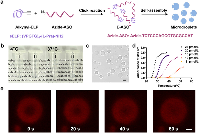
The E-ASO conjugate was synthesized via copper(Ⅰ)-catalyzed azide-alkyne cycloaddition (CuAAC), linking an alkyne-functionalized sELP30 (Table S1 in Supporting information) with an azide-functionalized ASO targeting the B-cell lymphoma-2 (Bcl-2) gene (18 nt, broad oncogene such as melanoma and hypopharyngeal carcinoma) (Fig. 1a and Table S2 in Supporting information). The E-ASO conjugate was confirmed by polyacrylamide gel electrophoresis (PAGE) and mass spectrometry (Fig. S1 in Supporting information). Our previous studies demonstrated that conjugates of sELP and oligonucleotides exhibited temperature-dependent LLPS behavior [48]. The phase transition of E-ASOs was investigated subsequently. Figs. 1b and c showed that the E-ASO solution (20 µmol/L) turned turbid at 37 ℃, and microsized particles (∼2 µm) were observed under microscopy. The temperature-dependent LLPS of E-ASO conjugates was monitored via a turbidity assay, which measured the absorbance changes at 350 nm during the cooling process. Fig. 1d revealed that the transition temperature (Tt) of E-ASO conjugates increased as the concentration decreased.
Figure 1
Figure 1.
Construction and characterization of E-ASO coacervates. (a) Schematic illustration of the synthesis and LLPS-driven formation of E-ASO coacervates. (b) Phase transition behavior of E-ASO conjugate solution under heating (37 ℃) and cooling (4 ℃). (ⅰ) E-ASO solution; (ⅱ) PBS. (c) CLSM images of E-ASO microdroplets. Scale bar: 2 µm. (d) Turbidity profiles (absorbance at 350 nm) of E-ASO at various concentrations. (e) Time-lapse CLSM images of FRAP of a single E-ASO coacervate microdroplet. Scale bar: 2 µm.
Specifically, the coacervates were formed at concentrations of 20 and 25 µmol/L under physiological conditions. Dynamic light scattering (DLS) analysis at 37 ℃ indicated that the average diameter of E-ASO coacervates was approximately 2.1 µm, with minimal variation across different concentrations (Fig. S2 in Supporting information). Therefore, the E-ASO coacervate system (20 µmol/L) was selected for subsequent experiments. Moreover, the fluorescence recovery after photobleaching (FRAP) was evaluated using tetramethylrhodamine (TAMRA) labeled E-ASO coacervate. The time-lapsed confocal laser scanning microscopy (CLSM) images showed that the rapid fluorescence recovery was observed within E-ASOTAMRA coacervate (Fig. 1e), demonstrating the liquid-like and dynamic molecular exchange properties. In addition, the E-ASO coacervates exhibited spreading behavior on glass substrates within minutes, which was consistent with their liquid-like characteristic (Fig. S3 in Supporting information).
Initially, the cellular uptake capacities of E-ASO coacervate were assessed. The 5-carboxyfluorescein (FAM)-labeled ASO strand was covalently conjugated with sELP to generate E-ASOFAM coacervates. Considering the high expression level of the Bcl-2 gene in melanoma cells, B16 cells were selected for this experiment [50,51]. The B16 cells were incubated with E-ASOFAM coacervates, and the CLSM imaging was performed at different incubation time points. The results showed that the E-ASOFAM coacervates were internalized within 6 h, and fluorescence intensity of the E-ASOFAM coacervates reached its peak at 24 h (Fig. S4 in Supporting information). Consequently, a 24-h incubation period was chosen for subsequent gene silencing evaluations. We then compared the cellular uptake of Free ASO and E-ASO. Confocal imaging demonstrated that Free ASO was barely internalized, whereas E-ASO exhibited higher uptake efficiency (Fig. S5 in Supporting information). Subsequently, the internalization mechanism was investigated. To avoid potential effects from gene regulation, coacervates prepared with random oligonucleotide sequences (Table S2), termed E-oligo coacervates, were used. HeLa cells were pretreated with four endocytosis inhibitors: Chlorpromazine (CPZ, clathrin-mediated endocytosis inhibitor), amiloride (AML, pinocytosis inhibitor), sodium azide (NaN3, energy-dependent endocytosis inhibitor), and methyl-β-cyclodextrin (MβCD, lipid raft-mediated pathway inhibitor). FAM-labeled E-oligo coacervates were incubated with HeLa cells. CLSM imaging (Fig. S6 in Supporting information) showed that AML pretreatment had little effect on uptake, while NaN3 and CPZ caused modest reductions. MβCD treatment largely inhibited internalization, suggesting that lipid raft-mediated endocytosis plays a major role. These findings are consistent with previous study, indicating that coacervate uptake can be cholesterol-dependent and largely independent of classical clathrin-mediated pathways [45]. The limited effect of NaN3 implies that membrane fluidity and passive, lipid-raft-associated processes may contribute to internalization [49,52]. Although the precise molecular mechanism remains to be fully clarified, these observations suggest that E-ASO coacervates can potentially bypass endosomal entrapment and deliver cargo into the cytosol.
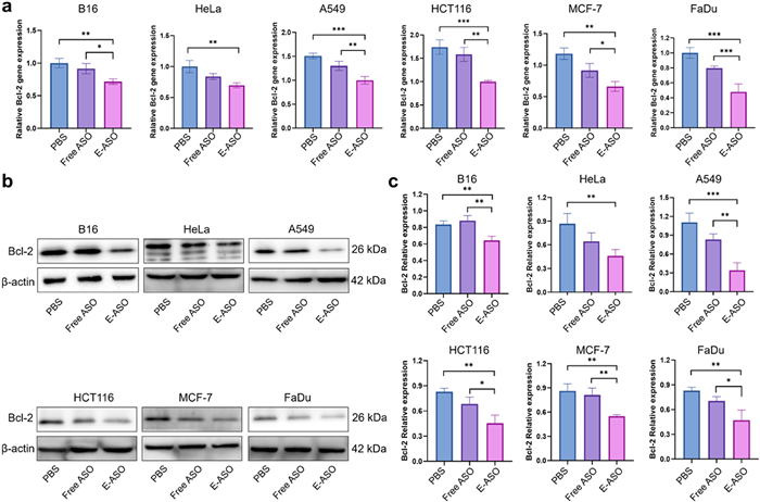
The gene silencing efficacy of E-ASO coacervates was then evaluated in B16 cells at both the mRNA and protein levels. The naked ASO solution (named Free ASO) (Table S2) was employed for comparison. Following a 24-h incubation of E-ASO coacervates, total RNA of B16 cells was extracted and analyzed via reverse transcription quantitative PCR (RT-qPCR) analysis. As shown in Fig. 2a, compared to the phosphate buffered saline (PBS) and Free ASO-treated groups, the E-ASO coacervate-treated group demonstrated the most efficient gene silencing of the Bcl-2 gene in terms of RNA level. Western blot (WB) analysis yielded consistent findings, indicating that the Bcl-2 protein was predominantly downregulated in the E-ASO coacervate-treated group (Fig. 2b and Fig. S7 in Supporting information). Grayscale quantification analysis further confirmed a statistically significant reduction in Bcl-2 protein levels between the groups treated with Free ASO and E-ASO coacervates in B16 cells (Fig. 2c).
Figure 2
Figure 2.
Validation of Bcl-2 silencing by E-ASO coacervates in six distinctive tumor cell lines of B16, HeLa, A549, HCT116, MCF-7, and FaDu. (a) RT-qPCR analysis of Bcl-2 expression levels in six cell lines following PBS, naked ASO (Free-ASO), and coacervate (abbreviated as E-ASO) treatment, respectively. (b) WB analysis of intracellular Bcl-2 expression in six cell lines after PBS, Free-ASO, and E-ASO treatment. (c) Quantification analysis of Bcl-2 protein expression based on densitometric analysis using ImageJ. Data are presented as mean ± standard deviation (SD) (n = 3). Statistical significance was determined using a one-way ANOVA test. *P < 0.05, **P < 0.01, ***P < 0.001.
Since Bcl-2 is a widely prevalent oncogene across various cancers, to validate the universality of gene silencing by E-ASO coacervates, five additional tumor cell lines, HeLa, A549, HCT116, MCF-7, and FaDu cells, all of which overexpress Bcl-2, were subsequently examined. The results showed that compared with the PBS and free ASO-treated groups, E-ASO coacervates consistently induced a significant downregulation of Bcl-2 in A549, HCT116, MCF-7, and FaDu cells at both gene and protein levels, while HeLa cells showed no significant response (Fig. 2). The lack of response in HeLa cells may reflect cell line-dependent differences in coacervate uptake or processing. The potent gene silencing ability of E-ASO coacervate across diverse cell lines shows its potential to perform therapeutic efficacy for cancer treatment in a gene silencing manner.
The membraneless characteristic of the coacervate system allows for the enrichment of environmental molecules efficiently. Here, the loading of the immune stimulator, cGAMP, by E-ASO coacervate, was carried out (Fig. S8a in Supporting information). The E-ASO coacervates were incubated with cGAMP for 2 h. Then the unloaded cGAMP molecules were removed through centrifugation. The loading efficiency of cGAMP was calculated to be 26.1% (Figs. S9a and b in Supporting information). The release profile of cGAMP from the coacervates was evaluated by high performance liquid chromatography (HPLC), showing a cumulative release of approximately 40.5% over 24 h (Fig. S10 in Supporting information). Treatment with E-ASO-cGAMP subsequently led to increased intracellular cGAMP accumulation relative to free cGAMP (Fig. S9c in Supporting information). Next, the STING pathway activation capacity of E-ASO-cGAMP coacervate was assessed in B16 and FaDu cells. These two cell lines were selected as they represent difficult-to-treat tumour models, providing a need to evaluate the outcome of combined therapeutic. As shown in Fig. S8b (Supporting information), treatment with E-ASO-cGAMP coacervates in B16 cells resulted in significant activation of the STING pathway, as evidenced by the presence of phosphorylation-STING (p-STING), p-TBK1, and p-IRF3 signals. In contrast, phosphorylation signals were considerably weaker in both PBS and the naked cGAMP solution (named Free cGAMP)-treated groups. In the FaDu cell, similar activated signals of the STING pathway were obtained (Fig. S8c in Supporting information). Meanwhile, Bcl-2 protein expression was assessed in B16 cells. It was observed that three experiment groups: E-ASO, cGAMP and E-ASO-cGAMP all effectively decreased the expression of Bcl-2, of which E-ASO-cGAMP showed the strongest inhibition efficiency of Bcl-2 (Fig. S11 in Supporting information). In addition, the cGAMP treatment could also decrease Bcl-2 expression, of which the underlying mechanism remains unclear. These results demonstrate that the efficacy of E-ASO-cGAMP coacervates in combined gene silencing and immune activation for synergistic antitumor therapy.
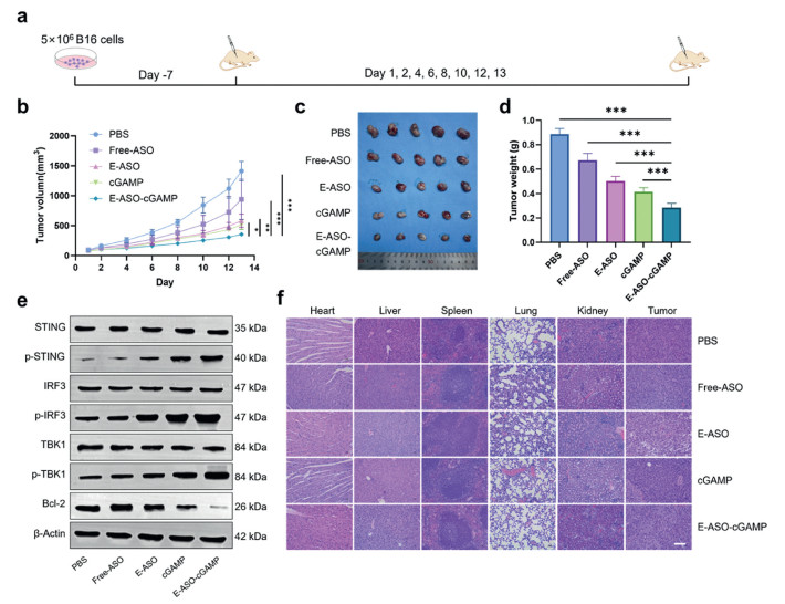
The in vivo combined therapeutic effect of E-ASO-cGAMP coacervate was investigated. All animal procedures were approved by the Ethics Committee of Xinhua Hospital Affiliated to Shanghai Jiao Tong University School of Medicine (approval No. XHEC-SHDC-2025-006). The subcutaneous B16 melanoma mouse model was employed, which were randomly divided into 5 experimental groups (n = 5) when the tumor volumes reached 100 mm3: PBS, free ASO, E-ASO coacervates, cGAMP, and E-ASO-cGAMP coacervates. The drug was administered via intratumor injection (Fig. 3a). In all five groups, the body weights and tumor volumes of tumor-bearing mice were recorded on days 1, 2, 4, 6, 8, 10, 12, and 13. It was observed that the body weight of mice did not decrease in all groups during the treatment period (Fig. S12 in Supporting information), indicating that treatment did not affect the growth of mice. As shown in Figs. 3b–d, free ASO treatment showed no significant therapeutic effect, likely due to low delivery efficiency. In contrast, treatment with E-ASO, cGAMP, or E-ASO-cGAMP all induced significant tumor suppression. As expected, the E-ASO-cGAMP treatment demonstrated the strongest antitumor effect among the five groups, confirming synergistic efficacy compared to either E-ASO or cGAMP alone. The gene silencing of Bcl-2 and the STING pathway activation were examined systematically via WB. The WB results (Fig. 3e and Fig. S13 in Supporting information) showed that treatment with cGAMP or E-ASO-cGAMP significantly upregulated phosphorylated STING (p-STING) levels, indicating STING pathway activation. The downstream proteins, p-IRF3 and p-TBK1 were also significantly elevated. Furthermore, E-ASO-cGAMP treatments markedly reduced anti-apoptotic Bcl-2 expression, suggesting apoptosis induction. Interestingly, cGAMP also appeared to enhance Bcl-2 silencing, although the underlying mechanism remains unknown. The safety of the treatment was also evaluated in mice. Hematoxylin and eosin (H&E) staining of heart, liver, spleen, lung, and kidney tissues from all groups revealed no significant pathological changes (Fig. 3f), indicating no obvious toxicity or side effects to normal organs.
Figure 3
Figure 3.In vivo therapeutic efficacy of E-ASO-cGAMP coacervates in B16 melanoma mouse model. (a) Schematic illustration of the treatment regimen in C57BL/6 mice. (b) Tumor growth curves for each treatment group. (c) Representative images of dissected tumors at the study end point. (d) Quantification of tumor weights across different groups. (e) WB analysis of cGAS-STING pathway-related proteins and Bcl-2 expression in tumor tissues post-treatment. (f) H&E staining of major organs (heart, liver, spleen, lung, kidney) and tumor tissues. Scale bar: 100 µm. Data are presented as mean ± SD (n = 5). Statistical significance was determined using one-way ANOVA (d) or two-way ANOVA (b). *P < 0.05, **P < 0.01, ***P < 0.001.
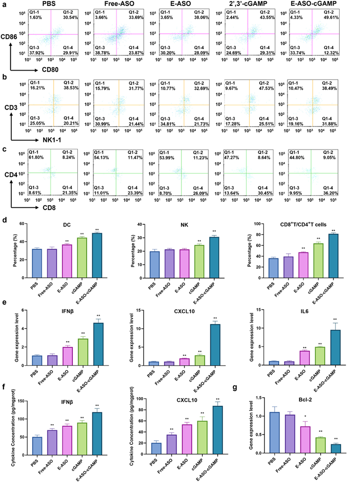
Immunological analysis was further performed to evaluate the immune activation elicited by different treatment groups. Flow cytometry analysis revealed a significantly increased proportion of mature dendritic cells (CD80+CD86+) in the E-ASO-cGAMP group, indicating enhanced antigen presentation (Fig. 4a and Fig. S14a in Supporting information). Natural killer (NK) cell activation was also markedly elevated (Fig. 4b and Fig. S14b in Supporting information). Furthermore, the proportions of CD8+T cells significantly increased in the E-ASO-cGAMP group, suggesting enhanced adaptive immune activation and improved T cell-mediated antitumor responses (Fig. 4c and Fig. S14c in Supporting information). These results were consistent across quantitative analysis (Fig. 4d). STING activation was assessed via the expression of STING-responsive genes, including IFNβ, CXCL10 and IL6. E-ASO-cGAMP induced significantly higher expression of these STING-related genes in tumors compared to the other four groups, indicating enhanced STING pathway activation within the tumor microenvironment (Fig. 4e). Consistent with these findings, ELISA analysis confirmed elevated IFN-β and CXCL10 levels, further validating the enhanced STING activation (Fig. 4f). Additionally, significant Bcl-2 downregulation supported tumor cell apoptosis induction and cooperative immune responses inhibiting tumor growth (Fig. 4g). Collectively, these results demonstrate that the combined gene silencing and immune activation synergistically enhance antitumor efficacy compared to monotherapies.
Figure 4
Figure 4.
Evaluation of immune activation at the tumor site following treatment. (a) Flow cytometry analysis of dendritic cell (DC) infiltration in tumor tissues across different treatment groups. (b) Flow cytometry analysis of NK cell infiltration. (c) Flow cytometry analysis of CD8+T cell infiltration. (d) Quantification of DCs, NK cells, and CD8+T/CD4+T cells in tumor tissues. (e) RT-qPCR analysis of IFNβ, CXCL10, and IL6 mRNA expression in tumor tissues. (f) ELISA-based measurement of IFNβ and CXCL10 cytokine levels in tumor tissues. (g) RT-qPCR analysis of Bcl-2 expression in tumor tissues. Data are presented as mean ± SD (n = 5). Statistical significance was assessed using one-way ANOVA. *P < 0.05, **P < 0.01.
Building upon the synergistic antitumor efficacy demonstrated in the melanoma model, E-ASO-cGAMP coacervates were evaluated in a second, highly challenging tumor type: Hypopharyngeal carcinoma. This aggressive and debilitating cancer is notoriously resistant to monotherapies [52-56]. As shown in Figs. S15 and S16 (Supporting information), in tumor-bearing mice randomized into five treatment groups (Fig. S15a), E-ASO-cGAMP again elicited the most potent tumor growth inhibition (Fig. S15b), with no significant body weight changes indicating minimal toxicity (Fig. S16). WB analysis confirmed STING pathway activation (elevated p-STING, p-TBK1, p-IRF3) and effective Bcl-2 downregulation (Fig. S15c). CLSM immunofluorescence further validated the downregulated Bcl-2 and STING, and upregulated p-STING proteins (Fig. S15d). Consistent with the melanoma findings, H&E staining revealed no organ damage (Fig. S15e), underscoring systemic safety. Collectively, the recapitulation of potent tumor suppression and safety across immunologically distinct melanoma and hypopharyngeal carcinoma models demonstrates the broad applicability of this therapeutic coacervate platform. By concurrently enabling Bcl-2 downregulation-induced apoptosis and STING-mediated immune activation, this strategy offers a translatable approach for combined antitumor treatment across diverse solid tumors.
This study establishes an LLPS-driven coacervate platform for synergistic integration of gene silencing and immunotherapy. By conjugating Bcl-2-targeting ASOs with phase-separating short ELPs, E-ASO coacervates were engineered that bypass endocytic trafficking to achieve efficient cytoplasmic delivery and potent Bcl-2 suppression across diverse cancer cells. Incorporation of the STING agonist cGAMP further enhanced intracellular accumulation and concurrently activated the STING pathway, remodeling the immunosuppressive tumor microenvironment. These combined effects led to significant tumor suppression exceeding monotherapies in both melanoma and hypopharyngeal carcinoma models. This platform offers a strategy for improving delivery of nucleic acid drugs and immunotherapeutics and provides a promising approach for antitumor treatment.
CRediT authorship contribution statement
Jianmian Fu: Writing – review & editing, Writing – original draft, Visualization, Validation, Software, Methodology, Formal analysis, Data curation. Shule Hou: Visualization, Validation, Software, Formal analysis, Data curation. Rui Huang: Writing – original draft, Validation, Formal analysis, Data curation. Bin Wang: Formal analysis, Data curation, Conceptualization. Yuxuan Wu: Visualization, Validation, Software. Hui Dong: Funding acquisition. Jun Yang: Supervision, Project administration, Funding acquisition. Weiping Zhu: Supervision, Funding acquisition. Yufang Xu: Supervision, Funding acquisition. Penghui Chen: Writing – review & editing, Writing – original draft, Supervision, Funding acquisition, Conceptualization. Jin Xie: Supervision, Funding acquisition. Xuhong Qian: Supervision, Conceptualization. Yangyang Yang: Writing – review & editing, Writing – original draft, Supervision, Project administration, Investigation, Conceptualization.
Declaration of competing interest
The authors declare that they have no known competing financial interests or personal relationships that could have appeared to influence the work reported in this paper.
Acknowledgments
This work was supported by the National Key Research and Development Programs of China (Nos. 2022YFD1700400 and 2023YFD1700300), Shanghai Hospital Development Center (No. SHDC2020CR6011).
Supplementary materials
Supplementary material associated with this article can be found, in the online version, at doi:10.1016/j.cclet.2025.112311.
G. Nishimura, H. Takahashi, D. Sano, et al., Int. J. Clin. Oncol. 29 (2024) 103–114. doi: 10.1007/s10147-023-02433-8
[56]
A. Bozec, G. Poissonnet, O. Dassonville, D. Culié, J. Clin. Med. 12 (2023) 1237. doi: 10.3390/jcm12031237
Figure 1
Construction and characterization of E-ASO coacervates. (a) Schematic illustration of the synthesis and LLPS-driven formation of E-ASO coacervates. (b) Phase transition behavior of E-ASO conjugate solution under heating (37 ℃) and cooling (4 ℃). (ⅰ) E-ASO solution; (ⅱ) PBS. (c) CLSM images of E-ASO microdroplets. Scale bar: 2 µm. (d) Turbidity profiles (absorbance at 350 nm) of E-ASO at various concentrations. (e) Time-lapse CLSM images of FRAP of a single E-ASO coacervate microdroplet. Scale bar: 2 µm.
Figure 2
Validation of Bcl-2 silencing by E-ASO coacervates in six distinctive tumor cell lines of B16, HeLa, A549, HCT116, MCF-7, and FaDu. (a) RT-qPCR analysis of Bcl-2 expression levels in six cell lines following PBS, naked ASO (Free-ASO), and coacervate (abbreviated as E-ASO) treatment, respectively. (b) WB analysis of intracellular Bcl-2 expression in six cell lines after PBS, Free-ASO, and E-ASO treatment. (c) Quantification analysis of Bcl-2 protein expression based on densitometric analysis using ImageJ. Data are presented as mean ± standard deviation (SD) (n = 3). Statistical significance was determined using a one-way ANOVA test. *P < 0.05, **P < 0.01, ***P < 0.001.
Figure 3In vivo therapeutic efficacy of E-ASO-cGAMP coacervates in B16 melanoma mouse model. (a) Schematic illustration of the treatment regimen in C57BL/6 mice. (b) Tumor growth curves for each treatment group. (c) Representative images of dissected tumors at the study end point. (d) Quantification of tumor weights across different groups. (e) WB analysis of cGAS-STING pathway-related proteins and Bcl-2 expression in tumor tissues post-treatment. (f) H&E staining of major organs (heart, liver, spleen, lung, kidney) and tumor tissues. Scale bar: 100 µm. Data are presented as mean ± SD (n = 5). Statistical significance was determined using one-way ANOVA (d) or two-way ANOVA (b). *P < 0.05, **P < 0.01, ***P < 0.001.
Figure 4
Evaluation of immune activation at the tumor site following treatment. (a) Flow cytometry analysis of dendritic cell (DC) infiltration in tumor tissues across different treatment groups. (b) Flow cytometry analysis of NK cell infiltration. (c) Flow cytometry analysis of CD8+T cell infiltration. (d) Quantification of DCs, NK cells, and CD8+T/CD4+T cells in tumor tissues. (e) RT-qPCR analysis of IFNβ, CXCL10, and IL6 mRNA expression in tumor tissues. (f) ELISA-based measurement of IFNβ and CXCL10 cytokine levels in tumor tissues. (g) RT-qPCR analysis of Bcl-2 expression in tumor tissues. Data are presented as mean ± SD (n = 5). Statistical significance was assessed using one-way ANOVA. *P < 0.05, **P < 0.01.


 DownLoad:

 下载:


 下载:

