State Key Laboratory of Bioactive Molecules and Druggability Assessment, College of Pharmacy, Jinan University, Guangzhou 510632, China
b.
Key Laboratory of Pesticide and Chemical Biology of Ministry of Education, Hubei Key Laboratory of Genetic Regulation and Integrative Biology, School of Life Sciences, Central China Normal University, Wuhan 430079, China
c.
Key Laboratory of Tropical Biological Resources of Ministry of Education and Hainan Engineering Research Center for Drug Screening and Evaluation, School of Pharmaceutical Sciences, Hainan University, Haikou 570228, China
d.
Institute for Safflower Industry Research, Key Laboratory of Xinjiang Phytomedicine Resource and Utilization (Ministry of Education), School of Pharmacy, Shihezi University, Shihezi 832003, China
e.
State Key Laboratory of Bioreactor Engineering, Shanghai Frontiers Science Center of Optogenetic Techniques for Cell Metabolism, Frontiers Science Center for Materiobiology and Dynamic Chemistry, Shanghai Key Laboratory of New Drug Design, School of Pharmacy, East China University of Science and Technology, Shanghai 200237, China
Received Date:
09 October 2025 Accepted Date:
19 December 2025 Revised Date:
18 December 2025 Available Online:
15 December 2026
Abstract:
WD repeat domain 5 (WDR5), a WD40 repeat protein, promotes oncogenic processes in various human cancers through dynamic interactions with key partners such as the mixed lineage leukemia (MLL) complex and cellular Myc (c-Myc). Although small-molecule inhibitors targeting WDR5–protein interfaces hold therapeutic potential, the lack of specific molecular probes for real-time monitoring has limited the mechanistic investigation of WDR5 dynamics. Here, we report the rational design of P3, a first-in-class fluorescent probe derived from the WDR5 inhibitor OICR-9429 scaffold. Structural characterization via X-ray crystallography and surface plasmon resonance (SPR) confirmed micromolar binding affinity with WDR5, while systematic linker optimization elucidated structure–optical property relationships essential for bioimaging. P3 enabled in situ visualization of WDR5 distribution in cancer cells and facilitated tumor-selective intravital imaging in xenograft models. This probe offers high spatiotemporal resolution for tracking WDR5 pathobiology, directly linking its dysregulated localization to malignant progression through multimodal imaging validation.
WD repeat domain 5 (WDR5) is a member of the WD40 repeat protein family, characterized by tryptophan-aspartic acid (WD) motifs. It serves as a crucial scaffolding component in assembling various protein complexes, thereby facilitating their enzymatic activity in epigenetic and transcriptional regulation [1]. WDR5 adopts a donut-shaped, seven-bladed β-propeller structure, which provides multiple interfaces for protein–protein interactions (PPIs) [2,3]. Notably, WDR5 contains two major PPI sites: The WDR5-binding motif (WBM) and the WDR5-interaction motif (WIN) [4,5]. The WIN site binds mixed lineage leukemia 1 (MLL1)/SET family proteins to form a histone methyltransferase complex, which catalyzes histone H3 lysine 4 di- and trimethylation (H3K4me2 and H3K4me3) [2], leading to the aberrant expression of oncogenes [6,7]. The WBM site is involved in recruiting and localizing the oncogenic transcription factor MYC to chromatin, thereby promoting tumorigenesis [8,9]. Recent studies highlight the critical role of the MLL complex in maintaining oncogenic gene expression in human leukemias with MLL rearrangement (MLL-r) [10-13], and disruption of the WDR5-MLL1 complex has been shown to suppress MLL-r leukemia growth [14]. Moreover, the WDR5-MYC axis plays a pivotal role in tumorigenesis in pancreatic cancer and Burkitt’s lymphoma [8,15,16]. Thus, targeting WDR5’s PPI sites to modulate gene activity represents a promising therapeutic strategy for cancer treatment.
Recent efforts have focused on developing inhibitors targeting the WIN and WBM sites of WDR5 [17]. Initial inhibitors were primarily designed for the WIN site, resulting in high-affinity compounds such as the peptidomimetic MM-589 and the small molecule OICR-9429 [18-23]. These inhibitors disrupt WDR5-MLL1 PPIs by binding to the WIN site, thereby inhibiting MLL1 activity and exerting antiproliferative effects in vitro and in vivo. In addition to the WIN site, the WBM site has emerged as a target for inhibitors that block WDR5-MYC interactions, thereby suppressing MYC-mediated transcriptional functions [24-27]. Moreover, a WDR5-targeted proteolysis-targeting chimera (PROTAC) has been developed by conjugating an optimized OICR-9429 scaffold to von Hippel–Lindau (VHL), cereblon (CRBN), and DDB1- and CUL4-associated factor 1 (DCAF1) E3 ligase ligands, providing an alternative strategy for WDR5 degradation [28-30]. These PROTACs induce WDR5 degradation in cell and mouse models, effectively inhibiting tumor growth. Despite WDR5’s recognition as an important cancer therapy target and tumor marker, effective detection tools for monitoring WDR5 in vivo remain lacking. Developing such tools is essential for a better understanding of the molecular mechanisms underlying tumor development and the action of WDR5-targeting compounds.
Fluorescent probes, known for their high selectivity, sensitivity, biocompatibility, and superior spatial–temporal resolution, are powerful tools for monitoring biomolecules and studying biological processes [31-35]. However, no fluorescent probes specifically targeting WDR5 have been reported to date. In this study, we designed a series of environment-sensitive fluorescent probes by conjugating the environmentally responsive fluorophore 4-nitro-7-aminobenzoxadiazole (NBD) with the WDR5 ligand OICR-9429 [20], and identified P3 as the pioneering candidate. P3 exhibits excellent biocompatibility, rapid and specific binding to WDR5, and stable fluorescence in both cellular and in vivo contexts. In tumor-bearing mice, P3 selectively accumulates in WDR5-overexpressing tumors with minimal off-target distribution, outperforming the non-specific dye NBD. These findings establish P3 as a robust and selective probe for WDR5 imaging in live cells and in vivo applications.
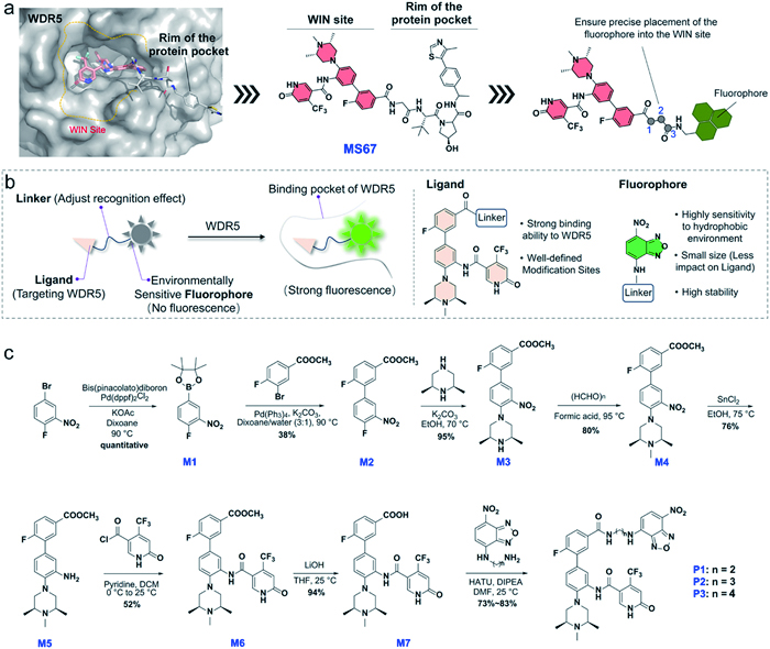
Guided by structural insights, we adopted a turn-on probe design that integrates an environmentally sensitive fluorophore with the WIN site-binding pharmacophore (Figs. 1a and b). The NBD fluorophore was selected based on its exceptional quantum yield, microenvironment responsiveness (enabling fluorescence modulation upon target engagement), and synthetic versatility for rational modification [36]. We prioritized the WIN site, a deep arginine-binding pocket essential for WDR5′s chromatin recruitment, over the shallower WBM site, allowing strategic fluorophore placement to leverage spatial confinement effects for signal amplification [1,17]. OICR-9429, a potent ligand that selectively targets the WIN site of WDR5, is widely used as a chemical probe in chemical biology research. In previous work, OICR-9429 was conjugated with a VHL ligand to generate the PROTAC molecule MS67, which induces WDR5 degradation (Fig. 1a) [28]. Inspired by the design of MS67, we introduced an NBD fluorophore via a flexible linker at the amide position, anticipating that the fluorophore would bind to the WIN site and exhibit strong fluorescence upon target engagement (Fig. 1b) [37-39].
Figure 1
Figure 1.
Design and synthesis of WDR5 fluorescent probes. (a) Design of the WDR5 fluorescent probe inspired by MS67. (b) Schematic diagram of the design strategy of WDR5 environmentally sensitive fluorescent probe. (c) Synthesis route of fluorescent probes P1–P3.
To optimize specificity while retaining WDR5 binding, we systematically varied the linker length and incorporated carboxylic acid modifications. This effort yielded probes P1–P3 through iterative medicinal chemistry optimization (Fig. 1c). All compounds were thoroughly characterized by spectroscopic methods (1H/13C nuclear magnetic resonance (NMR) spectroscopy and high-resolution mass spectrometry (HRMS) data have been provided in Supporting information), confirming their structural identity.
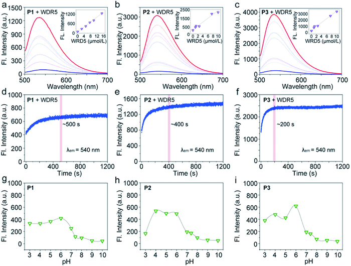
Following the synthesis of probes P1–P3, we systematically evaluated their molecular recognition capabilities through ultraviolet-visible (UV–vis) absorption spectroscopy. As depicted in Fig. S1 (Supporting information), all probes (10 µmol/L) exhibited distinct absorption maxima at ~480 nm in the free state. Upon incubation with recombinant WDR5 protein (10 µmol/L), a notable enhancement at 480 nm was observed, indicative of enhanced structural rigidity induced by ligand-protein complexation. Subsequent fluorescence titration experiments revealed concentration-dependent emission enhancement at 540 nm (λex = 480 nm) upon progressive addition of WDR5 (Figs. 2a–c). The results exhibited pronounced structural dependency: P3 demonstrated maximal fluorescence enhancement (28-fold), significantly outperforming P1 (10-fold) and P2 (18-fold). In addition, the limit of detection (LOD) of the P3 is calculated as the 9.2 nmol/L, which is the lowest among these probes (P1, 60 nmol/L; P2, 34.5 nmol/L). This hierarchy (P3 > P2 > P1) strongly correlated with alkyl linker length (C4 > C3 > C2), suggesting optimal spatial accommodation of the C4 spacer for WDR5 binding-induced fluorophore dequenching.
Figure 2
Figure 2.
Optical evaluation of all probes. (a–c) Fluorescence spectra of probe P1, P2 and P3 after the addition of WDR5 protein (0–30 µmol/L), all probes were dilutes to 10 µmol/L. Inset: fluorescent intensity changes at 540 nm of these probes upon the addition of different concentrations of WDR5 protein (0–30 µmol/L); (d–f) Time course of fluorescence intensity of probes P1, P2 and P3 at 540 nm after the addition of 10 µmol/L WDR5 (time range 0–1200 s). (g–i) Fluorescence spectra of probes P1, P2 and P3 in PBS buffer with different pH values from 3 to 10.
Real-time monitoring of fluorescence kinetics (λem = 480 nm) upon saturation with WDR5 (10 µmol/L) uncovered striking differences in binding speed (Figs. 2d–f). Probe P3 achieved signal plateau within 200 s, demonstrating ~2.5-fold faster equilibration than P1 (500 s) and P2 (400 s). These data corroborated the critical role of linker flexibility in accelerating molecular recognition kinetics, with shorter linkers imposing higher energy barriers for optimal binding conformation. To validate detection specificity, we quantified pH susceptibility across physiologically relevant ranges from pH 3 to 10. All probes maintained stable emission intensities (≤3-fold variation) under acidic conditions (pH 3–6) (Figs. 2g–i), with signal fluctuations significantly dwarfed by WDR5-induced responses (28-fold for P3). This environmental insensitivity confirmed the reliability of P1–P3 for biological applications under heterogeneous pH microenvironments.
Further, to rigorously validate target specificity, we challenged probe P3 against a structurally diverse panel of 16 proteins spanning (1) epigenetic reader proteins (NSD1, NSD2, DNMT3B and SETDB1), (2) chromatin reader proteins (CBX5, MPP8, SGF29, SMN, CDY1, CDYL2, CDYL1B and PHF1), (3) RNA-binding regulators (FMRP, FXR1 and TDRD3), and (4) metabolic enzyme (ALDH2) (Fig. S2 in Supporting information). Strikingly, P3 exhibited <5% fluorescence enhancement when incubated with non-target proteins at 10 µmol/L, comparable to background signals in protein-free buffer. In contrast, WDR5 binding induced a robust ~28-fold emission enhancement, demonstrating exceptional discrimination capability. Comprehensive photophysical evaluation established P3 as the optimal candidate, exhibiting superlative sensitivity, rapid equilibration kinetics, and robust environmental tolerance. The C4 alkyl linker was identified as a critical structural determinant, balancing molecular flexibility for rapid target engagement with sufficient rigidity for maximal fluorescence activation. Based on these findings, P3 was advanced for subsequent cellular and in vivo investigations.
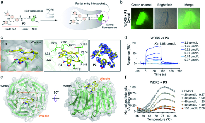
To further investigate the binding mechanism of P3 to WDR5, we employed X-ray crystallography, surface plasmon resonance (SPR), and differential scanning fluorimetry (DSF). As previously reported, the NBD fluorophore does not fluoresce in free solution but emits fluorescence when bound to a protein and restricted in rotation (Fig. 3a). We incubated WDR5 protein crystals with P3 and observed green fluorescence under the green channel, indicating successful binding of P3 to WDR5 (Fig. 3b). Subsequently, we collected diffraction data from the WDR5 crystals and resolved the complex structure, confirming that P3 binds to the WIN site as expected. The crystal structure also revealed that the NBD fluorophore is positioned outside the WIN pocket, forming hydrophobic interactions with Y131, which restricts its rotation and enhances fluorescence (Fig. 3c). This finding explains why P3 exhibits superior fluorescence properties compared to P2 and P1, as the flexible linker between the guiding moiety and NBD in P3 allows effective binding to the WIN site (Fig. 3d). Additionally, SPR analysis demonstrated a binding affinity of P3 to WDR5 with a Kd value of 1.06 µmol/L (Fig. 3e). DSF experiments further confirmed the interaction, revealing a positive protein melting temperature shift within the concentration range of 20–100 µmol/L (Fig. 3f). Collectively, these results provide a comprehensive understanding of the binding mechanism of P3 to WDR5.
Figure 3
Figure 3.P3 binding to WDR5. (a) Mechanism of P3 binding to the WIN site of WDR5. (b) Fluorescence and bright-field images of the P3–WDR5 protein crystal. (c) Interactions between P3 and WDR5, and the 2Fo–Fc omit electron density map contoured at 1.0 σ. (d) Binding mode of P3 at the WIN site of WDR5. (e) SPR analysis of the binding affinity between P3 and WDR5. (f) DSF plot showing P3 (20–100 µmol/L) binding to WDR5.
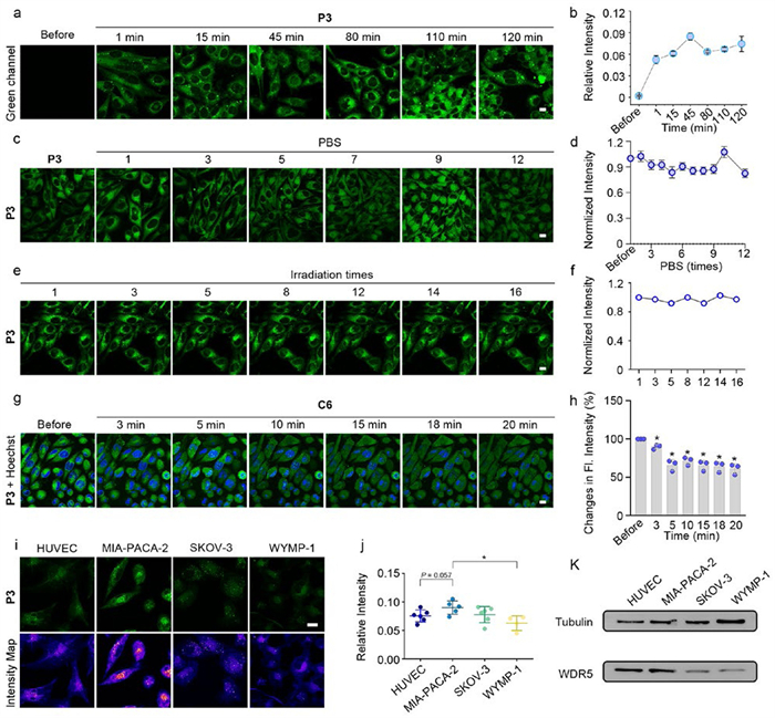
Prior to cellular experiments, we systematically evaluated the cytotoxicity of probe P3 across multiple cell lines, including HUVECs, MIA-PACA-2, SKOV-3, and WYMP-1. As illustrated in Fig. S3 (Supporting information), no significant cytotoxicity was observed at a concentration of 2.5 µmol/L, demonstrating excellent biocompatibility. This concentration was thus selected for subsequent cellular experiments. Cellular kinetic experiments were performed to assess the binding dynamics of probe P3 to WDR5 post-uptake. Upon administration of P3, distinct fluorescence signals emerged within 1 min, rapidly reaching saturation within ~15 min (Figs. 4a and b). These results indicate rapid transmembrane permeability and efficient binding to WDR5. Notably, no apparent weakness of fluorescence was detected over a 2 h observation period, highlighting robust fluorescence stability. To evaluate the binding affinity between P3 and WDR5, cells were subjected to sequential phosphate buffer saline (PBS) washing steps (up to 12 cycles). Fluorescence intensity remained unaltered despite repeated washing (Figs. 4c and d), confirming high-affinity binding of P3 to WDR5.
Figure 4
Figure 4.
Imaging experiment of P3 in cells. (a, b) Green channel fluorescence and relative intensity of probe P3 (2.5 µmol/L) in MIA-PACA-2 cells under varying incubation durations. Green channel: λex/λem = 488 nm/520–560 nm. (c, d) Green channel fluorescence imaging and the normalized intensity of MIA-PACA-2 cells (fixed cell) incubated with P3 (2.5 µmol/L) after PBS washing for different times. (e, f) Green channel fluorescence imaging and the normalized intensity of MIA-PACA-2 cells incubated with P3 (2.5 µmol/L) after exposure to irradiation for different times. (g, h) Fluorescence imaging and the normalized intensity of MIA-PACA-2 cells incubated with P3 (2.5 µmol/L) after administration of the WDR5 inhibitor C6 (2.5 µmol/L). (i) Green channel and fluorescence intensity map of probe P3 (2.5 µmol/L) incubated with different cells. (j) The relative intensity of fluorescence in green channel of P3 (2.5 µmol/L) incubated with different cells. (k) Relative content of WDR5 protein in different cell lines. Data are presented as mean ± SEM. *P < 0.05, **P < 0.01, ***P < 0.001 (unpaired t-test). Scale bar: 50 µm.
Photostability was assessed via continuous excitation cycles (16 iterations). Fluorescence signals exhibited negligible attenuation (Figs. 4e and f), underscoring exceptional resistance to photo-bleaching, a critical feature for long-term imaging applications. Further, to verify that fluorescence signals originated from specific P3-WDR5 interactions, a competitive assay was conducted using C6, a known WDR5 ligand [23]. Upon addition of C6 (2.5 µmol/L), fluorescence intensity decreased markedly within 3 min and continued to diminish over time (Figs. 4g and h). This competitive displacement unequivocally corroborated the specificity of P3 for WDR5. Finally, WDR5 expression levels and corresponding P3 fluorescence intensities were compared across cell lines. HUVECs and MIA-PACA-2 cells, which exhibit high endogenous WDR5 expression (Figs. 4i–k), displayed significantly stronger fluorescence signals relative to other cell types, with maximal intensity observed in MIA-PACA-2. These findings further validate the capacity of P3 to selectively report WDR5 expression levels in live cells.
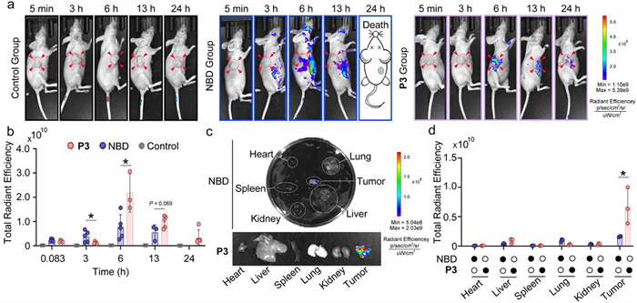
To further evaluate the detection capacity of probe P3 for WDR5 in vivo, we established a tumor-bearing mouse model using MIA-PACA-2 cells and performed live fluorescence imaging. All animal experiments were conducted in accordance with protocols approved by the Institutional Animal Care and Use Committee (IACUC Approval No. HPIACUC2025007) and under its supervision. Mice were divided into three groups: (1) Control group (no probe administration), (2) NBD group (imaged with the 20 mg/kg fluorescent molecule NBD-Cl, 4-chloro-7-nitrobenzofurazan), and (3) P3 group (imaged with probe 20 mg/kg P3). Intraperitoneal injection of probes was followed by longitudinal fluorescence imaging at predefined time points. As shown in Fig. 5a, no significant fluorescence signal was detected in the control group throughout the observation period. In the NBD group, diffuse fluorescence signals were observed at 3 h post-injection, peaking at 6 h and gradually declining thereafter. In contrast, the P3 group exhibited tumor-focused fluorescence starting at 6 h, with signal intensity sustained until 13 h and partially retained at 24 h (Figs. 5a and b). Notably, all mice in the NBD-treated group died within 24 h after administration. This acute mortality can be explained by the well-documented and inherent cytotoxicity of NBD-based compounds, which have been explored for their anticancer activity in previous studies [40,41]. The strong and non-selective covalent reactivity of NBD-Cl likely leads to severe systemic toxicity. By integrating the NBD fluorophore into a WDR5-binding ligand, the reactive chlorine atom is replaced. Consequently, the resulting conjugate, P3, benefits from enhanced tumor targeting and a significantly improved safety profile, as evidenced in both cellular and in vivo experiments (Fig. S3). Ex vivo imaging of dissected organs further validated the targeting precision of P3. In the NBD group, only weak fluorescence was detected in tumor tissues, indistinguishable from background signals in other organs (heart, liver, spleen, lung, and kidney). Conversely, the P3 group displayed intense fluorescence selectively localized to tumors, with minimal off-target accumulation (Figs. 5c and d). Quantitative analysis revealed a > 5-fold higher tumor-to-organ fluorescence ratio in the P3 group compared to the NBD group. The rapid mortality in the NBD group underscores the systemic toxicity of non-targeted fluorescent agents, whereas the prolonged survival and tumor-specific retention of P3 highlights its biocompatibility and WDR5-dependent binding. The sustained fluorescence stability of P3in vivo aligns with its superior photostability observed in cellular assays, reinforcing its utility for longitudinal tumor monitoring. These findings conclusively demonstrate that probe P3 achieves WDR5-specific detection in vivo, combining high tumor-targeting fidelity, prolonged signal retention, and minimal off-target toxicity. The stark contrast with NBD validates the design rationale of P3 as a theranostic tool for WDR5-overexpressing malignancies.
Figure 5
Figure 5.
Fluorescence imaging using P3in vivo. (a) Representative whole-body imaging in mice in different groups at different times after injection of P3 (20 mg/kg) and NBD (20 mg/kg), respectively. (b) Quantitative analysis of the fluorescence imaging of mice from parallel images (including a). (c) Representative images of ex vivo imaging of five major organs (heart, liver, spleen, lung and kidney) and tumors harvested from mice in different group at 24 h after injection of the P3. (d) Quantitative analysis of the fluorescence imaging of major organs and tumors from parallel images (including c). n = 3 mice for each group. Data are presented as mean ± SEM. *P < 0.05, **P < 0.01, ***P < 0.001 (unpaired t-test).
WDR5 has solidified its position as a critical oncology target through its dual role in orchestrating epigenetic regulation via MLL1 complexes and sustaining oncogenic transcription through MYC partnerships. Despite remarkable progress in developing therapeutic modalities such as competitive inhibitors and proteolysis-targeting chimeras to disrupt these interactions, the field has lacked robust tools for direct visualization of WDR5′s dynamic behavior in living systems. In this study, we addressed this gap by designing and characterizing the NBD-based fluorescent probe P3, which demonstrates high specificity, biocompatibility, and in vivo stability. P3 selectively accumulates in WDR5-overexpressing tumors, enabling real-time imaging of WDR5 in both cellular and animal models. The demonstrated success of this probe not only expands the chemical biology toolkit for epigenetic targets but also provides a blueprint for developing precision molecular diagnostics in oncology, thereby creating new synergies between basic research and therapeutic innovation.
CRediT authorship contribution statement
Xin Chen: Writing – original draft, Methodology, Investigation, Data curation. Linchong He: Writing – original draft, Methodology, Investigation, Data curation. Xinhang Lv: Writing – original draft, Methodology, Investigation, Data curation. Siying Liu: Investigation. Jie Song: Investigation. Jinrong Min: Validation, Investigation. Pinghua Sun: Validation, Investigation. Jian Li: Validation, Investigation. Donglei Shi: Writing – review & editing, Supervision, Conceptualization. Yunyuan Huang: Writing – review & editing, Supervision, Conceptualization.
Declaration of competing interest
The authors declare that they have no known competing financial interests or personal relationships that could have appeared to influence the work reported in this paper.
Acknowledgments
We gratefully appreciate the financial support from the National Natural Science Foundation of China (Nos. 82404418, 22407038), the Fundamental Research Funds for the Central Universities (No. 21624352), the Science and Technology Projects in Guangzhou (No. 2025A04J3241), the Hainan Provincial Natural Science Foundation of China (No. 825MS073), the China Postdoctoral Science Foundation (No. 2024M751137), Self-determined Research Funds of Central China Normal University from the Colleges’ Basic Research and Operation of Ministry of Education (No. CCNU25HD022), and the National Funding Postdoctoral Fellowship Program of CPSF (No. GZC20230972). We acknowledge the staff of beamline BL10U2 at the Shanghai Synchrotron Radiation Facility for assistance during the data collection.
Supplementary materials
Supplementary material associated with this article can be found, in the online version, at doi:10.1016/j.cclet.2025.112300.
[1]
A. Schuetz, A. Allali-Hassani, F. Martín, et al., EMBO J. 25 (2006) 4245–4252. doi: 10.1038/sj.emboj.7601316
H.H. Sha, Z. Wang, S.C. Dong, et al., Biosci. Rep. 38 (2018) BSR20171440. doi: 10.1042/BSR20171440
[41]
F. Pellizzari Tregno, A. Sau, S. Pezzola, et al., Eur. J. Cancer 45 (2009) 2606–2617. doi: 10.1016/j.ejca.2009.06.033
Figure 1
Design and synthesis of WDR5 fluorescent probes. (a) Design of the WDR5 fluorescent probe inspired by MS67. (b) Schematic diagram of the design strategy of WDR5 environmentally sensitive fluorescent probe. (c) Synthesis route of fluorescent probes P1–P3.
Figure 2
Optical evaluation of all probes. (a–c) Fluorescence spectra of probe P1, P2 and P3 after the addition of WDR5 protein (0–30 µmol/L), all probes were dilutes to 10 µmol/L. Inset: fluorescent intensity changes at 540 nm of these probes upon the addition of different concentrations of WDR5 protein (0–30 µmol/L); (d–f) Time course of fluorescence intensity of probes P1, P2 and P3 at 540 nm after the addition of 10 µmol/L WDR5 (time range 0–1200 s). (g–i) Fluorescence spectra of probes P1, P2 and P3 in PBS buffer with different pH values from 3 to 10.
Figure 3P3 binding to WDR5. (a) Mechanism of P3 binding to the WIN site of WDR5. (b) Fluorescence and bright-field images of the P3–WDR5 protein crystal. (c) Interactions between P3 and WDR5, and the 2Fo–Fc omit electron density map contoured at 1.0 σ. (d) Binding mode of P3 at the WIN site of WDR5. (e) SPR analysis of the binding affinity between P3 and WDR5. (f) DSF plot showing P3 (20–100 µmol/L) binding to WDR5.
Figure 4
Imaging experiment of P3 in cells. (a, b) Green channel fluorescence and relative intensity of probe P3 (2.5 µmol/L) in MIA-PACA-2 cells under varying incubation durations. Green channel: λex/λem = 488 nm/520–560 nm. (c, d) Green channel fluorescence imaging and the normalized intensity of MIA-PACA-2 cells (fixed cell) incubated with P3 (2.5 µmol/L) after PBS washing for different times. (e, f) Green channel fluorescence imaging and the normalized intensity of MIA-PACA-2 cells incubated with P3 (2.5 µmol/L) after exposure to irradiation for different times. (g, h) Fluorescence imaging and the normalized intensity of MIA-PACA-2 cells incubated with P3 (2.5 µmol/L) after administration of the WDR5 inhibitor C6 (2.5 µmol/L). (i) Green channel and fluorescence intensity map of probe P3 (2.5 µmol/L) incubated with different cells. (j) The relative intensity of fluorescence in green channel of P3 (2.5 µmol/L) incubated with different cells. (k) Relative content of WDR5 protein in different cell lines. Data are presented as mean ± SEM. *P < 0.05, **P < 0.01, ***P < 0.001 (unpaired t-test). Scale bar: 50 µm.
Figure 5
Fluorescence imaging using P3in vivo. (a) Representative whole-body imaging in mice in different groups at different times after injection of P3 (20 mg/kg) and NBD (20 mg/kg), respectively. (b) Quantitative analysis of the fluorescence imaging of mice from parallel images (including a). (c) Representative images of ex vivo imaging of five major organs (heart, liver, spleen, lung and kidney) and tumors harvested from mice in different group at 24 h after injection of the P3. (d) Quantitative analysis of the fluorescence imaging of major organs and tumors from parallel images (including c). n = 3 mice for each group. Data are presented as mean ± SEM. *P < 0.05, **P < 0.01, ***P < 0.001 (unpaired t-test).


 DownLoad:

 下载:


 下载:

