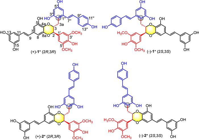
Figure 1.
Structures of (+)–1/(–)–1 and (+)–2/(–)–2.

Discovery and biomimetic synthesis of the first stilbenolignan-stilbene hybrids from Heterosmilax yunnanensis inhibiting myocardial fibroblast activation
Ji-Ping Liao , Ji-Chao Zhou , Ying-Li Fan , Jian-Shuang Jiang , Xu Zhang , Xiang Yuan , Pei-Cheng Zhang , Xiao-Wei Zhang , Ya-Nan Yang


Stilbenolignans, formed by union of a stilbene unit (C6—C2—C6) and a phenylpropane unit (C6—C3), belong to a rare group of “none-conventional” lignans in plants [1]. To date, only 19 naturally-occurring stilbenolignans have been reported [2-13]. Among them, aiphanol (from Aiphanes aculeata by Kinghorn et al.) and 5-hydroxyaiphanol (from Syagrus romanzoffiana by Lee et al.) possessed a unique 1,4-benzodioxane skeleton and showed significantly pharmacological activities [2,14]. Aiphanol functions as potent inhibitors against cyclooxygenase-1 (COX-1) and COX-2 with half-maximal inhibitory concentration (IC50) values of 1.9 and 9.9 µmol/L, respectively, and against vascular endothelial growth factor receptor (VEGFR) to inhibit angiogenesis and generation of lymphatic vessels [15]. These studies highlight the great value of discovering this type of rarely natural products.
Heterosmilax yunnanensis Gagnep. (Liliaceae), also named “Bai-Tu-Ling”, has been commonly used as a folk medicine for detoxifying, dehumidifying, clearing heat, and relieving joint pain and muscle pain [16]. Our previous research of the EtOAc extract of this medicinal plant revealed four stilbenoid dimers with rare benzo bicyclo[3.3.1] and [4.2.1] nonene scaffolds [17]. As a continuous investigation of H. yunnanensis for more bioactive molecules with intriguing structures, two racemic pairs of unprecedented stilbenolignan-stilbene hybrids, (+)/(–)–1 and (+)/(–)–2, were isolated and identified. Compounds (+)/(–)–1 and (+)/(–)–2 featured a unique 1,4-benzodioxane core with one stilbene unit and one phenylpropane unit, and further connected through a rare C(sp3)–C(sp2) bond with another stilbene, which represented the first examples of hybrids composed of stilbenolignan and stilbene (Fig. 1). Interestingly, both 1 and 2 exhibited potent anti-fibrotic effects in transforming growth factor-β1 (TGF-β1)-induced HL-1 cells by inhibiting cytoskeletal remodeling, cell migration, and the expression of fibrotic markers. Furthermore, molecular docking and Western blot (WB) analysis demonstrated that (+)–2 could dose-dependently and significantly suppress the TGF-β1-induced upregulation of macrophage migration inhibitory factor (MIF) protein expression in HL-1 cells, indicating that this class of compounds likely exerts their anti-cardiac fibrotic effects by targeting MIF. Cultivated by their fascinating structures and remarkable bioactivities, we designed and realized the biomimetic synthesis of compounds 1 and 2 via the key Friedel-Crafts (F–C) alkylation catalyzed by p-toluenesulfonic acid (p-TSA) and cyclization reaction mediated by silver oxide (Ag2O). This synthetic method conducted with a protecting-group-free strategy and proceeded with high regioselectivity and stereoselectivity, providing valuable insights for developing new methodology of building molecules with 1,4-benzodioxane scaffolds.
(+)/(–)–Heterosmilphanol A (1), isolated as a light brownish amorphous powder, had the molecular formula of C39H34O11 with 23 unsaturation degrees, as established by its protonated high resolution electrospray ionization mass spectroscopy (HRESIMS) ion peak at m/z 679.2174 [M + H]+ (calcd. 679.2174). The infrared resonance (IR) absorptions at 3416, 1604, 1514, 1463, 1146, 1115, 839 cm−1 indicated the presences of hydroxyl, phenyl, vinyl and ether functional groups. The 1H nuclear magnetic resonance (NMR) data (Table S1 in Supporting information) of 1 showed protons of an AA'BB' system [δH 7.27 (dd, J = 8.5, 2.5 Hz, H-10′′/H-14′′) and 6.73 (dd, J = 8.5, 2.5 Hz, H-11′′/H-13′′)], an A2X system [δH 6.39 (d, J = 2.0 Hz, H-12/H-16) and 6.12 (t, J = 2.0 Hz, H-14)], two AX systems [δH 6.66 (d, J = 2.5 Hz, H-8), 6.62 (d, J = 2.5 Hz, H-6) and 6.43 (d, J = 2.5 Hz, H-6′′), 6.23 (d, J = 2.5 Hz, H-4′′)] and two trans-coupled olefins [δH 7.20 (d, J = 16.5 Hz, H-7′′), 6.76 (d, J = 16.5 Hz, H-8′′) and 6.84 (d, J = 16.5 Hz, H-9), 6.76 (d, J = 16.5 Hz, H-10)]. Signals of methylene [δH 2.87 (dd, J = 14.0, 9.0 Hz, Hα−3a), 2.73 (dd, J = 14.0, 3.5 Hz, Hβ−3a)] and methoxy [δH 3.71 (s, 6H)] were observed. Meanwhile, two aliphatic protons at δH 4.94 (d, J = 5.5 Hz, H-2), 4.49 (m, H-3) suggested the existence of a 1,4-benzodioxane ring. The 13C NMR associated with heteronuclear single quantum coherence (HSQC) spectra demonstrated thirty-nine carbons in 1 (Table S1), including seventeen sp2 methylene carbons [δC 129.6, 127.9 (2C), 127.5, 126.9, 124.5, 115.6 (2C), 106.6, 106.1, 105.3 (2C), 104.5 (2C), 103.4, 102.0, 101.6], seventeen sp2 quaternary carbons [δC 158.6 (2C), 157.2, 156.7, 156.4, 147.8 (2C), 146.5, 144.1, 139.4, 139.0, 135.8, 131.6, 129.2, 128.5, 128.0, 112.9] and five sp3 aliphatic carbons (δC 77.9, 76.6, 56.0, 56.0, 27.8). In the heteronuclear multiple-bond correlation (HMBC) spectrum, the correlations of H-2 to C-1′/C-3, H-3 to C-2/C-3a, H-3a to C-2/C-3, and H-2′/H-6′ to C-2 implied a phenylpropane unit, which constituted as part of the 1,4-benzodioxane fragment as deduced from the long-range correlations from H-2 to C-8a and H-3 to C-4a (Fig. 2). Moreover, the HMBC correlations from H-9 to C-6/C-7/C-8, H-10 to C-11/C-12/C-16, and H-6 to C-9, H-12/H-16 to C-10 approved the presence of a penta-substituted stilbene unit. The abovementioned fragment enabled the deduction of 5-hydroxyaiphanol [14], a scarcely natural stilbenolignan loaded with a 1,4-benzodioxan scaffold. Moreover, a resveratrol unit was obviously observed by the HMBC correlations from H-7′′ to C-1′′/C-2′′/C-6′′, H-8′′ to C-9′′/C-10′′/C-14′′. The connection between 5-hydroxyaiphanol fragment and resveratrol fragment via a C(sp3)–C(sp2) bond was rationalized by the key HMBCs from H-3 to C-2′′ and H-3a to C-2′′/C-3′′. Thus, the planar structure of 1 was undoubtedly established (Fig. 1), the first stilbenolignan-stilbene hybrid composed of a 1,4-benzodioxan-based stilbenolignan fragment and a resveratrol unit linked through a unique C(sp3)–C(sp2) bond.
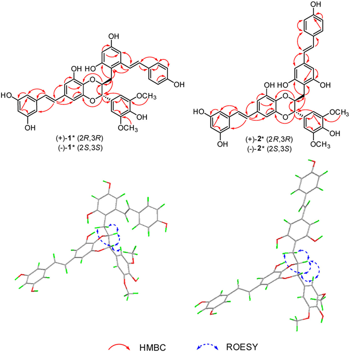
The relative stereochemistry of C-2 and C-3 could not be determined from the coupling constants comparisons (5.5 Hz in 1 vs. 8.3 Hz in 5-hydroxyaiphanol). Then the rotating frame overhauser spectroscopy (ROESY) experiment was conducted and confirmed the trans configuration of C-2, C-3 by the key correlations from H-2 to H-3a and H-3 to H-2′/H-6′. The optical inactivity of 1 indicated it existed as racemates and a subsequent chiral HPLC analysis enabled separation of 1 into a pair of enantiomers, (+)–1 and (–)–1, equally. Their absolute configurations were elucidated by electronic circular dichroism (ECD) calculations using time-dependent density functional theory (TD-DFT) at the CAM-B3LYP/6–31+g (d, p) level (Supporting information). As shown in Fig. 3, the calculated ECD curves of (2R, 3R) agreed well with the experimental one of (+)–1, while that of (2S, 3S) was in accordance with (–)–1, allowing the absolute configurations of (+)–1 and (–)–1 to be determined as (2R, 3R) and (2S, 3S), respectively.
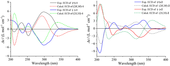
(+)/(–)–Heterosmilphanol B (2), isolated as a light brownish amorphous powder, had the same molecular formula of C39H34O11 as that of 1, deduced based on its HRESIMS ion peak at m/z 679.2173 [M + H]+ (calcd. 679.2174). The 1H NMR data of 2 was quite similar with that of 1 (Table S1), with the only difference being the replacement of an AX system [δH 6.43 (d, J = 2.5 Hz, H-6′′) and 6.23 (d, J = 2.5 Hz, H-4′′)] in 1 as an A2 system [δH 6.43 (s, H-2′′/H-6′′)] in 2. This discrepancy indicated a symmetric substituted position of the methylene on the resveratrol unit, supported by the key HMBC correlations from H-3a (δH 2.60, 2.51) to C-4′′ (δC 109.9), C-3′′/C-5′′ [δC 156.7 (2C)], and H-3 (δH 4.64) to C-4′′ (δC 109.9) (Fig. 2). Hence, compound 2 was characterized as a positional isomer of 1, in which the methylene at C-3a of the stilbenolignan fragment linked to C-4′′ of the resveratrol unit via C(sp3)–C(sp2) bond (Fig. 1). The relative configuration of 2R*, 3R* was confirmed by the mutual correlations from H-2 to H-3a and H-3 to H-2′/H-6′ in the ROESY spectrum. Chiral HPLC analysis also separated 2 into a pair of enantiomers, (+)–2 and (–)–2. The absolute configurations of (+)–2 and (–)–2 were assigned as (2R, 3R) and (2S, 3S), respectively, through comparisons of calculated ECD curves with the experimental ECD spectra (Fig. 3).
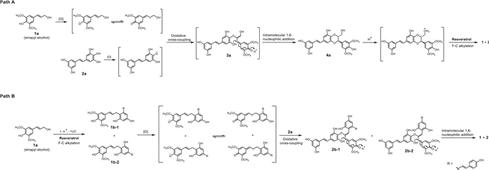
Biogenetically, compounds 1 and 2 could be generated via two plausible pathways (Scheme 1). For path A, sinapyl alcohol and 3,3′,4,5,5′-pentahydroxy stilbene were first oxidated to offer free radical intermediates, which then underwent oxidative cross-coupling to yield the key intermediate 3a. Later, an intramolecular 1,6-nucleophilic addition occurred spontaneously to provide 5-hydroxyaiphanol (intermediate 4a) equipped with a 1,4-benzodioxane skeleton. Finally, upon protonation, an F–C alkylation-like reaction between protonated 4a and resveratrol enabled the construction of the C(sp3)–C(sp2) bond, thus resulting in the production of compounds 1 and 2. Path B, on the contrary, started from the formation of C(sp3)–C(sp2) bond between protonated sinapyl alcohol and resveratrol to give intermediate 1b–1 and 1b–2, which were oxidated to generate the corresponding free radicals, and then reacted with oxidated 3,3′,4,5,5′-pentahydroxy stilbene (2a) via the oxidative cross-coupling, intramolecular 1,6-nucleophilic addition to finally obtain the expected compounds with a 1,4-benzodioxane skeleton.
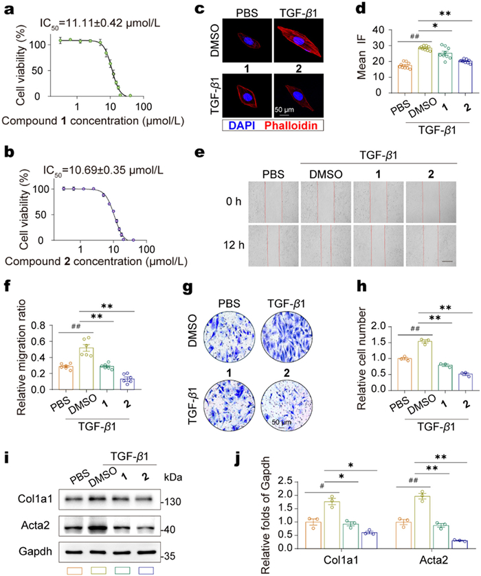
To systematically investigate the antifibrotic effect of compounds 1 and 2, we investigated whether 1 and 2 could inhibit the proliferation, migration and activation of HL-1 cells in vitro [18]. Since effective anti-fibrotic drugs require selective inhibition of fibroblast activation at subtoxic doses, we first established the safety profiles of 1 and 2. Using Alamar Blue assay, we determined the IC50 values of 1 (11.11 ± 0.42 µmol/L) and 2 (10.69 ± 0.35 µmol/L) in HL-1 cells (Figs. 4a and b). Subtoxic doses (5 µmol/L for 1 and 2.5 µmol/L for 2) were selected for further studies to ensure cell viability. Phalloidin staining revealed that TGF-β1 stimulation (at a dosage of 10 ng/mL) induced pronounced F-actin polymerization compared to the phosphate-buffered saline (PBS) control group. Notably, both compounds significantly attenuated this fibrotic transformation, and compound 2 displayed more robust pharmacological activity (Figs. 4c and d). Given the pivotal role of aberrant cell migration in fibrotic progression, we conducted scratch wound healing and Transwell migration assays to quantitatively assess the compounds' effects on the motility of HL-1 cells. In the scratch assay, TGF-β1 treatment enhanced HL-1 cell migration, and reduced the wound area by 82% after 12 h compared to PBS control (Fig. 4e). Both 1 and 2 significantly inhibited TGF-β1-induced migration, restoring migration rates to near-baseline levels (Fig. 4f). Similarly, in the Transwell assay, TGF-β1 increased cell migration by approximately 1.5-fold compared to PBS control, while treatments of 1 and 2 significantly reduced the number of migrated cells (Figs. 4g and h). Then, we assessed the expression of fibrotic markers Col1a1 (collagen type I alpha 1 chain) and Acta2 (alpha-smooth muscle actin) by WB analysis. TGF-β1 treatment significantly upregulated Col1a1 (1.76 ± 0.177) and Acta2 (1.97 ± 0.104) protein levels compared to PBS control. Notably, both 1 and 2 treatments substantially attenuated these effects (Figs. 4i and j). These findings collectively demonstrate that 1 and 2 exert potent anti-fibrotic effects in TGF-β1-induced HL-1 cells by inhibiting cytoskeletal remodeling, cell migration, and the expression of fibrotic markers.
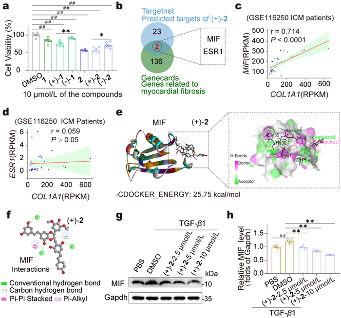
To explore the antifibrotic mechanism of compounds, we systematically evaluated the effects of compounds 1, 2, and their enantiomers ((+)–1/(–)–1, (+)–2/(–)–2) on the suppression of fibroblast activation at the cellular level. The Alamar Blue assay revealed that (+)–2 exhibited the most potent activity in inhibiting cardiac fibroblast activation among the tested compounds (Fig. 5a). Thus, it was selected as the primary focus for subsequent investigations into the antifibrotic mechanism. A Venn diagram analysis was performed by intersecting myocardial fibrosis-associated genes from the GeneCards database (
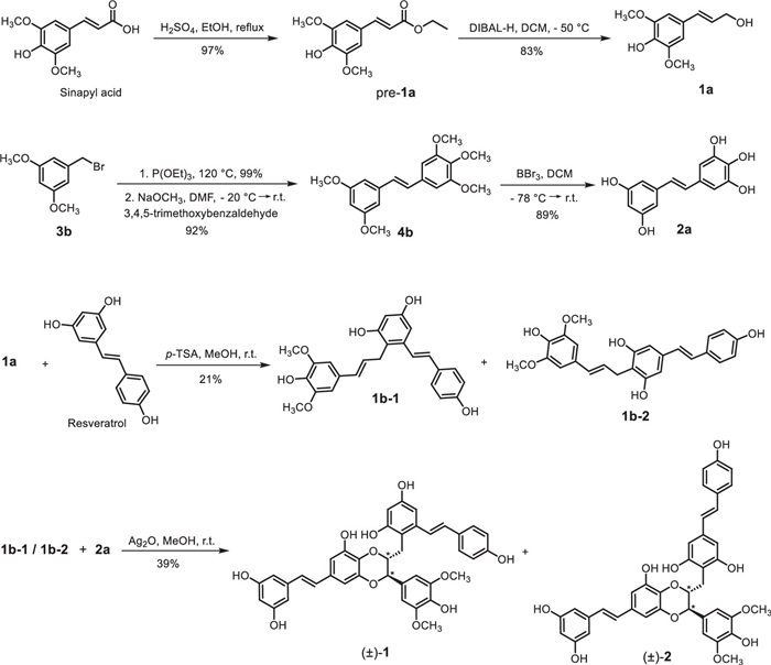
Cultivated by the intriguing structures of 1 and 2 and their potent anti-fibrotic effects in TGF-β1-induced HL-1 cells, we sought to achieve their total synthesis based on aforementioned biogenetic pathways (Scheme 1, path A and B). We speculated that 1 and 2 could be gained either through oxidative cross-coupling of sinapyl alcohol 1a and pentahydroxy stilbene 2a to construct the 1,4-benzodioxane core (4a), followed by F−C alkylation with resveratrol (Scheme 1, path A), or by F–C alkylation to generate the phenol 1b-1 and 1b-2 first between resveratrol and sinapyl alcohol 1a, and then underwent oxidative cross-coupling with 2a (Scheme 1, path B). The key intermediates 1b-1 and 1b-2 were expected to be synthesized via F−C alkylation between resveratrol and sinapyl alcohol 1a, while the latter could be attained by reduction of sinapic acid. Another key intermediate 4b could be prepared from 3,5-dimethoxy benzyl bromide and 3,4,5-trimethoxybenzaldehyde by classic Witting-Horner reaction and then demethylation.
According to the retrosynthetic analysis, the key intermediate 2a needed to be first prepared. Therefore, 4b was synthesized via Witting-Horner reaction in the presence of 3b and 3,4,5-trimethoxybenzaldehyde, the former could be obtained by Arbuzov reaction starting from commercially available 3,5-dimethoxy bromide with triethyl phosphite [19]. Then demethylation of 4b by boron tribromide was applied to yield 2a [20]. The key substrate pre-1a was prepared from sinapic acid through esterification by concentrated sulfuric acid in ethanol, followed by reduction mediated by the reducing reagent DIBAL-H [21,22].
With the key intermediates 1a and 2a in hand, we sought to synthesize 4a according to the reported method for the construction of 1,4-benzodioxane skeleton using silver-based catalyst [23]. However, two sets of peaks in the 1H NMR spectrum of 4a were observed, indicating this strategy exerted no regio-/stereo-selectivity, similar to the synthesis of aiphanol by Ulbrich et al. (Supporting information) [24]. Therefore, we turned our focus to another strategy (Scheme 1, path B), in which the C(sp3)–C(sp2) bond was constructed prior to the assembling of the 1,4-benzodioxane scaffold. We speculated that the replacement of –OH on the methylene of sinapyl alcohol 1a by resveratrol unit furnished with sufficiently steric hindrance would enable better regioselectivity and stereoselectivity, avoiding the production of unexpected regio-isomers in the next step. In recent years, mild and efficient BrØnsted-acid or lewis-acid catalyzed F–C alkylation of alcohols with other nucleophiles have been successfully applied in the synthesis of bioactive natural products with C(sp3)–C(sp2) bond [25-27]. Considering the primary alcohol 1a bearing a π-system of styrene and dihydroxylation by acid would generate stable carbon cation intermediates, we attempted p-TSA to catalyze the dehydrated cross-coupling of alcohol 1a and resveratrol. Gratifyingly, 1b-1 and 1b-2 were obtained simultaneously with moderate yield (Scheme 2). Finally, the oxidative couplings between substrates 1b-1, 1b-2, and 2a were efficiently mediated by Ag2O in methanol as the solvent under ambient temperature [28]. Remarkably, this condition proceeded with outstanding regio- and stereo-selectivity, affording products 1 and 2 exclusively without the formation of any detectable positional or stereoisomers. This method also provides a notably efficient and operationally simple approach for the selective construction of similar complex molecular architectures.
In summary, two racemic pairs of unprecedented stilbenolignan derivatives, (+)/(–)–heterosmilphanols A and B (1 and 2), representing the first examples of stilbenolignan-stilbene hybrids, were discovered from H. yunnanensis. Compounds 1 and 2 are positional isomers possessing a rare 1,4-benzodioxane core and further connected with resveratrol through a unique C(sp3)–C(sp2) bond. Next, biogenetic analysis of 1 and 2 facilitated their synthesis via key p-TSA-catalyzed and Ag2O-mediated reactions in seven concise steps by protecting-group-free strategy. This strategy proceeds smoothly with high regioselectivity and stereoselectivity, providing new insights for the synthesis of other molecules endowed with 1,4-benzodioxane scaffolds. Pharmacological tests demonstrated their potent anti-fibrotic effects in TGF-β1-induced HL-1 cells by inhibiting cytoskeletal remodeling, cell migration, and the expression of fibrotic markers. A preliminary mechanism exploration demonstrated this class of compounds likely exerts their anti-cardiac fibrotic effects by targeting MIF. Overall, this study not only enriches the library of naturally-occurring stilbenolignan derivatives but also offers potential inhibitors for myocardial fibrosis therapy. Moreover, the established biomimetic synthesis ensures a sufficient supply for subsequent druggability research.
Ji-Ping Liao: Writing – original draft, Validation, Methodology, Investigation, Formal analysis, Data curation, Conceptualization. Ji-Chao Zhou: Writing – original draft, Methodology, Investigation, Data curation, Conceptualization. Ying-Li Fan: Methodology, Investigation, Data curation. Jian-Shuang Jiang: Validation. Xu Zhang: Validation. Xiang Yuan: Validation. Pei-Cheng Zhang: Writing – review & editing, Validation, Funding acquisition. Xiao-Wei Zhang: Writing – review & editing, Visualization, Validation, Supervision, Funding acquisition. Ya-Nan Yang: Writing – review & editing, Visualization, Validation, Supervision, Project administration.
The authors declare that they have no known competing financial interests or personal relationships that could have appeared to influence the work reported in this paper.
This work was financially supported by the CAMS Innovation Fund for Medical Sciences (CIFMS, Nos. 2021-I2M-1–028, 2022-I2M-1–014, China). The ECD calculations were supported by Biomedical High Performance Computing Platform, Chinese Academy of Medical Sciences.
Supplementary material associated with this article can be found, in the online version, at doi:
S.A. Begum, M. Sahai, A.B. Ray, Fortschr. Chem. Org. Naturst. 93 (2010) 1–70. doi: 10.1007/978-3-7091-0140-7_1
D. Lee, M. Cuendet, J.S. Vigo, et al., Org. Lett. 3 (2001) 2169–2171. doi: 10.1021/ol015985j
S.H. Lam, S.S. Lee, Phytochemistry 71 (2010) 792–797. doi: 10.1016/j.phytochem.2010.01.013
M. Lin, J.B. Li, B. Wu, et al., Phytochemistry 30 (1991) 4201–4203. doi: 10.1016/0031-9422(91)83507-H
M. Kobayashi, T. Mahmud, N. Yoshika, et al., Chem. Pharm. Bull. 44 (1996) 2249–2253. doi: 10.1248/cpb.44.2249
K. Kawazoe, N. Shimogai, Y. Takaishi, et al., Phytochemistry 44 (1997) 1569–1573. doi: 10.1016/S0031-9422(96)00768-6
Z. Ali, T. Tanaka, I. Iliya, et al., J. Nat. Prod. 66 (2003) 558–560. doi: 10.1021/np020532o
X.M. Li, M. Lin, Y.H. Wang, et al., Planta Med. 70 (2004) 160–165. doi: 10.1055/s-2004-815494
C.S. Yao, M. Lin, L. Wang, Chem. Pharm. Bull. 54 (2006) 1053–1057. doi: 10.1248/cpb.54.1053
Y.Q. Cheng, R. Jiang, W. Huang, et al., RSC Adv. 4 (2014) 28901–28907. doi: 10.1039/C4RA03371J
L. Pan, L. Fu, X.G. Jia, et al., J. Asian Nat. Prod. Res. 23 (2021) 627–636. doi: 10.1080/10286020.2021.1918119
D.V. Tarbeeva, D.V. Berdyshev, E.A. Pislyagin, et al., Molecules 28 (2023) 2593. doi: 10.3390/molecules28062593
G.X. Yang, C.Q. Hu, Chin. J. Org. Chem. 23 (2003) 873–876.
S. Xu, M.Y. Shang, G.X. Liu, et al., Molecules 18 (2013) 5265–5287. doi: 10.3390/molecules18055265
J. Zeng, Q. Deng, Z. Chen, et al., Bioorg. Chem. 146 (2024) 107278. doi: 10.1016/j.bioorg.2024.107278
H. Shen, Z. Qu, Y. Harata-Lee, et al., Front. Oncol. 9 (2019) 632. doi: 10.3389/fonc.2019.00632
J.P. Liao, J. Li, W.J. Qin, et al., Chin. Chem. Lett. 36 (2025) 110687. doi: 10.1016/j.cclet.2024.110687
S.X. Guo, X.H. Qi, L.Z. Zhang, et al., Int. Immunopharmacol. 148 (2025) 114086. doi: 10.1016/j.intimp.2025.114086
M. Wang, Y. Jin, C.T. Ho, et al., J. Agric. Food Chem. 47 (1999) 3974–3977. doi: 10.1021/jf990382w
G.R. Pettit, M.P. Grealish, M.K. Jung, et al., J. Med. Chem. 45 (2002) 2534–2542. doi: 10.1021/jm010119y
J.T. Sanderson, H. Clabault, C. Patton, et al., Bioorg. Med. Chem. 21 (2013) 7182–7193. doi: 10.1016/j.bmc.2013.08.057
Y. Lv, Y. Feng, J. Dai, et al., Org. Lett. 24 (2022) 2694–2698. doi: 10.1021/acs.orglett.2c00797
L. Merlini, A. Zanarotti, Tetrahedron Lett. 16 (1975) 3621–3622. doi: 10.1016/S0040-4039(00)91340-6
M.G. Banwell, A. Bezos, S. Chand, et al., Org. Biomol. Chem. 1 (2003) 2427–2429. doi: 10.1039/B305106D
L. Tan, T. Xu, X. Zhang, et al., Org. Lett. 25 (2023) 2600–2605. doi: 10.1021/acs.orglett.3c00554
X. Pan, T. Sheng, Z.Z. Liu, Chin. Chem. Lett. 36 (2025) 110913. doi: 10.1016/j.cclet.2025.110913
S.M. Chen, T.J. Zhang, Z.H. Xu, et al., Chin. Chem. Lett. 34 (2023) 108130. doi: 10.1016/j.cclet.2023.108130
L.I. Pilkington, D. Barker, Nat. Prod. Rep. 32 (2015) 1369–1388. doi: 10.1039/C5NP00048C

Figure 4 Compounds 1 and 2 suppress TGF-β1-induced cardiac fibrosis in HL-1 cells. (a, b) Dose-response curves for 1 (a) and 2 (b) in HL-1 cells. (c) Representative immunofluorescence images of F-actin (phalloidin, red) and nuclei (4′,6-diamidino-2-phenylindole (DAPI), blue) in HL-1 cells. (d) Quantification of mean fluorescence intensity of phalloidin staining. (e) Representative images of the scratch assay in HL-1 cells at 0 and 12 h. Scale bar: 250 µm. (f) Quantification of relative migration ratio from the scratch assay. (g) Representative images of Transwell migration assay in HL-1 cells. (h) Quantification of relative cell number from the Transwell assay. (i) WB analysis of Col1a1 and Acta2 protein expression in HL-1 cells. (j) Quantification of relative protein expression levels of Col1a1 and Acta2 compared with glyceraldehyde-3-phosphate dehydrogenase (Gapdh). # P < 0.05, ## P < 0.01 vs. the PBS control group; *P < 0.05, **P < 0.01 vs. the TGF-β1-treated group. Data are presented as mean ± standard error of the mean (SEM) (n ≥ 3).

Figure 5 (+)–2 Inhibits TGF-β1-Induced Myocardial Fibrosis by Targeting MIF. (a) Cell viability assay of compounds 1, 2, and their enantiomers in HL-1 cells (concentration 10 µmol/L). (b) Venn diagram analysis of the intersection between genes related to myocardial fibrosis from GeneCards and predicted targets of (+)–2 from Targetnet. (c, d) Correlation analysis between MIF/ESR1 and COL1A1 in the GSE116250 dataset (n = 14 for non-heart failure, n = 13 for ICM). Molecular docking model of (+)–2 with MIF, including 3D structure (e) and 2D interaction diagram (f) of the docking results. (g, h) WB analysis and quantitative analysis of MIF (1:1000, Sigma-Aldrich) protein expression in HL-1 cells. ## P < 0.01 vs. the PBS control group; *P < 0.05, **P < 0.01 vs. the TGF-β1-treated group. Data are presented as mean ± SEM (n ≥ 3).

 扫一扫看文章


扫一扫关注我们
 DownLoad:

 下载:


 下载: