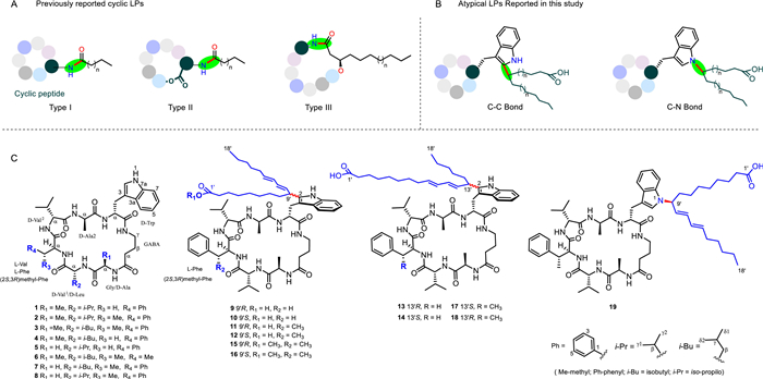
Figure 1.
(A) Typical LPs produced from NRPS assembly lines. (B) Atypical LPs reported in this study. (C) The chemical structures of 1‒19.

Novel lipopeptides featuring unusual C–C and C–N linkages between cyclic peptides and fatty acyl chains
Weixia Ye , Kai Liu , Wei Liu , Xianbo Chen , Xuemin Ding , Haiyan Tian , Wenjun Zhang , Changsheng Zhang


Lipopeptides (LPs) are a unique class of natural products characterized by a hydrophobic fatty acyl chain linked to a hydrophilic polypeptide core, and exhibit diverse biological properties including antibacterial, antiviral, antitumor, anti-infection, and immunosuppressant activities [1,2]. Biogenetically, the hydrophobic fatty acyl chains of LPs are derived either from primary metabolism via fatty acid synthases, or are assembled by an iterative polyketide synthase (PKS) [1-3], which are activated and subsequently assembled through the actions of nonribosomal peptide synthetase (NRPSs) or a hybrid machinery that combines NRPSs/PKS [1,4]. The structural diversity of LPs can be expanded through the attack of different side chain nucleophiles on the carbonyl of the terminal residue. However, nearly all fatty acids are linked to the N-terminal of the peptide through an amide bond (Fig. 1A) [3].
Unguisins A‒K, a small group of cyclic heptapeptides characterized by the presence of non-proteinogenic amino acid γ-aminobutyric acid (GABA) and D-amino acid residues, were naturally isolated from a marine-derived Emericella unguis [5,6], Aspergillus strains [7-10], endophytic fungus Mucor irregularis [11], or discovered through a genome mining approach [12]. The stereochemistry of individual amino acids in unguisins was determined using Marfey’s method. However, the configuration of the β-methylphenylalanine (β-MePhe) remains unassigned. In our efforts to screen bioactive natural products from marine-derived fungi, the fungal strain Aspergillus sp. SCSIO 40435, associated with the sponge Halichondria panicea collected from the South China Sea, attracted our attention for the richness in metabolites with antimicrobial activities. Aspergillus genus is a significant source for the discovery of novel natural products and bioactive compounds [13,14]. Previously, we reported four p-terphenyl compounds from Aspergillus sp. SCSIO 40435 [15]. In subsequent work guided by MS2-based molecular networking (MS2-MN), we obtained a series of atypical LPs (Fig. 1B). Here, we describe the isolation and characterization of these compounds, comprising lipounguisins (LPUs) A‒K (9‒19), four new heptapeptides, unguisins L‒O (5‒8), alongside four known analogues, unguisins A, E, F and K (1‒4) (Fig. 1C). The LPUs represent a rare class of lipopeptides, featuring an unguisin core linked to an octadecadienoic acid moiety via unusual C‒C (C9’-C2 or C13’-C2) or C‒N (C9’-N1) bonds. The fatty acid chains possess distinct diene configurations (either trans Δ10, Δ12 or trans Δ9, Δ11). The absolute configuration of β-MePhe in unguisins was determined through a combination of J-HMBC experiments, hydrolysis reaction, and optical rotation analysis. Notably, the LPUs exhibited superior biological activity compared to their unguisin precursors. Some of them showed bioactivities including anti-Gram-positive bacteria, inhibitory activity of α-glucosidase, and cytotoxicity to Huh-7 (half-maximal inhibitory concentration (IC50) = 5.42 µmol/L). In addition, we present a bioinformatic analysis of the biosynthetic gene cluster (BGC) of unguisins and proposed putative mechanisms for the formation of these novel LPUs.
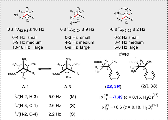
Compounds 1‒4 were characterized to be unguisin A (1) [5], unguisin E (2) [7], unguisin F (3) [11], and unguisin K (4) [10], respectively, by comparing their high-resolution electrospray ionization mass spectrometry (HRESIMS) and nuclear magnetic resonance (NMR) data with literatures (Tables S1 and S2, Figs. S1–S5 in Supporting information). However, the stereochemistry the β-MePhe residue in unguisin E (2) and unguisin F (3) remains unassigned due to the lack of a standard [7,11]. To address this, the configuration of the β-MePhe was analyzed by the J-based configurational analysis (JBCA) [16] and comparison of the optical rotation (OR) [17]. For this purpose, 3
JH,H and 3
JC,H values of 2 were determined by 1H spectroscopy and J-heteronuclear multiple bond correlation (J-HMBC) experiments, respectively (Figs. S2D and E). The typical criteria of large (L), medium (M), and small (S) 3
JH,H and 3
JC,H values were adopted (Fig. 2) [16]. The rotational conformer concerning the C-2(C-α)-C-3(C-β) bond was considered an alternating system due to its medium value (5.0 Hz) of 3
JH-2,H-3. Additionally, the J values of 2.6 Hz for 3
JC-1,H-3 and 2.2 Hz for 3
JC-4,H-2 were regarded as small values [8,16], indicating a gauche interaction between H-3/COOH and H-2/Ph. Hence, based on the JBAC analysis, the threo rotamers pair, corresponding to A-1 and A-3 (Fig. 2), met all established criteria. Finally, the absolute configuration of the β-MePhe residue was assigned as (2S,3R) by comparing the optical rotation value of β-MePhe (
Compounds 5‒8 were identified as new unguisin analogues, designated unguisins L‒O. Compound 5 differed from 1 by having an additional Gly residue instead of Ala residue. Compound 6 differed from unguisin B by the replacement of a Leu with Val. Compound 7 is a derivative of 6, featuring a Gly residue in place of Ala. Compound 8 is an analogue of 7, where a Leu residue in 7 was replaced with Val residue in 8. These assignments were confirmed through comprehensive 2D NMR analysis (Tables S3 and S4, Figs. S6–S9 in Supporting information). The absolute configurations of the amino acid residues in 5‒8 were assigned according the advanced Marfey’s method or biosynthetic pathway (Fig. S10 in Supporting information), and the β-MePhe in 6 and 7 were tentatively assigned as 2S,3R, following that in 3.
Compound 9 was obtained as colorless powder. The molecular formula of 9 was established as C58H84N8O9 by high-resolution mass spectrometry (HRMS) (electrospray ionization/quadrupole time-of-flight (ESI/Q-TOF) m/z: [M + H]+ calcd. for C58H85N8O9, 1037.6434; found 1037.6456), indicating 21 degrees of unsaturation. The 1H NMR spectrum (Fig. S11 and Table S5 in Supporting information) contained signals for seven amide NH signals at δH 8.59 (1H, d, J = 8.3 Hz), 8.43 (1H, br s), 8.11 (1H, d, J = 3.1 Hz), 8.01 (1H, br s), 7.88 (1H, d, J = 9.6 Hz), 7.79 (1H, d, J = 5.8 Hz), 7.66 (1H, t, J = 5.1 Hz) and overlapping methylene protons at 1.20–1.26 ppm. The 13C NMR data of 9 exhibited eight carbonyl signals at δC 171.0 and five methine signals between 48.3 and 61.2, typical for a peptide. The 13C NMR spectrum (Fig. S11 and Table S6 in Supporting information) also showed a group of signals for methylene carbons around δC 29.2, which was indicative of a putative fatty acid unit and suggested 9 as a lipopeptide.
The correlation spectroscopy (COSY) and HMBC spectra of 9 (Fig. 3) allowed the assignment of a heptapeptide unit, the same as that of 1. In addition, 2D NMR data also revealed the presence of a lipid unit, which was characterized as 10,12-octadecadienoic acid unit by COSY correlations of H3–18′/H2–17′, H2–8′/H-9′/H-10′/H-11′/H-12′/H-13′/H2–14′/H2–15′, H2–2′/H2–3′, and HMBC correlations from H3–18′ to C-16′/C-17′, H2–15′ to C-17′, H2–8′ to C-7′, and H2–2′/H2–3′ to C-1′/C-4′. The linkage of C-9′ (δC 39.4) of the lipid to C-2 (δC 139.2) of the indole ring of the heptapeptide was suggested by HMBC correlations from H-9′ (δH 3.62) to C-2 (δC 139.2) and C-3 (δC 106.9) (Fig. 3A). Thus, the planar structure of 9 was assigned and designated as LPU A (9), which was an unprecedented hybrid of a cyclic heptapeptide and a lipid linked by a C‒C bond (Fig. 1B). The E configurations of the double bond Δ10′ and Δ12′ in 9 were assigned by the large 3 JH-10′,H-11′ (14.3 Hz) and 3 JH-12′,H-13′ (14.2 Hz).
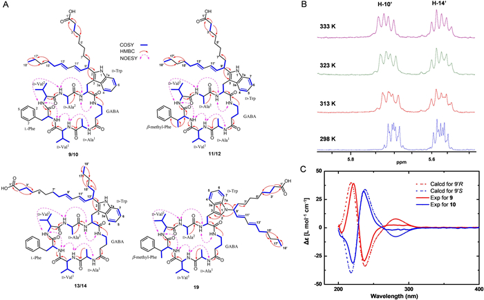
Compound 10 was obtained as colorless powder. The molecular formula of 10 was established as C58H84N8O9 by HRMS (ESI/Q-TOF m/z: [M + H]+ calcd. for C58H85N8O9, 1037.6434; found 1037.6440), the same as that of 9. The 1H and 13C NMR spectra of 10 (Tables S5 and S6, Fig. S12 in Supporting information) were highly similar to those of 9, suggesting that 10 was a stereoisomer of 9. This suggestion was confirmed by detailed comparison of NMR data of 10 and 9. Thus, the planar structure of 10 was assigned and designated as LPU B. The complex coupling patterns of Δ10′ and Δ12′ in 10 hampered the direct determination of the geometric configurations of conjugated double bonds in 10 by the coupling constant analysis. A careful analysis of NMR data of 10 revealed that the chemical shifts of C-9′ (δC 39.4) and C-14′ (δC 32.5) in 10 was almost identical to those in 9 (δC 39.7, C-9′; 32.4, C-14′), respectively. These data suggested that the complex splitting of Δ10’ and Δ12′ in 10 might be related to conformational changes compared to 9 [18,19]. Then, we sorted to test variable-temperature (VT) 1H NMR experiments with 10. The 1H NMR spectrum of 10 recorded at 313 K resulted in a set of well-distinguished peaks, which could be further improved at 333 K (Fig. 3B). By this way, the coupling constants for Δ10′ and Δ12′ in 10 could be resolved to be 3 JH-10′,H-11′ = 13.7 Hz and 3 JH-12′,H-13′ = 13.9 Hz, respectively, which assigned both of the double bonds Δ10′ and Δ12′ as an E configuration.
The aforementioned analysis implied that 9 and 10 only differed from each other in the configuration at C-9′. To resolve the absolute configurations of C-9′ in 9 and 10, the time-dependent density functional theory-electronic circular dichroism (TDDFT-ECD) calculations were performed (Tables S7–S13 in Supporting information). The experimental ECD of 9 was in good agreement with calculated ECD spectra of 9′R (Fig. 3C and Fig. S13 in Supporting information), while the experimental ECD of 10 was in good agreement with calculated ECD spectra of 9′S (Fig. 3C). Thus, the absolute configurations of C-9′ were assigned as 9′R in 9 and 9′S in 10.
Compounds 11 and 12 were identified as a pair of isomers (Tables S14 and S15, Figs. S14 and S15 in Supporting information). Their structures are closely related to the isomeric pair 9 and 10, with the difference by having 2 as a heptapeptide unit, instead of 1 (Fig. 3A). The geometries of Δ10′ and Δ12′ and the absolute configuration of C-9′ in 11 and 12 were assigned by coupling constants or compared their circular dichroism (CD) spectra with those of 9/10 (Fig. S16 in Supporting information). Compound 13/14 differed from 9/10 by having a 9,11-octadecadienoic acid unit (18:2 Δ9′,11′), instead of a 10,12-octadecadienoic acid (18:2 Δ10′,12′) unit in 9, which was supported by COSY correlations of H3–18′/H2-17′, H2-15′/H2-14′/H-13′ (δH 3.63)/H-12′ (δH 5.77)/H-11′ (δH 6.00), H-10′ (δH 6.00)/H-9′ (δH 5.60) and HMBC correlations from H–12′ to C-10′ (δC 130.5), and from H-9′ to C-11′(δC 130.3) (Fig. 3A, Tables S16 and S17, Figs. S17–S19 in Supporting information). Additionally, 13/14 differed from 9/10 in the C‒C linkage between the fatty acid chain and the Trp residue. Specifically, the C‒C linkage occurred via C-13′ to C-2 in compounds 13/14, whereas the C-9′‒C-2 linkage was observed in 9/10. This assignment was supported by HMBC correlations from H-13′ (δH 3.63) to C-2 (δC 139.2) and C-3 (δC 107.0) (Fig. 3A). The geometries of Δ10′ and Δ12′ were assigned by coupling constants (Table S16). The absolute configurations of C-13′ in 13/14 were assigned by comparing their CD spectra with those of 9/10 (Fig. S20 in Supporting information), respectively.
Compounds 15‒18 were also identified as lipopeptides. Compounds 15/16 were distinct from 11/12 by having one methyl ester group at C-1′ Compounds 17/18 differed from 13/14 by the substitution of phenylalanine (Phe) with β-methyl-Phe (MePhe). These assignments were confirmed through comprehensive 2D NMR analysis (Figs. S21–S25 in Supporting information). To determine the absolute configuration of C-9′ (15, 16) or C-13′ (17, 18), their ECD spectra were compared with those of 9/10 (Fig. S26 in Supporting information). The NMR data for 15–18 can be found in Tables S18–S21 (Supporting information).
Compound 19 was obtained as colorless powder. The molecular formula of 19 was established as C59H86N8O9 by HRMS spectrum (ESI/Q-TOF m/z: [M + H]+ calcd. for C59H87N8O9, 1051.6591; found 1051.6603), identical to that of 11/12. Careful analysis of NMR data of 19 (Fig. S27 and Table S22 in Supporting information) revealed that it differed from 11/12 by the C‒N connection between the fatty acid chain and the Trp residue. Specifically, the C‒N linkage was observed from C-9′ to N-1 in compound 19, whereas an C-2‒N-1 linkage was found in 11/12. This observation was supported by HMBC correlations from H-9′ (δH 4.95) to C-2 (δC 124.2), and C-7 (δC 110.5) (Fig. 3A). Detailed 2D NMR assignment confirmed the planar structure of 19. The E configuration of Δ10′ and Δ12′ were assigned by the large 3 JH-10′/H-11′ (15.1 Hz), and 3 JH-12′/H-13′ (15.0 Hz), respectively. The absolute configuration of C-9′ in 19 was assigned as 9′S by comparing its CD spectra with those of 9 and 10 (Fig. S28 in Supporting information).
Analysis of the genomic sequence Aspergillus sp. SCSIO 40435 using AntiSMASH (version 6.1.1) [20] led to the identification of a putative biosynthetic gene cluster (BGC) (lug) responsible for the formation of unguisins (Tables S23 and S24 in Supporting information) [12,21]. However, no genes surrounding the lug BGC were found to be related to the generation of the lipid side chain in LPUs. Moreover, no BGCs were identified in Aspergillus sp. SCSIO 40435 for the production of polyunsaturated fatty acids (PUFAs). These observations suggest that the PUFAs (18:2 Δ9,11 or Δ10,12) in LPUs likely originate from primary metabolism such as the fatty acid pathway (Fig. S29 in Supporting information) [22].
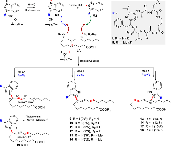
We propose two plausible radical-mediated pathways for the biosynthesis of these LPUs. In the first pathway (Scheme 1), a hydrogen atom transfer (HAT) from the N1 position of the Trp residue in cyclic peptides 1 or 2 is initiated, potentially catalyzed by an unknown cytochrome P450 enzyme [23,24]. A typical compound I species (FeIV=O+•) could abstract a hydrogen atom to yield an N1-centered radical (M1), which then undergoes intramolecular radical migration to C2, generating a C2 • radical (M2). Either M1 or M2 could then react with the lipid-derived radical acceptor at positions C9′ or C13′ to afford the observed C9′–N1, C9′–C2, or C13′–C2 linkages. The resulting Δ10, cis-Δ12 dienes are proposed to isomerize spontaneously to the observed trans-Δ10, Δ12 configuration, supported by TDDFT calculations (ΔE = −2.1 kcal/mol) (Experimental section in Supporting information). In the alternative pathway, the reaction initiates with HAT from the bis-allylic position of the linoleic acid (LA) moiety by a reactive oxygen species (ROS), such as a hydroxyl radical generated via the Haber–Weiss reaction [25]. This yields a carbon-centered pentadienyl radical (Int1), which in turn abstracts a hydrogen atom from the N1 of Trp to form radical M1. Subsequent M1 generates the C2 • radical M2. The coupling between Int1 and M1 (or M2) then affords the observed lipid–peptide conjugates via radical–radical recombination (Fig. S30 in Supporting information).
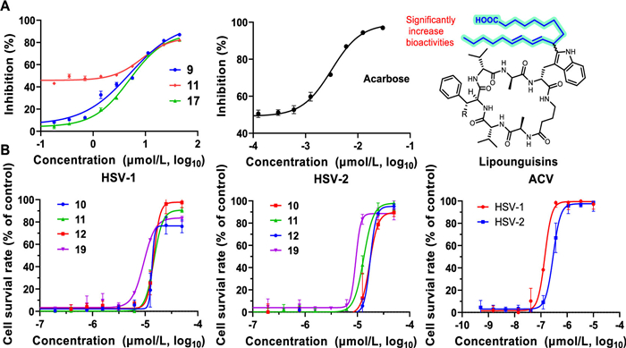
To explore the biological significance of these novel lipopetides, we evaluated their anti-bacterial, α-glucosidase inhibitory, cytotoxic, as well as anti-herpes simplex virus (HSV) activities. Remarkably, LPUs were found to be superior to unguisins in bioactivities. For instance, LPUs 10 and 12 exhibited significant inhibitory activity against Micrococcus luteus SCSIO ML01, with minimum inhibitory concentration (MIC) values of 4 µg/mL, while unguisins showed no such activities (Table S25 in Supporting information). Similarly, LPUs 17, 14 and 11 displayed prominent inhibitory activity against α-glucosidase, a target for diabetes treatment [26], with IC50 values of 5.0, 5.1 and 8.5 µmol/L, respectively (Fig. 4). Additionally, LPU 9 displayed selective cytotoxicity against human liver cancer cell line Huh-7 with an IC50 value of 5.42 µmol/L, in comparison to human breast cancer cell line MDA-MB-231 and human pancreatic cancer cell line Pancl with IC50 values of 25.9 and 16.4 µmol/L, respectively. LPU 17 showed weak inhibitory activity against Huh-7, with an IC50 value of 19.5 µmol/L, while the other compounds showed no antitumor activities (Fig. S31 in Supporting information). Furthermore, none of unguisins showed anti-HSV-1 activity, but LPUs 10‒12 and 19 demonstrated moderate anti-HSV-1 activity, with half-maximal effective concentration (EC50) values of 18.6, 15.0, 15.6, and 9.2 µmol/L, respectively (Fig. 4). Notably, all of these compounds showed no cytotoxic activity with a cytotoxic concentration (CC50) value greater than 50 µmol/L. Collectively, these results demonstrate that the lipid chain plays a critical role in mediating the bioactivity of LPUs.
In conclusion, guided by MS2-MN, we identified 15 new compounds, unguisins L‒O (5‒8) and LPUs A‒K (9‒19), from Aspergillus sp. SCSIO 40435. The stereochemistry of β-methyl-Phe was determined for the first time and found to be differ from the previously reported (2S,3S)-β-MePhe in mannopeptimycin, a glycopeptide antibiotic derived from an actinomycete [27]. This stereochemical divergence suggests that a distinct methylation mechanism operates in the biosynthetic pathway of fungal natural products. LPUs represent a distinct class of LPs, featuring rare C‒C or C‒N linkages, rather than the conventional amide bond, connecting the fatty acid chain to the cyclic heptapeptides core. Their biosynthesis is proposed to involve cross-pathway assembly through enzymatic or non-enzymatic mechanisms, underscoring the intricacy of their chemical structures. Notably, LPUs exhibited a broader range of biological activities compared to cyclic heptapeptides, making them a compelling subject for in-depth exploration into their structure-activity relationship and function mechanisms.
Weixia Ye: Writing – original draft, Investigation. Kai Liu: Formal analysis, Data curation. Wei Liu: Methodology, Investigation. Xianbo Chen: Investigation, Data curation. Xuemin Ding: Methodology, Investigation. Haiyan Tian: Writing – review & editing, Writing – original draft, Methodology, Data curation. Wenjun Zhang: Writing – original draft, Methodology, Investigation, Funding acquisition. Changsheng Zhang: Writing – review & editing, Writing – original draft, Supervision, Project administration, Funding acquisition.
The authors declare that they have no known competing financial interests or personal relationships that could have appeared to influence the work reported in this paper.
This work is supported in part by National Natural Science Foundation of China (Nos. 42176127 and 82473818), Natural Science Foundation of Guangdong Province of China (No. 2024A1515011056), Guangzhou Key Research and Development Program (No. 2025B03J0088), Guangdong Basic and Applied Basic Research Foundation (Nos. 2021A1515110284, 2021A1515010839, 2022A1515110883). We are grateful to the analytical facilities of SCSIO for recording spectroscopic data. W. Zhang thanks Sanqiang Gong (School of Marine Sciences, Guangxi University, Nanning 530004, China) for identifying the coral species used in the study. C. Zhang appreciates the support from China-Sri Lanka Joint Center for Education and Research, Chinese Academy of Sciences.
Supplementary material associated with this article can be found, in the online version, at doi:
S. Zhang, Y. Chen, J. Zhu, et al., Nat. Prod. Rep. 40 (2023) 557–594. doi: 10.1039/D2NP00044J
S.M. Sreedharan, N. Rishi, R. Singh, Microbiol. Res. 271 (2023) 127363. doi: 10.1016/j.micres.2023.127363
S. Götze, P. Stallforth, Nat. Prod. Rep. 37 (2020) 29–54. doi: 10.1039/c9np00022d
R. Aoki, E. Kumagawa, K. Kamata, et al., Commun. Chem. 8 (2025) 17. doi: 10.1038/s42004-024-01379-w
J. Malmstrøm, J. Nat. Prod. 62 (1999) 787–789. doi: 10.1021/np980539z
J. Malmstrøm, A. Ryager, U. Anthoni, et al., Phytochemistry 60 (2002) 869–872. doi: 10.1016/S0031-9422(02)00150-4
S. Liu, Y. Shen, Chem. Nat. Compd. 47 (2011) 786–788. doi: 10.1007/s10600-011-0059-2
W. Li, F.W. Jiao, J.Q. Wang, et al., Tetrahedron Lett. 61 (2020) 152322. doi: 10.1016/j.tetlet.2020.152322
S. Neupane, M. Rodrigues de Amorim, E. Skellam, Beilstein J. Org. Chem. 20 (2024) 321–330. doi: 10.3762/bjoc.20.32
W. Diao, W. Zhang, X. Zhang, et al., Mar. Drugs 23 (2025) 219. doi: 10.3390/md23050219
S.H. Akone, G. Daletos, W. Lin, et al., Z. Naturforsch C 71 (2016) 15–19. doi: 10.1515/znc-2015-0137
X. Wei, T.K. Chan, C.T.D. Kong, et al., J. Nat. Prod. 86 (2023) 416–422. doi: 10.1021/acs.jnatprod.2c01065
A. Fu, C. Chen, Q. Li, et al., Chin. Chem. Lett. 35 (2024) 109100. doi: 10.1016/j.cclet.2023.109100
H. Zhang, C.P. Li, L.L. Wang, et al., Chin. Chem. Lett. 35 (2024) 109351. doi: 10.1016/j.cclet.2023.109351
W. Ye, M. Zhao, L. Wang, et al., Acta Microbiol. Sin. 62 (2022) 1819–1831.
N. Matsumori, D. Kaneno, M. Murata, et al., J. Org. Chem. 64 (1999) 866–876. doi: 10.1021/jo981810k
M. Alı́as, M.a.P. López, C. Cativiela, Tetrahedron 60 (2004) 885–891. doi: 10.1016/j.tet.2003.11.044
J. Foote, C. Milstein, Proc. Natl. Acad. Sci. U. S. A. 91 (1994) 10370–10374. doi: 10.1073/pnas.91.22.10370
R. Radeglia, H. Poleschner, G. Haufe, Magn. Reson. Chem. 31 (1993) 1054–1056. doi: 10.1002/mrc.1260311205
K. Blin, S. Shaw, A.M. Kloosterman, et al., Nucl. Acids Res. 49 (2021) W29–W35. doi: 10.1093/nar/gkab335
Y.T. Huang, S.Y. Lyu, P.H. Chuang, et al., ChemBioChem 10 (2009) 2480–2487. doi: 10.1002/cbic.200900351
R.E. Minto, B.J. Blacklock, Prog. Lipid Res. 47 (2008) 233–306. doi: 10.1016/j.plipres.2008.02.002
J. Liu, X. Xie, S.M. Li, Angew. Chem. Int. Ed. 58 (2019) 11534–11540. doi: 10.1002/anie.201906891
V.V. Shende, Y. Khatri, S.A. Newmister, et al., J. Am. Chem. Soc. 142 (2020) 17413–17424. doi: 10.1021/jacs.0c06312
J.P. Kehrer, Toxicology 149 (2000) 43–50. doi: 10.1016/S0300-483X(00)00231-6
C. Yang, C. Huang, C. Fang, et al., J. Org. Chem. 86 (2021) 11019–11028. doi: 10.1021/acs.joc.0c02517
H. He, R.T. Williamson, B. Shen, et al., J. Am. Chem. Soc. 124 (2002) 9729–9736. doi: 10.1021/ja020257s

Figure 1 (A) Typical LPs produced from NRPS assembly lines. (B) Atypical LPs reported in this study. (C) The chemical structures of 1‒19.

Figure 3 (A) The key HMBC, COSY, and NOESY correlations of 9‒14 and 19. (B) Variable temperature NMR experiment of 10. (C) Comparison of the experimental ECD of 9 and 10 with those calculated for (R/S) enantiomers.

Figure 4 Biological activities of LPUs. (A) α-Glucosidase inhibitory of 9, 11, 17. Acarbose: positive control. (B) Antiviral activities of 10, 11, 12 and 19 against HSV-1 and HSV-2. ACV (Acyclovir): positive control. On the right, the chemical structures of LPUs are shown, highlighting the importance of lipid chain. Data are presented as mean ± standard deviation (SD) (n = 3).

 扫一扫看文章


扫一扫关注我们
 DownLoad:
 下载:


 下载: