Citation:
Ruipeng Shi, Zhangfeng Zhong, LuXi Wang, Yifei Mao, Ying Tang, Yan Wang, Jinchao Wei, Beibei Xie, Peng Li. Assembly of peptide guided precise accumulation and retention of nano-herb medicine for enhanced immunotherapy of colorectal cancer[J]. Chinese Chemical Letters,
2026, 37(12): 112292.
doi:
10.1016/j.cclet.2025.112292
Assembly of peptide guided precise accumulation and retention of nano-herb medicine for enhanced immunotherapy of colorectal cancer
English
Assembly of peptide guided precise accumulation and retention of nano-herb medicine for enhanced immunotherapy of colorectal cancer
pli1978@hotmail.com (P. Li). 1 These authors contributed equally to this work.
Received Date:
22 May 2025 Accepted Date:
16 December 2025 Revised Date:
15 December 2025 Available Online:
15 December 2026
Abstract:
The application of nano-encapsulation-based drug delivery systems has become increasingly prevalent in oncological therapy. However, the suboptimal accumulation and retention of nanomedicines within tumor cells pose significant challenges to enhance therapeutic efficacy. Herein, we engineered peptide-capped mesoporous silica nanoparticles (MSNs) capable of intracellular self-aggregation into micron-sized clusters, thereby facilitating specific accumulation and retention of encapsulated drugs within tumor cells. In this innovative design, polydopamine (PDA) was in situ polymerized on the surface of MSNs loaded with berberine (BBR) and evodiamine (EVO), respectively. A peptide was conjugated to PDA via esterification. The coating of the colon cancer cell membrane could further enhance the drug encapsulation and targeting ability of MSNs. The resultant nanomedicine could assemble into micron-sized MSN clusters through peptide self-assembly, resulting in a heightened concentration and prolonged retention of nano-herb medicine. The combination of acid-responsive, sustainable release of nano-herb medicine from these nanocarriers and immunotherapy with an immune checkpoint inhibitor (anti-programmed death-ligand 1 (anti-PD-L1)) had been demonstrated to be effective in the treatment of colon cancer. This study not only presents a pragmatic approach to the treatment of colon cancer but also contributes novel perspectives to the development of intelligent drug delivery systems.
Colorectal cancer (CRC) is the third most prevalent malignancy and the fourth leading cause of cancer-related mortality worldwide [1,2]. Chemotherapy, either as a monotherapy or in combination with surgery and radiotherapy, remains the primary therapeutic strategy for CRC, but the lack of specificity of chemotherapeutic drugs towards tumor cells often results in severe side effects and suboptimal efficacy [3,4].
The advent of nano-encapsulation-based drug delivery systems could significantly improve the delivery efficiency and stability of chemotherapeutic drugs [5,6]. Notably, small nanoparticles (NPs) with tissue penetration capabilities have been widely used as drug carriers due to their ability to reach deep tissues. Despite their potential, small NPs (< 100 nm) are prone to rapid clearance, leading to inadequate accumulation and short retention times in target tissue and cells [7]. In contrast, larger NPs assemblies (> 500 nm) tend to exhibit prolonged retention in tumor and slower clearance [8]. Therefore, the targeted uptake and intracellular self-assembly of small NPs into large NPs assemblies represent a rational design strategy to enhance retention time and optimize therapeutic efficacy.
The self-assembly of peptides has emerged as an effective approach to induce small NPs to aggregate into large NPs assemblies [9,10]. By mimicking natural proteins and designing specific amino acid sequence, nanostructures can be fine-tuned into well-defined morphologies and structures [11,12]. The peptide motif Lys-Leu-Val-Phe-Phe (KLVFF) originating from the segment of amyloid β (Aβ) has been identified as the key sequence involved in β-sheet fibril formation [13,14].
Natural plants contain abundant bioactive components and have a wide range of therapeutic effects [15,16]. Zuojinwan, a well-known traditional Chinese medicine, is composed of two herbal medicines, namely, Coptidis Rhizoma (Huanglian in Chinese) and Euodiae Fructus (Wuzhuyu in Chinese), in a ratio of 6:1 (w/w) [17]. Berberine (BBR) and evodiamine (EVO) are the primary bioactive compounds in Zuojinwan. BBR, an isoquinoline alkaloid, exhibits antibacterial, anti-inflammatory, and anticancer properties [18]. EVO, an indole alkaloid, functions as an anticancer, anti-inflammatory, and anti-obesity agent, has been shown to induce apoptotic activity, cell cycle arrest, and inhibit metastasis and angiogenesis [19]. Compared with other chemotherapeutic drugs, the BBR and EVO demonstrate dual effects of promoting cancer cell death while protecting normal cells, with minimal side effects [20].
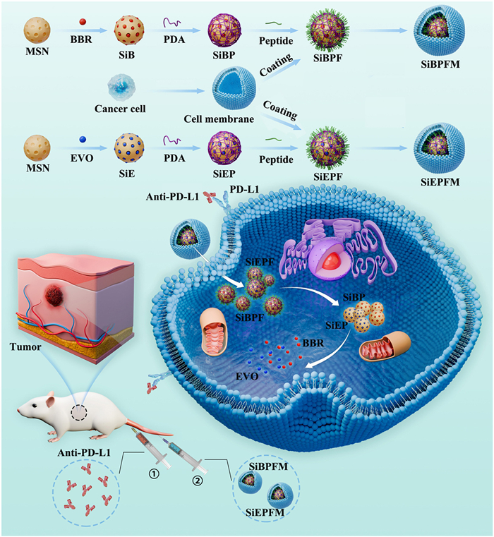
In this study, we engineered a cancer cell-mimetic, peptide-modified drug delivery system to significantly enhance the retention time and aggregation concentration of nano-herb medicine in tumors. As shown in Fig. 1, mesoporous silica nanoparticles (MSNs) with high drug-loading capacity and biocompatibility were utilized as carriers for BBR (SiB) and EVO (SiE), respectively. Polydopamine (PDA) was employed to encapsulate SiB (SiBP) and SiE (SiEP) through polymerization of dopamine (DA), respectively. A small peptide, FFVLK, capable of self-assembling into β-sheet structures in aqueous solutions [13], was conjugated to SiBP (SiBPF) and SiEP (SiEPF) via esterification, respectively. To facilitate rapid recognition and internalization by CRC cells, the cell membrane (CM) of CRC cells (HT-29 cells) was used to further encapsulate SiBPF (SiBPFM) and SiEPF (SiEPFM). Upon uptake by HT-29 cells, the CM is disrupted during transmembrane transport, exposing FFVLK. Free FFVLK could self-assemble into β-sheet structures through hydrogen bonding, π-π stacking, and hydrophobic interactions [21,22], inducing the self-aggregation of SiBPF and SiEPF into micron-sized clusters. This process could significantly improve the specific enrichment and retention of nanomedicines in HT-29 cells. The moderately acidic microenvironment in HT-29 cells could disrupt the PDA chains, resulting in locally sustained release of BBR and EVO from MSN aggregates. The combination of immunotherapy with an immune checkpoint inhibitor, anti-programmed death-ligand 1 (anti-PD-L1) and the peptide-modified drug delivery systems could achieve effective therapy for CRC.
Figure 1
Figure 1.
Schematic representation of the synthesis and functionalization of SiBPFM and SiEPFM, delineating the intracellular self-assembly mechanism of SiBPF and SiEPF, and the subsequent release of BBR and EVO from MSNs aggregates within the context of CRC treatment.
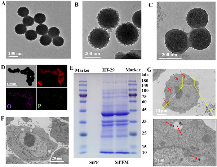
Monodispersed MSNs exhibited a porous architecture (Fig. 2A) with an average diameter of 140 nm (Fig. S1 in Supporting information). After PDA coating, the mean diameter escalated from 140 nm for MSNs to 160 nm for SiP (Fig. 2B and Fig. S1). Proton nuclear magnetic resonance (1H NMR) spectra (Figs. S2 and S3 in Supporting information) and the decreased zeta potential (Fig. S4 in Supporting information) confirmed the successful modification of PDA on SiP. The synthesis of FFVLK modified SiP (SiPF) was achieved through an esterification reaction between Fmoc-FFVLK-Fmoc and PDA (Fig. S5 in Supporting information). The coating of cancer cell membranes on NPs has been shown to substantially augment NP stability under physiological conditions and to enhance tumor accumulation through homing effects [23]. Consequently, the CM of HT-29 cells was coated onto the surface of SiPF utilizing an ultrasonication technique to yield cell-mimetic SiPF (SiPFM). The mean diameter expanded from 165 nm for SiPF to 190 nm for SiPFM (Fig. S1), indicating the CM layer thickness estimated to be approximately 25 nm (Fig. 2C). Elemental mapping of Si, O, P in SiPFM further demonstrated the homogeneous CM coating on SiPF (Fig. 2D). Protein banding patterns of SiPFM, acquired via Coomassie brilliant blue (CBS) staining, were congruent with those of CM (Fig. 2E), further validating the successful CM coating on SiPF.
Figure 2
Figure 2.
TEM images depicting the morphological progression of (A) MSN, (B) SiP, (C) SiPFM and (D) corresponding elemental mapping images of oxygen (O), silicon (Si), and phosphorus (P) for SiPFM. (E) CBS staining of protein bands for a molecular weight marker, CM, SiPF and SiPFM. Bio-TEM images of HT-29 cells treated with (F) SiP and (G) SiPFM for 12 h.
FFVLK exhibits solubility in organic solvents but was prone to self-assembly into β-sheet structures upon exposure to aqueous solution [29,30]. This self-assembly propensity could trigger the self-aggregation of MSNs. As shown in Fig. S6 (Supporting information), SiPF exhibited a clear and transparent dispersion in methanol (Fig. S6A), while a flocculent precipitate indicative of SiPF aggregation was observed when transferred from methanol solvent to phosphate buffered saline (PBS) solution (Fig. S6B). Under the protection of CM coating, the SiPFM maintained a clear and transparent dispersion in PBS solution (Fig. S6C). High-power ultrasonication was used to disrupt the CM, resulted in the appearance of a flocculent precipitate due to peptide self-assembly (Fig. S6D).
The intracellular self-assembly and subsequent aggregation of SiPFM within HT-29 cells were further investigated using biological transmission electron microscopy (bio-TEM). In contrast to PBS-treated HT-29 cells (Fig. S7 in Supporting information), a negligible amount of MSNs was observed within HT-29 cells treated with SiP (Fig. 2F). In stark contrast, distinct MSNs micrometer aggregates were evidently observed in HT-29 cells treated with SiPFM (Fig. 2G), indicating the intracellular aggregation of SiPFM induced by the self-assembly of peptide.
The successful encapsulation of BBR (Fig. S2 in Supporting information) and EVO (Fig. S3 in Supporting information) was ascertained through the 1H NMR spectra. Leveraging the porous architecture and substantial specific surface area of MSN, the loading efficiency for BBR and EVO within SiPB and SiPE NPs reached 34.9% and 31.9%, respectively. The loading efficiencies for BBR and EVO in the SiPBFM and SiPEFM were 21.8% and 21.1%.
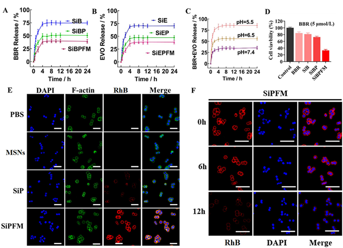
Under neutral environments (pH 7.4), approximately 75% of BBR (Fig. 3A) and EVO (Fig. 3B) were released from SiBE and SiBP within 24 h, respectively. The release of BBR (Fig. 3A) and EVO (Fig. 3B) from SiP was reduced to 51%, attributed to the reduced nonspecific release facilitated by the PDA shell. Upon CM coating, the release of BBE and EVO from SiBPFM and SiEPFM was further diminished to 31% within 24 h. As shown in Fig. 3C, the combined formulation of SiBPFM and SiEPFM demonstrated pH-responsive drug release. In contrast to the minima drug release at pH 7.4, ~56% of the drugs were released from the mixture of SiBPFM and SiEPFM at pH 6.5, and 85% of the drugs were released at pH 5.5, underscoring the acid-responsive release profile of BBR and EVO from the mixture of SiBPFM and SiEPFM.
Figure 3
Figure 3.
(A) Release profiles of BBR from SiB, SiBP and SiBPFM in PBS solutions at pH 7.4. (B) Release profiles of EVO from SiE, SiEP and SiEPFM in PBS solutions at pH 7.4. (C) Cumulative release profiles of BBE and EVO from the mixture of SiBPFM and SiEPFM in PBS solutions across a range of pH values. (D) Viability assessment of HT-29 cells following treatment with BBR in various NP formulations. (E) Confocal laser scanning microscopy (CLSM) images of HT-29 cells after 12 h cellular internalization of different MSN formulations. (F) CLSM images depicting the internalization of SiPFM by HT-29 cells after 6 h, followed by incubation with fresh medium for 0, 6 and 12 h. Scale bar: 40 µm. DAPI: 4′, 6-Diamidino-2-phenylindole. Data are presented as mean ± standard deviation (SD) (n = 3).
SiPFM exhibited minimal cytotoxicity against 293T cells (Fig. S8 in Supporting information) even at high concentrations, indicating the excellent biocompatibility of SiPFM. Compared with SiBPFM (Fig. 3D) and SiEPFM (Fig. S9A in Supporting information), the mixture of SiBPFM and SiEPFM (Fig. S9B in Supporting information) displayed enhanced cytotoxicity against HT-29 cells, attributable to the synergistic effect of BBR and EVO. Flow cytometry analysis of HT-29 cells incubated with SiPFM revealed rapid cellular uptake (Figs. S10A and B in Supporting information). Subsequently, rhodamine B (RhB), a fluorophore with high fluorescence intensity, was employed as a surrogate for the drugs to further assess the cellular uptake of nanomaterials by HT-29 cells. As shown in Fig. 3E, HT-29 cells treated with SiP exhibited stronger fluorescence compared to those MSNs- treated HT-29 cells, due to the reduced leakage of RhB from MSNs facilitated by the PDA coating. HT-29 cells treated with SiPFM displayed the strongest fluorescence of RhB among all the groups, due to the enhanced internalization by CM coating and reduced efflux by the aggregation of MSNs induced by self-assembly of peptide.
The impact of MSNs self-aggregation on intracellular retention was also investigated in HT-29 cells. As shown in Fig. S11 (Supporting information), the red fluorescence of RhB in HT-29 cells treated with MSN (Fig. S11A) and SiP (Fig. S11B) was significantly diminished, with nearly no fluorescence observed after 12 h of co-incubation. In contrast, HT-29 cells treated with SiPFM (Fig. 3F) still maintained high RhB fluorescence intensity even after incubation for 12 h of incubation, suggesting that MSNs self-aggregation could significantly enhance intracellular retention. Collectively, these findings confirmed that cellular-mimetic, peptide-modified MSNs promote recognition and uptake by HT-29 cells and subsequently self-assemble into micron-sized MSNs aggregates, thereby prolonging the intracellular retention time of MSNs.
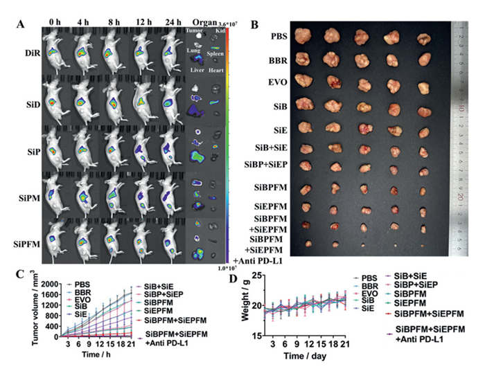
The bio-distribution of various formulations loaded with 1 equiv. of DiR was investigated in an HT-29 tumor-bearing mice model. In the free DiR group, a substantial accumulation of DiR was observed in the spleen and liver, but no fluorescence was detected in tumors (Fig. 4A). Mice treated with free SiP showed brighter DiR fluorescence in tumor sites than that of the MSNs group, due to the reduced efflux by PDA coating. Mice treated with SiPM exhibited higher DiR fluorescence in tumors than those treated with SiP, suggesting that the CM could enhance encapsulation, permeability and targeting. Among all groups, the highest fluorescence intensity of DiR was observed in tumors of mice treated with SiPFM (Fig. S12 in Supporting information), confirming the superior targeting ability of this formulation, which is attributed to enhanced encapsulation, permeability and targeting mediated by CM, as well as the enhanced permeability and retention induced by the self-assembly of MSNs.
Figure 4
Figure 4.
(A) In vivo fluorescence imaging of HT-29 tumor-bearing mice with intravenous injection with various MSNs loaded with DiR and ex vivo imaging of their principal organs (heart, liver, spleen, lung, kidney and tumor). (B) Digital photographs of harvested tumors from all groups (n = 5). Evolution of (C) tumor volume and (D) body weight of HT-29 tumor-bearing mice throughout treatment period. Data are presented as mean ± SD.
The in vivo antitumor efficacy of various formulations was assessed in mice model bearing HT-29 tumors. All animal procedures were performed in accordance with the Guidelines for Care and Use of Laboratory Animals of Zunyi Medical University and approved by the Animal Ethics Committee of Zunyi Medical University (No. ZHSC-2-[2024]001). The tumor-bearing mice were randomly allocated into 11 groups (n = 5 per group). The various formulations were administered intravenously via the tail vein on days 1, 3, 6, and 9. Analysis of the final tumor images (Fig. 4B), the tumor volume progression curves (Fig. 4C) and the weight of tumors (Fig. S13 in Supporting information) revealed that the PBS-treated group experienced the most rapid tumor growth. The free BBR and EVO group also exhibited minimal antitumor activity. Compared to the free BBR or EVO groups, the SiB or SiE group demonstrated modest antitumor effects, likely due to the enhanced permeability afforded by the MSNs carriers. The group treated with a combination of SiBP and SiEP (SiBP+SiEP) exhibited more pronounced anti-proliferative effects than SiBP group and SiEP group, attributable to the synergistic antitumor effects of the combined BBR and EVO. Benefiting from the synergistic antitumor effects of BBR and EVO, the encapsulation, permeability and targeting capabilities conferred by the CM, as well as the enhanced permeability and retention effects induced by the self-assembly of peptide, the group treated with a combination of SiBPFM and SiEPFM (SiBPFM+SiEPFM) displayed even higher anti-proliferative effects. Among all groups, the group treated with SiBPFM+SiEPFM in combination with anti-PD-L1 (SiBPFM+SiEPFM+anti-PD-L1) exhibited the most significant anti-proliferative effects, suggesting a potentiated antitumor response due to the immunotherapeutic strategy. In addition, the moderate change in body weight of the mice in the SiBPFM+SiEPFM-treated group indicates that SiBPFM+SiEPFM was much less toxic (Fig. 4D).
Dendritic cells (DCs) process antigenic materials and present them to T cells, thereby activating the immune response. Consequently [24], the level of DCs cells within the tumor was assessed [25–27]. Immunofluorescent stainings of CD3 and CD8 were conducted on tumor sections (Fig. S14 in Supporting information), an obvious increase in intratumor infiltration was observed in mice treated with SiBPFM+SiEPFM+anti-PD-L1, confirming that the anti-PD-L1 could enhance immune cell infiltration into tumor tissues. Moreover, the flow cytometry analysis (Fig. S15 in Supporting information) and cytokine (interferon-gamma (IFN-γ)) level analysis (Fig. S16 in Supporting information) further confirmed the combination of BBR/EVO with anti-PD-L1 could synergistically induce the immune infiltration and trigger the effective maturation of DCs.
Hematoxylin and eosin (H&E) staining of the main organs from all treated mice exhibited negligible organ toxicity for all formulations (Fig. S17 in Supporting information). Hematological analysis revealed that levels of albumin (ALB), alkaline phosphatase (ALP), alanine aminotransferase (ALT), aspartate aminotransferase (AST), blood urea nitrogen (BUN), creatinine (CRE), glucose (GLU) and total cholesterol (T-CHO) in mice treated with SiBEPFM+SiEPFM (Fig. S18 in Supporting information) were all within the normal range, suggesting that the SiBEPFM+SiEPFM were generally safe and could not induce damage to the heart, liver and kidneys.
In summary, a cancer cell-mimetic, peptide-modified MSNs loaded with nano-herb medicine (BBR and EVO) was developed to achieve specific recognition and intracellular internalization by CRC cells. This approach resulted in slowed clearance and prolonged retention of BBR and EVO within the cells, facilitated by the intracellular self-assembly of MSNs induced by the self-assembly of peptide. The sustained release of BBR and EVO from the MSNs aggregates, coupled with an immune checkpoint inhibitor, significantly inhibited tumor growth, achieving effective therapy of CRC. This study not only provides a practical methodology for the development of intelligent drug delivery systems but also offers novel insights into achieving effective treatment strategies for CRC.
CRediT authorship contribution statement
Ruipeng Shi: Writing – original draft, Methodology, Formal analysis, Data curation, Conceptualization. Zhangfeng Zhong: Formal analysis, Data curation, Conceptualization. LuXi Wang: Methodology, Investigation. Yifei Mao: Software, Resources. Ying Tang: Validation, Project administration. Yan Wang: Formal analysis, Data curation. Jinchao Wei: Visualization, Validation, Supervision. Beibei Xie: Writing – review & editing, Writing – original draft, Supervision, Methodology, Conceptualization. Peng Li: Writing – review & editing, Writing – original draft, Supervision.
Declaration of competing interest
The authors declare that they have no known competing financial interests or personal relationships that could have appeared to influence the work reported in this paper.
Acknowledgments
This study was supported by the National Natural Science Foundation of China (No. 32401119), Macao Science and Technology Development Fund (Nos. 0056/2023/ITP2, 0047/2025/ITP1, 0008/2023/RIC, 0123/2022/A, 001/2023/ALC, and 0006/2020/AKP), Elite Medical Professionals Project of China-Japan Friendship Hospital (No. ZRJY2023-QM24), the Research Fund of University of Macau (Nos. MYRG-GRG2023–00198-ICMS and SRG2022–00052-ICMS), and the Research Fund of Zhongzhi Pharmaceutical Group (No. CP-031–2023). We also sincerely thank Prof. Guizhen Cui and Prof. Kun Feng (Zhuhai Campus of Zunyi Medical University) for his kind help in all animal procedures.
Supplementary materials
Supplementary material associated with this article can be found, in the online version, at doi:10.1016/j.cclet.2025.112292.
[1]
I. Marmol, C. Sanchez-de-Diego, A. Pradilla Dieste, et al., Int. J. Mol. Sci. 18 (2017) 197–225. doi: 10.3390/ijms18010197
B. Xie, H. Zhao, Y.F. Ding, et al., Adv. Sci. 10 (2023) 2304407. doi: 10.1002/advs.202304407
[24]
B. Chen, L. Wang, R. Xie, et al., Adv. Healthc. Mater. 14 (2025) 2500954. doi: 10.1002/adhm.202500954
[25]
T. Nie, Y. Fang, R. Zhang, et al., Bioact. Mater. 47 (2025) 51–63.
[26]
T. Nie, H. Liu, Z. Fang, et al., ACS Nano 17 (2023) 10925–10937. doi: 10.1021/acsnano.3c02803
[27]
D. Wang, T. Nie, Y. Fang, et al., Adv. Healthc. Mater. 14 (2025) 2403979. doi: 10.1002/adhm.202403979
Figure 1
Schematic representation of the synthesis and functionalization of SiBPFM and SiEPFM, delineating the intracellular self-assembly mechanism of SiBPF and SiEPF, and the subsequent release of BBR and EVO from MSNs aggregates within the context of CRC treatment.
Figure 2
TEM images depicting the morphological progression of (A) MSN, (B) SiP, (C) SiPFM and (D) corresponding elemental mapping images of oxygen (O), silicon (Si), and phosphorus (P) for SiPFM. (E) CBS staining of protein bands for a molecular weight marker, CM, SiPF and SiPFM. Bio-TEM images of HT-29 cells treated with (F) SiP and (G) SiPFM for 12 h.
Figure 3
(A) Release profiles of BBR from SiB, SiBP and SiBPFM in PBS solutions at pH 7.4. (B) Release profiles of EVO from SiE, SiEP and SiEPFM in PBS solutions at pH 7.4. (C) Cumulative release profiles of BBE and EVO from the mixture of SiBPFM and SiEPFM in PBS solutions across a range of pH values. (D) Viability assessment of HT-29 cells following treatment with BBR in various NP formulations. (E) Confocal laser scanning microscopy (CLSM) images of HT-29 cells after 12 h cellular internalization of different MSN formulations. (F) CLSM images depicting the internalization of SiPFM by HT-29 cells after 6 h, followed by incubation with fresh medium for 0, 6 and 12 h. Scale bar: 40 µm. DAPI: 4′, 6-Diamidino-2-phenylindole. Data are presented as mean ± standard deviation (SD) (n = 3).
Figure 4
(A) In vivo fluorescence imaging of HT-29 tumor-bearing mice with intravenous injection with various MSNs loaded with DiR and ex vivo imaging of their principal organs (heart, liver, spleen, lung, kidney and tumor). (B) Digital photographs of harvested tumors from all groups (n = 5). Evolution of (C) tumor volume and (D) body weight of HT-29 tumor-bearing mice throughout treatment period. Data are presented as mean ± SD.


 DownLoad:

 下载:


 下载:

