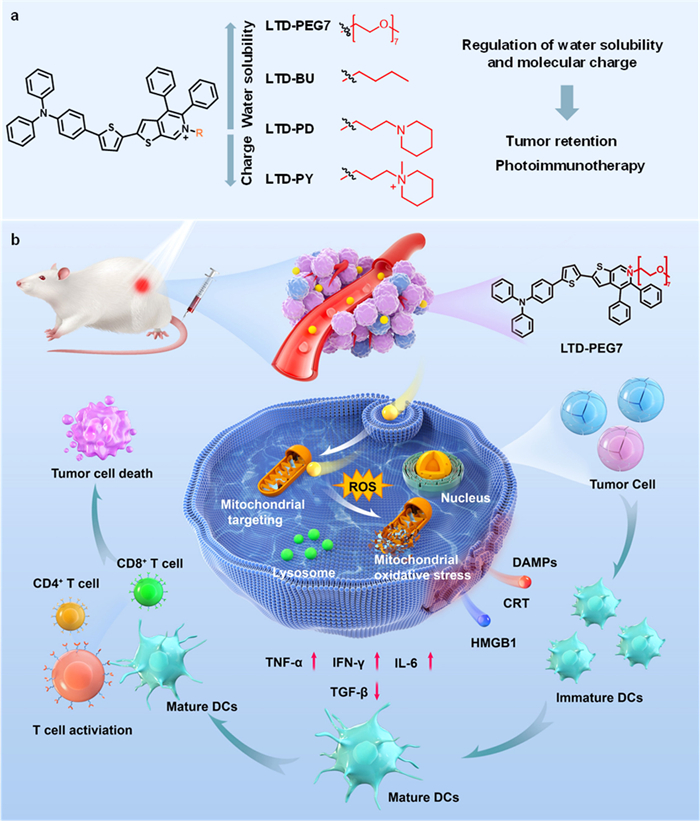
Scheme 1.
The structural design of the PSs and the schematic diagram of the anti-tumor immune response induced by LTD-PEG7.

Boosting tumor retention via water solubility and molecular charge engineering for enhanced photoimmunotherapy
Xiu Pan , Kai-Hui Yan , Meng-Die Wang , Ling-Xiao Zhou , Kun-Mei Liu , Jin-Yu An , Yan Jiang , Shun Feng , Ming-Yu Wu


The global cancer burden continues to rise dramatically, with approximately 20 million new cases and 9.7 million deaths reported in 2022 [1]. Although conventional therapies remain clinical mainstays, their limitations, including drug resistance, metastatic recurrence, and severe side effects, have driven increasing interest in alternative approaches [2,3]. Immunotherapy has emerged as a transformative strategy for anti-tumor therapy, offering a new avenue to combat cancer recurrence and metastasis [4,5]. Among the various immunotherapy strategies, photo-activated immunogenic cell death (ICD) stands out for its unique advantages, including minimal invasiveness, spatiotemporal precision, and low risk of resistance [6]. This approach utilizes light and a photosensitizer (PS) to generate reactive oxygen species (ROS), inducing inflammasome activation and releasing damage-associated molecular patterns (DAMPs), such as surface-exposed calreticulin (CRT) and extracellular high-mobility group box 1 (HMGB1) [7]. These signals facilitate antigen uptake by dendritic cells (DCs), promoting cytotoxic T lymphocyte activation and establishing a durable anti-tumor immune response [8,9].
The immunogenicity of photo-activated ICD depends critically on the PS, influenced by multiple factors including targeting efficiency, ROS generation efficiency, and ROS type. To enhance tumor targeting, numerous strategies have been explored, including antibody conjugation, targeted peptides, and in situ activation approaches [10-14]. To address tumor hypoxia and associated immunosuppression, efforts have focused on improving type I ROS generation through enhanced donor-acceptor (D-A) systems, supramolecular self-assembly, or heavy-atom effects [15-22]. Additionally, organelle targeting PSs (e.g., mitochondria, lysosome, or endoplasmic reticulum) have been developed to enhance ICD efficacy [23-26]. However, these strategies primarily address cellular-level challenges. In vivo, PSs must overcome multiple biological barriers: (1) Rapid clearance by the mononuclear phagocyte system (MPS), limiting tumor accumulation; (2) dense extracellular matrix impedes tissue penetration; and (3) poor cellular internalization efficiency. These barriers collectively compromise PS retention in tumors [27-31]. Most PSs exhibit strong hydrophobicity, which not only impairs cellular uptake and tissue penetration but also shortens blood circulation and accelerates clearance by the metabolic system, resulting in bad tumor retention and diminished immunogenicity [32,33].
Achieving an optimal hydrophile–lipophile balance is critical for tumor retention. Common strategies focus on improving water solubility of organic PS. For example, a PS with three hydrophilic oligomeric polyethylene glycol (PEG) chains and double cationic moiety was developed with 6-h tumor retention via prolonged circulation time and electrostatic-driven tumor targeting [34]. However, such retention remains inadequate for clinical translation. Therefore, improving the retention of PS within tumors remains a significant challenge.
Herein, we rationally designed and synthesized a series of amphiphilic cationic thiophenopyridine-based PSs featuring D-A architectures with triphenylamine as an electron donor, thiophene as a π-bridge, and thiophenopyridine as a dual π-extended/electron acceptor unit. The amphiphilic cationic design enhances cellular uptake and targeting. Water solubility was fine-tuned by introducing piperidine, linear butane, or oligomeric ethylene glycol side chains. To investigate the role of charge on cellular uptake and tumor retention, we incorporated cationic quaternary ammonium group. Systematic study revealed that increasing the water solubility promoted cellular internalization and extended tumor retention while shifting organelle targeting from the plasma membrane to intracellular compartments (e.g., mitochondria and lysosomes). In contrast, although increasing the positive charge enhanced water solubility but promoted serum protein binding, reducing retention time. Reasonably optimizing the water solubility or moderate charge reduction maximized cellular uptake and tumor retention. The lead compound, LTD-PEG7, achieved exceptional long-term tumor retention and potently inhibited tumor growth via photo-induced ICD. Mechanistically, DAMPs (e.g., CRT, HMGB1) promoted DC maturation, driving T-cell activation and differentiation into CD4+ and CD8+ T cells. At the same time, the pro-inflammatory cytokine secretion amplified anti-tumor immunity (Scheme 1).
In previous studies, Wu et al. systematically constructed isoquinolinium-based PS for high fidelity imaging and long-term tracking of mitochondrial, and photodynamic anticancer with the advantages of simple synthesis, high yields, reactant flexibility, and ease of modification with multifunctional application [35-37]. Building on these works, they further advanced the research by employing a thiophenopyridine-based core, to construct PSs featuring a D-A system, which enhanced the excitation/emission wavelengths and ROS generation efficiency. Notably, the PS named LIQ-TPA-DTZ was successfully applied for both mitochondrial autophagy imaging and photodynamic anticancer therapy [38]. However, it still suffered from poor cellular uptake and tumor retention due to its high lipophilicity. To address this issue, we introduced piperidine, straight-chain alkanes, PEG side chains, and cationic quaternary ammonium moieties at the pyridine group’s tail to modulate the water solubility and molecular charge of LIQ-TPA-DTZ. These modifications were designed to regulate protein adsorption, thereby avoiding recognition and clearance by the MPS. Additionally, the optimized hydrophilic-lipophilic balance and charge distribution promoted intracellular internalization and tissue penetration, leading to prolonged tumor retention. Therefore, we rational designed and synthesized LTD-PEG7, LTD-BU, LTD-PD and LTD-PY via a one-pot multicomponent reaction (Scheme S1 in Supporting information), which were characterized by 1H nuclear magnetic resonance (NMR), 13C NMR, and high-resolution mass spectrometry (HRMS) (Figs. S1–S12 in Supporting information).
The photophysical properties of the PSs were systematically investigated, including their n-octanol-water partition coefficient (LogP), protein adsorption rates and absorption/emission wavelengths (Table S1 in Supporting information). To assess the influence of side chain modification on water solubility, LogP values were first determined. As shown in Fig. S13 (Supporting information), the LogP values were 0.47 (LTD-PEG7), 0.72 (LTD-BU), 0.86 (LTD-PD), and 0.39 (LTD-PY), respectively. Subsequently, the binding affinities of these PSs to human serum albumin (HSA) were evaluated. Fig. 1A reveals that the protein adsorption rates increased sequentially: LTD-PEG7 (3.89%), LTD-BU (8.93%), LTD-PD (10.69%), and LTD-PY (17.26%). These results demonstrated that increasing the water solubility could efficiently decrease the protein binding ability. Nevertheless, even having the lowest LogP value, LTD-PY with two positive charges showed the highest protein adsorption rate, indicating that increasing the positive charges highly heighten the protein adsorption ability. This is mainly attributed to its stronger cationic properties, which can bind to proteins more effectively through electrostatic interaction. Next, we further investigated the absorption and emission spectra of the series of PSs. As shown in Fig. 1B, these PSs showed maximum absorption peaks between 470 nm and 490 nm and maximum emission peaks between 740 nm and 790 nm in dimethyl sulfoxide (DMSO).
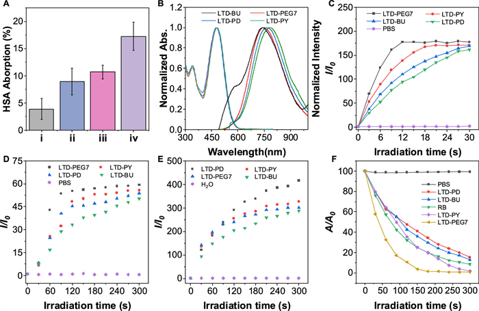
The ROS generation capacity of the PSs was assessed using 2′,7′-dichlorodihydrofluorescein diacetate (DCFH-DA). Upon irradiation under white light for 0–30 s, the fluorescence intensity of DCFH-DA at 525 nm increased by 177-, 169-, 161-, and 170-fold in the presence of LTD-PEG7, LTD-BU, LTD-PD and LTD-PY, respectively. By contrast, negligible fluorescence change was observed in the absence of any PS, confirming the efficient ROS generation capability of all the synthesized PSs. (Fig. 1C and Fig. S14 in Supporting information). To measure superoxide anion (O2•−) generation, DHR123 was used as utilized as the indicator. DHR123 was mixed with or without PS in phosphate-buffered saline (PBS) and then exposed to light illumination for 0–300 s. The fluorescence intensity at 529 nm was then recorded to evaluate the efficiency of O2•− generation. Without PS, the fluorescence signal of DHR123 hardly changed. In contrast, once LTD-PEG7, LTD-BU, LTD-PD or LTD-PY was added, the fluorescence intensity at 529 nm increased by 59-, 50-, 53- or 55-fold respectively (Fig. 1D and Fig. S15 in Supporting information). We next evaluated the generation of •OH using hydroxyphenylfluorescein (HPF) as indicator. After being exposed for 0–300 s, the fluorescence intensity change of only the HPF group was negligible. However, when HPF was separately mixed with LTD-PEG7, LTD-BU, LTD-PD and LTD-PY, the fluorescence intensity of HPF at 515 nm increased by 300-, 286-, 415- and 327-fold, respectively (Fig. 1E and Fig. S16 in Supporting information). Furthermore, when LTD-PEG7, LTD-BU, LTD-PD and LTD-PY were mixed with 9,10-anthracene diphenyl-bis(methylene) dimalonic acid (ABDA) and irradiated for 0–300 s, the absorbance at 378 nm decreased to 0.85%, 12%, 15% and 1.7% respectively (Fig. 1F and Fig. S17 in Supporting information). In contrast, the absorbance of the commercially available photosensitizer Rose Bengal (RB) decreased to only 8% under the same conditions, indicating that all PSs can efficiently generate 1O2. The generation of •OH and 1O2 by LTD-PEG7 were further verified through electron paramagnetic resonance (EPR) spectroscopy (Fig. S18 in Supporting information). Collectively, all four PSs designed in this study can efficiently produce ROS including O2•−, •OH and 1O2 and under white light irradiation through both type I and type II mechanism.
Thanks to their common rigid thienopyridine framework structure, the introduction of different R groups did not change the electron cloud distribution and conjugation characteristics, all PSs exhibited comparable ROS generation ability [39]. This feature makes this series of PSs with different water solubilities and charge properties an ideal research system for exploring tumor retention and the efficacy of photoimmunotherapy.
Cellular uptake of the PSs was evaluated by flow cytometry. After incubating cells with the PSs for varying durations, fluorescence intensity was measured (Fig. S19 in Supporting information). The results revealed a strong correlation between water solubility and cellular uptake efficiency. Specifically, the intracellular fluorescence rapidly reached to the top within 1.5 h for LTD-PY and LTD-PEG7. However, LTD-BU and LTD-PD still climbed at 4 h. Notably, despite exhibiting comparable water solubility, LTD-PEG7 displayed significantly higher fluorescence intensity than LTD-PY, indicating a higher cellular internalization rate. We hypothesize that the two positive charges on LTD-PY promote nanoparticle formation via electrostatic interactions with serum proteins, thereby shifting its uptake mechanism from direct membrane penetration to endocytosis [40].
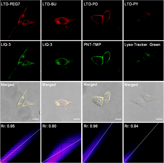
Subsequently, subcellular localization of the PSs was further investigated in human cervical cancer cells (HeLa). All PSs exhibited red fluorescence in cells. Due to the amphiphilic cationic characteristics, these PSs predominantly accumulate in cell membrane and mitochondria with negative membrane potential [41]. As depicted in Fig. 2, LTD-PD showed preferential localization at the cell membrane (Pearson’s correlation coefficient, Rr = 0.96). Notably, increasing the hydrophilicity was be beneficial to the affinity to mitochondria. The Rr of LTD-BU and LTD-PEG7 with mitochondria targeting probe LIQ-3 were 0.80 and 0.95, respectively. Interestingly, LTD-PY predominantly accumulated in lysosomes (Rr = 0.84), indicating that increasing the positive charges did not improve the electrostatic interaction with the cell membrane or mitochondria. This may attribute to the strong binding with serum protein to form nanoparticles, which were uptaked by the cells through endocytosis [42]. These results demonstrate that modulating the water solubility and charge properties of PSs can significantly alter their organelle-targeting specificity. Such precise subcellular localization is critical for overcoming the inherent limitations of ROS-based therapies, including their short half-life and restricted diffusion range, thereby improving the spatiotemporal precision and efficacy of photoimmunotherapy [43,44].
Cell counting kit-8 (CCK-8) assay was used to assess the photodynamic anti-cancer performance of the PSs. As shown in Figs. 3A and B, when human breast cancer cells (4T1) were incubated with 5.0 µmol/L of LTD-PEG7, LTD-BU, LTD-PD or LTD-PY for 30 min, >80% cell viability was maintained. However, upon white light irradiation, all PSs showed significant concentration-dependent phototoxicity, reducing cell viability to 0.48%, 5.28%, 4.79%, and 4.14% for LTD-PEG7, LTD-BU, LTD-PD and LTD-PY, respectively. Notably, LTD-PEG7 exhibited superior anti-cancer performance. Even as low as 1.25 µmol/L, >96.5% cells were killed due to its excellent cell uptake efficiency and high ROS generation efficiency. To evaluate the broad-spectrum anticancer potential of LTD-PEG7, we assessed its photodynamic activity against multiple cancer cell lines, including HeLa cells, human breast cancer cells (MCF-7), human liver cancer cells (HepG2), and mouse melanoma cells (B16). Remarkably, at a low concentration of 0.5 µmol/L with white light irradiation, LTD-PEG7 reduced cell viability to below 20% in all tested cell lines (Fig. S20 in Supporting information), confirming its potent and wide-ranging anti-tumor efficacy.
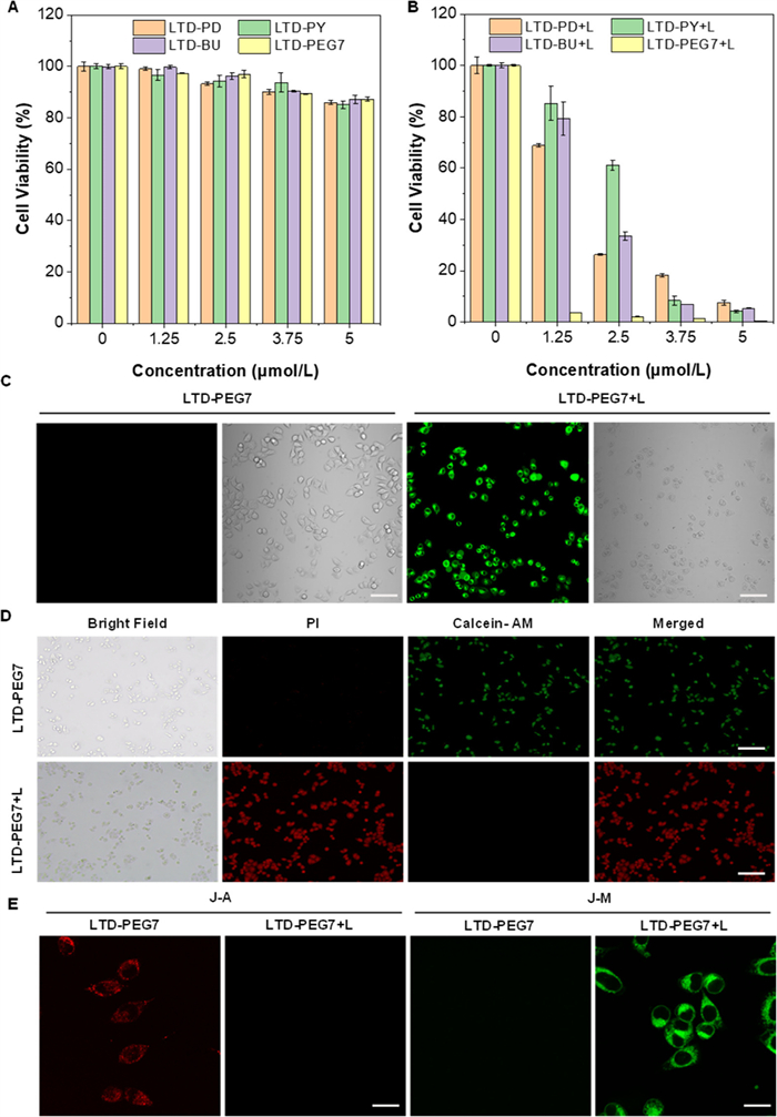
To investigate whether the cell death was mediated by the cytotoxic ROS generated under the light exposure, we used DCFH-DA as an indicator to access the intracellular ROS generation of LTD-PEG7. As shown in Fig. 3C, without light irradiation, no green fluorescence signal was observed in the cells after incubated with LTD-PEG7. However, upon white light irradiation, LTD-PEG7 turned on the green fluorescence of DCFH-DA, confirming that it produced cytotoxic ROS upon light activation. The anti-cancer activity was further investigated by the calcein-AM/PI cell cytotoxicity assay kit. After incubated with LTD-PEG7, red fluorescence was observed in the photo-treated cells, while only green fluorescence was observed in the dark environment (Fig. 3D).
Mitochondria, the central hubs of cellular energy metabolism and oxidative stress regulation, play a pivotal role in determining cell fate [45-47]. Our previous studies demonstrated that LTD-PEG7 can efficiently target mitochondria and induce intracellular ROS accumulation. Since mitochondrial oxidative stress represents a potent strategy for triggering ICD, we hypothesized that mitochondria-targeted LTD-PEG7 could significantly enhance tumor cell immunogenicity [48-50]. To test this hypothesis, we systematically evaluated the impact of LTD-PEG7 on mitochondrial function. JC-1 was used as a mitochondrial membrane potential (MMP) indicator to investigate the death mechanism of LTD-PEG7. When MMP is high, JC-1 aggregates in the mitochondrial matrix, forming polymers (J-A) that emit red fluorescence. When MMP is low, JC-1 remains monomeric (J-M) and emits green fluorescence [51]. As shown in Fig. 3E, cells treated with LTD-PEG7 in the dark maintained high MMP, as evidenced by predominant red fluorescence. However, following white light irradiation, we observed complete loss of red fluorescence concomitant with intense green fluorescence, demonstrating ROS-mediated mitochondrial membrane depolarization. These results confirm that photo-activated LTD-PEG7 generates cytotoxic ROS, leading to mitochondrial dysfunction and subsequent cell death.
To determine whether mitochondria-targeted LTD-PEG7 could induce ICD, we systematically evaluated two hallmark ICD markers: CRT surface translocation and HMGB1 release. Immunofluorescence analysis revealed significantly enhanced CRT exposure on the cell surface in the LTD-PEG7+L group compared to both the Control and LTD-PEG7 groups (Figs. 4A and B). Consistent with these findings, enzyme-linked immunosorbent assay (ELISA) demonstrated a substantial increase in extracellular HMGB1 levels following LTD-PEG7 treatment with light irradiation (Fig. 4C). These results collectively confirm that light-activated LTD-PEG7 effectively induces ICD at the cellular level.
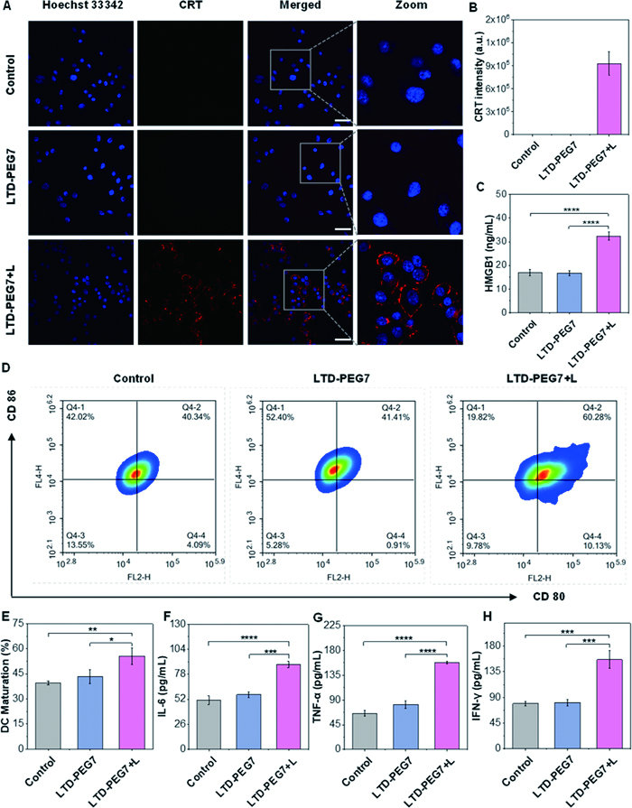
Given that DCs activation and maturation are crucial for initiating antigen-specific immune responses, we next investigated the impact of LTD-PEG7-treated tumor cells on DCs maturation [52]. Bone marrow-derived dendritic cells (BMDCs) were isolated from the femur and tibia of BALB/c female mice and differentiated with granulocyte-macrophage colony-stimulating factor (GM-CSF) and interleukin-4 (IL-4) [53]. Successful generation of immature BMDCs was confirmed by the high CD11c expression (58.3%) via flow cytometry (Fig. S21 in Supporting information). Subsequently, 4T1 cells were pre-treated with LTD-PEG7, and the supernatant was collected and co-cultured with immature BMDCs. The percentage of mature DCs (CD80+/CD86+) was measured by flow cytometry (Figs. 4D and E). The proportion of mature DCs in the LTD-PEG7+L group (55.54%) was significantly higher than that in the LTD-PEG7 (43.34%) and control groups (39.5%). Additionally, supernatants from different groups were collected and analyzed by ELISA to quantify the levels of pro-inflammatory cytokines. As shown in Figs. 4F-H, tumor necrosis factor-alpha (TNF-α), IL-6, and interferon-gamma (IFN-γ) levels were significantly upregulated in the LTD-PEG7+L group compared to LTD-PEG7 and Control groups. All these results collectively indicate that LTD-PEG7 and light irradiation can definitely and efficiently induce ICD at the cellular level.
In order to investigate the retention behavior, PSs were intravenously injected into 4T1 tumor-bearing mice, and fluorescence imaging images were recorded. All animal experiments were approved by the Animal Ethics Committee of Southwest Jiaotong University (No. SWJTU-2409-KJT(180)). As shown in Fig. 5A and Fig. S22 (Supporting information), the tumor was lit up for LTD-PEG7 after 8 h injection. With the extension to 48 h, the fluorescence intensity at the tumor reached the strongest, which could still be clearly observed at 96 h, indicating that it has excellent long-term tumor retention. In contrast, the non-specific accumulations in the liver and spleen were observed for LTD-BU and LTD-PD. Only a small number of signals can be observed in the tumors, and the retention time was all <22 h. This may due to form the protein-PS complexes, which were absorbed by the liver and subsequently cleared by MPS [54-56]. Compared with LTD-PD, LTD-PY has an increased positive charge and was more likely to undergo protein adsorption action through electrostatic interaction, which led to its retention time in tumors being <12 h.
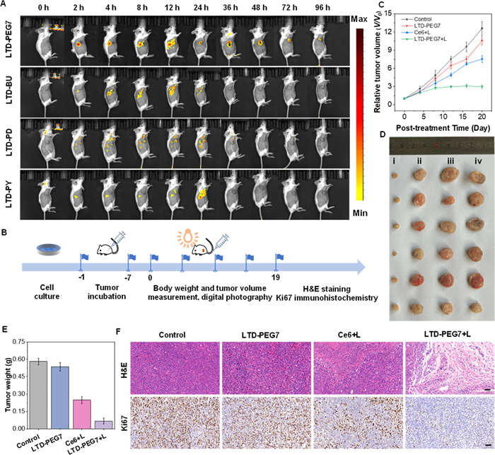
Benefit for the long-term retention ability in tumor, LTD-PEG7 was selected for further in vivo antitumor studies. Prior to therapeutic assessment, hemolysis assays confirmed its outstanding biocompatibility, demonstrating below 5% hemolysis even at 20 µmol/L (Fig. S23 in Supporting information). Subsequently, the therapeutic efficacy of LTD-PEG7 was systematically evaluated in 4T1 tumor-bearing mice. The treatment protocol is illustrated in Fig. 5B. 4T1 tumor-bearing mice were randomly assigned to 4 groups: PBS group, LTD-PEG7 group, Ce6+L group and LTD-PEG7+L group. All four drugs were administered via tail vein injection every 3 days. After 48 h of administration, the Ce6+L group and the LTD-PEG7+L group were locally exposed to white light for 30 min. Throughout the study, body weight and tumor volume were monitored as key indicators of treatment efficacy and safety. After the treatment, the mice in each group were sacrificed for further analysis. Remarkably, the average tumor volume increased by 12.60-fold for PBS group, 10.57-fold for LTD-PEG7 group, 7.57-fold for Ce6+L group, only 2.98-fold for LTD-PEG7+L group (Fig. 5C and Fig. S24 in Supporting information). Consistent with these observations, excised tumors from the LTD-PEG7+L group displayed the smallest mass (Figs. 5D and E). The experimental results show that LTD-PEG7 has excellent antitumor performance in vivo under white light irradiation.
To further evaluate the antitumor efficacy of LTD-PEG7, we performed histological analyses of tumor sections using hematoxylin and eosin (H&E) staining and Ki67 immunohistochemistry. As demonstrated in Fig. 5F, both the Ce6+L and LTD-PEG7+L groups exhibited significant tumor tissue necrosis and reduced Ki67 expression relative to the PBS and LTD-PEG7 groups. Most strikingly, the LTD-PEG7+L group demonstrated the most pronounced therapeutic effect, characterized by extensive tumor necrosis and minimal Ki67-positive cells. These results definitely confirm the superior of LTD-PEG7+L in vivo antitumor efficacy compared to the commercial PS, Ce6. To evaluate treatment safety, we monitored body weight changes of the mice during the treatment period (Fig. S25 in Supporting information), observing no significant fluctuations in any group. Furthermore, H&E staining of major organs (heart, liver, spleen, lung, and kidney) revealed no apparent pathological abnormalities or tissue damage (Fig. S26 in Supporting information). These results further confirm the excellent biocompatibility of LTD-PEG7 in vivo photoimmunotherapy.
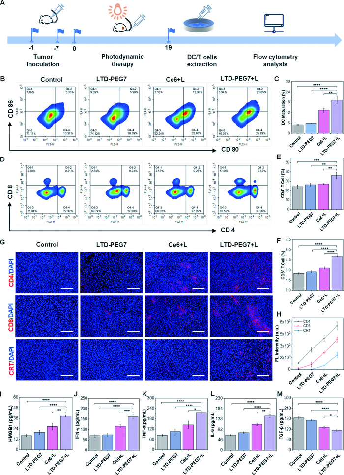
Based on the excellent anti-tumor performance of LTD-PEG7 both in vivo and in vitro, we next investigated its immune mechanism in vivo, and the experimental schematic diagram was shown in Fig. 6A. DCs and T cells were extracted from spleens of mice in each group and analyzed by flow cytometry [57]. As shown in Figs. 6B and C, the DCs maturation rate in the LTD-PEG7+L group reached 19.29%, significantly exceeding the positive control Ce6+L group (13.41%), while remaining at only 5.88% and 5.05% in the LTD-PEG7 group and the Control group, respectively. Furthermore, the proportion of CD4+ T cells was significantly elevated in the LTD-PEG7+L group (36.19%) compared to the LTD-PEG7 group (27.1%), Ce6+L group (26.37%), and control group (24.3%). Similarly, the proportion of CD8+ T cells in the LTD-PEG7+L group (5.01%) was markedly higher than that in the LTD-PEG7 (3.32%), Ce6+L (2.76%), and control (2.53%) groups (Figs. 6D–F). These results suggested that LTD-PEG7 successfully induced immune responses upon light exposure. We further evaluated the infiltration of CRT, CD4+ T and CD8+ T cells in tumor by immunofluorescence staining. As shown in Figs. 6G and H, strong red fluorescence signals for CRT, CD4, and CD8 were observed in the LTD-PEG7+L group. In contrast, the Ce6+L group exhibited much weaker red fluorescence signals compared to the LTD-PEG7+L group, which however were barely detectable in both the LTD-PEG7 and Control groups. In addition, ELISA kits were used to detect the expressions of cytokines, such as HMGB1, IFN-γ, TNF-α, TGF-β, and IL-6 in serum to evaluate the immune responses. As shown in Figs. 6I–M, the levels of pro-inflammatory cytokines and HMGB1 in the serum of mice in the LTD-PEG7+L group were significantly higher than those in the PBS, Ce6+L and LTD-PEG7 groups. TGF-β was significantly lower than that of the other three groups. To sum up, LTD-PEG7 can induce oxidative stress in cancer cells and enhance tumor immunogenicity, thereby promoting DCs activation and triggering robust T-cell-dependent immune responses. This not only endows it with excellent antitumor efficacy, but also effectively stimulates adaptive antitumor immunity.
In summary, we developed a series of amphipathic cationic PSs via a one-pot multicomponent reaction, tuning water solubility and positive charge to boost tumor retention for enhancing photoimmunotherapy. These PSs demonstrated excellent type I and type II ROS generation capabilities. Our studies revealed that enhancing water solubility while reducing positive charge significantly improved cellular uptake and extended tumor retention time. Notably, higher water solubility shifted organelle localization from the plasma membrane to mitochondria, whereas excessive positive charge promoted HSA binding, redirecting lysosomal accumulation. The optimized PS, LTD-PEG7, achieved ultralong tumor retention (>96 h) and efficiently generated •OH under illumination, inducing mitochondrial oxidative stress to trigger ICD. This process promoted DAMPs release (CRT exposure, HMGB1 secretion) and elicited a robust anti-tumor immune response. In 4T1 tumor-bearing mice, LTD-PEG7-mediated photoimmunotherapy achieved potent tumor suppression. Collectively, our findings establish design principles for long-term tumor retention PS and high-efficiency photoimmunotherapy.
Xiu Pan: Writing – original draft, Methodology, Investigation, Data curation. Kai-Hui Yan: Methodology, Investigation. Meng-Die Wang: Investigation. Ling-Xiao Zhou: Investigation. Kun-Mei Liu: Methodology. Jin-Yu An: Investigation. Yan Jiang: Supervision, Conceptualization. Shun Feng: Supervision, Funding acquisition. Ming-Yu Wu: Writing – review & editing, Supervision, Funding acquisition.
The authors declare that they have no known competing financial interests or personal relationships that could have appeared to influence the work reported in this paper.
This work was financially supported by the National Natural Science Foundation of China (Nos. 22577085 and 22174117), and the Fundamental Research Funds for the Central Universities (No. YJ202419).
Supplementary material associated with this article can be found, in the online version, at doi:
B. Han, R. Zheng, H. Zeng, et al., J. Natl. Cancer Cent. 4 (2024) 47–53.
S. Tohme, R.L. Simmons, A. Tsung, Cancer Res. 77 (2017) 1548–1552.
T. Kitano, T. Sasaki, Y. Gon, et al., Thromb. Haemost. 120 (2020) 714–723. doi: 10.1055/s-0040-1708484
P. Sharma, J.P. Allison, Science 1979 348 (2015) 56–61. doi: 10.1126/science.aaa8172
Y. Guo, S.Z. Wang, X. Zhang, et al., Nat. Commun. 13 (2022) 6534. doi: 10.1038/s41467-022-33831-7
J. Liu, X. Li, R. Xia, et al., Chin. Chem. Lett. 35 (2024) 109619. doi: 10.1016/j.cclet.2024.109619
R. Zheng, Z. Chen, Q. Li, N. Yang, et al., Chin. Chem. Lett. 36 (2025) 110865. doi: 10.1016/j.cclet.2025.110865
D.V. Krysko, A.D. Garg, A. Kaczmarek, et al., Nat. Rev. Cancer 12 (2012) 860–875. doi: 10.1038/nrc3380
L. Galluzzi, A. Buqué, O. Kepp, et al., Nat. Rev. Immunol. 17 (2017) 97–111. doi: 10.1038/nri.2016.107
D. Ni, C.A. Ferreira, T.E. Barnhart, et al., J. Am. Chem. Soc. 140 (2018) 14971–14979. doi: 10.1021/jacs.8b09374
X. Wu, S. Yao, Q. Huang, et al., ACS Appl. Mater. Interfaces 16 (2024) 41939–41948. doi: 10.1021/acsami.4c09588
X. Duan, P. Wang, L. He, et al., Adv. Mater. 36 (2024) 2311548. doi: 10.1002/adma.202311548
X. Xia, R. Wang, Y. Hu, et al., Adv. Funct. Mater. 35 (2025) 2422823. doi: 10.1002/adfm.202422823
J.G Jia, Y.Q. Jiang, X.Y. Li, Pharm. Sci. Adv. 2 (2024) 100048. doi: 10.1016/j.pscia.2024.100048
X. Yang, X. Wang, X. Zhang, et al., Adv. Mater. 36 (2024) 2402182. doi: 10.1002/adma.202402182
J. Yang, B. Ren, X. Yin, et al., Adv. Mater. 36 (2024) 2402720. doi: 10.1002/adma.202402720
D. Liu, A.M. El-Zohry, M. Taddei, et al., Angew. Chem. Int. Ed. 59 (2020) 11591–11599. doi: 10.1002/anie.202003560
H. Gu, W. Sun, J. Du, et al., Smart Mol. 2 (2024) e20230014. doi: 10.1002/smo.20230014
R. Wang, M. He, Z. Zhang, et al., Smart Mol. 2 (2024) e20240010. doi: 10.1002/smo.20240010
X. Chen, L. Shi, X.Y. Ran, et al., Adv. Funct. Mater. 34 (2024) 2400728. doi: 10.1002/adfm.202400728
M.Y. Wu, Z.J. Wu, J.L. Wang, et al., ACS Mater. Lett. 6 (2024) 4533–4544. doi: 10.1021/acsmaterialslett.4c01214
L. Wei, A. Dao, G. Yuan, et al., Acc. Chem. Res. 58 (2025) 2640–2651. doi: 10.1021/acs.accounts.5c00455
Y. Xu, D. An, T. Zhang, et al., Adv. Mater. 37 (2025) 2418894. doi: 10.1002/adma.202418894
K.H. Li, H.Y. Zhao, D.D. Wang, et al., Chin. Chem. Lett. 35 (2024) 108882. doi: 10.1016/j.cclet.2023.108882
Y. Li, W. Han, D. Gong, et al., Chem. Sci. 14 (2023) 5106–5115. doi: 10.1039/D3SC00455D
Y. Chen, T. Xiong, M. Gu, et al., J. Am. Chem. Soc. 147 (2025) 17915–17925. doi: 10.1021/jacs.5c02764
Y.N. Zhang, W. Poon, A.J. Tavares, et al., J. Control Release 240 (2016) 332–348. doi: 10.1016/j.jconrel.2016.01.020
P. Decuzzi, R. Pasqualini, W. Arap, et al., Pharm. Res. 26 (2009) 235–243. doi: 10.1007/s11095-008-9697-x
H. Maeda, H. Nakamura, J. Fang, Adv. Drug Deliv. Rev. 65 (2013) 71–79. doi: 10.1016/j.addr.2012.10.002
Y. Matsumura, H. Maeda, Cancer Res. 46 (1986) 6387–6392.
E. Blanco, H. Shen, M. Ferrari, Nat. Biotechnol. 33 (2015) 941–951. doi: 10.1038/nbt.3330
Y. Xu, J. Zhang, Z. Wang, et al., Biomaterials 314 (2025) 122847. doi: 10.1016/j.biomaterials.2024.122847
Z. Zhao, S. Shi, Y. Huang, et al., ACS Appl. Mater. Interfaces 6 (2014) 8878–8885. doi: 10.1021/am501608c
D. Zhang, K.X. Teng, L. Zhao, et al., Adv. Mater. 35 (2023) 2209789. doi: 10.1002/adma.202209789
L. Liu, Q. Zou, J.K. Leung, et al., Chem. Commun. 55 (2019) 14681–14684. doi: 10.1039/c9cc07775h
M.Y. Wu, J.K. Leung, L. Liu, et al., Angew. Chem. Int. Ed. 59 (2020) 10327–10331. doi: 10.1002/anie.201916718
M. Gu, M.Y. Wu, Y. Yuan, et al., Mater. Chem. Front. 6 (2022) 3042–3053. doi: 10.1039/d2qm00630h
M.Y. Wu, L.J. Wang, C.M. Qin, et al., Sens. Actuators B: Chem. 381 (2023) 133471. doi: 10.1016/j.snb.2023.133471
M.M.S. Lee, E.Y. Yu, D. Yan, et al., ACS Nano 17 (2023) 17004–17020. doi: 10.1021/acsnano.3c04266
S. Zhang, H. Gao, G. Bao, ACS Nano 9 (2015) 8655–8671. doi: 10.1021/acsnano.5b03184
X. Zheng, D. Wang, W. Xu, et al., Mater. Horiz. 6 (2019) 2016–2023. doi: 10.1039/c9mh00906j
J. Mosquera, I. García, L.M. Liz-Marzán, Acc. Chem. Res. 51 (2018) 2305–2313. doi: 10.1021/acs.accounts.8b00292
R. Wang, X. Li, J. Yoon, ACS Appl. Mater. Interfaces 13 (2021) 19543–19571. doi: 10.1021/acsami.1c02019
X. Dai, T. Du, K. Han, ACS Biomater. Sci. Eng. 5 (2019) 6342–6354. doi: 10.1021/acsbiomaterials.9b01251
F. Chen, Y. Xue, W. Zhang, et al., Cancer Metastasis Rev. 43 (2024) 1419–1443. doi: 10.1007/s10555-024-10211-9
L. Yang, P. Gao, Y. Huang, et al., Chin. Chem. Lett. 30 (2019) 1293–1296. doi: 10.1016/j.cclet.2019.03.032
X. Pan, Y. Zhao, J.L. Wang, et al., J. Mater. Chem. B 13 (2025) 177–183. doi: 10.1039/d4tb01785d
R. Bai, J. Cui, Cancer Lett. 564 (2023) 216223. doi: 10.1016/j.canlet.2023.216223
K. Shi, Z. Ye, H. Zhu, et al., Acta Biomater. 203 (2025) 535–549. doi: 10.1016/j.actbio.2025.07.035
C. Chen, X. Ni, S. Jia, et al., Adv. Mater. 31 (2019) 1904914. doi: 10.1002/adma.201904914
Y. Wang, W. Wang, R. Gu, et al., Adv. Sci. 10 (2023) 2300286. doi: 10.1002/advs.202300286
D.V. Krysko, K.S. Ravichandran, P. Vandenabeele, Nat. Commun. 9 (2018) 4644. doi: 10.1038/s41467-018-06807-9
X. Huang, M. Gao, H. Xing, et al., Small 18 (2022) 2202728. doi: 10.1002/smll.202202728
C.D. Walkey, J.B. Olsen, H. Guo, et al., J. Am. Chem. Soc. 134 (2012) 2139–2147. doi: 10.1021/ja2084338
J. Ren, N. Andrikopoulos, K. Velonia, et al., J. Am. Chem. Soc. 144 (2022) 9184–9205. doi: 10.1021/jacs.2c02277
R. Cai, C. Chen, Adv. Mater. 31 (2019) 1805740. doi: 10.1002/adma.201805740
J.L. Wang, X. Pan, X. Li, et al., Adv. Mater. 37 (2025) 2411468. doi: 10.1002/adma.202411468

Scheme 1 The structural design of the PSs and the schematic diagram of the anti-tumor immune response induced by LTD-PEG7.

Figure 1 Photophysical property of LTD-PEG7, LTD-BU, LTD-PD and LTD-PY. (A) Protein adsorption rates of PSs (i: LTD-PEG7, ii: LTD-BU, iii: LTD-PD, iv: LTD-PY). (B) Absorption and photoluminescence (PL) spectra of PSs in DMSO. (C) ROS generation ability of PSs. Relative changes in fluorescence intensity (I/I0) of DCFH-DA at 525 nm in the absent or present of PSs at different time points under white light illumination. (D) O2•− generation ability of PSs. Relative changes in fluorescence intensity (I/I0) of DHR123 at 529 nm in the presence or absence of PSs at different time points under white light illumination (E) •OH radical generation ability of PSs. Relative changes in fluorescence intensity (I/I0) of HPF at 515 nm in the presence or absence of PSs at different time points under white light illumination. (F) ABDA decomposition rates under white light irradiation in the presence or absence of PSs. A0 and A represent the initial absorbance and final absorbance of ABDA at 378 nm, respectively. Data are presented as mean ± standard deviation (SD) (n = 3).

Figure 2 Cellular organelle targeting of PSs. Colocalization analysis of LTD-PEG7, LTD-BU, LTD-PD and LTD-PY with mitochondrial-targeted LIQ-3, cell membrane-targeted PNT-TMP and lysosomes targeted Lyso-Tracker Green. LTD-PEG7, LTD-BU, LTD-PD and LTD-PY: λex = 488 nm, λem = 650–750 nm, LIQ-3: λex = 405 nm, λem = 500–580 nm, lysotracker Green: λex = 488 nm, λem = 500–600 nm, PNT-TMP: λex = 405 nm, λem = 440–600 nm. Scale bar: 10 µm.

Figure 3 Photodynamic anticancer activity of PSs. (A, B) Dark toxicity and phototoxicity of LTD-PEG7, LTD-BU, LTD-PD and LTD-PY (0–5 µmol/L) against 4T1 cells. (C) CLSM images of ROS generation in LTD-PEG7-treated HeLa cells stained with DCFH-DA. DCFH-DA: λex = 488 nm, λem = 500–550 nm. Scale bar: 100 µm. (D) Live/dead cell staining of HeLa cells treated with LTD-PEG7 using calcein-AM and PI (calcein-AM: λex = 488 nm, λem = 500–550 nm; PI: λex = 561 nm, λem = 600–650 nm). Scale bar: 100 µm. (E) Fluorescence images of HeLa cells co-stained with LTD-PEG7 and JC-1. J-A: λex = 560 nm, λem = 590 nm; J-M: λex = 488 nm, λem = 529 nm. Scale bar: 40 µm. Data are presented as mean ± SD (n = 3).

Figure 4 Photon-actived ICD by LTD-PEG7 in cells. (A) Immunofluorescence images of CRT in 4T1 cells with different treatment. Scale bar: 50 µm. (B) Quantification analysis of CRT fluorescence intensity in A. (C) ELISA for the quantification of HMGB1 expression. (D) Flow cytometry analysis of DCs maturation. (E) Quantification of DCs maturation in D. (F-H) Cytokine secretion profiles of (F) IL-6, (G) TNF-α, and (H) IFN-γ measured by ELISA. Data are presented as mean ± SD (n = 3). *P < 0.05, **P < 0.01, ***P < 0.001, ****P < 0.0001.

Figure 5 Photoimmunotherapy of tumor with LTD-PEG7 in vivo. (A) In vivo imaging of mice at different time points after caudal vein injection of PSs. (B) Schematic diagram of the in vivo photodynamic anticancer experiment. (C) Tumor growth curves showing relative volume changes in different treatment groups. (D) Images of excised tumors from each group. (E) Quantitative analysis of tumor weights after resection. (F) H&E staining and Ki67 immunohistochemical staining of tumor tissues from each group. Scale bar: 20 µm. i: LTD-PEG7+L, ii: Ce6+L, iii: LTD-PEG7, iv: control. Data are presented as mean ± SD (n = 6).

Figure 6 In vivo antitumor immunity with LTD-PEG7. (A) Schematic diagram of in vivo immunity experiment. (B) Flow cytometric analysis of CD80+ and CD86+ cell populations with different treatment. (C) Quantitative analysis of DCs maturation in B. (D) Flow cytometric analysis of CD4+ T and CD8+ T cells populations following different treatments. (E) Quantification analysis of CD4+ T cells in D. (F) Quantification analysis of CD8+ T cells in D. (G) Fluorescence microscope images of CRT, CD4+ T and CD8+ T cells expression in tumor tissue detected by immunofluorescence. Scale bar: 100 µm. (H) Quantification analysis of mean fluorescence intensity of CRT, CD4+ T and CD8+ T cells in tumor tissue infiltration in G. (I–M) Serum levels of HMGB1 (I), IFN-γ (J), TNF-α (K), IL-6 (L), and TGF-β (M) measured by ELISA. Data are presented as mean ± SD (n = 3). *P < 0.05, **P < 0.01, ***P < 0.001, ****P < 0.0001.

 扫一扫看文章


扫一扫关注我们
 DownLoad:
 下载:
 下载: