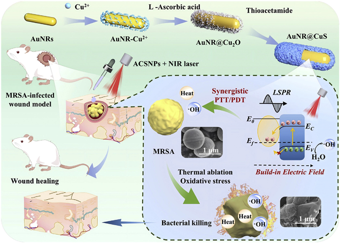
Figure 1.
Schematic illustration of the preparation and antibacterial effect of AuNR@CuS hybrid nanomaterials as PTT/PDT synergistic therapy.

Interfacial-engineered AuNR@CuS nanohybrids for photothermal and photodynamic synergistic therapy against MRSA wound infections
Chiyin Zhang , Xiwen Geng , Hang Xu , Xinyu Gu , Shaokui Cao , Zhanrong Li , Jingguo Li , Jun Shi


Infectious diseases caused by microbial pathogens have become a severe global health challenge [1,2]. The rapid emergence of antibiotic resistance, especially in bacteria, has exacerbated this crisis, rendering traditional antibiotic development strategies inadequate to counter the accelerated evolution of microbial resistance [3]. In particular, methicillin-resistant Staphylococcus aureus (MRSA) significantly reduces cell membrane permeability by mecA/PBP2a, forming a robust biofilm to provide a physical barrier, and possessing complex immune evasion mechanisms, making it one of the most common and dangerous pathogens in both hospital-acquired and community-acquired infections [4–8]. Consequently, developing novel therapeutic approaches beyond conventional antibiotics has become imperative. Phototherapy, including photothermal therapy (PTT) and photodynamic therapy (PDT), has emerged as a potent alternative for combating bacterial infections, due to its non-specific bactericidal mechanisms that are less likely to induce drug resistance [9,10]. Unlike antibiotics, which function through precise biochemical targeting of specific pathways (e.g., penicillin-binding proteins or DNA gyrase) and incur high evolutionary selection pressure for resistance, phototherapy operates via a fundamentally distinct mechanism. PTT physically eliminates bacteria via high temperatures generated by photothermal conversion [11], while PDT utilizes reactive oxygen species (ROS) to exert oxidative damage [12]. These non-traditional antimicrobial mechanisms effectively circumvent resistance pathways, positioning phototherapy as a promising anti-infective strategy.
Inorganic nanomaterials demonstrate significant advantages in antimicrobial applications due to their high specific surface area, tunable surface ligand architecture, and outstanding optical and catalytic properties [13–15]. Representative materials include carbon-based nanomaterial [16], noble metals (e.g., Au, Ag, Pt) [17], and transition metal compounds (e.g., TiO2, ZnO, Cu2O, CuS) [18,19]. Among these, copper sulfide (CuS) nanoparticles, as a p-type semiconductor, have stimulated concentrated research efforts for combating multidrug-resistant pathogens due to their excellent near-infrared absorption and photothermal conversion capabilities [20,21]. Under near-infrared (NIR) irradiation, CuS converts light energy into thermal energy, elevating local temperatures that cause bacterial protein denaturation, membrane structural disruption, and metabolic inactivation [22]. Additionally, the photodynamic mechanism of CuS nanomaterials likely stems from abundant hole carriers reacting with water molecules to generate hydroxyl radicals (·OH) [23]. Although its photothermal properties can be partially enhanced by adjusting size, morphology, and surrounding medium, the photothermal conversion efficiency and photodynamic activity of CuS alone remain a significant limitation [24]. The limitations of single-component CuS in both photothermal and photodynamic properties present fundamental barriers to its antibacterial applications.
To overcome these limitations, integrating CuS with plasmonic Au has been explored [24–26]. The localized surface plasmon resonance (LSPR) coupling effect between CuS and Au significantly enhances photothermal efficiency [27], broadening their applications in photocatalysis and biomedicine. Although Au-CuS hybrids have been extensively studied for photothermal tumor therapy with promising results [28–30], their application in eradicating tenacious pathogens remains less explored and often suffers from suboptimal synergy. For example, Zhang et al. constructed Au/Pt modified hollow CuS nanoenzymes for the treatment of Staphylococcus aureus (S. aureus) wound infection [31]. However, the minimum inhibitory concentration required for this material was high, which is 128 µg/mL. Gold nanorods (AuNRs) core matching the laser wavelength, result in stronger absorption and superior photothermal performance. Wang et al. developed a hydrogel based on Au-CuS nanoparticles for eliminating S. aureus and Escherichia coli at 50 µg/mL under NIR light [32]. However, robust antioxidant defenses and complex infectious microenvironment of MRSA demand a significantly higher and more sophisticated therapeutic threshold [33]. Most existing systems lack a deliberate structural design to maximize the interplay between PTT and PDT, which is crucial for achieving potent antibacterial action at low biocompatible doses.
In this study, we innovatively synthesized AuNR@CuS core-shell hybrid nanoparticles (ACSNPs) using a micelle template method for MRSA-infected wounds. This strategy was pivotal for directing the in-situ growth of a porous and loose CuS shell, which improved the phototherapeutic output by enhancing LSPR coupling and forming a metal-semiconductor heterojunction with a tight interface. This structure was designed to enhance both photothermal and photodynamic effects, thereby achieving synergistic elimination of MRSA and promoting wound healing. The LSPR coupling effect of this structure significantly enhanced the absorption capacity for NIR light. Simultaneously, interface engineering induced an electric field that promotes photogenerated charge separation, synergistically enhancing both photothermal and photodynamic properties. As shown in Fig. 1, ACSNPs-mediated combination of PTT/PDT achieves effective eradication of MRSA in vitro and promotes wound healing in vivo at a relatively low dose. This work highlights the critical role of precision interface engineering in antibacterial nano-platforms and offers a more efficient and potentially safer therapeutic strategy against drug-resistant bacterial infections.
Figs. 2a and b clearly reveal the core-shell structure of ACSNPs. The average particle size was measured to be 55.48 nm (Fig. S1 in Supporting information). High-resolution transmission electron microscopy in Fig. 2c confirmed lattice spacings of 0.20 and 0.19 nm, corresponding to the crystal planes of AuNRs [30] and CuS [34], respectively. Elemental mapping (Fig. 2d) revealed the presence and distribution of Au, Cu, and S elements, further confirming the core-shell structure. As depicted in Fig. 2e, the longitudinal LSPR peak of AuNRs within ACSNPs exhibited a red shift, increased full width at half maximum, and enhanced absorbance in the NIR region. These changes became more pronounced with increasing CuS shell thickness (Fig. 2f), indicating plasmonic coupling effects between the core and shell [35]. However, an excessively thick CuS shell partially quenched the intensity and frequency of the LSPR of AuNRs, reducing the absorbance of ACSNPs [23]. As the Au: CuS feed ratio increased from 1:2 to 4:1, the shell thickness progressively decreased, accompanied by a corresponding reduction in average particle size (Fig. S2 in Supporting information). Thus, structural regulation was effectively achieved simply by limiting epitaxial growth. As displayed in Fig. S3a (Supporting information), the LSPR peak of AuNRs was initially blue-shifted, proving the formation of Cu2O [36]. Upon thioacetamide addition, the peak red shifted to 800–1000 nm. The absorbance increased with the extension of sulfide time, confirming successful deposition of CuS on the AuNRs. This control strategy establishes a foundation for optimizing the photothermal performance of nanomaterials.
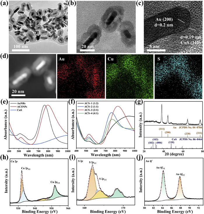
The zeta potential values of all components were measured to be less than +20 mV (Fig. S3b in Supporting information), indicating good colloidal stability. X-ray diffraction was performed to further study the crystalline structure of the binary plasmonic hybrids. As shown in Fig. 2g, the peaks at 38.2°, 44.4°, 64.6°, and 77.6° are indexed to the (111), (200), (220), and (311) planes of face-centered cubic Au (JCPDS No. 04–0784) [30]. Additionally, the characteristic peaks at 29.3°, 31.8°, and 47.9° correspond to the covellite phase of CuS (JCPDS No. 06–0464) [37]. These results confirm the successful formation of the binary hybrid material. X-ray photoelectron spectroscopy (XPS) analysis was conducted to investigate the composition and chemical states of ACSNPs. The survey spectrum (Fig. S3c in Supporting information) revealed the presence of Au, Cu, and S elements in the binary hybrids. In the Cu 2p spectra (Fig. 2h), the peaks of CuS at 932.04 and 952.04 eV were assigned to Cu 2p3/2 and Cu 2p1/2 of Cu2+ [24]. Additionally, the S 2p spectrum (Fig. 2i) was fitted with peaks at 161.95 and 163.94 eV, assignable to the S 2p3/2 and S 2p1/2 signals of S2− [38], respectively, confirming the formation of Cu−S bonds. Furthermore, the peak observed at 168.50 eV was attributed to surface oxidation of the sample [38]. The Au 4f spectrum displayed characteristic peaks at Au 4f7/2 and Au 4f5/2 (Fig. 2j), consistent with metallic gold [39]. Collectively, these XPS results conclusively demonstrate the successful synthesis of the ACSNPs core-shell nanostructure.
To evaluate the photothermal conversion performance of ACSNPs, we first compared the heating effects of ACSNPs with different feed ratios. As the CuS proportion increased (Fig. 3a), the thickness of the CuS shell grew, and the photothermal performance of ACSNPs exhibited a trend of initial improvement followed by decline: ACS-3 > ACS-2 > ACS-4 > ACS-1. The initial performance enhancement likely stemmed from increased NIR light absorption capacity. However, beyond a certain shell thickness, excess CuS reduced the photothermal efficiency of the hybrid materials. This degradation can be attributed to the overly thick shell hindering light absorption by the AuNR core. At a mixing ratio of 2:1 (ACS-3), the sample exhibited optimal photothermal conversion efficiency, indicating that an optimal interface engineering equilibrium point was achieved to maximize synergistic effects, consistent with conclusions from previous studies [23].
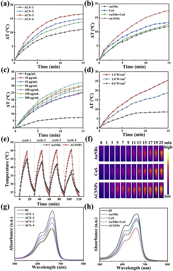
Then we compared the heating profiles of AuNRs, CuS, their physical mixture, and ACSNPs (ACS-3) under irradiation from an 808 nm laser (1.0 W/cm2). The physical mixture (Fig. 3b) exhibited a heating effect similar to the sum of its individual components. In contrast, ACSNPs under the same conditions achieved a temperature increase of 17.6 ℃, demonstrating a heating behavior far exceeding that of any single component or the physical mixture. This result strongly confirms that the AuNR@CuS hybrid synergistically enhances photothermal performance. As shown in Figs. 3c and d, the temperature change of the solution was positively correlated with both the ACSNPs concentration and the laser power density. The ACSNPs induced effective temperature elevation even at low concentrations (50 µg/mL) and power densities (1.0 W/cm2), demonstrating their significant potential as highly efficient photothermal agents.
Photothermal stability is a key indicator for evaluating the practical application value of photothermal formulations. As shown in Fig. 3e, the maximum temperature achieved by AuNRs decreased significantly after four laser on/off cycles. In contrast, ACSNPs maintained their maximum temperature without obvious degradation after repeated cycling, demonstrating excellent photothermal stability of ACSNPs. The enhanced stability is due to the protective effect of the CuS shell, which helps maintain structural integrity and functional performance under prolonged laser exposure. Meanwhile, the temperature distribution of each sample after laser irradiation was visually captured using an infrared thermal imaging camera (Fig. 3f). Under the same irradiation duration, the ACSNPs exhibited the brightest thermal signal, consistent with the temperature rise curves. As shown in Figs. S4a-c (Supporting information), the photothermal conversion efficiency of ACSNPs achieved 46.6%, which was significantly higher than that of AuNRs (33.0%) and CuS (35.8%), further confirming its superiority as a highly efficient photothermal platform. In summary, ACSNPs exhibit significantly enhanced light absorption, improved photothermal conversion efficiency, and increased photostability, collectively endowing this nanocomposite with highly efficient, durable, and reliable potential for PTT in antimicrobial applications.
CuS nanoparticles are known to generate ·OH under NIR irradiation [20]. When the incident light wavelength matches the LSPR absorption peak of the Au core, it promotes the generation of holes within the CuS, further enhancing ·OH production [23]. We employed the methylene blue method to detect ·OH generation. The characteristic absorption peak of methylene blue at 664 nm gradually diminishes as it is degraded by ·OH [5]. As shown in Fig. 3g, ACSNPs with different shell thicknesses exhibit regular variations in photodynamic performance similar with their photothermal behavior, with the ACS-2 and ACS-3 groups demonstrating the optimal photodynamic performance. This can be attributed to the fact that their absorption peak in the Ultraviolet-visible-near infrared region most closely matches that at 808 nm (Fig. 2f) [23,32]. As shown in Fig. 3h, the absorbance of the ACSNPs group decreased most significantly compared to other groups, indicating that the introduction of the AuNRs core effectively enhanced the ·OH generation capacity. The time-dependent degradation of methylene blue, indicated by the decrease in its characteristic absorption peak under prolonged illumination (Fig. S4d in Supporting information), along with the corresponding electron spin resonance analysis (Fig. S4e in Supporting information), collectively confirm that the generation of ·OH increases progressively with irradiation time extending. In summary, ACSNPs exhibit outstanding photothermal and photodynamic properties, offering a highly promising synergistic strategy for eradicating drug-resistant bacteria.
To elucidate the phototherapy mechanism of ACSNPs, density functional theory (DFT) calculations were employed to investigate charge transfer behavior at the Au-CuS interface. First, the electron extraction capabilities of each component material were assessed via work function calculations. As shown in Figs. S5a and b (Supporting information), Au exhibits a work function of 5.34 eV, lower than that of CuS (6.03 eV). When a metal contacts a semiconductor, electrons spontaneously flow from the side with the lower work function to the side with the higher work function [40,41]. Therefore, at the ACSNPs interface, electrons migrate from the Au core to the CuS shell. As shown in Fig. 4a, the work function of ACSNPs was 6.20 eV. This value indicates that the formation of the interface promotes electron redistribution and transfer, facilitating electron enrichment toward the CuS. Additionally, the band structure in Fig. 4b revealed that ACSNPs exhibit metallic properties with excellent conductivity. This enhances the separation and migration efficiency of photogenerated electron-hole pairs, thereby boosting the synergistic photothermal and photodynamic performance. The partial density of states (Fig. 4c) further confirms the metallic nature of ACSNPs, with the S 3p orbital serving as the primary contributor near the Fermi energy. Differential charge density analysis (Fig. 4d) provides electron accumulation on CuS and electron dissipation on Au at the interface, confirming the directional charge transfer. This process results in the formation of a positive charge center on the Au and a negative charge center on the CuS, thereby establishing an internal electric field directed from Au toward CuS.
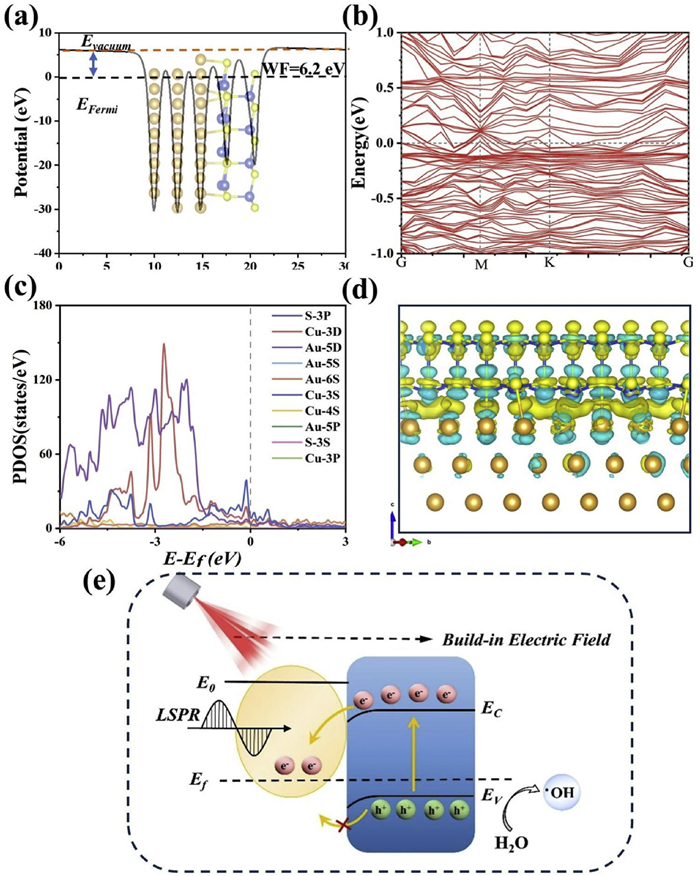
As a p-type semiconductor, CuS exhibits a Fermi level close to the valence band edge [20]. The formation of the internal electric field induces a downward bending of the band structure from the bulk to the interface on CuS (Fig. 4e) [42]. This band bending creates a barrier for the majority carriers of the p-type semiconductor migrating toward Au, effectively impeding hole diffusion [43]. Under laser irradiation, the built-in electric field drives photo-generated electrons to migrate and become confined within the CuS valence band. This spatial charge separation effectively suppresses electron-hole pair recombination, prolonging carrier lifetime [44]. This separation mechanism significantly enhances the photothermal conversion performance of ACSNPs [23]. Concurrently, the enrichment of holes in the CuS surface region promotes reactions with water molecules to generate ·OH, thereby intensifying the photodynamic effect. To experimentally verify these theoretical predictions regarding charge separation, we performed photocurrent response measurements. As shown in Fig. S5c (Supporting information), the ACSNPs heterostructure exhibited a dramatically enhanced photocurrent compared to the individual components, AuNRs and CuS. This significantly stronger photocurrent provides direct and tangible evidence for the markedly improved separation and migration of photogenerated electron-hole pairs in ACSNPs, thereby functionally validating the charge transfer mechanism and built-in electric field effect revealed by our DFT calculations. The above findings theoretically elucidate the underlying mechanism by which ACSNPs synergistically enhance PTT/PDT effects, providing crucial mechanistic support for achieving highly efficient and rapid bactericidal performance in antimicrobial applications, particularly against highly drug-resistant pathogens. The synergy in our ACSNPs operates through complementary mechanisms: An intrinsic charge separation enabled by rational interface design, and external thermal effects that improve ROS delivery and reaction kinetics.
Based on their superior photothermal and photodynamic properties, the antibacterial capabilities in vitro of ACS-2 and ACS-3 were further evaluated. Under identical conditions (50 µg/mL, 1.0 W/cm2, 10 min) the ACS-3 treated group was completely eradicated of MRSA (Fig. 5a), whereas ACS-2 did not (Fig. S6 in Supporting information). This is attributed to the stronger photothermal conversion capability of ACS-3. Therefore, ACS-3 nanoparticles were selected for subsequent experiments and were uniformly designated as ACSNPs. The standard plate count method was employed to investigate the antibacterial efficacy of ACSNPs against MRSA. As shown in Figs. 5a and b, the antimicrobial effect exhibited concentration and irradiation time dependence. At a low concentration of 5 µg/mL, the antibacterial activity of ACSNPs was negligible, and the bacterial survival rate was 18.5% when exposed to NIR light for 10 min. When the concentration was increased to 10 µg/mL, the antibacterial effect improved, achieving approximately 86.5% bactericidal rate after 10 min irradiation. Notably, ACSNPs at a concentration of 20 µg/mL exhibited excellent, nearly complete antibacterial performance, achieving a bactericidal rate of 99.0% and 100% after 5 and 10 min irradiation, respectively. Furthermore, the time required for effective sterilization was significantly reduced at higher concentrations. At concentrations of 20 and 50 µg/mL, the necessary irradiation time for achieving strong antibacterial activity was reduced to 5 and 3 min, respectively. The antibacterial efficacy of ACSNPs was significantly enhanced by increasing the nanoparticle concentration and irradiation time. Rapid and complete elimination of MRSA was achieved at a relatively low concentration (20 µg/mL) and short-time irradiation (10 min). The antibacterial mechanism was attributed to the PTT/PDT synergy, as evidenced by a dramatic increase in intracellular ROS and concurrent severe leakage of intracellular proteins and nucleic acids (Fig. S7 in Supporting information). Additionally, cell viability assay results showed that after treatment with 20 µg/mL ACSNPs, the survival rate of mouse fibroblast NIH/3T3 cells reached 85.4%, and even after 10 min of laser exposure, the survival rate remained over 80%, indicating the good biocompatibility and low cytotoxicity of ACSNPs at 20 µg/mL (Fig. S8 in Supporting information). The antibacterial efficacy of ACSNPs was further assessed through co-staining with SYTO 9 and propidium iodide (PI). The green-fluorescent SYTO 9 dye labels all bacteria due to its cell permeability, in contrast to the red-fluorescent PI, which enters and labels only bacteria with damaged membranes.
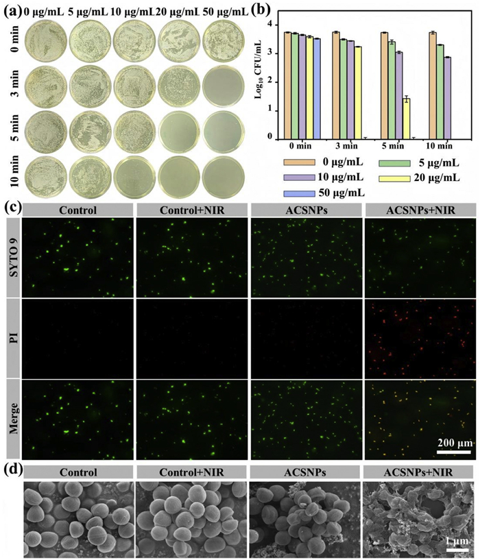
The control, control+NIR, and ACSNPs groups exhibited strong green fluorescence without red fluorescence (Fig. 5c), indicating that the bacteria maintained their physiological integrity. In contrast, the ACSNPs+NIR group showed a significantly higher proportion of dead bacteria, consistent with the colony count results. Scanning electron microscope images were used to analyze morphological changes in MRSA after different treatments. Bacteria in the control group maintained stable morphology before and after NIR laser treatment (Fig. 5d). Without light exposure, the ACSNPs group showed that bacterial structures remained relatively intact, indicating that ACSNPs had no significant effect on bacterial growth without NIR irradiation conditions. However, after 10 min of NIR irradiation, MRSA bacteria ruptured and exhibited incomplete morphology, further demonstrating the exceptional phototherapy antibacterial performance of ACSNPs. The in vitro experimental results above demonstrate that ACSNPs exhibited outstanding efficacy in eliminating MRSA. This is primarily attributed to the built-in electric field engineered through interfacial modifications (Fig. 4e), which effectively enhances their photothermal and photodynamic properties. This synergistic effect achieves highly efficient bacterial clearance, showcasing promising application prospects for treating MRSA-infected wounds.
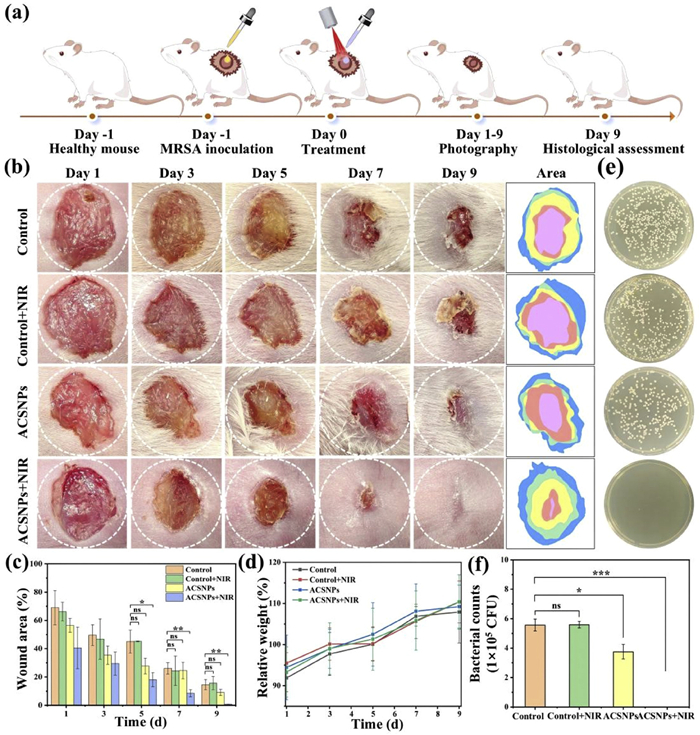
To further investigate the therapeutic effect of ACSNPs against real bacterial infections in vivo, we established a mouse MRSA skin wound infection model. ACSNPs in vivo with NIR laser and the surgical procedures are depicted in Fig. 6a. All animal experiments were approved by the Experimental Animal Ethics Committee of Henan Eye Hospital. To assess the therapeutic efficacy of ACSNPs, we took representative photographs of the wound surfaces in each group on days 1–9 and performed measurements. Real-time infrared thermal imaging revealed that the local temperature of the infected wound treated with ACSNPs under 808 nm laser irradiation (1.0 W/cm2) rapidly increased to approximately 50 ℃ (Fig. S9 in Supporting information), which demonstrated the generation of the photothermal effect. Importantly, no adverse effects such as redness, swelling, blistering, or necrosis were observed at the irradiation site during the treatment. By calculating the percentage change in wound area compared to the initial state, the healing process was reflected (Fig. 6b). On the 5th day after treatment, quantitative statistical results (Fig. 6c) indicated that the wound area of ACSNPs+NIR group was significantly smaller than the other three groups, with statistically significant differences. Further observation revealed that the three control groups still exhibited large wound areas with relatively slow healing progress on day 9. In contrast, the ACSNPs+NIR group achieved a healing rate of 99.2% at this point, representing 13.8% improvement over the control groups. This outcome can be primarily attributed to the superior synergistic antibacterial effect of ACSNPs in PTT/PDT, further confirming its highly efficient antibacterial and wound-healing capabilities. As shown in Fig. 6d, the body weights were similar across all groups, indicating that ACSNPs did not exhibit significant toxic side effects in vivo. Additionally, quantitative analysis of bacterial residual levels at the wound site on day 9 was performed using plate counting (Figs. 6e and f). Bacterial counts showed no significant difference between the Control groups. The ACSNPs group showed no significant change in the colony counts, whereas the ACSNPs+NIR group exhibited a substantial reduction of 99.9%. This strongly demonstrates the key role of NIR-induced PTT/PDT synergistic effects in vivo antibacterial performance of ACSNPs. The in vivo results confirm the synergistic PTT/PDT antibacterial performance of ACSNPs against MRSA infection under NIR irradiation. This performance stems from the built-in electric field, which enhances the photothermal conversion efficiency and ·OH radical generation capacity of ACSNPs. Specifically, the localized high heat not only accelerates bacterial metabolism and enhances sensitivity to ROS but also disrupts bacterial cell membrane structures, thereby facilitating ROS penetration into the bacterial interior and achieving a synergistic photothermal-photodynamic antibacterial effect [45]. Such synergistic interaction significantly boosts the bactericidal efficacy of ACSNPs, highlighting their superior capability for treating MRSA-infected wounds.
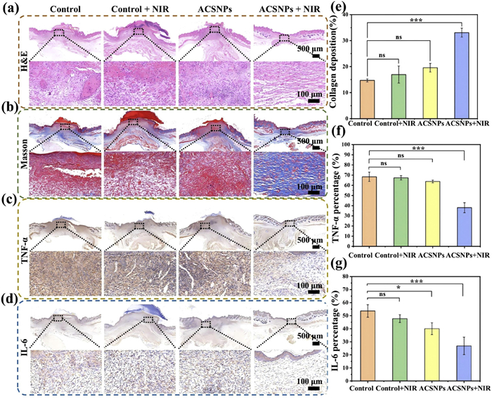
In addition, to further examine the rate of wound healing in the different groups, the main markers of infection in the wound tissue were assessed by histological analysis. In the H&E staining results on day 9, the ACSNPs + NIR group exhibited optimal tissue recovery and most intact skin structure among all experimental groups (Fig. 7a), including clearly recognizable epidermal, dermal and appendage structures, showing a good state of tissue repair compared with normal tissue morphology. In contrast, the other groups had partial scab formation and larger incomplete healing areas. This result can be attributed to the synergistic antimicrobial effects of ACSNPs by PTT/PDT, which effectively cleared the MRSA infection and attenuated inflammatory damage, and created a microenvironment for tissue regeneration. Collagen deposition was assessed by Masson staining (Fig. 7b). Compared with the other groups, the stained area in the ACSNPs + NIR group was amount of blue, indicating that a large amount of new collagen was formed (Fig. 7e). These findings corroborated the efficient antibacterial and anti-inflammatory abilities, suggesting that ACSNPs significantly accelerated the healing process through a synergistic mechanism under laser excitation. Immunohistochemical analysis was performed to detect the expression of key inflammatory mediators TNF-α and IL-6 (Figs. 7c and d). Compared with the Control group, significantly lower expression of both factors was observed in the ACSNPs+NIR group (Figs. 7f and g), further confirming that this therapeutic strategy effectively mitigates excessive inflammatory responses in infected wounds. These advantages stem from the built-in electric field at interface, which promotes carrier separation under NIR irradiation, thereby enhancing the effects of PTT and PDT. This synergistic strategy efficiently eliminates MRSA, shortens the inflammatory phase, and promotes collagen synthesis and deposition, accelerating the healing process of infected wounds.
In this study, an interface-engineered AuNR@CuS core-shell nanohybrid was developed. Through precise interface engineering, we constructed a built-in electric field at the Au-CuS interface that significantly facilitated photogenerated charge separation, resulting in synergistic enhancement of PTT/PDT effects. ACSNPs exhibited a photothermal conversion efficiency of 46.6% and efficiently produced ·OH. It effectively overcame the limitations of insufficient heat production and low ROS production efficiency of traditional phototherapy agents in antibacterial therapy. Complete eradication of MRSA was achieved in vitro at a concentration of 20 µg/mL ACSNPs, 808 nm laser irradiation for 10 min. In vivo, the ACSNPs+NIR treatment group shows a 99.9% reduction in bacterial load and a 13.8% increase in wound healing rate. This study not only provides a synergistic therapeutic approach for the treatment of resistant bacterial infections, but also demonstrates the critical value of interface engineering strategies in improving phototherapy performance.
Chiyin Zhang: Writing – review & editing, Writing – original draft, Software, Methodology, Investigation, Formal analysis, Data curation, Conceptualization. Xiwen Geng: Software, Resources, Methodology, Investigation, Formal analysis. Hang Xu: Software, Resources. Xinyu Gu: Software, Methodology, Investigation, Data curation. Shaokui Cao: Supervision, Project administration, Conceptualization. Zhanrong Li: Project administration, Funding acquisition, Conceptualization. Jingguo Li: Writing – review & editing, Writing – original draft, Visualization, Supervision, Project administration, Funding acquisition, Conceptualization. Jun Shi: Writing – review & editing, Writing – original draft, Supervision, Project administration, Conceptualization.
The authors declare that they have no known competing financial interests or personal relationships that could have appeared to influence the work reported in this paper.
The work is supported by the National Natural Science Foundation of China (No. 52173143), the Natural Science Foundation of Henan Province of China (Nos. 242300421018 and 252300421024), Henan Province Science and Technology Research and Development Plan Joint Fund Project (No. 242301420010), Henan Province Youth Health Science and Technology Innovation Talent Training Project (No. LJRC2024003) and Medical Science and Technology Project of Henan Province of China (No. LHGJ20230042). The analytical and testing assistance from Advanced Analysis & Computation Center, Zhengzhou University and Shiyanjia Lab (
Supplementary material associated with this article can be found, in the online version, at doi:
U. Theuretzbacher, Nat. Rev. Microbiol. 23 (2025) 491–508. doi: 10.1038/s41579-025-01169-8
B. Xue, X. Geng, H. Cui, et al., Chin. Chem. Lett. 34 (2023) 108140. doi: 10.1016/j.cclet.2023.108140
E.M. Darby, E. Trampari, P. Siasat, et al., Nat. Rev. Microbiol. 21 (2023) 280–295. doi: 10.1038/s41579-022-00820-y
D. Lim, N.C.J. Strynadka, Nat. Struct. Biol. 9 (2002) 870–876.
Y.X. Huang, J.L. Li, Z.H. Yu, et al., Adv. Mater. 36 (2024) 2414111. doi: 10.1002/adma.202414111
Z. Guo, W. Liu, T. Liu, et al., Chin. Chem. Lett. 35 (2024) 109060. doi: 10.1016/j.cclet.2023.109060
H. Tang, X. Qu, W. Zhang, et al., Adv. Mater. 34 (2022) 2107300. doi: 10.1002/adma.202107300
F. Gao, Y. Wu, L. Yang, et al., Chin. Chem. Lett. 36 (2025) 109917. doi: 10.1016/j.cclet.2024.109917
B.Q. Yu, Q.R. Liu, J.D. Sun, et al., Chem. Eng. J. 487 (2024) 150705. doi: 10.1016/j.cej.2024.150705
L. Mei, Y.F. Zhang, K.X. Wang, S.J. Chen, T. Song, Mater. Today Bio 29 (2024) 101354. doi: 10.1016/j.mtbio.2024.101354
H. Zhou, D. Gao, G. Cheng, et al., J. Mater. Sci. Technol. 208 (2025) 252–262. doi: 10.1016/j.jmst.2024.04.055
X.J. Hu, H. Zhang, Y.T. Wang, et al., Chem. Eng. J. 450 (2022) 138129. doi: 10.1016/j.cej.2022.138129
I.E. Mba, E.I. Nweze, World J. Microbiol. Biotechnol. 37 (2021) 108. doi: 10.1007/s11274-021-03070-x
H.L. Xin, Y.Y. Liu, Y.N. Xiao, et al., Adv. Funct. Mater. 34 (2024) 2402607. doi: 10.1002/adfm.202402607
Y. Zhao, L. Chen, Y. Wang, et al., Nano Res. 14 (2021) 4417–4441. doi: 10.1007/s12274-021-3417-4
A. Serrano-Aroca, K. Takayama, Y.K. Mishra, C. de la Fuente-Nunez, Adv. Funct. Mater. 34 (2024) 2402023. doi: 10.1002/adfm.202402023
B. Ouyang, D. Wei, B. Wu, et al., Small 20 (2023) 2303153.
M.M. Khan, S.N. Matussin, A. Rahman, Bioprocess. Biosyst. Eng. 46 (2023) 1231–1249. doi: 10.1007/s00449-023-02878-1
F. Hu, B. Song, X. Wang, et al., Chin. Chem. Lett. 33 (2022) 308–313. doi: 10.1016/j.cclet.2021.07.018
W.J. Chan, S. Urandur, H.T. Li, V.S. Goudar, Nanomedicine 18 (2023) 2185–2204. doi: 10.2217/nnm-2023-0202
H.C. Han, J.J. Yang, X.Y. Li, et al., Nano Res. 14 (2021) 2512–2534. doi: 10.1007/s12274-021-3293-3
W. Chung, B. Lee, Y. Kim, Prog. Org. Coat. 198 (2025) 108909. doi: 10.1016/j.porgcoat.2024.108909
Y. Chang, Y. Cheng, Y.L. Feng, et al., Nano Lett. 18 (2018) 886–897. doi: 10.1021/acs.nanolett.7b04162
X.R. Deng, K. Li, X.C. Cai, et al., Adv. Mater. 29 (2017) 1701266. doi: 10.1002/adma.201701266
L. Wang, Y. Chang, Y. Feng, et al., Nano Lett. 19 (2019) 6800–6811. doi: 10.1021/acs.nanolett.9b01869
R. Huang, C. Zhang, Y. Bu, et al., Biomaterials 277 (2021) 121088. doi: 10.1016/j.biomaterials.2021.121088
M.Q. Sun, X.Q. Fu, K.X. Chen, H. Wang, ACS Appl. Mater. Interfaces 12 (2020) 46146–46161. doi: 10.1021/acsami.0c13420
Q. Yu, T.Y. Peng, J.F. Zhang, et al., Small 18 (2022) 2103174. doi: 10.1002/smll.202103174
Z.R. Xu, R. Aalinkeel, S.D. Mahajan, et al., Adv. Therap. 6 (2023) 2200248. doi: 10.1002/adtp.202200248
Z. Wang, N. Yu, X. Li, et al., Chem. Eng. J. 381 (2020) 122613. doi: 10.1016/j.cej.2019.122613
Y. Zhang, C. Wen, Y. Liu, et al., Chem. Eng. J. 470 (2023) 144345. doi: 10.1016/j.cej.2023.144345
L. Wang, Z. Hussain, P.H. Zheng, et al., J. Mater. Chem. B 11 (2023) 2166–2183. doi: 10.1039/d2tb02403a
W. Xiu, L. Wan, K. Yang, et al., Nat. Commun. 13 (2022) 3875. doi: 10.1038/s41467-022-31479-x
H. Xue, W.F. Li, Y.M. Long, Colloids Surfaces A: Physicochem. Eng. Asp. 703 (2024) 135287. doi: 10.1016/j.colsurfa.2024.135287
D. Zheng, X.Y. Zhao, Y. Wang, et al., Chem. Eng. J. 486 (2024) 150271. doi: 10.1016/j.cej.2024.150271
L.L. Arjona, J.I. Garcia-Peiro, M. Haro, J.L. Hueso, E.J. Juarez-Perez, J. Alloy. Compd. 1016 (2025) 179030. doi: 10.1016/j.jallcom.2025.179030
P.P. Yang, W.H. Chen, J.G. Li, et al., Int. J. Biol. Macromol. 253 (2023) 127150. doi: 10.1016/j.ijbiomac.2023.127150
H.S. Kang, J.W. Zou, Y. Liu, et al., Adv. Funct. Mater. 33 (2023) 2303911. doi: 10.1002/adfm.202303911
W.Y. Wang, J.Y. Wang, J.G. Li, S.K. Cao, J. Shi, Mater. Today Commun. 37 (2023) 107448. doi: 10.1016/j.mtcomm.2023.107448
W. Monch, Rep. Prog. Phys. 53 (1990) 221–278. doi: 10.1088/0034-4885/53/3/001
X.H. Li, M. Antonietti, Chem. Soc. Rev. 42 (2013) 6593–6604. doi: 10.1039/c3cs60067j
Z. Zhang, J.T. Yates, Chem. Rev. 112 (2012) 5520–5551. doi: 10.1021/cr3000626
D. Xu, S.N. Zhang, J.S. Chen, X.H. Li, Chem. Rev. 123 (2023) 1–30.
Q. Wu, T. Deng, J. Cheng, et al., ACS Nano 18 (2024) 11988–12009. doi: 10.1021/acsnano.4c02433
C. Mao, Y. Xiang, X. Liu, et al., ACS Appl. Mater. Interfaces 11 (2019) 17902–17914. doi: 10.1021/acsami.9b05787

Figure 1 Schematic illustration of the preparation and antibacterial effect of AuNR@CuS hybrid nanomaterials as PTT/PDT synergistic therapy.

Figure 2 Characterization of ACSNPs. (a, b) TEM images. (c) High-resolution transmission electron microscopy image displaying lattice fringes of the Au and CuS phases. (d) Elemental mapping. (e) Ultraviolet-visible-near infrared absorption spectra of AuNRs, CuS and ACSNPs. (f) Ultraviolet-visible-near infrared absorption spectra of ACSNPs with different Au: CuS feed ratios. (g) X-ray diffraction pattern of ACSNPs. High-resolution XPS spectra in (h) Cu 2p, (i) S 2p, and (j) Au 4f regions of ACSNPs.

Figure 3 Photothermal and photodynamic effect of ACSNPs. (a) Photothermal heating performance of ACSNPs with different composition ratios under laser irradiation (1.0 W/cm2, 15 min). (b) Temperature rise curves of AuNRs, CuS, the physical mixture (AuNRs + CuS), and ACSNPs after 808 nm laser irradiation (1.0 W/cm2, 15 min). (c) Temperature rise curves of ACSNPs at different concentrations after laser irradiation (1.5 W/cm2, 15 min). (d) Temperature changes of ACSNPs under different laser power densities over 15 min. (e) Temperature changes of AuNRs and ACSNPs during four on/off cycles of laser irradiation (1.5 W/cm2, 30 min per cycle). (f) Infrared thermal imaging of AuNRs, CuS, and ACSNPs under 0.5 W/cm2 laser irradiation. (g) Degradation of MB using catalysts with different Au: CuS feed ratios. (h) The degradation of MB in different groups under NIR laser irradiation (1.5 W/cm2).

Figure 4 Density functional theory calculation of ACSNPs. (a) Electrostatic potential along the c-axis of Au-CuS interfaces. (b) Band structure of ACSNPs. (c) Partial density of states. (d) Electron localization analysis. (e) Schematic illustration of the mechanism for synergistically enhanced PTT and PDT.

Figure 5 In vitro antibacterial effect of ACSNPs. (a) Photographs of MRSA treated with different concentrations of ACSNPs (0, 5, 10, 20, 50 µg/mL) on agar plates, with varying NIR laser (808 nm, 1.0 W/cm2) irradiation times. (b) The survival rate of MRSA in (a) was calculated using the colony count method (Data are presented as mean ± standard deviation (SD) (n = 3). (c) Confocal images of MRSA treated with phosphate buffered saline and ACSNPs (20 µg/mL), with or without NIR laser (808 nm, 1.0 W/cm2) irradiation for 10 min. (d) Scanning electron microscope images of different groups.

Figure 6 In vivo antimicrobial assessment of ACSNPs in the MRSA infectious wound model. (a) Scheme of infectious wound model establishment and drug administration process. (b) Representative photographs of the wound area of different treatment groups, and (c) corresponding quantitative results (n ≥ 3). (d) Changes in body weight of mice in each group at different times. (e) Images of plant colonies of residual bacteria on day 9, and (f) the corresponding statistics (n ≥ 3). Data are presented as mean ± SD. ns: not significant. *P < 0.05, **P < 0.01, ***P < 0.001.

Figure 7 Histological and immunohistochemical analysis of skin tissues on day 9. (a) H&E staining, (b) Masson trichrome. Immunohistochemical analysis of (c) TNF-α and (d) IL-6. Quantitative results of (e) collagen deposition, (f) TNF-α, and (g) IL-6 (mean ± SD, n = 3). *P < 0.05, ***P < 0.001.

 扫一扫看文章


扫一扫关注我们
 DownLoad:
 下载:
 下载: