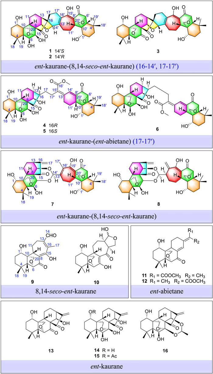
Figure 1.
Chemical structures of compounds 1–16.

Unprecedented ent-kaurane heterodimers from Icacina oliviformis: Potent anti-Helicobacter pylori leads targeting enoyl-ACP reductase FabI
Mingming Xu , Binglei Li , Junfei Zhou , Xudong Hang , Chuanlin Jin , Chujie Lu , Wei Lin , Xinyi Wei , Er-Xin Shang , Liping Zeng , Monday M. Onakpa , Oluwatosin O. Johnson , Chun-Tao Che , Jin-Ao Duan , Hongkai Bi , Ming Zhao


The relentless global spread of antibiotic resistance has severely compromised conventional therapies for Helicobacter pylori, a ubiquitous pathogen implicated in gastritis, peptic ulcers, and gastric malignancy. With infection rates reaching 50%–70% worldwide and exceeding 90% in developing regions, H. pylori pose a substantial global health burden [1–3]. Current first-line regimens combining broad-spectrum antibiotics (e.g., clarithromycin-amoxicillin) are increasingly failing due to the rapid emergence of multidrug-resistant strains, leading to alarmingly low eradication rates [4,5]. This therapeutic crisis underscores the urgent need for new antibacterial agents with distinct mechanisms of action to circumvent existing resistance pathways. While narrow-spectrum antibiotics have recently gained traction for their potential to minimize microbiome disruption and resistance selection [6–8], the discovery pipeline for species-selective agents targeting H. pylori remains critically underdeveloped. The absence of mechanistically innovative H. pylori-specific therapeutics therefore represents a major unmet need in antimicrobial discovery, demanding immediate scientific attention.
Against this backdrop, the bacterial fatty acid biosynthesis Ⅱ (FASII) pathway emerges as a particularly promising target for H. pylori-specific intervention. As an essential metabolic pathway for bacterial viability that is absent in humans, FASII offers an exceptional therapeutic window for selective antimicrobial development [9,10]. Within this pathway, enoyl-acyl carrier protein (ACP) reductase (FabI) represents a critical intervention point, catalyzing the rate-limiting final step in fatty acid elongation that is indispensable for bacterial membrane biogenesis [10–14]. Although FabI catalysis is functionally conserved across bacteria, its notable structural divergence among species offers an opportunity to design highly selective inhibitors. This structural variability could enable precise targeting of H. pylori FabI while sparing commensal microbiota, a crucial advantage over conventional broad-spectrum antibiotics [9,15]. Despite FabI's extensive validation as an antibacterial target in other pathogens, the lack of H. pylori-selective FabI inhibitors highlights both an untapped therapeutic opportunity and a key challenge in developing precision antibacterials against this globally prevalent organism.
Natural products remain an invaluable and underexploited source of FabI-targeting agents, providing unparalleled structural diversity and novel modes of enzyme inhibition [11,15–17]. Our research program has explored the anti-H. pylori potential of medicinal plants endemic to West and Central Africa, with particular focus on the diterpenoid-rich Icacina genus [18,19]. Through these investigations, we have characterized 56 diterpenoids encompassing ten distinct carbon skeletons, demonstrating the exceptional scaffold diversity of this botanical source [18–27]. Notably, a substantial subset of these compounds exhibited potent anti-H. pylori activity (minimum inhibitory concentration (MIC) = 8–16 μg/mL), validating their promise as lead candidates for mechanism-driven drug discovery.
Building on this foundation, we focused our investigation on Icacina oliviformis as a source of structurally novel FabI-targeting agents. This exploration yielded twelve previously undescribed diterpenoids (1–12), including two unprecedented dimeric architectures: ent‑kaurane-(8,14-seco‑ent‑kaurane) (16–14′,17–17′) and ent‑kaurane-(ent‑abietane) (17–17′) dimers, alongside four known ent‑kaurane analogs (13–16) (Fig. 1). Notably, several isolates demonstrated potent and selective anti-H. pylori activity, with oliviformisone A (1) emerging as the most promising candidate. Through an integrated approach combining enzymatic kinetics, microscale thermophoresis, target overexpression, and site-directed mutagenesis, we unequivocally established compound 1 as a competitive FabI inhibitor with nanomolar binding affinity. This work represents the first report of a structurally unique, mechanism-defined FabI inhibitor derived from a natural product with species-selective activity against H. pylori. While ent‑kaurane diterpenoids are widely distributed across the plant kingdom, their anti-H. pylori potential has remained largely unexplored. Our findings not only reveal the untapped pharmacological value of this structural class but also significantly expand their known target diversity. Collectively, these results deliver a validated lead compound for anti-H. pylori therapy and establish a solid framework for mechanism-guided exploration of ent‑kaurane diterpenoids as a privileged scaffold for species-selective antimicrobial development.
Oliviformisone A (1), colorless crystals, [α]D20 −20.0 (c 0.02, MeOH), possessed a molecular formula of C40H52O10 as determined by its high-resolution electrospray ionization mass spectrometry (HRESIMS) data at m/z 693.3695 [M + H]+ (calcd. for C40H53O10+, 693.3638), indicating 15 degrees of unsaturation. The proton nuclear magnetic resonance (1H NMR) spectrum of 1 (Table S1 in Supporting information) exhibited resonances attributable to 4 methyl groups, 2 oxygenated methylenes, and 6 oxygenated methines. Analysis of the 13C NMR spectrum (Table S1) of 1, combined with the distortionless enhancement by polarization transfer 135° (DEPT135) and heteronuclear single quantum coherence (HSQC) data, revealed the presence of 40 carbon resonances, including 4 methyls, 12 methylenes (2 oxygenated), 11 methines (6 oxygenated), as well as thirteen nonprotonated carbons (4 olefinic, 1 oxygenated, and 2 carbonyls). The unusually large carbon number (C40), 15 degrees of unsaturation, and the observation of partially duplicated or closely related NMR signals implied that compound 1 might be composed of two diterpenoid-like subunits. Therefore, detailed 2D NMR analyses were performed to elucidate the structures of these subunits and their mutual linkage.
Subsequent analysis of the 1H and 13C NMR data revealed that one portion of 1 closely resembled an ent‑kaurane-type diterpenoid, which was designated as unit Ⅰ (Fig. 2A). The 1H and 13C NMR data of unit Ⅰ in 1 were similar to those of oridonin (14), except for the presence of a quaternary carbon (δC 61.3, C-16) and a methylene (δC 17.8, C-17) in 1, rather than two olefinic carbons (δC 153.4, C-16; δC 120.6, C-17) in 14. The chemical shift of C-14 (δC 82.4) in unit Ⅰ was upfield-shifted by ca. 8.3 ppm compared with that of 14 (δC 74.1), suggesting that C-14 is involved in a linkage to unit Ⅱ, likely via an oxygen bridge, which alters its electronic environment. Thus, the gross structure of unit Ⅰ in 1 was further determined via the proton–proton correlation spectroscopy (1H–1H COSY) and heteronuclear multiple bond correlation (HMBC) correlations as depicted in Fig. 2A.
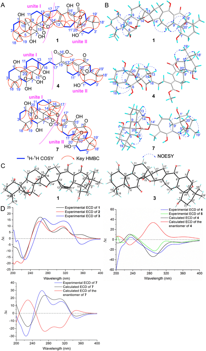
The remaining signals in the NMR spectra corresponded to a second diterpenoid unit, hereafter referred to as unit Ⅱ (Fig. 2A). The NMR data of unit Ⅱ in 1 (Table S1) were further confirmed by the 2D NMR spectra including the 1H–1H COSY correlations of H-1′/H2–2′/H2–3′, H-5′/H2–6′, and H-9′/H2–11′/H2–12′, and the HMBC correlations were employed to assemble the planar structure of unit Ⅱ in 1 from these three partial fragments. Specifically, the key HMBC correlations from two gem‑dimethyls CH3–18′/CH3–19′ to C-3′/C-4′/C-5′ suggested the direct connections of C-3′/C-5′/C-18′/C-19′ toward C-4′, the connection of C-1′/C-5′/C-9′/C-20′ through C-10′ was confirmed by the HMBC correlations from H2–20′ to C-1′/C-5′/C-9′/C-10′, and the HMBC correlations from H-6′ to C-7′, from H-9′ to C-7′/C-8′/C-15′, from H2–17′ to C-13′/C-14′/C-15′, and H-14′ to C-12′/C-13′/C-14′ established the planar framework of unit Ⅱ as a 6/6/7 fused tricyclic system. Moreover, the presence of a 6′,20′-epoxy group could be verified by the downfield chemical shifts of C-6′ (δC 81.2) and C-20′ (δC 68.9), together with a key HMBC correlation from H2–20′ to C-6′.
After the structures of both subunits were independently established, the linkage between unit Ⅰ and unit Ⅱ was then determined from the 2D NMR data. The connections between units Ⅰ and Ⅱ via covalent C–C single bonds (C-16/C-14′ and C-17/C-17′) were supported by the 1H–1H COSY correlation of H2–17/H2–17′, along with HMBC cross-peaks from H-14′ to C-13/C-15/C-16. Additionally, the notably downfield-shifted chemical shifts of CH-14 (δH 5.33, δC 82.4) and CH-14′ (δH 4.51, δC 76.7), compared to those of typical hydroxylated methines, further corroborated the presence of an oxygen bridge between C-14 and C-14′. These correlations demonstrated that 1 is a heterodimer composed of units Ⅰ and Ⅱ connected through C-16/C-14′, C-17/C-17′, and an additional C-14–O–C-14′ bridge. Collectively, the planar structure of 1 was fully elucidated.
The relative configuration of 1 was ascertained via the interpretation of its nuclear Overhauser effect spectroscopy (NOESY) data (Fig. 2B). So far, all reported ent‑kauranes possessed 5β-H, thus, H-5 of unit Ⅰ in 1 also could be assumed as β-oriented. The NOESY correlations between H-5 (δH 1.86) and H-1 (δH 3.66)/H-9 (δH 1.42)/CH3–18 (δH 1.20), indicated that H-1, H-9, and CH3–18 were all β-oriented. Also, the α-orientations of H-6, H-13, H-14, CH3–19 were deduced from the NOESY correlations of CH3–19 (δH 1.11) and H-6 (δH 4.11), H-6 (δH 4.11) and H-20b (δH 4.38), H-20a (δH 4.56) and H-14 (δH 5.33) as well as H-14 (δH 5.33) and H-13 (δH 2.43). Likewise, the key NOESY cross-peaks of H-13 and H-17a, H-17b and H-14′, H-14′ and H-11′b, H-11′b and H-9′, H-9′ and H-5′ together with H-5′ and H-1′/H-6′ proved that H-1′, H-5′, H-6′, H-9′, and H-14′ were all β-oriented.
Finally, the structure of 1 was unequivocally confirmed by single-crystal X-ray diffraction using Cu Kα radiation, and the absolute configuration of 1 was determined as 1S,5R,6S,7S,8R,9S,10S,13S,14R,16R,1′S,5′R,6′R,9′S,10′S,14′S with a Flack parameter of 0.14(7) (CCDC 1438196, Fig. 2C) [28]. Structurally, oliviformisone A (1) represents the first example of an ent‑kaurane-(8,14-seco‑ent‑kaurane) (16–14′,17–17′) spiroheterodimer, bearing an unprecedented 6/6/6/5/6/7/6/6-fused octocyclic core featuring a rare 2-oxatricyclo[6.2.1.03,8]hendecane spirolinker segment.
Oliviformisone D (4) was obtained as an amorphous powder. Its molecular formula was determined as C41H56O11 by a [M + H]+ ion at m/z 725.3894 (calcd for C41H57O11+, 725.3896) in the HRESIMS and 13C NMR data, requiring fourteen double-bond equivalents. The 1H NMR spectrum of 4 (Table S2 in Supporting information) exhibited resonances attributable to four methyl groups at δH (1.07, s, CH3–18; 1.12, s, CH3–19; 0.97, s, CH3–18′; 1.03, s, CH3–19′), a methoxy at δH (3.80, s, CH3O-16′), and an olefinic proton at δH 7.58 (dd, H-14′). The 13C NMR and DEPT spectroscopic data (Table S2) of 4 highlighted the presence of 41 carbon resonances, including five methyls (one oxygenated), twelve methylenes (two oxygenated), twelve methines (one olefinic and five oxygenated) together with twelve nonprotoned carbons (three olefinic, an oxygenated, and three carbonyls). Following the approach used for 1, compound 4 was analyzed to identify its two subunits (units Ⅰ and Ⅱ, Fig. 2A).
Detailed analysis of the 1H and 13C NMR spectroscopic data (Table S2) for unit Ⅰ indicated that its structure resembled that of oridonin (14), the major difference was the presence of a methine (δC 57.0, C-16) and a methylene (δC 30.5, C-17) in unit Ⅰ instead of a double bond (δC 153.4, C-16; 120.6, C-17) in 14. Additionally, the NMR data of unit Ⅱ in 4 (Tables S2 and S4 in Supporting information) was similar to those of oliviformisol D (12), the major difference included the presence of a methylene (δH 2.84, 2.28; δC 28.0, CH2–17′) in unit Ⅱ of 4, replacing a methyl group (δH 2.05; δC 15.2, CH3–17) in 12. The connectivity between units Ⅰ and Ⅱ was established through 2D NMR correlations. The linkage via a C-17/C-17′ single bond was supported by 1H–1H COSY correlations between H2–17 (δH 1.62, 1.30) and H2–17′ (δH 2.84, 2.28). In addition, the NOESY cross-peak between H-14′ and 16′-OCH3 confirmed the cis-configuration of Δ13′,15′. These data demonstrated that compound 4 is a heterodimer composed of ent‑kaurane (unit Ⅰ) and ent‑abietane (unit Ⅱ) moieties linked through a C-17–C-17′ single bond (Figs. 2A and B).
The relative configuration of 4 was ascertained via the interpretation of its NOESY data. Herein, H-5 and H-5′ were both assumed to be β-oriented based on all reported ent‑kauranes and abietanes featuring 5β-H up to now. The NOESY correlations of H-5/H-1/H-9/CH3–18 and H-5′/H-1′/H-9′/CH3–18′ suggested that they were all β-oriented. The α-orientations of H-6/H-13/H-14/H-16 were deduced from the key NOESY correlations of H-6/CH3–19/H-20a and H-20b/H-14/H-13. The absolute configuration of 4 was then assigned as 1S,5R,6R,8R,9S,10S,13S,14R,16R,1′S,5′R,6′R,9′S,10′R based on the good agreement between the experimental and calculated electronic circular dichroism (ECD) curves (Fig. 2D) [29]. Consequently, compound 4 was established as depicted and was given a trial name oliviformisone D. It is the first report of an ent‑kaurane-(ent‑abietane) (17–17′) dimeric diterpenoid furnished by forming an undescribed C-17–C-17′ linkage.
Oliviformisone G (7) gave an elemental composition of C40H52O10 based on its HRESIMS data at m/z 693.3655 [M + H]+ (calcd. for C40H53O10+, 693.3639), requiring fifteen indices of hydrogen deficiency. Its 1H NMR spectrum (Table S3 in Supporting information) displayed signals for 5 methyl groups (δH 1.31, CH3–18; 1.18, CH3–19; 2.16, CH3–17′; 0.84, CH3–18′; 1.06, CH3–19′), 2 olefinic protons (δH 6.21, 5.41, H2–17), and 8 oxygenated protons (δH 3.63, H-1; 4.32, H-6; 5.00, H-14; 4.76, 4.41, H2–20; 3.94, H-1′; 4.47, 4.28, H2–20′). The 13C NMR, DEPT135, and HSQC data (Table S3) disclosed the presence of 40 carbon signals belonging to 5 methyl groups, 11 methylenes (2 oxygenated and 1 olefinic), 11 methines (five oxygenated), 13 nonprotoned carbons (5 olefinic, 1 oxygenated, and 2 carbonyls). The 1H and 13C NMR spectroscopic data of 7 mostly appeared in pairs combined with its molecular formula, suggesting that compound 7 was a dimeric diterpenoid comprising unit Ⅰ, identified as oridonin (14). The remaining NMR signals for unit Ⅱ in 7 showed similar to those of oliviformisol A (9), except for the presence of an acetal group (δH 6.59, δC 94.0, CH-14′) in 7 instead of an aldehyde (δH 10.25, δC 191.9, CH-14′) in 9. Also, a 1,3-dioxane moiety linked units Ⅰ and Ⅱ in 7 through the key HMBC cross-peaks from H-14′ (δH 6.59) to C-7 (δC 95.8)/C-14 (δC 77.4). Therefore, the planar structure of 7 was constructed as shown in Fig. 2A.
Additionally, H-5/H-5′ were proposed to be β-oriented based on the acknowledged biosynthetic pathway of ent‑kauranes. The NOESY correlations of CH3–18/H-5/H-1/H-9 and H-6′/H-5′/H-1′/H-9′ indicated that they were all β-oriented, and the α-orientations of H-6/H-13/H-14/H-14′ could be proved by the NOESY cross-peaks of H-6/CH3–19/H-20b/H-20a/H-14/H-13 and H-14/H-14′ (Fig. 2B). Consequently, the calculated ECD spectra of (1S,5R,6S,7R,8R,9S,10S,13S,14R,1′S,5′R,6′R,9′S,10′S,14′S)-7 were in good agreement with the experimental results (Fig. 2D). All in all, the structure of 7 was defined as depicted and it was given a trial name oliviformisone G. This is the first example of a diterpenoid dimer consisting of a strobic acid-like segment and an ent‑kaurane unit featuring an undescribed 1,3-dioxane linkage [30].
The detailed structure elucidation of compounds 2, 3, 5, 6, and 8–12 were depicted in Supporting information. Among them, compounds 9 and 10 represented the first examples of 8,14-seco‑ent‑kaurane diterpenoids possessing 6,20-epoxy motifs. Recently, the Sun group isolated four novel 8,14-seco‑ent‑kaurane diterpenoids, named glutinosasins A-D, which featured a β-carbonyl enol moiety from the aerial parts of Isodon glutinosus [31]. This discovery suggested that the two distantly-related plants, I. oliviformis and Isodon glutinosus, exhibited a unique pattern of convergent evolution. The other known ent‑kauranes (13–16) (Fig. 1) were confirmed as kauranone (13) [32], oridonin (14) [33], lasiokaurin (15) [34], and enmedol (16) [34] by comparing the NMR spectroscopic characteristics with the literature, respectively. Notably, 13 was obtained from natural sources for the first time, and the absolute configurations of 11, 12, 15, and 16 were first corroborated by the single-crystal X-ray crystallography method (Fig. S15 in Supporting information).
So far, over forty naturally occurring ent‑kaurane dimers featuring eleven carbon architectures have been identified from Isodon species in Lamiaceae family (Fig. S1 in Supporting information). The formation of these dimers is mainly supposed to involve an intermolecular Diels-Alder addition and oxidative cleavage reactions [35–37]. Herein, the plausible biosynthetic pathways of 1, 2, 4, and 5 are proposed in Fig. S16 (Supporting information). In path A, the biosynthetic precursors of 1 and 2 could be traced back to the co-isolated 9 and 14. First, the α,β-unsaturated ketone moiety in 14 was acid-catalyzed to form a carbocation center located at C-17 in Ⅰ, and the tautomer of 9, intermediate Ⅱ, could undergo a nucleophilic addition reaction with Ⅰ to generate the C-17-C-17′ linkage in intermediate Ⅲ, which is the key step to form the new carbon skeleton. Subsequently, Ⅲ undergoes successive deprotonation and protonation to yield Ⅴ, which could be converted into Ⅵ through an aldol reaction to produce the C-16-C-14′ linkage. Finally, a pair of 14′-epimers, compounds 1 and 2, could be established by deprotonation and intramolecular dehydration of Ⅵ. In path B, compounds 12 and 14 could be regarded as precursors of 4 and 5. Similarly, intermediate Ⅷ, the converted product of 12, could attack a carbocation center located at C-17 in Ⅰ to yield Ⅸ. Consequently, Ⅸ undergoes successive deprotonation and isomerization to afford a pair of 16-epimers (4 and 5).
All isolates from the leaves of I. oliviformis were evaluated for antimicrobial activity against H. pylori strains using broth microdilution assays, with metronidazole as a positive control. The antimicrobial results are shown in Table S12 (Supporting information). Except for compounds 4–6, all tested diterpenoids displayed potent antibacterial activity against H. pylori reference strains 26695 and G27, as well as a multiple-drug resistant clinical isolate Hp159, with minimum inhibitory concentration (MIC) values ranging from 2 μg/mL to 64 μg/mL. Notably, compounds 1, 3, and 16 demonstrated superior antimicrobial efficacy against H. pylori 26695, displaying an MIC of 2 μg/mL each. In particular, the inhibitory activity of 16 against the G27 strain was comparable to that of metronidazole, with an MIC of 2 μg/mL. Preliminary structure-activity relationship analysis revealed that the introduction of an acetyl group at C-1 (15, MIC = 4 μg/mL) improved antibacterial activity over 14 (MIC = 8 μg/mL), while enmedol (16, MIC = 2 μg/mL) demonstrated further enhancement with its acetal group, suggesting its potential role as a key pharmacophore. Furthermore, oliviformisones A (1) and C (3) displayed an additive effect when combined with amoxicillin or clarithromycin against H. pylori G27 strain (Tables S13 and S14 in Supporting information). The most pronounced additive action was observed between 3 and amoxicillin, yielding a fractional inhibitory concentration index (FICI) of 0.625 (Table S14). Time-kill assays further revealed that compound 1 exhibited rapid and concentration-dependent bactericidal activity against H. pylori G27, achieving a > 3 log10 reduction in viable counts within 24 h at 4×MIC, comparable to metronidazole, thereby confirming its bactericidal nature (Fig. S18 in Supporting information).
Encouraged by their potent activity, we further assessed the antimicrobial selectivity of 1 and 3 against a panel of representative bacterial and fungal strains. Neither compound exhibited detectable activity against non-Helicobacter species (MIC > 32 μg/mL, Table S15 in Supporting information), underscoring their selective antibacterial profiles. Consistently, compound 1 showed low cytotoxicity toward normal human gastric mucosal epithelial GES-1 cells (half-maximum inhibitory concentration (IC50) = 194.4 μmol/L), indicating a wide therapeutic window and good selectivity for H. pylori over mammalian cells (Fig. S19 in Supporting information).
Based on both their unique structures and selective potency, compounds 1 and 3 emerged as promising candidates for further mechanistic study. Compound 1 was chosen as the representative probe owing to its higher natural yield and suitability for target validation assays. To identify the putative molecular target of compound 1, we conducted large-scale molecular docking using AutoDock Vina 1.1.2 against all 1502 proteins encoded by the H. pylori G27 genome (Fig. 3A) [38]. Among the top-ranked candidates, enoyl-acyl carrier protein (ACP) reductase FabI, a key enzyme in the bacterial type Ⅱ fatty acid synthesis (FASII) pathway, was prioritized for further study. This prioritization was based on its essentiality, structural druggability, and well-established role as an antimicrobial target in other pathogens, in contrast to other top-ranked proteins that are non-essential for H. pylori viability [39,40]. To validate FabI as a direct target, we expressed and purified recombinant H. pylori FabI and assessed whether 1 inhibits its enoyl-ACP reductase activity [41]. In vitro assays monitoring NADH consumption (Fig. 3B) revealed that, while the control reaction showed a time-dependent decrease in OD340, reflecting active NADH oxidation, the presence of compound 1 (32 μg/mL) nearly abolished this activity, indicating strong inhibition of FabI. As shown in Fig. 3C, 1 inhibited in vitro activity of H. pylori FabI in a dose-dependent manner with an apparent IC50 of 3.11 μmol/L. We also measured the binding affinity using microscale thermophoresis (MST) [42–44]. The result of this analysis revealed strong binding of FabI with 1, allowing the determination of a dissociation constant (Kd) of 6.34 ± 0.3 nmol/L (Fig. 3D). The kinetics mechanism was then analyzed by Lineweaver-Burk plots, which indicated that 1 is a competitive inhibitor of crotonoyl-CoA with an inhibitory constant Ki value of 8.9 μmol/L (Fig. 3E), and an uncompetitive inhibitor with respect to NADH (Ki = 7.03 μmol/L) (Fig. 3F). FabI catalysis proceeds through an ordered mechanism in which NADH first binds to the enzyme, followed by the substrate crotonoyl-CoA. Compound 1 binds within the acyl-substrate channel, overlapping the crotonoyl-CoA site, which explains its competitive behavior toward the substrate. However, because it preferentially associates with the NADH-bound form of the enzyme, it displays uncompetitive inhibition with respect to NADH. This means that NADH binding stabilizes the conformation required for 1 attachment. Such a mechanism locks FabI in an inactive, NADH-bound state, resulting in tight and selective inhibition that is consistent with the observed strong antibacterial activity against H. pylori. This dual inhibition pattern further indicates that compound 1 stabilizes the NADH-bound conformation of FabI, thereby preventing catalytic turnover and effectively trapping the enzyme in an inactive form, a mechanistic feature consistent with its potent and selective bactericidal activity.
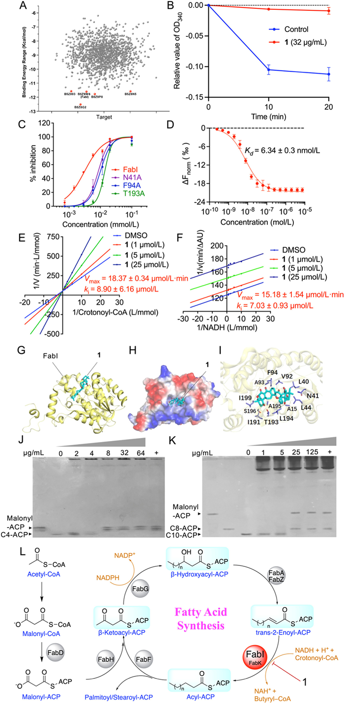
To further probe the molecular mechanism underlying FabI inhibition by compound 1, we investigated its binding interactions at the atomic level. Molecular docking was performed using the FabI-NADH-triclosan/diclosan co-crystal structures (PDB IDs: 2PD3 and 2PD4) as templates. The resulting model suggested that Asn41, Thr193, and Ser196 form hydrogen bonds with the hydroxyl groups of 1, while multiple hydrophobic residues (Ala15, Leu40, Leu44, Val92, Ala93, Phe94, Ile191, Leu194, Ala195, and Ile199) contribute additional stabilization through hydrophobic contacts and van der Waals interactions. Together, these residues define a putative binding pocket for 1 within the FabI active site (Figs. 3G-I). In comparison, a docking model with the human fatty acid synthase ketoacyl-reductase domain showed much weaker binding affinity (−27.77 kcal/mol) and loss of key hydrogen bonds, consistent with the species-selective inhibitory profile of 1.
To experimentally validate the predicted binding mode, we generated nine single-point FabI mutants (L40A, N41A, L44A, V92A, F94A, I191A, T193A, S196A, and I199A) and evaluated their sensitivity to compound 1. Most mutations led to a measurable reduction in inhibitory potency (Table S16 in Supporting information). Notably, N41A, F94A, and T193A exhibited significantly diminished sensitivity, with IC50 values of 8.63, 10.08, and 13.75 μmol/L, representing 2.8-, 3.2-, and 4.4-fold increases compared to wild-type FabI (Fig. 3C). These results support the functional relevance of the predicted hydrogen-bonding interactions and underscore the importance of Asn41, Phe94, and Thr193 in mediating binding of 1 to the FabI active site.
To further validate FabI as the functional intracellular target of compound 1, we next assessed whether modulation of FabI expression alters cellular susceptibility to 1 in H. pylori. We constructed an overexpression strain of H. pylori G27 harboring a plasmid (pHel3) encoding fabI and measured changes in antimicrobial sensitivity. Compared to the parental strain carrying an empty vector, the fabI-overexpressing strain exhibited a 4-fold increase in MIC (64 vs. 16 μg/mL), indicating reduced susceptibility to compound 1 (Table S18 in Supporting information). In contrast, both strains showed identical MIC values for amoxicillin (0.063 μg/mL) and metronidazole (1 μg/mL), serving as negative controls. These results further support FabI as the cellular target of 1 and suggest that overexpression confers resistance by titrating the compound's inhibitory effect.
To functionally corroborate the role of FabI inhibition in disrupting fatty acid metabolism, we next evaluated the impact of compound 1 on the reconstituted H. pylori FASII pathway. Given that FabI catalyzes the rate-limiting step of the FASII cycle [15], we assessed whether compound 1 impairs fatty acid synthesis in vitro. Using a reconstructed FASII system composed of FabH/FabF (3-oxoacyl-ACP synthases), FabG (3-ketoacyl-ACP reductase), FabZ (3-hydroxyacyl-ACP dehydratase), and FabI, we examined the effects of 1 on both initiation (acetyl-CoA + malonyl-ACP → acetoacetyl-ACP → butyryl-ACP) and elongation (octanoyl-ACP + malonyl-ACP → acetooctanoyl-ACP → decanoyl-ACP) steps of fatty acid biosynthesis [44]. Conformationally sensitive gel electrophoresis revealed that compound 1 completely blocked the initiation and elongation reactions at concentrations of 8 and 25 μg/mL, respectively (Figs. 3J and K). These results demonstrate that inhibition of FabI by compound 1 disrupts H. pylori fatty acid biosynthesis in vitro. Together with the kinetic, binding, and genetic data, these findings establish a coherent mechanistic framework linking FabI inhibition to disrupted fatty acid synthesis and bactericidal activity. A proposed mechanistic model summarizing the role of FabI within the FASII pathway and its inhibition by 1 is illustrated in Fig. 3L
Our phytochemical investigation of Icacina oliviformis leaves yielded twelve novel diterpenoids, including three distinct classes of unprecedented dimers: (8,14-seco‑ent‑kaurane) (16–14′,17–17′) spiroheterodimers 1–3 featuring a unique 6/6/6/5/6/7/6/6-fused octocyclic scaffold with rare 2-oxatricyclo[6.2.1.03,8]hendecane spirolinker segment; 4–6 as the first ent‑kaurane-(ent‑abietane) dimers connected through a novel C-17-C-17′ bond; 7 and 8 containing an unprecedented 1,3-dioxane-bridged ent‑kaurane-(8,14-seco‑ent‑kaurane) structure. Comprehensive structural characterization was achieved through integrated spectroscopic and crystallographic analyses. The isolates 1 and 3 demonstrated selective anti-H. pylori activity (MIC = 2–16 μg/mL), with 3 showing synergistic potential when combined with amoxicillin (FICI = 0.625). Genetic and biochemical analyses demonstrated that compound 1 binds FabI with high affinity (Kd = 6.34 nmol/L) and competitively inhibits enoyl-ACP reductase activity (Ki = 8.9 μmol/L) by displacing crotonoyl-CoA. Further kinetic, mutagenic, and pathway reconstitution assays confirmed that 1 selectively targets H. pylori FabI, disrupts FASII-mediated fatty acid biosynthesis, and exhibits minimal cytotoxicity toward human gastric cells, highlighting its favorable therapeutic profile. As the first reported natural product inhibitor of FabI, compound 1 is a structurally unique inhibitor exhibiting potent and selective antibacterial activity against H. pylori. These findings underscore the untapped pharmacological potential of ent‑kaurane diterpenoids in H. pylori treatment, expanding their structural diversity and target repertoire while reinforcing their status as privileged scaffolds. Collectively, this work delivers a rigorously validated lead compound and key mechanistic insights to advance the development of species-selective antibacterials against H. pylori.
Mingming Xu: Writing – original draft, Investigation, Conceptualization. Binglei Li: Formal analysis, Data curation. Junfei Zhou: Writing – review & editing, Funding acquisition, Data curation. Xudong Hang: Investigation, Data curation. Chuanlin Jin: Investigation. Chujie Lu: Investigation. Wei Lin: Methodology. Xinyi Wei: Investigation. Er-Xin Shang: Investigation. Liping Zeng: Investigation. Monday M. Onakpa: Resources. Oluwatosin O. Johnson: Resources. Chun-Tao Che: Writing – review & editing. Jin-Ao Duan: Supervision, Funding acquisition. Hongkai Bi: Supervision, Funding acquisition. Ming Zhao: Writing – review & editing, Supervision, Funding acquisition.
The authors declare that they have no known competing financial interests or personal relationships that could have appeared to influence the work reported in this paper.
This work was supported by the National Natural Science Foundation of China (No. 82204549 to J. Zhou and No 82473974 to H. Bi), the Jiangsu Collaborative Innovation Center of Chinese Medicinal Resources Industrialization (No. ZDXM-2020–17), the Jiangsu Specially Appointed Professor, and Jiangsu Province "Innovative and Entrepreneurial Team" Program.
Supplementary material associated with this article can be found, in the online version, at doi:
Y. Duan, Y. Xu, Y. Dou, et al., J. Hematol. Oncol. 18 (2025) 10. doi: 10.1117/12.3053830
J.K.Y. Hooi, W.Y. Lai, W.K. Ng, et al., Gastroenterology 153 (2017) 420–429. doi: 10.1053/j.gastro.2017.04.022
P. Malfertheiner, A. Link, M. Selgrad, Nat. Rev. Gastroenterol. Hepatol. 11 (2014) 628–638. doi: 10.1038/nrgastro.2014.99
T.C. Hong, E.M. El-Omar, Y.T. Kuo, et al., Lancet Gastroenterol. Hepatol. 9 (2024) 56–67. doi: 10.1016/S2468-1253(23)00281-9
J.J.C. Ho, M. Navarro, K. Sawyer, et al., Am. J. Gastroenterol. 117 (2022) 1221–1230. doi: 10.14309/ajg.0000000000001828
J. Casado, I. Olivan-Muro, S. Algarate, et al., Int. J. Mol. Sci. 25 (2024) 10175. doi: 10.3390/ijms251810175
Q. Tong, C. Yin, X. Hang, et al., Antimicrob. Agents Chemother. 68 (2024) e00314 24.
C. Lettl, F. Schindele, A.R. Mehdipour, et al., Cell Chem. Biol. 30 (2023) 499–512. doi: 10.1016/j.chembiol.2023.04.003
F.J. Asturias, J.Z. Chadick, I.K. Cheung, et al., Nat. Struct. Mol. Biol. 12 (2005) 225–232. doi: 10.1038/nsmb899
R.J. Heath, S.W. White, C.O. Rock, Appl. Microbiol. Biotechnol. 58 (2002) 695–703. doi: 10.1007/s00253-001-0918-z
P. Rana, S.M. Ghouse, R. Akunuri, et al., Eur. J. Med. Chem. 208 (2020) 112757. doi: 10.1016/j.ejmech.2020.112757
D.J. Payne, W.H. Miller, V. Berry, et al., Antimicrob. Agents Chemother. 46 (2002) 3118–3124. doi: 10.1128/AAC.46.10.3118-3124.2002
W.H. Miller, M.A. Seefeld, K.A. Newlander, et al., J. Med. Chem. 45 (2002) 3246–3256. doi: 10.1021/jm020050+
H. Lu, P.J. Tonge, Acc. Chem. Res. 41 (2008) 11–20. doi: 10.1021/ar700156e
T.G. Inci, E. Ugurel, M. Orlenco, et al., Mol. Biotechnol. 68 (2026) 846–853. doi: 10.1007/s12033-025-01411-x
S. Dinat, A. Orchard, S. Van Vuuren, J. Ethnopharmacol. 301 (2023) 115698. doi: 10.1016/j.jep.2022.115698
C. Sousa, R. Ferreira, N.F. Azevedo, et al., Crit. Rev. Microbiol. 48 (2022) 376–396. doi: 10.1080/1040841x.2021.1975643
M. Xu, J. Zhou, D. Heng, et al., J. Nat. Prod. 85 (2022) 1029–1038. doi: 10.1021/acs.jnatprod.1c01163
M. Xu, J. Zhou, L. Zeng, et al., Org. Chem. Front. 8 (2021) 3014–3022. doi: 10.1039/d1qo00374g
M. Zhao, J. Cheng, B. Guo, et al., J. Agric. Food Chem. 66 (2018) 7859–7872. doi: 10.1021/acs.jafc.8b02602
M. Zhao, B. Guo, M.M. Onakpa, et al., J. Nat. Prod. 80 (2017) 3314–3318. doi: 10.1021/acs.jnatprod.7b00668
M. Zhao, M.M. Onakpa, B.D. Santarsiero, et al., J. Nat. Prod. 78 (2015) 2731–2737. doi: 10.1021/acs.jnatprod.5b00688
M. Zhao, M.M. Onakpa, W.L. Chen, et al., J. Nat. Prod. 78 (2015) 789–796. doi: 10.1021/np5010328
M. Zhao, M.M. Onakpa, B.D. Santarsiero, et al., Org. Lett. 17 (2015) 3834–3837. doi: 10.1021/acs.orglett.5b01806
J. Zhou, Z. Wu, B. Guo, et al., Org. Chem. Front. 7 (2020) 355–367. doi: 10.1039/c9qo01320b
M. Sun, B. Guo, M. Xu, et al., J. Nat. Prod. 84 (2021) 949–955. doi: 10.1021/acs.jnatprod.9b01131
M. Xu, D. Di, L. Fan, et al., Phytochemistry 214 (2023) 113804. doi: 10.1016/j.phytochem.2023.113804
H.D. Flack, G. Bernardinelli, Chirality 20 (2008) 681–690. doi: 10.1002/chir.20473
G. Pescitelli, L. Di Bari, N. Berova, Chem. Soc. Rev. 43 (2014) 5211–5233. doi: 10.1039/C4CS00104D
D.F. Zinkel, B.P. Spalding, Tetrahedron Lett. 27 (1971) 2459–2462.
Y.Y. Fu, K. Hu, S.Y. Hou, et al., Org. Chem. Front. 12 (2025) 4247–4254. doi: 10.1039/d5qo00356c
E. Fujita, T. Fujita, H. Katayama, et al., Chem. Commun. (1967) 252–254.
H. Yao, S. Xie, X. Ma, et al., J. Med. Chem. 63 (2020) 8157–8178. doi: 10.1021/acs.jmedchem.0c00408
X.L. Yan, J.S. Zhang, J.L. Huang, et al., Phytochemistry 166 (2019) 112064. doi: 10.1016/j.phytochem.2019.112064
M. Liu, W.G. Wang, H.D. Sun, et al., Nat. Prod. Rep. 34 (2017) 1090–1140. doi: 10.1039/C7NP00027H
H.D. Sun, S.X. Huang, Q.B. Han, Nat. Prod. Rep. 23 (2006) 673–698. doi: 10.1039/b604174d
Y.H. Yu, Y. Peng, W.D. Li, Chin. Chem. Lett. 36 (2025) 111137. doi: 10.1016/j.cclet.2025.111137
S. Forli, R. Huey, M.E. Pique, et al., Nat. Protoc. 11 (2016) 905–919. doi: 10.1038/nprot.2016.051
N.R. Salama, B. Shepherd, S. Falkow, J. Bacteriol. 186 (2004) 7926–7935. doi: 10.1128/JB.186.23.7926-7935.2004
C.D. Radka, C.O. Rock, Annu. Rev. Microbiol. 8 (2022) 281–304. doi: 10.1146/annurev-micro-041320-110408
E.N. Parker, B.N. Cain, B. Hajian, et al., ACS Cent. Sci. 8 (2022) 1145–1158. doi: 10.1021/acscentsci.2c00598
S.A. Seidel, C.J. Wienken, S. Geissler, et al., Angew. Chem. Int. Ed. 51 (2012) 10656–10659. doi: 10.1002/anie.201204268
J. Jia, M. Zheng, C. Zhang, et al., Sci. Adv. 9 (2023) eadg5995. doi: 10.1126/sciadv.adg5995
Q. Zhang, L. Sun, L. Wu, et al., Chin. Chem. Lett. 36 (2025) 110795. doi: 10.1016/j.cclet.2024.110795

Figure 2 (A) Key 1H–1H COSY and HMBC correlations of 1, 4, and 7. (B) Key NOESY correlations of 1, 4, and 7. (C) X-ray ORTEP drawing of 1 and 3. (D) The experimental ECD spectra for 1–8 and the calculated ECD spectra for 4 and 6–8, and their enantiomers.

Figure 3 Inhibitory activity of 1 against H. pylori FabI and its interaction studies. (A) The molecular docking score distribution of 1502 proteins in H. pylori G27 using batch molecular docking. (B) Verification of the inhibitory activity of 1 against HpFabI in vitro by monitoring OD340 absorbance changes. (C) Determination of the IC50 of 1 against HpFabI and its mutant proteins. The IC50 values are 3.11 ± 0.09 μmol/L for FabI, 8.63 ± 1.62 μmol/L for N41A, 10.08 ± 1.15 μmol/L for F94A, and 13.75 ± 0.69 μmol/L for T193A. (D) MST assay demonstrating the in vitro binding affinity of 1 to HpFabI. (E) Inhibitory kinetic analysis of 1 against the FabI reaction substrate crotonoyl-CoA. (F) Inhibitory kinetic analysis of 1 against the FabI reaction substrate NADH. (G) The overall structure of FabI in complex with 5-CH3-THF (left panel). Compound 1 is drawn in stick mode, colored in cyan. FabI is shown as a cartoon and colored in yellow. (H) Surface electrostatic potential of FabI in complex with compound 1. (I) Stereo-view shows the predicted potential key residues of FabI involved in the interaction with 1. Color codes are the same as those in panel A. (J) Conformationally sensitive gel electrophoresis analysis verifying the in vitro inhibitory effect of 1 on the initiation reaction of the H. pylori type Ⅱ fatty acid synthesis pathway. (K) Conformationally sensitive gel electrophoresis analysis verifying the in vitro inhibitory effect of 1 on the elongation reaction of the H. pylori type Ⅱ fatty acid synthesis pathway. (L) Schematic diagram of 1 targeting HpFabI in the fatty acid synthesis pathway. Data are presented as mean ± standard deviation (SD) (n = 3).

 扫一扫看文章


扫一扫关注我们
 DownLoad:

 下载:
 下载: