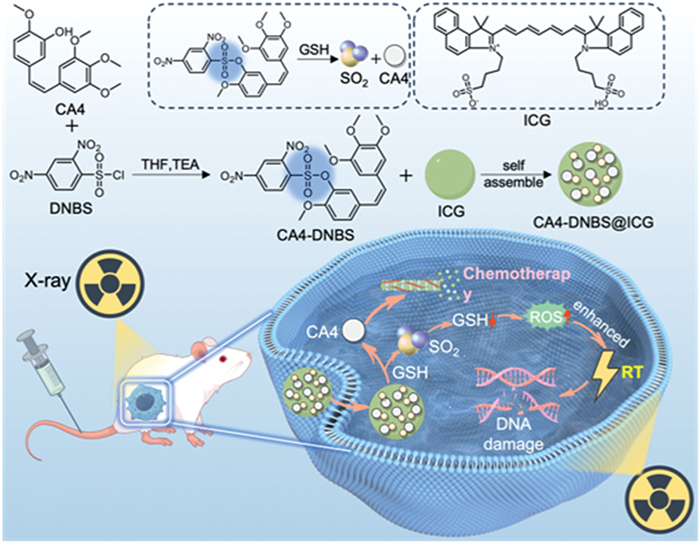
Scheme 1.
Schematic illustration of the preparation and antitumor mechanisms of CA4-DNBS@ICG nanoplatforms.

SO2 gas potentiator engineered for self-amplifying chemo-radiotherapy in breast tumors
Fanchun Hu , Yuhan Ding , Caiting Deng , Jingjing Zhang , Wei Qu , Yanqin Rong , Nandi Chen , Mengjiao Zhou , Feifei An


Radiotherapy employs particle beams or high-energy photon to generate a series of photoelectric effects and is well-established for radical cancer treatment [1]. Its efficacy stems from not only direct DNA damage, but also the production of various free reactive oxygen species (ROS), such as hydroxyl free radicals (OH•) and singlet oxygen (1O2), which enhance intracellular oxidative stress and inhibit tumor progression [2]. However, the ubiquitous hypoxic conditions (the oxygen (O2) level is < 1%) and robust DNA repair pathways significantly limit radiotherapy’s benefits and contribute to tumor recurrence [3]. Furthermore, radiotherapy-induced ROS generation and glutathione (GSH) depletion can be counteracted by upregulated intracellular antioxidant in some tumors, thereby drastically compromising the tumor treatment efficacy [4]. To date, various adjuvants and radiosensitizers have been developed to amplify the radiotherapeutic responses [5].
Combretastatin A4 (CA4) is a potent natural anticancer agent, but its clinical use is hindered by low hydrophilicity and a short half-life [6]. While monotherapy presents challenges, CA4 holds promise for combination strategies, which can overcome resistance, reduce toxicity, and enhance efficacy [7]. Preliminary animal studies and a clinical trial with its water-soluble prodrug suggest synergistic effects when combined with radiotherapy [8]. Considering the limitations of both chemotherapy and radiotherapy, strategies improving therapeutic precision or generating free radicals are highly desirable to enhance their efficacy [9–12].
Gas therapy leveraging small molecules like sulfur dioxide (SO2), O2, nitric oxide, and hydrogen sulfide has emerged as a promising anticancer strategy, owing to their passive tumor diffusibility without active transport requirements [13–16]. While SO2 is conventionally recognized as a toxic pollutant causing oxidative biomacromolecular damage [17], its controlled therapeutic delivery via tumor microenvironment-responsive prodrugs can harness this cytotoxicity against cancer [18]. Upon cellular internalization, SO2 hydrolyzes to cytotoxic sulfite radicals (•SO3−). These radicals induce apoptosis via Bcl-2/Bax regulation and deplete GSH, while amplified ROS triggers oxidative cell death [19]. Consequently, SO2 has been integrated with chemo-, photo-, and sonodynamic therapies to enhance efficacy [20–22]. However, uncontrolled diffusion of gaseous SO2 risks systemic toxicity and limits therapeutic precision. To overcome this, it is attractive to incorporate SO2 into a triple-therapy regimen via a stimuli-responsive nanoplatform, enabling localized SO2 release at effective yet safe dosages.
Herein, we developed a GSH-responsive triple-therapy nanoprodrug (CA4-DNBS@ICG) that self-amplifies oxidative stress to eradicate breast tumors. This nanoprodrug integrates 2,4-dinitrobenzenesulfonyl (DNBS) with CA4, enabling tumor-specific co-release of SO2 gas and CA4 chemotherapy upon encountering elevated GSH (Scheme 1). Critically, the near-infrared dye indocyanine green (ICG) drives π-stacked self-assembly, enhancing solubility while caging SO2 to prevent leakage. Upon internalization, GSH triggers SO2 release, which depletes intracellular GSH via two pathways: Reacting with GSH through thiol-sulfite exchange and inhibiting glutathione reductase to block GSH regeneration. GSH depletion impairs ROS scavenging, while SO2 induces ROS by disrupting mitochondrial function, jointly disrupting redox homeostasis and amplifying oxidative stress [23,24]. Concurrently, X-ray radiotherapy induces DNA damage and further augments intracellular oxidative stress, while CA4 inhibits tubulin polymerization. This cascade significantly sensitizes 4T1 cancer cells to both chemotherapy and radiotherapy. In vivo study demonstrated the CA4-DNBS@ICG for efficient tumor accumulation and agent release. Upon X-ray irradiation, the combined gas/chemo/radiotherapy exhibited significantly enhanced effect in inhibiting tumor growth. Taking these advantages, this designed multifunctional nanoplatform demonstrates significant potential for combinational tumor therapy.
CA4 is a promising broad-spectrum anticancer agent, but its clinical translation is challenged by poor water solubility and inadequate tumor targeting. To improve solubility and achieve tumor-specific drug release, we synthesized the CA4-DNBS prodrug, which is specifically activated by high intratumoral GSH concentrations, releasing the active CA4 along with SO2 (Fig. 1A and Fig. S1 in Supporting information). Successful synthesis of CA4-DNBS was confirmed by nuclear magnetic resonance (NMR) and high-resolution mass spectrometry (HRMS) (Figs. S2-S4 in Supporting information). However, the poor water solubility of CA4-DNBS prodrug still limits its in vivo application. Inspired by the favorable biocompatibility and clinical approval of ICG, a dye extensively used in the nanoparticle assembly for cancer theranostics, we co-assembled CA4-DNBS with ICG via solvent exchange [25]. This strategy yielded CA4-DNBS@ICG nanoparticles (Fig. 1B) endowed with fluorescence imaging capabilities for in vitro and in vivo tracking (Fig. S5 in Supporting information). Dynamic light scattering (DLS) characterization revealed a mean particle size of 80.02 nm and a polydispersity index (PDI) of 0.25 for CA4-DNBS@ICG (Fig. 1C), confirming successful nanoparticle preparation. Over a 3-day period, the hydrodynamic size of CA4-DNBS@ICG NPs remained stable in ultrapure water, phosphate buffer saline (PBS, pH 7.4), DMEM, and DMEM supplemented with 10% fetal bovine serum (FBS), confirming that CA4-DNBS@ICG NPs possess good colloidal stability under both physiological conditions and cell culture-relevant environments (Fig. S6 in Supporting information). Transmission electron microscopy (TEM) further demonstrated spherical morphology (Fig. 1D), consistent with DLS results. The nanoparticles exhibited excellent stability, showing minimal change in size over 48 h (0, 24, 48 h). Additionally, CA4-DNBS@ICG possessed a zeta potential of −73 ± 7.60 mV (Fig. 1E), attributable to the sulfonic acid groups of the ICG component.
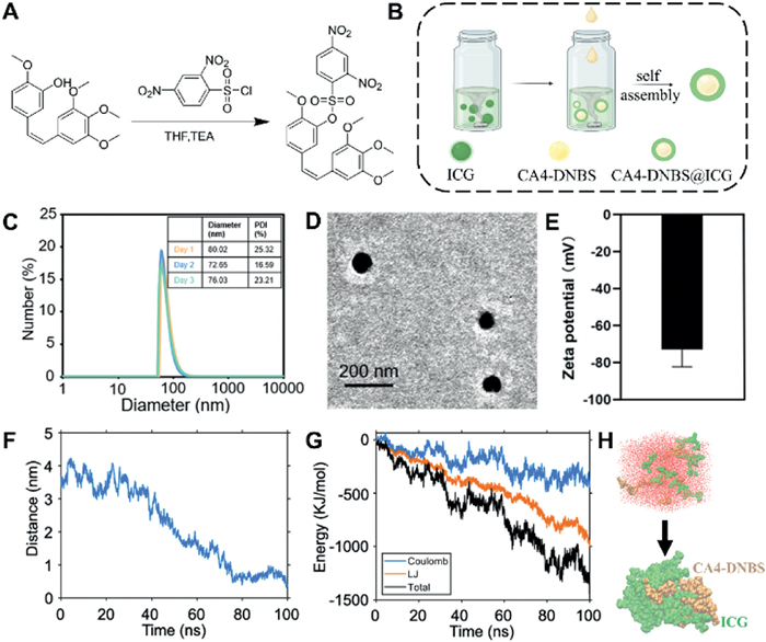
Subsequently, molecular dynamics simulations were employed to investigate the molecular interactions of ICG and CA4-DNBS. The simulations revealed that hydrophilic ICG molecules localized on the exterior of the aggregates, while hydrophobic CA4-DNBS was entrapped within the core of the aggregates. In detail, the average distance between ICG and CA4-DNBS was calculated to be approximately 3.69 Å, which was within the characteristic range of distances for π-π stacking (3.6–6.0 Å) (Fig. 1F). As shown in Fig. 1G, the electrostatic forces (ΔEelec = −366.8 kJ/mol) and van der Waals forces (ΔEvdw = −952.6 KJ/mol) were primary drivers of nanoparticle formation in CA4-DNBS@ICG, with the favorable van der Waals forces component primarily attributed to π-π stacking interactions. These evidences strongly support the proposed self-assembly mechanism of CA4-DNBS@ICG (Fig. 1H).
The higher levels of GSH in tumor site compared to normal tissue allow the prodrug to be specifically activated, releasing CA4 at the tumor site and thus reducing off-target toxic side effects. The DNBS group is well-established to release SO2 in response to GSH. To evaluate the GSH-responsive release capability of CA4-DNBS@ICG NPs, the free CA4-DNBS and CA4-DNBS@ICG NPs solution were analyzed using high performance liquid chromatography (HPLC). Following 8 h of incubation with GSH, a new peak corresponding to CA4 appeared while the CA4-DNBS peak decreased in the both solutions, confirming GSH-triggered activation of the prodrug to release CA4 (Fig. 2A). Subsequently, the CA4 release profile of CA4-DNBS@ICG under different conditions was systematically assessed. The weakly acidic tumor microenvironment is confirmed, with a lower pH than normal tissues and more significant subcellular pH differences. Based on this, the pH and GSH levels are adjusted to simulate the release of nanoparticles in different environments. CA4-DNBS@ICG was dispersed in PBS and divided into three groups simulating: (1) Solution simulated tumor microenvironment (pH 5.0 + GSH), (2) solution simulated physiological environment (pH 7.4), and (3) solution simulated acidic microenvironment (pH 5.0). As shown in Fig. 2B, cumulative CA4 release was minimal under physiological conditions (pH 7.4). This limited release is likely because ICG and CA4-DNBS molecules form stable nanoparticles via π-π stacking, guaranteeing its stability during blood circulation and benefitting its tumor accumulation; weakly acid conditions (pH 5.0) disrupt the π-π interactions, compromising the CA4-DNBS@ICG NPs’ structure and facilitating CA4 release. Owing to the combined effects of high GSH levels and an acidic environment, the solution simulating tumor microenvironment (pH 5.0 + GSH) achieved the highest cumulative CA4 release (98.39%) within 24 h. This near-complete release confirms the effective GSH-responsive drug release capability of CA4-DNBS@ICG.
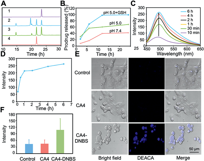
Subsequently, the capacity of CA4-DNBS prodrug to generate and release SO2 was investigated. CA4-DNBS was incubated with 4T1 breast cancer cells at 37 ℃, and 7-diethylaminocoumarin-3-aldehyde (DEACA) was used as the fluorescent probe for SO2 detection. The fluorescence intensity of the solution increased progressively over time, reaching an intensity at 6 h approximately 3.1-fold higher than at 0 min (Figs. 2C and D). These results preliminarily confirmed the prodrug’s ability to produce and release SO2. To assess SO2 generation in cells, 4T1 cells were incubated with CA4, CA4-DNBS, or PBS (control) for 3 h, followed by addition of the DEACA probe and observation via confocal laser scanning microscopy (CLSM). Cells treated with CA4-DNBS exhibited significantly stronger blue fluorescence compared to the Control and CA4 groups, with fluorescence intensity approximately three times higher (Figs. 2E and F). This phenomenon suggests that SO2 production resulted specifically from prodrug activation rather than endogenous physiological activities of cells. To further confirm CA4-DNBS’s GSH responsiveness, it was co-incubated with buthionine sulfoximine (BSO), a specific GSH synthesis inhibitor. Results showed notably reduced SO2 release in the CA4-DNBS + BSO group, directly verifying the prodrug’s GSH-dependent SO2 release (Fig. S7 in Supporting information). At the therapeutically relevant 24-h time point, the residual ratio of undecomposed CA4-DNBS was approximately 30.80%, confirming the majority of CA4-DNBS had undergone decomposition (Fig. S8 in Supporting information). Notably, CA4 and SO2 are co-released during CA4-DNBS decomposition (triggered by GSH-mediated cleavage of the DNBS linker); thus, the low residual ratio of undecomposed CA4-DNBS indirectly verifies efficient SO2 release. These results demonstrate that CA4-DNBS can be efficiently internalized by 4T1 cells and subsequently activated intracellularly to mediate SO2 release.
Precise tumor targeting and efficient drug delivery are critical for enhancing therapeutic efficacy while minimizing side effects. To evaluate the cellular uptake of CA4-DNBS@ICG, CLSM was utilized to perform cell fluorescence imaging on 4T1 cells. The characteristic fluorescence of ICG localized within the cytoplasm of the 4T1 cells (Fig. 3A), confirming effective NP internalization and enabling subcellular distribution analysis. Subsequent quantification of nanoparticles internalization in 4T1 cells was performed via flow cytometry using ICG fluorescence (APC-A750 channel). The results revealed that the internalization of CA4-DNBS@ICG increased proportionally with both concentration and incubation time, demonstrating time- and concentration-dependent uptake kinetics (Fig. 3B).
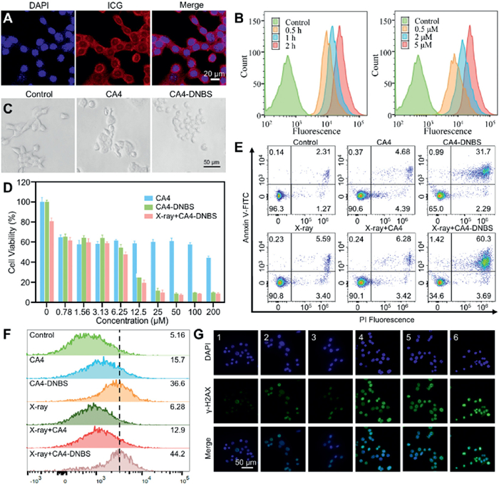
Inspired by the excellent internalization capability, we next assessed the prodrug’s synergistic antitumor effects in inhibiting the growth of 4T1 breast cancer cells. In cytomorphological studies, 4T1 cells of the Control group were completely stretched, while CA4-incubated 4T1 cells were shrunken and deformed. Notably, 4T1 cells incubated with CA4-DNBS were completely shrunken and rounded, suggesting that the combination of CA4 and SO2 possessed enhanced cytotoxicity (Fig. 3C). Furthermore, cytotoxicity of CA4-DNBS was assessed using (3-(4,5-dimethylthiazol-2-yl)-2,5-diphenyltetrazolium bromide) (MTT) assay. At concentrations < 12.5 µmol/L, CA4-DNBS and free CA4 showed comparable efficacy (Fig. 3D), likely due to delayed prodrug activation limiting CA4 release and SO2 generation. Notably, combining CA4-DNBS with X-ray irradiation (X-ray + CA4-DNBS) significantly reduced viability (half-maximum inhibitory concentration (IC50) = 5.8 µmol/L) versus CA4-DNBS alone (IC50 = 7.1 µmol/L) or free CA4 (IC50 = 154.9 µmol/L), demonstrating synergy between chemotherapy (CA4), radiotherapy, and SO2 gas therapy. To further validate the combinatorial effect of these three antitumor modalities, the annexin V conjugated with fluorescein isothiocyanate (Annexin V-FITC) and propidium iodide (PI) double staining were used to analyze the apoptosis of cancer cells by flow cytometry. Cells received X-ray irradiation (6 Gy) immediately after treatment with 15 µmol/L CA4 equivalents. The X-ray + CA4-DNBS group showed the highest late apoptosis rate (60.3%, Fig. 3E), further confirming synergistic anticancer activity between CA4, radiotherapy, and SO2.
To further elucidate the combinatorial antitumor mechanisms, 2′,7′-dichlorodihydrofluorescein diacetate (DCFH-DA) probe was utilized to assess the ROS generation ability. As shown in Fig. 3F, both CA4-DNBS and X-ray + CA4-DNBS groups exhibited significantly higher fluorescence intensity compared to other treatments, indicating enhanced ROS production. This increase is likely attributed to prodrug activation and subsequent SO2 release, which can trigger ROS generation while potentially depleting intracellular GSH. To verify the GSH depletion effect, GSH levels in 4T1 cells following CA4-DNBS@ICG treatment were further quantified, and a significant reduction in GSH content (~40.18%) was detected (Fig. S9 in Supporting information). In summary, these findings demonstrate that CA4-DNBS can induce intracellular ROS accumulation while consuming GSH, two mutually reinforcing effects that likely synergize to amplify its antitumor activity. DNA damage was further evaluated using γ-H2AX (phosphorylated histone H2AX), a biomarker of DNA double-strand breaks. While X-ray irradiation rapidly induces γ-H2AX phosphorylation, the X-ray + CA4-DNBS group showed significantly stronger γ-H2AX signal than X-ray + CA4 (Fig. 3G). This enhancement suggests SO2-mediated GSH downregulation sensitizes cancer cells to radiotherapy by impairing DNA repair. Although X-ray induces marked DNA damage, MTT assay shows limited synergistic proliferation inhibition by X-ray, possibly due to the short 24-h observation window of the assay. While γ-H2AX reflects early damage and cell cycle arrest, the cellular outcomes induced by X-ray have not yet fully manifested. These findings demonstrate that CA4 (chemotherapy), X-ray (radiotherapy), and SO2 (gas therapy) induce cellular damage through distinct yet synergistic mechanisms, significantly augmenting therapeutic efficacy against 4T1 cells.
To improve prodrug solubility for in vivo studies, ICG was adopted to form nanoparticles by co-assembly with CA4-DNBS molecules. TEM and DLS results confirmed the nanoparticle formation, with a particle size theoretically optimal for exploiting the enhanced permeability and retention (EPR) effect, thereby enabling passive tumor targeting. All animal experiments have been approved by the Biomedical Ethics Committee of Health Science Center, Xi’an Jiaotong University (No. 2023–1675). Following intravenous injection of CA4-DNBS@ICG (CA4-DNBS, 20 mg/kg equivalent) into 4T1-tumor bearing BALB/C mice (n = 3), the enrichment of CA4-DNBS@ICG in tumors was monitored using an optical imaging system. As shown in Fig. 4A, the fluorescence signal was evident in the tumor site at 2 h post-injection, with clear tumor/normal tissue demarcation emerging by 4 h. Moreover, the fluorescence signal at the tumors site increased progressively over time, indicating sustained CA4-DNBS@ICG accumulation. Subsequently, mice were sacrificed 48 h post-injection for ex vivo organ imaging. Fluorescence intensity in tumors significantly exceeded that in all other tissues (Figs. 4B and C), confirming the tumor-targeting capability of CA4-DNBS@ICG.
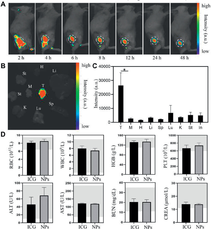
To evaluate biosafety, mice from different treatment groups were sacrificed on day 14. Hematoxylin and eosin (H&E) staining of heart, liver, spleen, lung, and kidney sections revealed no significant lesions or inflammation in the CA4-DNBS@ICG + X-ray group (Fig. S10 in Supporting information). Additionally, routine blood analysis 14 days post-injection showed all hematological parameters within normal ranges (Fig. 4D). These normal histopathology and blood biochemistry results indicate that liver and kidney functions of mice were normal, confirming the in vivo biosafety profile of CA4-DNBS@ICG. Encouraged by the excellent in vitro therapeutic efficacy of CA4-DNBS, we further evaluate the tumor inhibition capability of CA4-DNBS@ICG in 4T1 tumor-bearing BALB/c mice. When the tumor volumes reached ~70 mm3, BALB/C mice were randomly divided into 5 groups (n = 5 in each group): PBS, X-ray alone, free CA4, CA4-DNBS@ICG, and CA4-DNBS@ICG + X-ray. Mice in the CA4 and CA4-DNBS@ICG groups received intravenous drug administration (26 mg/kg CA4 equivalents) on days 1 and 7. Group designated for radiation received X-ray irradiation (6 Gy) immediately after each drug dose (Fig. 5A).

After 14 days, PBS-treated tumors exhibited significant growth. This growth was partially suppressed by free CA4 (19.4% inhibition) and X-ray alone (74.0% inhibition) (Figs. 5B-E). Strikingly, the CA4-DNBS@ICG + X-ray group achieved the highest tumor inhibition rate (87.8%), validating the in vivo synergistic anticancer effect of CA4-based chemotherapy, SO2 gas therapy, and radiotherapy. These results align with in vitro findings and confirm that combining X-ray with the SO2-generating CA4-DNBS@ICG nanoplatform potently inhibits tumor growth. Notably, no significant body weight changes occurred in any group during treatment (Fig. 5F), further indicating the favorable biosafety profile of CA4-DNBS@ICG.
Overall, we have developed a GSH-responsive nanoplatform (CA4-DNBS@ICG) for the targeted co-delivery of SO2 prodrugs and CA4 to tumor sites, aiming to synergistically enhance breast cancer therapy. The core innovations lie in the design of the intelligent drug delivery nanoplatform and the establishment of a triple-modal synergistic therapeutic cascade. Specifically, CA4-DNBS@ICG enables the targeted co-delivery of CA4/SO2 prodrugs and GSH-triggered release in tumor cells, thereby improving biosafety and therapeutic efficacy. Mechanistically, X-ray radiotherapy induces DNA damage and intracellular oxidative stress, CA4 inhibits tubulin polymerization, and SO2 depletes GSH in tumors while increasing ROS levels. This synergy overcomes chemoradiation resistance by regulating the redox balance in tumor cells, thereby significantly sensitizing 4T1 breast cancer cells to chemotherapy and radiotherapy. In vitro experiments validated this effect, as evidenced by enhanced DNA damage, promoted cell apoptosis, and a distinct ROS burst in tumor cells. In vivo, the CA4-DNBS@ICG + X-ray combination achieved an 87.8% tumor inhibition rate in 4T1 tumor-bearing mice after 14 days of treatment, which was markedly superior to that of free CA4 and X-ray monotherapy alone [26]. Future work will focus on expanding the application of CA4-DNBS@ICG to other cancer subtypes; modifying targeting ligands to optimize intratumoral delivery efficiency; integrating imaging agents for real-time monitoring; and evaluating long-term toxicity and immunomodulatory effects in large animal models to facilitate clinical translation.
Fanchun Hu: Methodology, Investigation. Yuhan Ding: Writing – original draft, Investigation. Caiting Deng: Writing – original draft, Methodology. Jingjing Zhang: Writing – original draft, Investigation. Wei Qu: Writing – original draft, Data curation. Yanqin Rong: Writing – original draft, Methodology. Nandi Chen: Writing – review & editing. Mengjiao Zhou: Writing – original draft. Feifei An: Writing – review & editing, Funding acquisition.
The authors declare that they have no known competing financial interests or personal relationships that could have appeared to influence the work reported in this paper.
The work received financial support from the National Natural Science Foundation of China (No. 81971637) and Natural Science Foundation of Top Talent of SZTU (No. GDRC202433). We thank the Instrumental Analysis Center of Xi’an Jiaotong University for their help in characterizations.
Supplementary material associated with this article can be found, in the online version, at doi:
M. Abdel-Wahab, F. Giammarile, M. Carrara, et al., Lancet Oncol. 25 (2024) 545–580. doi: 10.1016/S1470-2045(24)00207-9
P.G.S. Prasanna, M.M. Ahmed, J.A. Hong, et al., Lancet Oncol. 25 (2024) 501–511. doi: 10.1016/S1470-2045(24)00069-X
L. Yi, X. Jiang, Z.G. Zhou, et al., Adv. Mater. 36 (2024) 2304328. doi: 10.1002/adma.202304328
Q.Y. Li, Z.D. Wu, S.W. Chen, et al., ACS Nano 19 (2024) 1757–1770. doi: 10.3390/s24061757
Y. Liao, D.M. Wang, C.L. Gu, et al., Nat. Nanotechnol. 19 (2024) 1892–1902. doi: 10.1038/s41565-024-01784-1
C.Z. Gao, H.R. Jia, T.Y. Wang, et al., Chin. Chem. Lett. 36 (2025) 109840. doi: 10.1016/j.cclet.2024.109840
M. Zheng, H. Xu, Y. Huang, et al., J. Control. Release 365 (2024) 480–490. doi: 10.1016/j.jconrel.2023.11.054
Q. S Ng, H. Mandeville, V. Goh, et al., Ann. Oncol. 23 (2012) 231–237. doi: 10.1093/annonc/mdr332
H. Chen, C. Xing, H. Lei, et al., J. Control. Release 368 (2024) 637–649. doi: 10.1016/j.jconrel.2024.03.015
Z. Hu, H. Tan, Y. Ye, et al., Adv. Mater. 36 (2024) 2412227. doi: 10.1002/adma.202412227
Z. Wang, X. Ren, Y. Li, et al., ACS Nano 18 (2024) 10288–10301. doi: 10.1021/acsnano.4c01625
B. Chen, Y. Wang, M. Mu, et al., ACS Nano 19 (2025) 10147–10161. doi: 10.1021/acsnano.4c17148
P. Pan, T. Liu, L. Zhang, et al., Adv. Drug Deliv. Rev. 228 (2025) 115738.
D.R. Mink van der Molen, M.C.T. Batenburg, W. Maarse, et al., JAMA Oncol. 10 (2024) 464–474. doi: 10.1001/jamaoncol.2023.6776
F.J. Xu, M. J Wang, E. Dotse, et al., Angew. Chem. Int. Ed. 63 (2024) 202404561. doi: 10.1002/anie.202404561
H. L Zheng, H.D. Chen, Y.J. Cai, et al., Mol. Cell 84 (2024) 4016–4030. doi: 10.1016/j.molcel.2024.08.035
M. Chen, T. Xu, L. Song, et al., Theranostics 14 (2024) 5461–5491. doi: 10.7150/thno.98884
J. Liang, G. Cheng, L. Qiu, et al., Adv. Sci. 12 (2025) 2409442. doi: 10.1002/advs.202409442
W. Shen, W.G. Liu, H.L. Yang, et al., Biomaterials 178 (2018) 706–719. doi: 10.1016/j.biomaterials.2018.02.011
P. He, X. Ren, Y. Zhang, et al., Acta Biomater. 174 (2024) 91–103. doi: 10.1016/j.actbio.2023.12.011
S. Liu, J. Li, A. Wang, et al., Angew. Chem. Int. Ed. 64 (2025) 202422362. doi: 10.1002/anie.202422362
S.S. Zhao, Z.H. Qu, L.K. Wang, et al., ACS Appl. Mater. Inter. 17 (2025) 16681–16695. doi: 10.1021/acsami.5c01310
N. Zhang, W. Ping, M. Suo, et al., Adv. Sci. 11 (2024) e2405575. doi: 10.1002/advs.202405575
L.T. Wu, Y.J. Meng, X.M. Zheng, et al., Chin. Chem. Lett. 36 (2025) 110808. doi: 10.1016/j.cclet.2024.110808
Q.Y. Ye, Y.Y. Liang, Y.H. Yuan, et al., Chin. Chem. Lett. 36 (2025) 110432. doi: 10.1016/j.cclet.2024.110432
X. Gong, J. Li, X. Xu, et al., Biomaterials 290 (2022) 121855. doi: 10.1016/j.biomaterials.2022.121855

Scheme 1 Schematic illustration of the preparation and antitumor mechanisms of CA4-DNBS@ICG nanoplatforms.

Figure 1 Preparation and characterization of CA4-DNBS@ICG. (A) Synthetic procedure of CA4-DNBS. (B) Schematic illustration of CA4-DNBS@ICG preparation. (C) Hydrodynamic size distribution of CA4-DNBS@ICG. (D) TEM image of CA4-DNBS@ICG. (E) Zeta potential of CA4-DNBS@ICG. (F) Average stacking distances between molecules simulated using GROMACS. (G) Adsorption energy analysis between ICG and CA4-DNBS. (H) Molecular aggregation model of CA4-DNBS@ICG from computer simulations. Data are presented as mean ± standard deviation (SD) (n = 3).

Figure 2 Performance characterization of GSH-responsive release and SO2 generation. (A) GSH-responsive drug release from CA4-DNBS@ICG determined by HPLC (1: CA4-DNBS; 2: CA4-DNBS@ICG + GSH; 3: CA4-DNBS + GSH; 4: CA4). (B) Time-dependent CA4 release profiles from CA4-DNBS@ICG. (C) Time-dependent fluorescence spectrum of a DEACA solution (50 µmol/L; SO2 probe) containing CA4-DNBS (50 µmol/L) and GSH (1 mmol/L). (D) Fluorescence intensity of the DEACA solution (50 µmol/L) containing CA4-DNBS (50 µmol/L) and GSH (1 mmol/L) over time. (E) CLSM images of 4T1 cells incubated with PBS, CA4 or CA4-DNBS. (F) Quantification of the DEACA fluorescence signal in 4T1 cells. Data are presented as mean ± SD (n = 3).

Figure 3 In vitro antitumor activity of the CA4-DNBS prodrug. (A) CLSM images of ICG fluorescence in 4T1 cells after 2 h incubation with CA4-DNBS@ICG. (B) Cellular uptake of CA4-DNBS@ICG in 4T1 cells. (C) Morphology of 4T1 cells after 12 h incubation with PBS, CA4 (10 µmol/L), or, CA4-DNBS (10 µmol/L). (D) Cytoviability of 4T1 cells after 24 h incubation with different concentrations. (E) Apoptosis evaluation of 4T1 cells after different treatments analyzed by flow cytometry using Annexin V-FITC/PI staining. (F) Flow cytometric analysis of ROS levels in 4T1 cells stained with DCFH-DA following various treatments. (G) DNA double-stranded breaks in 4T1 cells assessed by immunofluorescence at 2 h after various treatments (1: control; 2: CA4; 3: CA4-DNBS; 4: X-ray; 5: X-ray + CA4; 6: X-ray + CA4-DNBS). DAPI, 4′, 6-diamidino-2-phenylindole dihydrochloride. Data are presented as mean ± SD (n = 4).

Figure 4 Biodistribution and biosafety of CA4-DNBS@ICG. (A) The fluorescence images of 4T1 tumor-bearing mice after tail vein injection of CA4-DNBS@ICG at different time points (T: tumor; M: muscle H: heart; Li: liver; Sp: spleen; Lu: lung; K: kidney; St: stomach; In: intestine). (B) Biodistribution and (C) quantification of fluorescence signal after injection of CA4-DNBS@ICG for 48 h. (D) Blood analysis of mice injected with CA4-DNBS@ICG. Data are presented as mean ± SD (n = 3).

Figure 5 In vivo tumor inhibition activity of CA4-DNBS@ICG. (A) The schematic treatment of 4T1 tumor-bearing mice. (B) Photographs of tumor-bearing mice on day 14 after different treatments. (C) Photographs of tumor tissues collected on day 14 after different treatments. (D) Tumor growth curves of 4T1 tumor-bearing mice after different treatments during 14 days. (E) The weight of tumor tissues at the end point of different treatments (1: PBS; 2: CA4; 3: CA4-DNBS@ICG; 4: X-ray; 5: CA4-DNBS@ICG + X-ray). (F) Body weight changes of mice with different treatments. Data are presented as mean ± SD (n = 5). *P < 0.05 was tested via a Student’s t-test.

 扫一扫看文章


扫一扫关注我们
 DownLoad:
 下载:
 下载: