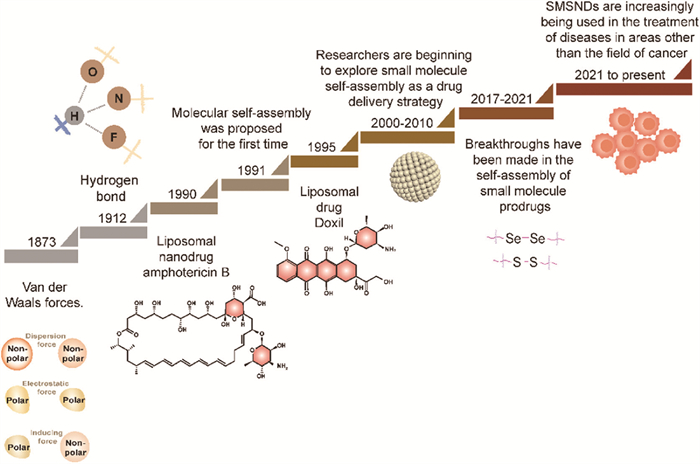
Figure 1.
The discovery of the interactions and the launch of traditional nanodrugs: The foundation and starting point for the development of SMSNDs.

Small-molecule self-assembled nanodrugs: Molecular design, assembly dynamics and biomedical applications
Yi Li , Jianbin Shi , Xinxin Sun , Zhixiao Zhang , Jin Sun , Zhonggui He , Cong Luo , Shenwu Zhang


Small-molecule drugs, characterized by their distinct chemical scaffolds and diverse pharmacodynamic profiles, are integral to contemporary pharmacotherapy, exhibiting notable clinical efficacy [1,2]. These agents are extensively employed in oncological treatments [3,4], neuropsychiatric disorders, and hematologic conditions [5,6], underscoring their broad therapeutic scope and significance in modern medicine [7]. In comparison to biologic agents such as recombinant proteins, monoclonal antibodies, and vaccines [8], small-molecule drugs confer unique pharmacokinetic and pharmacodynamic advantages [9,10], maintaining their pivotal role in clinical practice [11]. Nonetheless, they are associated with inherent limitations [12], including limited target selectivity due to their simple chemical structures, which can lead to off-target effects and adverse reactions [13]. Additionally, their pharmacokinetic properties, such as short systemic half-life, necessitate frequent administration, and issues like poor aqueous solubility [14] and suboptimal bioavailability can compromise therapeutic outcomes. These challenges have driven ongoing research into novel drug delivery systems and alternative therapeutic modalities.
The emergence of nanomedicine marks a pivotal advancement in overcoming the constraints of conventional small-molecule drugs [15-21]. The development of various nanomaterials, including liposomes [22], micelles [23], and proteins [24], has markedly improved pharmacokinetic profiles by prolonging systemic circulation, increasing bioavailability, and minimizing toxicity [25]. The emergence of traditional nanodrugs had also laid the foundation for the appearance of small-molecule self-assembled nanodrugs (SMSNDs) (Fig. 1). However, before the introduction of self-assembly technology, while nanomedicine made substantial strides in advancing the application of small-molecule drugs, certain limitations remained [26,27]. These included limited biocompatibility of complex nanostructures, suboptimal drug loading capacity with potential cytotoxicity, and insufficient targeting specificity, all of which impeded clinical translation [28-30]. Consequently, current research efforts are focused on developing innovative nanotechnologies and strategies to overcome these limitations inherent in traditional nanomedicine platforms [31].
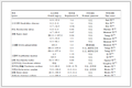
Self-assembly nanotechnology has revolutionized the field of nanomedicine for small-molecule drugs [32-35]. SMSNDs represent a significant clinical breakthrough [36,37], obviating the need for traditional delivery vectors, thereby enhancing drug payload capacity and mitigating systemic toxicity [38,39]. Furthermore, the integration of environmental trigger-sensitive linkers enables SMSNDs to selectively target pathophysiological microenvironments, facilitating high-concentration drug release at disease sites while sparing healthy tissues [40]. In their assembled configuration, SMSNDs not only facilitate the assembly of a single drug but also enable the co-assembly of multiple drugs, offering the potential for multi-drug collaborative delivery. By fine-tuning the ratio of different drugs, SMSNDs support personalized treatment and enhance precision medicine [41]. The comparison of the advantages and limitations of SMSNDs with traditional nanodrug delivery systems is shown in Table 1. These characteristics further underscore the tremendous potential of SMSNDs in nanomedicine delivery systems, positioning them as a promising avenue for future innovations in nanomedicine [42,43].
 DownLoad:
CSV
| Characteristics | Traditional nanodrugs | SMSNDs |
| Typical examples | Polymer nanoparticles Liposomal nanoparticles Inorganic nanomedicines |
Self-assembled prodrug nanodrug Self-assembled pure drug nanodrug Self-assembled hybrid nanodrug |
| Loading dose | Lower | Extremely high, close to 100% |
| Preparation process | Complex | Simple |
| Composition | Complex | Simple |
| Biocompatibility | Poor | Good |
| Design flexibility and intelligence | Medium | High |
| Current status of clinical translation | Mature | Emerging/leading-edge |
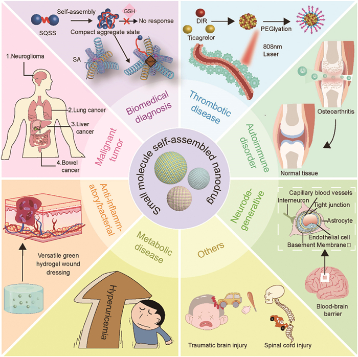
This review focuses on the increasing application of SMSNDs, which, owing to their unique advantages, have become a prominent modality in oncological therapeutics and are expanding into other biomedical domains, including cardiovascular pathology, neurodegenerative disorders, and thrombotic conditions. The capacity of SMSNDs to target complex pathologies positions them as a promising platform for the treatment of refractory diseases. Despite numerous existing reviews on drug self-assembly processes, there remains a significant gap in comprehensive analyses addressing the mechanisms of assembly and release, classification schemas, and diverse biomedical applications of SMSNDs. Most current studies tend to focus on specific assembly techniques or application contexts, often neglecting a systematic overview of the broader development landscape of SMSNDs. Consequently, an in-depth review of recent advancements in SMSND technology is essential to bridge these gaps, providing valuable insights for the rational design and clinical translation of next-generation nanotherapeutics. This review systematically examines the mechanisms underlying SMSND assembly and release, categorizes their structural types, and explores their advanced applications in oncology, biomedical imaging, bioactive molecule detection, as well as antimicrobial and anti-inflammatory therapies. Additionally, we highlight the broad potential of SMSNDs within biomedicine (Fig. 2). Finally, we analyze the design principles, therapeutic advantages, current challenges, and prospects of SMSNDs in clinical disease management. Although SMSNDs demonstrate significant potential, obstacles such as limited clinical translation despite extensive research and manufacturing scalability issues persist. This review aims to equip researchers with the theoretical framework and strategic insights necessary to address these challenges, promote technological advancement, and foster breakthroughs in disease treatment modalities.
SMSNDs demonstrate significant potential for therapeutic intervention in clinical pathologies, offering distinct advantages in enhancing drug encapsulation efficiency, optimizing pharmacodynamic outcomes, and mitigating systemic toxicity [44,45]. From a mechanistic standpoint, the formation of SMSNDs relies primarily on various non-covalent intermolecular interactions [46], which facilitate the organized aggregation of therapeutic agents and confer superior stability and functional attributes. Consequently, elucidating the molecular assembly mechanisms of SMSNDs is essential for establishing a robust framework to support their translational clinical deployment. This section focuses on the assembly and controllable release mechanisms of several representative SMSNDs. The remaining examples can be found in Table S1 (Supporting information).
Self-assembly is widely recognized as a spontaneous process in which disordered molecules interact to form ordered structures [47,48]. In the context of small-molecule nanodrugs, non-covalent interactions serve as the fundamental basis for self-assembly, including π-π stacking, hydrophobic force, hydrogen bonding, and van der Waals forces (Fig. S1 in Supporting information) [49,50]. These interactions can occur individually or coexist, depending on the structural characteristics of the small molecules involved. To illustrate this, Table S2 (Supporting information) systematically presents the assembly mechanisms of representative SMSNDs and examines the key molecular structural features closely related to these assembly processes. Although non-covalent bonds generally exhibit relatively weak affinity, a sufficient number of such bonds can promote stable self-assembly. These interactions play a critical role in the formation of nanodrugs. Therefore, in this section, we will discuss various combinations of interesting assembly mechanisms to provide more targeted guidance for SMSNDs design and optimization.
The proportion of hydrophobic drugs among small-molecule drugs is significant, and they are essential in diagnosing and treating clinical diseases due to their precise composition, structure, and favorable drug-forming properties. However, their limited solubility poses a major challenge for clinical application. Traditionally, many U.S. Food and Drug Administration (FDA)-approved small-molecule anticancer drugs with low water solubility have been encapsulated in nanocarriers to minimize the adverse effects of chemotherapy [51,52]. However, the extremely low drug loading capacity and premature drug leakage from the systemic circulation have significantly compromised the therapeutic efficacy. Fortunately, with a deeper understanding of small-molecule interactions, researchers have discovered that the hydrophobic force can drive the self-assembly of these drugs into nanodrugs, as shown in Fig. S1, hydrophobic force [53]. This discovery not only addressed the issue of poor drug solubility but also held great promise for enhancing pharmacokinetics, systemic drug distribution, and therapeutic efficacy. The hydrophobic anticancer drug venetoclax (Vtx) faced problems such as low solubility, poor bioavailability, and easy triggering of immune responses due to its inherent properties. To overcome these limitations, researchers constructed it with doxorubicin (DOX) as nanoparticles (DOX-Vtx NPs) through molecular self-assembly. These DOX-Vtx NPs not only significantly improved the solubility and stability of drugs but also prolonged the cycle time and promoted targeted accumulation of tumors through the enhanced permeability and retention (EPR) effect. Experiments showed that DOX-Vtx NPs effectively reduced systemic toxicity while enhancing anti-tumor activity, providing a new strategy for efficient delivery of hydrophobic drugs [54].
As a crucial non-covalent interaction, π-π stacking plays an extremely important role in the construction of nanostructures, especially in small molecules containing aromatic rings or benzene rings (Fig. S1). This interaction is crucial for the self-assembly of small-molecule drugs, and its unique physical and chemical properties can promote the ordered assembly of drug molecules, thereby significantly enhancing the synergistic effect of the drugs [55]. In the nanodrug composed of atorvastatin (ATO), new indocyanine green (IR820), and DOX, all three drug molecules contain π-electron conjugated structures. Therefore, π-π stacking played a key role in their self-assembly process. This non-covalent interaction mainly occurred between the pyrrole group of IR820 and the aromatic ring of ATO. It not only promoted the ordered arrangement of drug molecules and the formation of nanoparticles, but also enhanced the stability of the nanodrug, improved the drug loading efficiency and release characteristics, and made them show significant anti-tumor effects in radiotherapy [56].
It is essential to emphasize that the driving force for the assembly of small molecules is closely related to the Gibbs free energy (ΔG) change. The ΔG is a crucial thermodynamic parameter for determining whether a process is spontaneous. When ΔG < 0, the reaction or assembly process is spontaneous; when ΔG > 0, the process is non-spontaneous; and when ΔG = 0, the system is in an equilibrium state. In the process of small-molecule assembly, various forces act together to promote the orderly aggregation of molecules. These forces drive the assembly process by reducing the total energy of the system. For example, the formation of hydrogen bonding can significantly reduce the energy of the system, making ΔG more negative and promoting the spontaneous process of assembly. Similarly, Hydrophobic forces reduce the contact between molecules and water, lowering the entropy change (ΔS) of the system, but also reducing the enthalpy change (ΔH), thereby lowering ΔG under certain conditions. According to the ΔG formula, ΔG = ΔH − TΔS, the synergy of enthalpy change and entropy change determines the spontaneity of the assembly process. Therefore, understanding the relationship between these forces and the ΔG change is of great significance for designing and regulating the assembly process of small molecules. By adjusting the strength and type of forces, the direction and structure of the assembly process can be precisely controlled, thereby optimizing the material properties.
The stability and functionality of SMSNDs are crucial for optimal pharmacokinetics and therapeutic efficacy, as the drug release profile directly influences bioavailability and target site pharmacodynamics. Consequently, following the investigation of the self-assembly processes of small-molecule drugs, our focus shifted to the release behavior of these nanocarriers post-arrival at the pathological microenvironment (Fig. 3). During in vivo administration, SMSNDs must preserve structural integrity to prevent premature drug release in non-target tissues, thereby reducing off-target toxicity. Conversely, upon reaching the target site, these nanodrugs must rapidly degrade under specific stimulus conditions to enable drug release [57]. For instance, disulfide-bonded nanostructures can be cleaved within the highly REDOX-active tumor microenvironment, leading to disassembly and subsequent payload release. This stimulus-responsive mechanism enhances drug accumulation at the pathological site while minimizing collateral damage to healthy tissues. In this chapter, we will analyze the release mechanisms of SMSNDs to clarify their critical functions and impact in achieving precision nanodrug delivery.

The levels of glutathione (GSH) and reactive oxygen species (ROS) in tumor cells are significantly higher than those in normal cells [58,59]. This unique redox microenvironment provides a promising target for cancer treatments, sparking considerable interest in designing oxidation/reduction-responsive SMSNDs that specifically target tumor cells [60,61]. Chemical bonds that are sensitive to oxidation/reduction, such as those involving sulfur, selenium, or tellurium, are commonly used as linkers in the development of prodrugs that respond to these conditions (Fig. 3) [62-65]. These chemical bonds are sensitive to the elevated levels of GSH or ROS in the tumor microenvironment (TME), thereby triggering drug release specifically at the tumor site. This intelligent release mechanism ensures precise drug delivery, minimizing toxic side effects on normal cells and enhancing the therapeutic efficacy. Zhang et al. successfully developed an innovative curcumin (CUR) conjugate composed of two CUR molecules linked by a thioether bond (CUR-S-CUR@PEG NPs), engineered to exhibit reduction-responsive [66]. They conducted in-depth studies on the release behavior of the CUR-S-CUR@PEG NPs in response to reduction. The results demonstrated that the particle size of GSH-treated CUR-S-CUR@PEG NPs was significantly larger than that of non-GSH-treated nanoparticles, indicating responsive degradation behavior in the presence of GSH. This discovery confirmed its potential for achieving precise drug release within the TME, providing an important basis for the development of new tumor-targeted drug delivery systems [66].
Researchers have strategically leveraged the pathophysiological characteristics of pathological tissues to design nanodrugs that can specifically target and respond to certain pathological conditions [67]. These studies had been conducted by identifying biomarkers or microenvironmental changes specific to pathological tissues, such as pH changes, enzyme activity differences, temperature variations, and other relevant changes, and subsequently designing nanodrugs responsive to these stimuli [68-70]. For instance, researchers had successfully combined the A5 prodrug with Ce6 to form self-assembled nanoparticles (AKNP) [71]. Through π-π stacking and hydrophobic force, they achieved the stable self-assembly of the drug. These self-assembled nanoparticles not only enhanced the physiological stability of the drug, prolonged the drug retention time at the tumor site, but also achieved pH-responsive release. Specifically, AKNP exhibited excellent stability under physiological conditions (pH 7.4), while in the acidic TME (pH 5.0), the acid-sensitive hydrazone bond connection would break, resulting in the rapid release of the active drug A5. This pH-responsive release mechanism significantly increased the drug concentration at the tumor site, enhanced the therapeutic effect, and reduced the toxicity to normal tissues. Experimental results demonstrated that the drug release amount of AKNP in the acidic environment (pH 5.0) was significantly higher than that in the physiological condition (pH 7.4), further confirming its pH-responsive release characteristic [71].
SMSNDs currently face significant pharmacokinetic challenges, primarily characterized by inadequate in vivo stability and suboptimal distribution. The weak intermolecular forces within their self-assembled structures render SMSNDs susceptible to disassembly in the complex physiological environment, resulting in premature drug release and hindering their ability to penetrate biological barriers and reach target sites. Additionally, the non-specific accumulation of SMSNDs in non-target tissues not only diminishes therapeutic efficacy but also heightens the risk of adverse reactions. Nevertheless, investigations into the release behavior of SMSNDs have provided invaluable insights for optimizing future drug delivery systems. By thoroughly understanding their release kinetics and mechanisms, we can design more precise responsive carriers that adapt to diverse disease microenvironments. For instance, we can develop systems that trigger drug release under acidic pH conditions or in the presence of specific enzymes in tumor tissues. Such designs can enhance the in vivo stability of SMSNDs and improve their targeted distribution, thereby overcoming the limitations of conventional drug delivery methods. Future research should focus on further exploring and refining these mechanisms. On one hand, employing molecular engineering techniques allows us to precisely control the stability and response thresholds of self-assembled structures, thereby enhancing their ability to recognize pathological microenvironments. On the other hand, developing multimodal responsive systems that react to multiple microenvironmental factors, such as pH, ROS, and enzymes, can more accurately control the timing and location of drug release. This precise regulation significantly improves therapeutic outcomes and reduces toxicity to normal tissues by minimizing non-specific release.
Following an extensive analysis of the assembly and release mechanisms of SMSNDs, we have achieved a clearer understanding of how these nanodrugs form stable nanostructures via non-covalent molecular interactions and facilitate targeted drug release under specific physiological or pathological stimuli. These mechanisms provide a foundational framework for the rational design and optimization of SMSNDs, as well as a robust basis for expanding their therapeutic applications across various disease models. We will undertake a detailed classification of SMSNDs to systematically examine their structural diversity and explore their potential clinical utility (Fig. S3 in Supporting information). We classify SMSNDs into three major categories based on whether there is a structural modification: Pure drug, prodrug, and hybrid nanoassemblies, which play a crucial role in distinguishing differences in drug design strategies, therapeutic optimization, and clinical applications. Pure drug nanoassemblies rely on the self-assembly properties of the drug itself, preserving the active ingredients and making them suitable for drugs that inherently possess self-assembly characteristics. Prodrug nanoassemblies involve chemical modifications that enhance solubility and stability, addressing challenges associated with poorly soluble drugs. Hybrid nanoassemblies integrate both drug and non-drug components, conferring multifunctionality such as targeted delivery and theranostic capabilities, including imaging. facilitates the selection of the most appropriate SMSND type tailored to specific disease treatment protocols and clinical scenarios, thereby improving therapeutic efficacy and translational feasibility. In the supplementary information, we will systematically categorize and analyze various subtypes of SMSNDs, providing detailed insights into their structural features, functional properties, and potential biomedical applications.
The emergence of SMSNDs has markedly propelled biomedical research, providing innovative strategies for disease diagnostics, imaging, and treatment of diseases [72]. In clinical diagnostics, SMSNDs enable precise in vivo detection of bioactive biomarkers, facilitating early diagnosis and real-time disease monitoring. Their superior stability and biocompatibility afford enhanced accuracy in medical imaging, significantly improving visualization accuracy during disease assessment. In therapeutic applications, SMSNDs initially gained prominence in targeted oncologic treatments, delivering chemotherapeutic agents via the EPR effect to optimize therapeutic efficacy while reducing adverse effects. With advancements in nanoassembly technologies, the scope of SMSNDs has expanded beyond oncology. Ongoing research endeavors aim to further refine these nanostructures, with the potential to revolutionize early detection, precision medicine, and personalized therapeutic strategies. This section systematically reviews the diverse applications of SMSND in the field of biomedicine (Table S3 in Supporting information), and provides additional examples for each disease in the supporting information, to offer new insights, stimulating scientific innovation, accelerating clinical translation, and expanding the broader application prospects of modern biomedicine [73-75].
Variations in the concentration of specific bioactive analytes are intricately linked to the pathogenesis and progression of various diseases, often serving as early biomarkers for disease onset. Therefore, real-time biosensing of these biomarkers is essential for early diagnosis, precise clinical assessment, and targeted therapeutic intervention. Recently, SMSNDs have been effectively utilized for the detection of a broad spectrum of biomarkers, including metal ions, proteins, and key molecules such as GSH. Moreover, when small-molecule fluorescent probes undergo self-assembly into nanoplatforms, they exhibit considerable promise for oncological imaging, producing intense and highly specific fluorescence signals that facilitate tumor localization. Notably, fluorescence imaging extends beyond diagnostic applications to include real-time evaluation of therapeutic efficacy during oncological treatments, thereby underpinning clinical decision-making processes [76]. Supporting information will concentrate on the deployment of SMSNDs in bioactive analyte detection and imaging, emphasizing their substantial potential to introduce innovative diagnostic and therapeutic modalities in disease management.
SMSNDs have demonstrated significant potential for application in cancer, particularly in the treatment of malignant tumors such as breast cancer, liver cancer, lung cancer, and CRC [77-81]. Due to their unique targeting, drug delivery efficiency, and biocompatibility, SMSNDs offer novel strategies and approaches for clinical treatment [82-89]. This section will highlight recent innovations and notable applications of SMSNDs in cancer. For example, Shi et al. had developed a small molecule-based self-assembled nanodrug (BNN6@GBTC NPs) for targeted and synergistic treatment of cancer [90]. This nanodrug was formed by self-assembly of an amphiphilic prodrug molecule (GBTC) and a nitric oxide donor (BNN6), which had good stability, ROS responsiveness, and selective targeting ability against HepG2 hepatocellular carcinoma cells. After cell uptake, light irradiation triggered the nanodrug to produce ROS for photodynamic therapy, while breaking the thionadone bond in the prodrug molecule and releasing the chemotherapy drug CPT. Additionally, the released BNN6 was activated to produce nitric oxide, further enhancing the therapeutic effect. This multimodal synergistic treatment strategy not only improved the anti-cancer effect but also reduced damage to normal cells, demonstrating the unique advantages of SMSNDs in cancer treatment [90].
Vascular pathologies (VPs) represent some of the primary etiologies of mortality and morbidity worldwide, marked by elevated prevalence, considerable functional impairment, and high fatality rates. Current therapeutic modalities encounter numerous obstacles, including pharmacokinetic delivery barriers, insufficient treatment specificity, and challenges in multidisciplinary integration. SMSNDs, with their capacity for targeted delivery, responsive behavior, and facilitation of multi-modal synergistic interventions, offer promising innovations for vascular disease management. This section will examine the prospective applications of SMSNDs in the therapeutic landscape of VPs.
The correlation between arterial stenosis and thrombotic events is well-documented, with both pathologies synergistically contributing to the progression of thromboembolic diseases [91-93]. However, the application of SMSNDs in thrombosis is still underdeveloped, leaving much of its potential untapped. Recent technological advancements have demonstrated notable progress, emphasizing the distinctive advantages of SMSNDs in anticoagulation and thrombolysis [94,95]. In a pioneering investigation, researchers developed a self-propelled nano-puncture device capable of high drug payload and precise navigational control. This nano-puncture device consisted of photothermal photosensitizers (DiR) and BNN6 (Fig. S6 in Supporting information), providing an innovative approach to address the limitations inherent in conventional thrombolytic regimens. The propellant for the BNN6-DiR nano-penetrator was meticulously formulated to facilitate efficient nitric oxide (NO) generation while enabling laser-driven autonomous propulsion. In preclinical models of both arterial and venous thrombosis, this self-powered nanodevice autonomously homed to the thrombus site, achieved effective penetration, and delivered combined photothermal and mechanical thrombolytic therapy under fluorescence imaging guidance. Additionally, it effectively mitigated thrombus recurrence via a nitric oxide-mediated pathway. Compared to standard thrombolytic agents, this molecular self-propelled nano-penetrator markedly improved therapeutic outcomes and minimized hemorrhagic risks. This study introduced a simple, safe, and highly efficacious non-pharmacological modality for clinical thrombosis management and set the stage for future innovations in nanomedicine-based thrombolytic strategies [96].
SMSNDs have demonstrated significant potential in neurodegenerative disease therapeutics. A primary obstacle in this domain is the blood-brain barrier (BBB), which impedes the efficient translocation of numerous pharmacological agents, thereby restricting their central nervous system (CNS) bioavailability and therapeutic efficacy [97]. This issue has become a critical bottleneck in the development of effective therapies. Drawing inspiration from the self-assembly mechanisms of small molecules into SMSNDs, this nanomedicine platform offers a promising strategy to overcome BBB permeability limitations. Owing to their nanoscale dimensions, SMSNDs can facilitate enhanced transcytosis and targeted drug delivery to the CNS, thereby increasing drug concentration at pathological sites. Additionally, surface functionalization and targeting ligand modifications can further optimize SMSNDs’ ability to traverse the BBB and selectively bind to diseased neural cells. This chapter will analyze the application of SMSNDs in neurodegenerative disease treatment, thoroughly evaluating their therapeutic potential and advantages in disease modulation.
Alzheimer’s disease (AD) is a multifactorial neurodegenerative pathology characterized by cognitive decline, impaired learning ability, and memory decline [98-100]. Current treatments are limited by the intricate pathological mechanisms of the disease and the challenge of crossing the BBB. As such, the development of drugs capable of effectively crossing the BBB and providing multi-targeted, combination therapy is crucial for the treatment of AD. In this regard, researchers had developed a nano-regulator (NanoDS), formed through the self-assembly of donepezil and simvastatin [101]. Administered intranasally, NanoDS facilitated direct nose-to-brain delivery, ensuring targeted drug bioavailability and multimodal therapeutic actions. NanoDS not only demonstrated significant amelioration of AD-related cognitive deficits but also exhibited potential neuroprotective effects by enhancing synaptic plasticity, promoting amyloid-beta (Aβ) clearance, and exerting anti-inflammatory and antioxidant activities. This innovative approach presented a promising avenue for AD intervention, potentially overcoming current therapeutic limitations and offering renewed hope for affected patients [101].
The BBB has long been a formidable challenge for drug delivery to the central nervous system. However, the successful penetration of SMSNDs opens new avenues for more effective treatments, offering fresh hope for patients with AD and other neurodegenerative diseases.
With improving living standards, the incidence of metabolic diseases has progressively risen within the population. These disorders constitute a cluster of conditions intricately linked to dysregulation of metabolic pathways, with their etiology being multifactorial, encompassing genetic predispositions, environmental influences, and lifestyle behaviors. Typical metabolic conditions include diabetes mellitus, hyperuricemia, dyslipidemia, thyroid dysfunctions, and obesity [102]. Such disorders not only substantially compromise physiological health but also precipitate various complications, thereby adversely affecting psychological well-being and overall quality of life. Consequently, the development of efficacious therapeutic interventions and pharmacological agents remains a pivotal challenge for biomedical researchers. In this context, SMSNDs have demonstrated promising therapeutic potential in managing metabolic pathologies [103].
Chronic kidney disease (CKD), a metabolic disorder, poses a significant challenge to human health, with renal fibrosis being a progressive pathological process associated with CKD. Currently, the application of SMSNDs in CKD therapy remains limited, with sporadic investigations into the integration of microgels and self-assembled nanoparticles for co-delivery of therapeutics, aiming to enhance renal drug accumulation. For instance, Hou et al. developed a “hitchhiking” nanocarrier system targeting renal fibrosis [104]. Rhodopsin (EMO) and ascorbic acid (AA) demonstrated low bioavailability, resulting in suboptimal renal deposition. The 'hitchhiking' platform employed yeast cell wall particles (YCWPs) to transport self-assembled nanoparticles of EMO and AA, designed to improve bioavailability by facilitating cellular uptake. A chitosan/alginate (CS/SA) microgel was utilized to deliver these supramolecular yeast particles, which were also combined with probiotics. The researchers identified a strong correlation between renal fibrosis and gut microbiota dysbiosis, rendering this delivery system not only effective in enhancing renal targeting of EMO and AA but also in modulating gut microbiota to exert anti-fibrotic effects. This multifaceted strategy held considerable promise for both microbiota modulation and renal fibrosis mitigation, representing a novel therapeutic approach for CKD management [104].
Autoimmune diseases are a group of disorders characterized by an abnormal immune response of B cells and T cells against the body’s tissues [105]. These diseases, including rheumatoid arthritis (RA), systemic lupus erythematosus, multiple sclerosis, and type Ⅰ diabetes, place a significant long-term burden on patients’ health [106]. These diseases not only severely impact the quality of life of patients but also present a significant challenge to the pharmaceutical industry, as conventional treatments often fail to achieve the desired efficacy and may be accompanied by serious side effects. In this context, SMSNDs show great promise due to their unique nanostructure and drug delivery capabilities. SMSNDs are expected to facilitate targeted drug delivery, enhance drug efficacy, and reduce side effects on healthy tissues, offering a potentially transformative approach to treating autoimmune diseases.
RA is an autoimmune disorder characterized by chronic joint inflammation, primarily manifested by joint pain, swelling, and dysfunction, which severely impacts patients’ quality of life [107]. Methotrexate (MTX) and baricitinib (BTN) are commonly used first-line therapeutic agents in the treatment of RA, demonstrating significant synergistic effects when administered in combination. Nonetheless, combination therapy often results in adverse events such as gastrointestinal disturbances and hepatotoxicity. Additionally, the pharmacokinetic and pharmacodynamic profiles, including solubility and bioavailability of small-molecule therapeutics, are frequently suboptimal, leading to inefficient drug delivery and insufficient accumulation at inflamed synovial sites, thereby compromising therapeutic efficacy. To overcome these limitations, Xiong et al. engineered a co-assembled nanodrug integrating MTX and BTN (Fig. S8 in Supporting information) [108]. They employed hydrogen bonding and π-π stacking interactions to facilitate the co-assembly process. When combined at a 2:1 weight ratio, MTX and BTN formed a nanostructure characterized by nanobubbles (MB NVs (2:1)), which enhanced drug solubility, stability, and significantly extended systemic circulation time, thereby improving targeted delivery to inflamed tissues. In a collagen-induced arthritis (CIA) murine model, MB-NVs (2:1) demonstrated superior anti-arthritic efficacy and reduced toxicity relative to free drug formulations. Specifically, treatment with MB-NVs (2:1) markedly alleviated paw inflammation and edema in rats without notable adverse effects. These findings suggested that nanotechnology-facilitated co-assembly of MTX and BTN not only improved drug delivery efficiency and therapeutic outcomes but also mitigated side effects. The study underscored that employing self-assembly techniques to co-assemble MTX and ibuprofen enhanced pharmacological performance and safety profiles [108].
This review explores the transformative potential of SMSNDs in drug delivery systems. Due to their unique self-assembly properties, SMSNDs offer significant advantages across multiple key aspects and are poised to drive innovation in drug delivery technology. Firstly, the self-assembly properties of SMSNDs enable them to achieve impressive drug loading capacities. In comparison to traditional nanocarriers, SMSNDs can reach drug loading capacities of 50% or higher, a remarkable improvement over conventional carriers. This enhanced loading efficiency is crucial for the effectiveness of drug delivery. Secondly, SMSNDs demonstrate substantial safety benefits. Traditional nanocarriers often pose risks such as drug leakage and non-specific distribution, leading to severe side effects. In contrast, SMSNDs can effectively reduce drug distribution in non-target tissues by precisely targeting disease-specific microenvironments, thus significantly lowering the risk of toxicity. This characteristic is vital for improving patient tolerance to treatments. Furthermore, SMSNDs offer a controllable and responsive drug release mechanism, which represents another core advantage. By designing nanostructures that are sensitive to specific pathological microenvironments (such as TME), SMSNDs can release drugs in a targeted manner under specific conditions. This intelligent drug release system not only enhances treatment precision but also opens new possibilities for personalized and combination therapies. For instance, SMSNDs can carry multiple therapeutic drugs within the same nanostructure, facilitating synergistic treatment and further improving efficacy. In clinical applications, SMSNDs show broad potential across various fields, including oncology, cardiovascular diseases, neurological disorders, and thrombosis. In cancer treatment, SMSNDs can efficiently deliver chemotherapy drugs while minimizing toxicity; in cardiovascular diseases, SMSNDs can target the delivery of anti-inflammatory or anti-atherosclerotic drugs; in neurological diseases, SMSNDs are expected to penetrate the BBB and deliver neuroprotective drugs. These applications highlight the importance of SMSNDs in modern medicine and lay a solid foundation for the advancement of drug delivery systems in the future.
Our in-depth analysis of SMSNDs shows that, although they have great potential for drug delivery, the challenges to their clinical use go well beyond technical problems. A key issue is the instability of SMSND self-assembly during large-scale production, along with uncertainties about their long-term stability and breakdown in the body. These issues highlight the limits of current nanodrug design methods. Additionally, they emphasize the fragility of existing nanotechnologies in medical applications and expose significant gaps in understanding how nanomaterials interact with biological systems. The possible immunogenicity of SMSNDs adds further complexity, indicating that the way nanodrugs behave in the human body is much more complicated than our current knowledge suggests. These problems not only slow the transition of SMSNDs from lab research to clinical use but also call for a major rethinking of overall nanodrug development strategies. Achieving this requires more interdisciplinary teamwork and systematic research on how nanodrugs behave inside the human body. Only by tackling these issues can we overcome current obstacles and unlock the full clinical potential of SMSNDs.
The clinical translation of SMSNDs faces numerous challenges. Their poor in vivo stability leads to premature drug release, thereby reducing efficacy. Some materials have poor biocompatibility, which may trigger immune responses. The drug release is not precise enough, unable to be regulated according to TME signals (such as pH value changes, enzyme activity), affecting the therapeutic effect. Moreover, SMSNDs have a single function and lack integration of diagnostic imaging and biosensing functions. To address these issues, the following methods can be adopted: Optimize the chemical structure and surface modification of SMSNDs to enhance their stability and biocompatibility; design intelligent responsive SMSNDs to precisely control drug release according to TME signals; integrate multiple functions into the SMSNDs platform to achieve integration of drug delivery, diagnostic imaging, and biosensing; customize SMSNDs based on individual genetic characteristics and disease features to achieve precise treatment. These strategies offer valuable insights for the future development of SMSNDs and may accelerate their clinical application.
In our systematic analysis of SMSNDs, we identified key challenges related to stability, targeting, and clinical translation, while also exploring potential innovative directions. These challenges and opportunities together form the blueprint for the future development of SMSNDs. By introducing innovative concepts such as intelligent response mechanisms, multifunctional integrated designs, biocompatible materials, and personalized treatment strategies, SMSNDs are poised to achieve significant breakthroughs in drug delivery. These innovations not only provide a theoretical foundation for addressing current clinical challenges but also pave the way for the broader application and clinical translation of SMSNDs. As technology advances and research deepens, SMSNDs are expected to play an increasingly vital role in modern medicine, offering new solutions for precision medicine and personalized treatment.
Yi Li: Writing – review & editing, Writing – original draft, Visualization, Validation, Conceptualization. Jianbin Shi: Writing – review & editing, Writing – original draft, Visualization, Validation, Conceptualization. Xinxin Sun: Writing – review & editing, Validation. Zhixiao Zhang: Validation. Jin Sun: Validation. Zhonggui He: Validation. Cong Luo: Writing – review & editing, Writing – original draft, Validation, Funding acquisition, Conceptualization. Shenwu Zhang: Writing – review & editing, Writing – original draft, Validation, Funding acquisition, Conceptualization.
The authors declare that they have no known competing financial interests or personal relationships that could have appeared to influence the work reported in this paper.
This work was financially supported by the Liaoning Revitalization Talent Program (No. XLYC2403107), Shenyang Young and Middle-aged Science and Technology Innovation Talent Support Program (No. RC240393), the Excellent Youth Science Foundation of Liaoning Province (No. 2024JH3/10200046), and the Basic Scientific Research Project of Liaoning Provincial Department of Education (No. LJ212410163015).
Supplementary material associated with this article can be found, in the online version, at doi:
Y.R. Bai, D.J. Seng, Y. Xu, et al., J. Med. Chem. 276 (2024) 116706. doi: 10.1016/j.ejmech.2024.116706
P. Bhutani, G. Joshi, N. Raja, et al., J. Med. Chem. 64 (2021) 2339–2381. doi: 10.1021/acs.jmedchem.0c01786
L. Zhong, Y. Li, L. Xiong, et al., Signal Transduct. Target Ther. 6 (2021) 201. doi: 10.1038/s41392-021-00572-w
L. Fu, W. Jin, J. Zhang, et al., Acta Pharm. Sin. B 12 (2022) 532–557. doi: 10.1016/j.apsb.2021.09.006
H.H. Han, H. Tian, Y. Zang, et al., Chem. Soc. Rev. 50 (2021) 9391–9429. doi: 10.1039/d0cs01183e
P. Zhang, K. Li, A. Kamali, et al., Arthritis Res. Ther. 24 (2022) 105. doi: 10.1186/s13075-022-02785-y
Q. Wu, W. Qian, X. Sun, S. Jiang, J. Hematol. Oncol. 15 (2022) 143. doi: 10.1186/s13045-022-01362-9
H. He, Q. Liang, M.C. Shin, et al., Front. Chem. Sci. Eng. 7 (2013) 496–507. doi: 10.1007/s11705-013-1362-1
P.L. Bedard, D.M. Hyman, M.S. Davids, L.L. Siu, Lancet 395 (2020) 1078–1088. doi: 10.1016/S0140-6736(20)30164-1
J.S. Lasa, P.A. Olivera, S. Danese, L. Peyrin-Biroulet, Lancet Gastroenterol. Hepatol. 7 (2022) 161–170. doi: 10.1016/S2468-1253(21)00377-0
E. Tambuyzer, B. Vandendriessche, C.P. Austin, et al., Nat. Rev. Drug Discov. 19 (2019) 93–111.
A.M. Vargason, A.C. Anselmo, S. Mitragotri, Nat. Biomed. Eng. 5 (2021) 951–967. doi: 10.1038/s41551-021-00698-w
S. Hoelder, P.A. Clarke, P. Workman, Mol. Oncol. 6 (2012) 155–176. doi: 10.1016/j.molonc.2012.02.004
S. Karaosmanoglu, M. Zhou, B. Shi, et al., J. Control. Release 29 (2021) 805–832.
T. Lammers, M. Ferrari, Nano Today 31 (2020) 100853. doi: 10.1016/j.nantod.2020.100853
X. Xue, A. Lindstrom, H. Qu, Y. Li, Wiley Interdiscip. Rev. Nanomed. Nanobiotechnol. 12 (2019) e1607.
S. Van Herck, B.G. De Geest, Acta Pharmacol. Sin. 41 (2020) 881–894. doi: 10.1038/s41401-020-0425-3
Q. Sun, Z. Zhou, N. Qiu, Y. Shen, Adv. Mater. 29 (2017) 1606628. doi: 10.1002/adma.201606628
J. Sun, J. Li, X. Li, et al., Chin. Chem. Lett. 34 (2023) 107891. doi: 10.1016/j.cclet.2022.107891
F. Yang, Z. Zhao, B. Sun, et al., Trends Cancer 6 (2020) 645–659. doi: 10.1016/j.trecan.2020.05.001
S. Sindhwani, W.C.W. Chan, J. Int. Med. 290 (2021) 486–498. doi: 10.1111/joim.13254
N. Dhiman, R. Awasthi, B. Sharma, et al., Front. Chem. 9 (2021) 580118. doi: 10.3389/fchem.2021.580118
M. Ghezzi, S. Pescina, C. Padula, et al., J. Control. Release 332 (2021) 312–336. doi: 10.1016/j.jconrel.2021.02.031
E. Kianfar, J. Nanobiotechnol. 19 (2021) 159. doi: 10.1186/s12951-021-00896-3
A. Zhang, K. Meng, Y. Liu, et al., Adv. Colloid Interface Sci. 284 (2020) 102261. doi: 10.1016/j.cis.2020.102261
C. Zhang, L. Yan, X. Wang, et al., Nano Today 35 (2020) 101008. doi: 10.1016/j.nantod.2020.101008
J. Park, Y.K. Lee, I.K. Park, S.R. Hwang, Biomedicines 9 (2021) 85. doi: 10.3390/biomedicines9010085
L.H. Liu, X.Z. Zhang, Prog. Mater. Sci. 125 (2022) 100919. doi: 10.1016/j.pmatsci.2021.100919
F. Fang, X. Chen, ACS Nano 18 (2024) 23827–23841. doi: 10.1021/acsnano.4c09027
Z. Deng, S. Liu, J. Control. Release 326 (2020) 276–296. doi: 10.1016/j.jconrel.2020.07.010
S. Đorđević, M.M. Gonzalez, I. Conejos-Sánchez, et al., Drug Deliv. Transl. Res. 12 (2021) 500–525.
M. Mamuti, R. Zheng, H.W. An, H. Wang, Nano Today 36 (2021) 101036. doi: 10.1016/j.nantod.2020.101036
Y. Kuang, Z. Li, H. Chen, et al., Colloids Surf. B: Biointerfaces 237 (2024) 113838. doi: 10.1016/j.colsurfb.2024.113838
J. Manojlović, Facta Univ. Ser. Autom. Control Robot. 17 (2018) 123–132.
L. Zhao, Y. Xing, R. Wang, F. Yu, F. Yu, ACS Applied Bio. Mater. 3 (2019) 86–106.
Y. Ma, Q. Mou, D. Yan, X. Zhu, View 1 (2020) 20200062. doi: 10.1002/VIW.20200062
P. Mi, K. Miyata, K. Kataoka, H. Cabral, Adv. Ther. 4 (2020) 2000159.
G. Li, B. Sun, Y. Li, et al., Small 17 (2021) 2101460. doi: 10.1002/smll.202101460
C. Li, J. Lin, P. Wu, et al., Bioconjug. Chem. 29 (2018) 3495–3502. doi: 10.1021/acs.bioconjchem.8b00657
F. Li, Y. Qin, J. Lee, et al., J. Control. Release 322 (2020) 566–592. doi: 10.1016/j.jconrel.2020.03.051
H. Mei, S. Cai, D. Huang, et al., Bioact. Mater. 8 (2022) 220–240.
L.T. Varma, N. Singh, B. Gorain, et al., Curr. Drug Deliv. 17 (2020) 279–291. doi: 10.2174/1567201817666200210122340
Q. Zhang, Y.X. Deng, H.X. Luo, et al., J. Am. Chem. Soc. 141 (2019) 12804–12814. doi: 10.1021/jacs.9b05740
H. Wang, Z. Lu, L. Wang, et al., Cancer Res. 77 (2017) 6963–6974. doi: 10.1158/0008-5472.CAN-17-0984
H. Zhang, Y. Zhu, C. Sun, et al., Int. J. Pharm. 575 (2020) 118980. doi: 10.1016/j.ijpharm.2019.118980
X. Dong, H. Liu, H. Liu, X. Zhang, X. Deng, Giant 18 (2024) 100256. doi: 10.1016/j.giant.2024.100256
G.M. Whitesides, B. Grzybowski, Science 295 (2002) 2418–2421. doi: 10.1126/science.1070821
V. Percec, G. Ungar, M. Peterca, Science 313 (2006) 55–56. doi: 10.1126/science.1129512
W.J. Mullin, S.A. Sharber, S.W. Thomas, J. Polym. Sci. 59 (2021) 1643–1663. doi: 10.1002/pol.20210290
C. Rest, R. Kandanelli, G. Fernández, Chem. Soc. Rev. 44 (2015) 2543–2572. doi: 10.1039/C4CS00497C
S.R. Alizadeh, N. Savadkouhi, M.A. Ebrahimzadeh, Expert Opin. Drug Discov. 18 (2023) 1117–1132. doi: 10.1080/17460441.2023.2241366
D. Chenthamara, S. Subramaniam, S.G. Ramakrishnan, et al., Biomater. Res. 23 (2019) 20. doi: 10.1186/s40824-019-0166-x
D. Wang, Y. Huang, J. Yuan, et al., J. Control. Release 375 (2024) 47–59. doi: 10.1016/j.jconrel.2024.08.052
M. Mohankumar, S. Fernandes, F. Cavalieri, et al., ACS Nano 19 (2025) 13366–13380. doi: 10.1021/acsnano.5c01284
T. Chen, M. Li, J. Liu, Cryst. Growth Des. 18 (2018) 2765–2783. doi: 10.1021/acs.cgd.7b01503
R. Zhang, L. Guo, Q. Li, et al., ACS Nano 19 (2025) 16355–16371. doi: 10.1021/acsnano.4c15736
X. Dong, R.K. Brahma, C. Fang, S.Q. Yao, Chem. Sci. 13 (2022) 4239–4269. doi: 10.1039/d2sc01003h
L.H. Fu, Y. Wan, C. Qi, et al., Adv. Mater. 33 (2021) 2006892. doi: 10.1002/adma.202006892
C. Hou, N. Ma, Z. Shen, et al., Int. J. Nanomed. 15 (2020) 10417–10424. doi: 10.2147/ijn.s276470
D. Li, R. Zhang, G. Liu, Y. Kang, J. Wu, Adv. Healthc. Mater. 9 (2020) e2000605. doi: 10.1002/adhm.202000605
J. Guo, X. Zhang, F. Dong, et al., J. Colloid Interface Sci. 677 (2025) 941–952. doi: 10.1016/j.jcis.2024.08.026
E. Zhao, L. Li, J. Zhang, et al., Sci. China Chem. 68 (2024) 1185–1198.
Y. Wang, D. Liu, Q. Zheng, et al., Nano Lett. 14 (2014) 5577–5583. doi: 10.1021/nl502044x
S. Fu, C.M. Rempson, V. Puche, B. Zhao, F. Zhang, Methods 199 (2022) 67–79. doi: 10.1016/j.ymeth.2021.12.011
W. Jin, Z. Chen, S. Yang, et al., Chem. Commun. 58 (2022) 12584–12587. doi: 10.1039/d2cc04777b
H. Zhang, Y. Zhang, Y. Chen, et al., Int. J. Pharm. 549 (2018) 230–238. doi: 10.1016/j.ijpharm.2018.07.061
Q. Song, H. Wang, J. Yang, et al., Chin. Chem. Lett. 33 (2022) 1577–1583. doi: 10.1016/j.cclet.2021.08.113
H. Xiao, J. Liu, M. Huang, et al., Chem. Eng. J. 504 (2025) 158945. doi: 10.1016/j.cej.2024.158945
A.K. Jangid, D. Pooja, P. Jain, et al., RSC Adv. 11 (2021) 13928–13939. doi: 10.1039/d1ra01574e
W. Dai, P. Jin, X. Li, et al., Biomed. Pharmacother. 164 (2023) 114881. doi: 10.1016/j.biopha.2023.114881
M. Xiang, F. Yang, P. Chen, et al., Chin. Chem. Lett. 37 (2026) 111423. doi: 10.1016/j.cclet.2025.111423
C. Wang, S. Li, B. Qian, et al., Coord. Chem. Rev. 520 (2024) 216148. doi: 10.1016/j.ccr.2024.216148
T. Aili, J.b. Zong, Y.f. Zhou, et al., Theranostics 14 (2024) 7505–7533. doi: 10.7150/thno.100388
Y. Cong, Z.Y. Qiao, H. Wang, Adv. Ther. 1 (2018) 1800067. doi: 10.1002/adtp.201800067
R. Zhang, X. Yao, Q. Li, et al., J. Colloid Interface Sci. 683 (2025) 448–459. doi: 10.1016/j.jcis.2024.12.195
Y. Yang, C. Yue, Y. Han, et al., Adv. Funct. Mater. 26 (2016) 8735–8745. doi: 10.1002/adfm.201601369
K.G.M. Brown, M.J. Solomon, K. Mahon, S. O’Shannassy, BMJ 366 (2019) l4561. doi: 10.1136/bmj.l4561
M.M. Center, A. Jemal, R.A. Smith, E. Ward, CA Cancer J. Clin. 59 (2009) 366–378. doi: 10.3322/caac.20038
L.P. Zhao, R.R. Zheng, X.N. Rao, et al., Adv. Sci. 11 (2024) 2309204. doi: 10.1002/advs.202309204
K. Ma, J. Shi, Y. Pei, Z. Pei, J. Colloid Interface Sci. 609 (2022) 353–363. doi: 10.1016/j.jcis.2021.12.002
Y. Wang, X. Sun, J. Shi, et al., Chin. Chem. Lett. 37 (2026) 111609. doi: 10.1016/j.cclet.2025.111609
Y. Ma, Q. Mou, X. Zhu, D. Yan, Mater. Today Chem. 4 (2017) 26–39. doi: 10.1016/j.mtchem.2017.01.004
X. Sun, X. Yang, Y. Chen, et al., Chem. Eng. J. 466 (2023) 143365. doi: 10.1016/j.cej.2023.143365
H. Cabral, H. Kinoh, K. Kataoka, Acc. Chem. Res. 53 (2020) 2765–2776. doi: 10.1021/acs.accounts.0c00518
H. Kim, Y. Lee, S. Kang, et al., Nanotechnology 27 (2016) 48LT01. doi: 10.1088/0957-4484/27/48/48LT01
M. Bamburowicz-Klimkowska, M. Poplawska, I.P. Grudzinski, J. Nanobiotechnol. 17 (2019) 48. doi: 10.1186/s12951-019-0479-x
J. Cheng, S. Fu, Z. Qin, Y. Han, X. Yang, J. Mater. Chem. B 9 (2021) 2674–2687. doi: 10.1039/d0tb02995e
Y. Yang, D. Cai, Y. Shu, et al., Mater. Des. 225 (2023) 111435. doi: 10.1016/j.matdes.2022.111435
C. Wu, F. Zhang, B. Li, et al., Small 19 (2023) 2301600. doi: 10.1002/smll.202301600
J. Shi, K. Ma, Y. Yang, Y. Pei, Z. Pei, Chem. Eur. J. 31 (2024) e202403514.
H. Tian, L. Lin, Z. Ba, et al., Chin. Chem. Lett. 32 (2021) 3665–3674. doi: 10.1016/j.cclet.2021.05.070
Z. Zhao, F. Yang, X. Zhang, et al., Biomaterials 255 (2020) 120200. doi: 10.1016/j.biomaterials.2020.120200
A. Zenych, L. Fournier, C. Chauvierre, Biomaterials 258 (2020) 120297. doi: 10.1016/j.biomaterials.2020.120297
Q. Feng, M. Wang, E. Muhtar, Y. Wang, H. Zhu, Int. J. Nanomed. 16 (2021) 5777–5795. doi: 10.2147/ijn.s316863
H. Zhang, S. Zhang, S. Liu, et al., Adv. Funct. Mater. 35 (2024) 2416968.
H. Zhang, Z. Zhao, S. Sun, et al., Nat. Commun. 14 (2023) 255. doi: 10.1038/s41467-023-35895-5
B. Xiong, Y. Wang, Y. Chen, et al., J. Med. Chem. 64 (2021) 13152–13173. doi: 10.1021/acs.jmedchem.1c00910
M.A. DeTure, D.W. Dickson, Mol. Neurodegener. 14 (2019) 32. doi: 10.1186/s13024-019-0333-5
M. Goedert, M.G. Spillantini, Science 314 (2006) 777–781. doi: 10.1126/science.1132814
P. Scheltens, B. De Strooper, M. Kivipelto, et al., Lancet 397 (2021) 1577–1590. doi: 10.1016/S0140-6736(20)32205-4
W. Yang, Y. Shi, Y. Zhang, et al., ACS Nano 18 (2024) 29779–29793. doi: 10.1021/acsnano.4c09449
A. Gasmi, P.K. Mujawdiya, S. Noor, et al., Molecules 27 (2022) 6280. doi: 10.3390/molecules27196280
X. Jia, Z. Yuan, Y. Yang, et al., J. Nanobiotechnol. 20 (2022) 116. doi: 10.1186/s12951-022-01260-9
Y. Hou, L. Zhu, X. Ye, et al., J. Nanobiotechnol. 22 (2024) 305. doi: 10.1186/s12951-024-02586-2
F. Yuan, G.M. Thiele, D. Wang, Drug Dev. Res. 72 (2011) 703–716. doi: 10.1002/ddr.20479
G. Frazzei, R.F. van Vollenhoven, B.A. de Jong, et al., Front. Immunol. 13 (2022) 899372. doi: 10.3389/fimmu.2022.899372
M. Chen, K.A. Daddy JC, Y. Xiao, Q. Ping, L. Zong, Expert Opin. Drug Deliv. 14 (2017) 1141–1144. doi: 10.1080/17425247.2017.1372746
H. Xiong, H. Zhang, Y. Qin, et al., ACS Nano 18 (2024) 8337–8349. doi: 10.1021/acsnano.3c12692

Figure 1 The discovery of the interactions and the launch of traditional nanodrugs: The foundation and starting point for the development of SMSNDs.

Figure 2 Delving into small-molecule self-assembled nanodrugs: Properties, classification, and innovative biomedical applications.
Table 1. The comparison of the advantages and limitations of SMSNDs with traditional nanodrug delivery systems.
| Characteristics | Traditional nanodrugs | SMSNDs |
| Typical examples | Polymer nanoparticles Liposomal nanoparticles Inorganic nanomedicines |
Self-assembled prodrug nanodrug Self-assembled pure drug nanodrug Self-assembled hybrid nanodrug |
| Loading dose | Lower | Extremely high, close to 100% |
| Preparation process | Complex | Simple |
| Composition | Complex | Simple |
| Biocompatibility | Poor | Good |
| Design flexibility and intelligence | Medium | High |
| Current status of clinical translation | Mature | Emerging/leading-edge |
 下载: 导出CSV

 扫一扫看文章


扫一扫关注我们

 下载: